

The DOI matches its Crossref record; 98 of the 100 references it cites were found in a registry or an index, and 2 are not checked yet.
Dipy, a library for the analysis of diffusion MRI data
Open accessAbstract
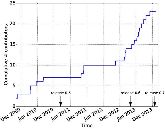
Diffusion Imaging in Python (Dipy) is a free and open source software project for the analysis of data from diffusion magnetic resonance imaging (dMRI) experiments. dMRI is an application of MRI that can be used to measure structural features of brain white matter. Many methods have been developed to use dMRI data to model the local configuration of white matter nerve fiber bundles and infer the trajectory of bundles connecting different parts of the brain. Dipy gathers implementations of many different methods in dMRI, including: diffusion signal pre-processing; reconstruction of diffusion distributions in individual voxels; fiber tractography and fiber track post-processing, analysis and visualization. Dipy aims to provide transparent implementations for all the different steps of dMRI analysis with a uniform programming interface. We have implemented classical signal reconstruction techniques, such as the diffusion tensor model and deterministic fiber tractography. In addition, cutting edge novel reconstruction techniques are implemented, such as constrained spherical deconvolution and diffusion spectrum imaging (DSI) with deconvolution, as well as methods for probabilistic tracking and original methods for tractography clustering. Many additional utility functions are provided to calculate various statistics, informative visualizations, as well as file-handling routines to assist in the development and use of novel techniques. In contrast to many other scientific software projects, Dipy is not being developed by a single research group. Rather, it is an open project that encourages contributions from any scientist/developer through GitHub and open discussions on the project mailing list. Consequently, Dipy today has an international team of contributors, spanning seven different academic institutions in five countries and three continents, which is still growing.
Overview
The claims and statistics of this paper haven’t been checked yet.
Run the check — free, no account needed. It reads the open-access text and checks the sources it cites.
Integrity signals
Registry
Cites
Statistics
Figures
Verification
Claims the paper makes
The claims and statistics of this paper haven’t been checked yet.
Run the check — free, no account needed. It reads the open-access text and checks the sources it cites.
Claims other papers make about this one
Each is checked against this paper’s own text: does it say what the citing paper says it does?
The cited source says what the claim attributes to it.
In the citing paper
“In contrast to projects such as DiPy (Garyfallidis et al., 2014) , this library does not provide functionality to manipulate raw image data within Python itself, but provides functionality to invoke and manage external high-performance executables to perform the actual computations.”
From MRtrix3: A fast, flexible and open software framework for medical image processing and visualisation (2019) · cites this paper as Garyfallidis et al., 2014
Self-check
Percentages, sums and reported differences re-computed from the paper’s own numbers.
The self-check needs the paper’s full text.
Citations
Cites
A Threshold Selection Method from Gray-Level Histograms
Otsu, N. (1979) · IEEE Transactions on Systems, Man and Cybernetics · doi:10.1109/tsmc.1979.4310076 · Cited by 44,708 · cited 1×
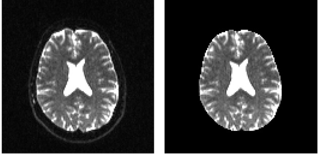
“…median_otsu (see Figure 3 ). from dipy.segment.mask import median_otsu mask, S0_mask = median_otsu(data [:, :, :, 0]) median_otsu uses first a median filter to smooth the S0 and then Otsu’s method, an automated histogram method (Otsu, 1979) to separate the brain (foreground) from its background.”
MethodAdvances in functional and structural MR image analysis and implementation as FSL
Smith, S. M., Jenkinson, M., Woolrich, M. W., Beckmann, C. F., Behrens, T. E. J., Johansen-Berg, H., Bannister, P. R., De Luca, M., Drobnjak, I., Flitney, D. E., et al. (2004) · NeuroImage · doi:10.1016/j.neuroimage.2004.07.051 · Cited by 13,630
Fischl, B. (2012) · NeuroImage · doi:10.1016/j.neuroimage.2012.01.021 · Cited by 9,526 · cited 4×
“…al., 2006), AFNI (Cox, 2012), MITK (Fritzsche et al., 2012), BrainVoyager QX (Goebel, 2012), DTI-Query/Quench (Sherbondy et al., 2005), FreeSurfer (Fischl, 2012), FSL-FDT (Smith et al., 2004), MedInria (Toussaint et al., 2007), MRtrix (Tournier et al., 2012), Diffusion Toolkit/Trackvis (Wang et…”
MethodInfluentialThe NumPy Array: A Structure for Efficient Numerical Computation
van der Walt, S., Colbert, S. C., & Varoquaux, G. (2011) · Computing in Science & Engineering · doi:10.1109/mcse.2011.37 · Cited by 9,381
MR diffusion tensor spectroscopy and imaging
Basser, P. J., Mattiello, J., & LeBihan, D. (1994) · Biophysical Journal · doi:10.1016/s0006-3495(94)80775-1 · Cited by 5,970 · cited 2×
“Another alternative is to make assumptions about the distribution of the PDF such as a Gaussian assumption, which leads to the popular diffusion tensor imaging (DTI) technique (Basser et al., 1994).”
BackgroundMethodThe WU-Minn Human Connectome Project: An overview
Van Essen, D. C., Smith, S. M., Barch, D. M., Behrens, T. E. J., Yacoub, E., & Ugurbil, K. (2013) · NeuroImage · doi:10.1016/j.neuroimage.2013.05.041 · Cited by 5,651
The basis of anisotropic water diffusion in the nervous system—A technical review.
Beaulieu, C. (2002) · NMR in Biomedicine · doi:10.1002/nbm.782 · Cited by 4,628
Basser, P. J., & Pierpaoli, C. (1996) · Journal of Magnetic Resonance, Series B · doi:10.1006/jmrb.1996.0086 · Cited by 4,306 · cited 3×
“…of the myelin wrapping the axons in the voxel may affect both MD and FA, changes in other factors, such as the distribution of directions of crossing of fiber populations through the voxel may also affect these measures (Basser and Pierpaoli, 1996; Jones et al., 2013; Wandell and Yeatman, 2013).”
BackgroundMethodMedical image analysis: progress over two decades and the challenges ahead
Duncan, J. S., & Ayache, N. (2000) · IEEE Transactions on Pattern Analysis and Machine Intelligence · doi:10.1109/34.824822 · Cited by 4,167
IPython: A System for Interactive Scientific Computing
Pérez, F., & Granger, B. (2007) · Computing in Science & Engineering · doi:10.1109/mcse.2007.53 · Cited by 4,129 · cited 1×
“There has been large growth in the number of Python users, there are many Python tools for scientific computing (Pérez and Granger, 2007; Pérez et al., 2011; McKinney, 2012), and there are complementary neuroimaging packages in Python1.”
Background
Research works printed but not linked: 9
- Chamberland, 2013, Explore the brain white matter networks in real-time: multi-sticks fiber tracking
- Garyfallidis, 2012, Towards an Accurate Brain Tractography
- Garyfallidis, 2011, Dipy - a novel software library for diffusion MR and tractography
- LeBihan, 1985, Imagerie de diffusion in vivo par résonance magnétique nucléaire, C. R. Acad. Sci. Paris, 301, 1109
- Pajevic, 1999, Color schemes to represent the orientation of anisotropic tissues from diffusion tensor data: application to white matter fiber tract mapping in the human brain, Magn. Reson. Med, 42, 526, 10.1002/(SICI)1522-2594(199909)42:3526::AID-MRM153.3.CO;2-A · DOI
- Schroeder, 2001, The Visualization Toolkit-User's Guide
- Vaillancourt, 2011, A fiber navigator for neurosurgical planning
- Wang, 2007, Diffusion toolkit: a software package for diffusion imaging data processing and tractography, Proc. Int. Soc. Magn. Reson. Med, 15, 3720
- Westin, 1997, Geometrical diffusion measures for MRI from tensor basis analysis, Proc. ISMRM, 97, 1742
Cited by
Sorted by citations
Items 101–200 of 1,000
Learn to Track: Deep Learning for Tractography
Poulin, P., Côté, M.-A., Houde, J., Petit, L., Neher, P., Maier-Hein, K. H., Larochelle, H., & Descoteaux, M. (2017) · Lecture notes in computer science · doi:10.1007/978-3-319-66182-7_62 · Cited by 62
Self-citationHigh Angular Resolution Diffusion Imaging ( HARDI )
Descoteaux, M. (2015) · Wiley eBooks · doi:10.1002/047134608x.w8258 · Cited by 80
Self-citationLongitudinal structural and molecular neuroimaging in agrammatic primary progressive aphasia
Tetzloff, K. A., Duffy, J. R., Clark, H. M., Strand, E. A., Machulda, M. M., Schwarz, C. G., Senjem, M. L., Reid, R. I., Spychalla, A. J., Tosakulwong, N., et al. (2017) · Brain · doi:10.1093/brain/awx293 · Cited by 62
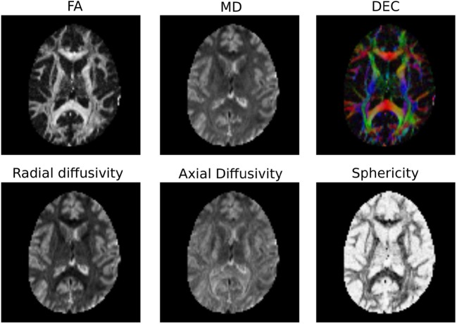
“Images were brain-extracted, and fractional anisotropy, mean diffusivity, radial diffusivity, and axial diffusivity were generated using weighted least-squares optimization with Dipy (Garyfallidis et al., 2014).”
MethodAxTract: Toward microstructure informed tractography
Girard, G., Daducci, A., Petit, L., Thiran, J., Whittingstall, K., Deriche, R., Wassermann, D., & Descoteaux, M. (2017) · Human Brain Mapping · doi:10.1002/hbm.23741 · Cited by 62
“Diffusion Tensor estimation and corresponding fractional anisotropy map generation were done using Dipy [Garyfallidis et al., 2014].”
MethodSelf-citationHasenstab, K. A., Yuan, N. F., Retson, T., Conrad, D. J., Kligerman, S. J., Lynch, D. A., Hsiao, A., & for the COPDGene Investigators (2021) · Radiology Cardiothoracic Imaging · doi:10.1148/ryct.2021200477 · Cited by 61
“Expiratory series were deformably registered to inspiratory series using a symmetric diffeomorphic registration algorithm (29,30).”
MethodDavis, S. W., Luber, B. M., Murphy, D. L. K., Lisanby, S. H., & Cabeza, R. E. L. (2017) · Human Brain Mapping · doi:10.1002/hbm.23803 · Cited by 63
“We used Dipy (Garyfallidis et al., 2014) to fit the data with the constant solid angle (CSA) model with fourth-order spherical harmonics, and generating generalized FA (GFA) maps from estimated orientation distribution functions.”
MethodSchilling, K. G., Tax, C. M. W., Rheault, F., Hansen, C. B., Yang, Q., Yeh, F.-C. F., Cai, L. Y., Anderson, A. W., & Landman, B. A. (2021) · NeuroImage · doi:10.1016/j.neuroimage.2021.118451 · Cited by 66
Annen, J., Heine, L., Ziegler, E., Frasso, G., Bahri, M. A., Perri, C. D., Stender, J., Martial, C., Wannez, S., D’Ostilio, K., et al. (2016) · Human Brain Mapping · doi:10.1002/hbm.23269 · Cited by 60
“All tensor calculations were performed with Dipy [Garyfallidis et al., 2014].”
MethodDifferences in brain morphology and working memory capacity across childhood
Bathelt, J., Gathercole, S. E., Johnson, A., & Astle, D. E. (2017) · Developmental Science · doi:10.1111/desc.12579 · Cited by 60
“0 implementation (Garyfallidis et al., 2014) of a non- local means de- noising algorithm (Coupe et al.”
MethodInfluentialBirchenall, J., Térémetz, M., Roca, P., Lamy, J., Oppenheim, C., Maier, M. A., Mas, J., Lamy, C., Baron, J., & Lindberg, P. G. (2018) · Neurophysiologie Clinique · doi:10.1016/j.neucli.2018.10.065 · Cited by 62
Raghavan, S., Przybelski, S. A., Reid, R. I., Graff‐Radford, J., Lesnick, T. G., Zuk, S. M., Knopman, D. S., Machulda, M. M., Mielke, M. M., Petersen, R. C., et al. (2020) · Neurobiology of Aging · doi:10.1016/j.neurobiolaging.2020.09.005 · Cited by 59
“Diffusion tensors were then fitted by nonlinearly minimizing the squared residuals (Garyfallidis et al., 2014), and FA and MD maps were calculated from the tensors.”
Structural and functional connectivity reconstruction with CATO - A Connectivity Analysis TOolbox
de Lange, S. C., Helwegen, K., & van den Heuvel, M. P. (2023) · NeuroImage · doi:10.1016/j.neuroimage.2023.120108 · Cited by 54
Tian, Q., Fan, Q., Witzel, T., Polackal, M. N., Ohringer, N. A., Ngamsombat, C., Russo, A. W., Machado, N., Brewer, K., Wang, F., et al. (2022) · Scientific Data · doi:10.1038/s41597-021-01092-6 · Cited by 59
Binette, A. P., Theaud, G., Rheault, F., Roy, M., Collins, D. L., Levin, J., Mori, H., Lee, J. H., Farlow, M. R., Schofield, P. R., et al. (2021) · eLife · doi:10.7554/elife.62929 · Cited by 60
Self-citationConnectivity‐based parcellation of normal and anatomically distorted human cerebral cortex
Doyen, S., Nicholas, P. J., Poologaindran, A., Crawford, L. S., Young, I. M., Romero-García, R., & Sughrue, M. E. (2021) · Human Brain Mapping · doi:10.1002/hbm.25728 · Cited by 56
“brain, and correction for gradient distortion (Garyfallidis et al., 2014).”
MethodInfluentialProgressive agrammatic aphasia without apraxia of speech as a distinct syndrome
Tetzloff, K. A., Duffy, J. R., Clark, H. M., Utianski, R. L., Strand, E. A., Machulda, M. M., Botha, H., Martin, P. R., Schwarz, C. G., Senjem, M. L., et al. (2019) · Brain · doi:10.1093/brain/awz157 · Cited by 55
Bernier, M., Croteau, É., Castellano, C., Cunnane, S. C., & Whittingstall, K. (2017) · NeuroImage · doi:10.1016/j.neuroimage.2017.01.055 · Cited by 52
“We replaced the usual 4–6 mm full-width half maximum Gaussian spatial smoothing by a non-local mean denoising, which improved the signal-to-noise ratio (SNR), particularly in subcortical areas (Bernier et al., 2014; Garyfallidis, 2014).”
MethodNath, V., Schilling, K. G., Parvathaneni, P., Huo, Y., Blaber, J. A., Hainline, A. E., Baraković, M., Romascano, D., Rafael‐Patiño, J., Frigo, M., et al. (2019) · Journal of Magnetic Resonance Imaging · doi:10.1002/jmri.26794 · Cited by 52
“40 The fiber orientation distribution (FOD) function was estimated using CSD41 implemented in DiPy.35 An in-house probabilistic tractography algorithm was used.42 Tractography parameters were: step size = 0.2 mm, maximal deviation = 20 , number of seeds per voxel = 3.”
InfluentialSelf-citationComparative evaluation of big-data systems on scientific image analytics workloads
Mehta, P., Dorkenwald, S., Zhao, D., Kaftan, T., Cheung, A., Bałazińska, M., Rokem, A., Connolly, A. J., VanderPlas, J. T., & AlSayyad, Y. (2017) · Proceedings of the VLDB Endowment · doi:10.14778/3137628.3137634 · Cited by 52
Self-citationAutomatic Early Diagnosis of Alzheimer’s Disease Using 3D Deep Ensemble Approach
Gamal, A., Elattar, M., & Selim, S. (2022) · IEEE Access · doi:10.1109/access.2022.3218621 · Cited by 56
“We used a nonlinear image registration algorithm based on diffusion imaging in Python (Dipy) [64].”
MethodA deep network for tissue microstructure estimation using modified LSTM units
Ye, C., Li, X., & Chen, J. (2019) · Medical Image Analysis · doi:10.1016/j.media.2019.04.006 · Cited by 51
Rafael‐Patiño, J., Romascano, D., Ramírez-Manzanares, A., Canales-Rodríguez, E. J., Girard, G., & Thiran, J. (2020) · Frontiers in Neuroinformatics · doi:10.3389/fninf.2020.00008 · Cited by 58
“To show qualitative results on the generated signals, the Diffusion Tensor (DT) estimation and the corresponding FA were computed using Dipy (Garyfallidis et al., 2014), as well as the ICVF maps for each of the three resolutions.”
MethodA collaborative resource platform for non-human primate neuroimaging
Messinger, A., Sirmpilatze, N., Heuer, K., Loh, K. K., Mars, R. B., Sein, J., Xu, T., Glen, D., Jung, B., Seidlitz, J., et al. (2021) · NeuroImage · doi:10.1016/j.neuroimage.2020.117519 · Cited by 52
Computational and Informatic Advances for Reproducible Data Analysis in Neuroimaging
Poldrack, R. A., Gorgolewski, K. J., & Varoquaux, G. P. (2019) · Annual Review of Biomedical Data Science · doi:10.1146/annurev-biodatasci-072018-021237 · Cited by 57
Whole-brain white matter organization, intelligence, and educational attainment
Bathelt, J., Scerif, G., Nobre, A. C., & Astle, D. E. (2019) · Trends in Neuroscience and Education · doi:10.1016/j.tine.2019.02.004 · Cited by 49
Neuroimaging and biomarker evidence of neurodegeneration in asthma
Rosenkranz, M. A., Dean, D., Bendlin, B. B., Jarjour, N. N., Esnault, S. J., Zetterberg, H., Heslegrave, A., Evans, M. D., Davidson, R. J., & Busse, W. W. (2021) · Journal of Allergy and Clinical Immunology · doi:10.1016/j.jaci.2021.09.010 · Cited by 51
White matter damage due to vascular, tau, and TDP-43 pathologies and its relevance to cognition
Raghavan, S., Przybelski, S. A., Reid, R. I., Lesnick, T. G., Ramanan, V. K., Botha, H., Matchett, B. J., Murray, M. E., Reichard, R. R., Knopman, D. S., et al. (2022) · Acta Neuropathologica Communications · doi:10.1186/s40478-022-01319-6 · Cited by 52
“The diffusion tensors were then fit using a nonlinear least squares fitting algorithm implemented in dipy [44].”
MethodRecent Developments in Fast Kurtosis Imaging
Hansen, B., & Jespersen, S. N. (2017) · Frontiers in Physics · doi:10.3389/fphy.2017.00040 · Cited by 51
“html), the Dipy library in python [71] (http://nipy.”
Asken, B. M., Tanner, J. A., VandeVrede, L. A., Casaletto, K. B., Staffaroni, A. M., Mundada, N. S., Fonseca, C. S., Iaccarino, L., Joie, R. L., Tsuei, T., et al. (2022) · Journal of Neurotrauma · doi:10.1089/neu.2022.0060 · Cited by 50
“41 Registration of diffusion data was accomplished through the DTI-TK software package.”
Maffei, C., Girard, G., Schilling, K. G., Aydogan, D. B., Adluru, N., Zhylka, A., Wu, Y., Mancini, M., Hamamcı, A., Sarica, A., et al. (2022) · NeuroImage · doi:10.1016/j.neuroimage.2022.119327 · Cited by 49
“…= 0.27) was obtained by Team 1, with a combination of the Robust and Unbiased Model-BAsed Spherical Deconvolution (Rumba-SD) method for orientation reconstruction ( Canales-Rodríguez et al., 2015 ) and probabilistic tractography ( Garyfallidis et al., 2014 ; Girard et al., 2014 ) on the DSI data.”
InfluentialRoles of ventral versus dorsal pathways in language production: An awake language mapping study
Riès, S., Piai, V., Perry, D. W., Griffin, S., Jordan, K., Henry, R. G., Knight, R. T., & Berger, M. S. (2019) · Brain and Language · doi:10.1016/j.bandl.2019.01.001 · Cited by 53
Lesh, T. A., Maddock, R. J., Howell, A. M., Wang, H., Tanase, C., Ragland, J. D., Niendam, T. A., & Carter, C. S. (2019) · Molecular Psychiatry · doi:10.1038/s41380-019-0428-y · Cited by 49
Multimodal Hippocampal Subfield Grading For Alzheimer’s Disease Classification
Hett, K., Ta, V.-T., Catheline, G., Tourdias, T., Manjón, J. V., Coupé, P., Initiative, A. D. N., Weiner, M. W., Aisen, P., Petersen, R., et al. (2019) · Scientific Reports · doi:10.1038/s41598-019-49970-9 · Cited by 49
Satpathi, S., Reid, R. I., Przybelski, S. A., Raghavan, S., Cogswell, P. M., Meyer, N. K., Lowe, V. J., Gunter, J. L., Petersen, R. C., Jack, C. R., et al. (2025) · Alzheimer s Research & Therapy · doi:10.1186/s13195-025-01842-3 · Cited by 48
“Diffusion tensors were estimated using nonlinear least squares fitting and used to calculate Fractional Anisot-ropy (FA) and Mean Diffusivity (MD) images in DIPY [30].”
Sarubbo, S., Petit, L., BENEDICTIS, A. D., Chioffi, F., Ptito, M., & Dyrby, T. B. (2019) · Brain Structure and Function · doi:10.1007/s00429-019-01856-2 · Cited by 48
“al (2014) and implemented in Dipy (Garyfallidis et al. 2014).”
MethodInfluentialStructural and functional brain imaging in acute HIV
Samboju, V., Philippi, C. L., Chan, P., Cobigo, Y., Fletcher, J. L. K., Robb, M. L., Hellmuth, J. M., Benjapornpong, K., Dumrongpisutikul, N., Pothisri, M., et al. (2018) · NeuroImage Clinical · doi:10.1016/j.nicl.2018.07.024 · Cited by 46
“Remaining tensors were then fi tted using DIPY with a non-linear least-squares approach derived fi tting model to construct FA, MD, RD and AD maps from standard equations (Garyfallidis et al., 2014; Pierpaoli & Basser, 1996).”
Ismail, A. A. O., Parker, D., Hernández-Fernández, M., Wolf, R., Brem, S., Alexander, S., Hodges, W., Pasternak, O., Caruyer, E., & Verma, R. (2020) · PLoS ONE · doi:10.1371/journal.pone.0233645 · Cited by 46
Poulin, P., Theaud, G., Rheault, F., St‐Onge, E., Boré, A., Renauld, E., Beaumont, L. D., Guay, S., Jodoin, P., & Descoteaux, M. (2022) · Scientific Data · doi:10.1038/s41597-022-01833-1 · Cited by 49
Self-citationRensonnet, G., Scherrer, B., Girard, G., Jankovski, A., Warfield, S. K., Macq, B. M. M., Thiran, J., & Taquet, M. (2018) · NeuroImage · doi:10.1016/j.neuroimage.2018.09.076 · Cited by 46
“dki_micro module from the DIPY software project4 (Garyfallidis et al., 2014).”
Cai, L. Y., Yang, Q., Kanakaraj, P., Nath, V., Newton, A. T., Edmonson, H. A., Luci, J. J., Conrad, B. N., Price, G. R., Hansen, C. B., et al. (2021) · Magnetic Resonance in Medicine · doi:10.1002/mrm.28926 · Cited by 50
Self-citationNecus, J., Sinha, N., Smith, F. E., Thelwall, P. E., Flowers, C. J., Taylor, P. N., Blamire, A. M., Cousins, D. A., & Wang, Y. (2019) · Journal of Affective Disorders · doi:10.1016/j.jad.2019.04.075 · Cited by 44
“11) (Garyfallidis et al., 2014) yielding an estimation of gFA for each brain voxel.”
Zacà, D., Corsini, F., Rozzanigo, U., Dallabona, M., Avesani, P., Annicchiarico, L., Zigiotto, L., Faraca, G., Chioffi, F., Jovicich, J., et al. (2018) · Frontiers in Human Neuroscience · doi:10.3389/fnhum.2018.00405 · Cited by 43
“The analyses pipeline was implemented using FSL and Dipy, an open source library for the analysis of diffusion MRI data (Jenkinson et al., 2002; Garyfallidis et al., 2014).”
MethodMitigating gyral bias in cortical tractography via asymmetric fiber orientation distributions
Wu, Y., Hong, Y., Feng, Y., Shen, D. G., & Yap, P. (2019) · Medical Image Analysis · doi:10.1016/j.media.2019.101543 · Cited by 43
Classifyber, a robust streamline-based linear classifier for white matter bundle segmentation
Bertò, G., Bullock, D. N., Astolfi, P., Hayashi, S., Zigiotto, L., Annicchiarico, L., Corsini, F., BENEDICTIS, A. D., Sarubbo, S., Pestilli, F., et al. (2020) · NeuroImage · doi:10.1016/j.neuroimage.2020.117402 · Cited by 46
Track-to-Learn: A general framework for tractography with deep reinforcement learning
Théberge, A., Desrosiers, C., Descoteaux, M., & Jodoin, P. (2021) · Medical Image Analysis · doi:10.1016/j.media.2021.102093 · Cited by 44
Self-citationLu, Q., Li, Y., & Ye, C. (2021) · Medical Image Analysis · doi:10.1016/j.media.2021.102094 · Cited by 43
Learning to estimate the fiber orientation distribution function from diffusion-weighted MRI
Karimi, D., Vasung, L., Jaimes, C. E., Machado‐Rivas, F., Warfield, S. K., & Gholipour, A. (2021) · NeuroImage · doi:10.1016/j.neuroimage.2021.118316 · Cited by 42
Mandonnet, E., Vincent, M., Valero‐Cabré, A., Facque, V., Barberis, M., Bonnetblanc, F., Rheault, F., Emmanuelle Volle, C., Descoteaux, M., & Margulies, D. S. (2020) · Cortex · doi:10.1016/j.cortex.2020.08.021 · Cited by 42
Self-citationInteraction of amyloid and tau on cortical microstructure in cognitively unimpaired adults
Vogt, N. M., Hunt, J. F. V., Adluru, N., Ma, Y., Hulle, C. A. V., Dean, D., Kecskemeti, S., Chin, N. A., Carlsson, C. M., Asthana, S., et al. (2021) · Alzheimer s & Dementia · doi:10.1002/alz.12364 · Cited by 42
Portegies, J. M., Fick, R., Sanguinetti, G., Meesters, S., Girard, G., & Duits, R. (2015) · PLoS ONE · doi:10.1371/journal.pone.0138122 · Cited by 39
The cingulum as a marker of individual differences in neurocognitive development
Bathelt, J., Johnson, A., Zhang, M., the CALM team, & Astle, D. E. (2019) · Scientific Reports · doi:10.1038/s41598-019-38894-z · Cited by 43
Evaluating arcuate fasciculus laterality measurements across dataset and tractography pipelines
Bain, J. S., Yeatman, J. D., Schurr, R., Rokem, A., & Mezer, A. (2019) · Human Brain Mapping · doi:10.1002/hbm.24626 · Cited by 43
“We used the SFM implemented in Dipy (Garyfallidis et al., 2014; http://nipy.org/dipy), and also available as a Docker image (https://hub.docker.com/r/arokem/dipy-sfm/), to create a brain map of the dispersion index (DI), a metric that quantifies the amount of fascicle crossing per voxel.”
MethodSelf-citationPhysical activity and cognitive and imaging biomarkers of Alzheimer's disease in down syndrome
Fleming, V., Piro‐Gambetti, B., Patrick, A., Zammit, M. D., Alexander, A. L., Christian, B. T., Handen, B. L., Cohen, A. D., Klunk, W. E., Laymon, C. M., et al. (2021) · Neurobiology of Aging · doi:10.1016/j.neurobiolaging.2021.07.016 · Cited by 41
Moltu, S. J., Nordvik, T., Rossholt, M. E., Wendel, K., Chawla, M. S., Server, A., Gunnarsdottir, G., Pripp, A. H., Domellöf, M., Bratlie, M., et al. (2023) · Clinical Nutrition · doi:10.1016/j.clnu.2023.11.037 · Cited by 42
Diffusion MRI Signal Augmentation: From Single Shell to Multi Shell with Deep Learning
Koppers, S., Haarburger, C., & Merhof, D. (2017) · Mathematics and visualization · doi:10.1007/978-3-319-54130-3_5 · Cited by 41
Genome-Wide Association Study of Brain Connectivity Changes for Alzheimer’s Disease
Elsheikh, S. S. M., Chimusa, E. R., Mulder, N., & Crimi, A. (2020) · Scientific Reports · doi:10.1038/s41598-020-58291-1 · Cited by 38
“In particular, the constant solid angle model was used 50 , and a deterministic algorithm called Euler Delta Crossings 49 was used stemming from 2,000,000 seed-points and stopping when the fractional anisotropy was smaller than 0 .”
Qi, X., & Arfanakis, K. (2020) · NeuroImage · doi:10.1016/j.neuroimage.2020.117462 · Cited by 40
“…et al., 2015), d) major white matter bundles (Table 1) were reconstructed from the streamlines of the tractogram using the RecoBundle approach (Garyfallidis et al., 2014; E. 2018) based on the Yeh et al., 2018 bundle models (and were made available at www.nitrc.org/projects/iit), and all…”
Karayumak, S. C., Zhang, F., Zurrin, R., Billah, T., Zekelman, L., Makris, N., Pieper, S., O’Donnell, L. J., & Yogesh Rathi, A. (2024) · Scientific Data · doi:10.1038/s41597-024-03058-w · Cited by 49
Uses of Human MR and PET Imaging in Research of Neurodegenerative Brain Diseases
Schwarz, C. G. (2021) · Neurotherapeutics · doi:10.1007/s13311-021-01030-9 · Cited by 40
“Some of the most popular include DTI-TK [61], DSI Studio [62], and multiple tools within the DIPY [63] and FSL packages [16, 64].”
MethodThe role of the arcuate and middle longitudinal fasciculi in speech perception in noise in adulthood
Tremblay, P., Perron, M., Deschamps, I., Kennedy‐Higgins, D., Houde, J., Dick, A. S., & Descoteaux, M. (2018) · Human Brain Mapping · doi:10.1002/hbm.24367 · Cited by 40
Self-citationYamashiro, K., Takabayashi, K., Kamagata, K., Nishimoto, Y., Togashi, Y., Yamauchi, Y., Ogaki, K., Li, Y., Hatano, T., Motoi, Y., et al. (2024) · Neurobiology of Disease · doi:10.1016/j.nbd.2024.106464 · Cited by 39
“FW maps of all study participants were calculated from multi-shell DWI data using the open-source software package Diffusion Imaging in Python (also known as Dipy) algorithm (dipy.org/) (Garyfallidis et al., 2014), based on a regularized bi-tensor model (Pasternak et al., 2009).”
MethodLe, H., Zeng, W., Zhang, H., Li, J., Wu, X., Xie, M., Yan, X., Zhou, M., Zhang, H., Wang, M., et al. (2020) · Frontiers in Aging Neuroscience · doi:10.3389/fnagi.2020.563595 · Cited by 38
A High-Throughput Pipeline Identifies Robust Connectomes But Troublesome VariabilityPreprint
Kiar, G., Bridgeford, E., Roncal, W. G., Chandrashekhar, V., Mhembere, D., Ryman, S. G., Zuo, X., Margulies, D. S., Craddock, R. C., Priebe, C. E., et al. (2017) · bioRxiv (Cold Spring Harbor Laboratory) · doi:10.1101/188706 · Cited by 38
The Mexican magnetic resonance imaging dataset of patients with cocaine use disorder: SUDMEX CONN
Ángeles-Valdéz, D., Rasgado‐Toledo, J., Issa‐Garcia, V., Balducci, T., Villicaña, V., Valencia, A., Gonzalez-Olvera, J. J., Reyes-Zamorano, E., & Garza‐Villarreal, E. A. (2022) · Scientific Data · doi:10.1038/s41597-022-01251-3 · Cited by 43
Boone, L., Biparva, M., Forooshani, P. M., Ramirez, J., Masellis, M., Bartha, R., Symons, S. P., Strother, S. C., Black, S. E., Heyn, C., et al. (2023) · NeuroImage · doi:10.1016/j.neuroimage.2023.120289 · Cited by 42
3D interactive tractography-informed resting-state fMRI connectivity
Chamberland, M., Bernier, M., Fortin, D., Whittingstall, K., & Descoteaux, M. (2015) · Frontiers in Neuroscience · doi:10.3389/fnins.2015.00275 · Cited by 37
“Next, the data were spatially smoothed using NLM denoising (Coupe et al., 2008; Bernier et al., 2014) implemented in Dipy (Garyfallidis et al., 2014) using default parameters, and band-pass filtered (0.008–0.08 Hz).”
MethodInfluentialSelf-citationREM sleep is associated with white matter integrity in cognitively healthy, older adults
Altendahl, M., Cotter, D. L., Staffaroni, A. M., Wolf, A. A., Mumford, P., Cobigo, Y., Casaletto, K. B., Elahi, F. M., Yack, L. M., Javed, S., et al. (2020) · PLoS ONE · doi:10.1371/journal.pone.0235395 · Cited by 38
“Diffusion tensors were calculated using a non-linear least-squares algorithm in Dipy [35].”
MethodLevakov, G., Faskowitz, J., Avidan, G., & Sporns, O. (2021) · NeuroImage · doi:10.1016/j.neuroimage.2021.118469 · Cited by 41
Badea, A., Wu, W., Shuff, J., Wang, M. S., Anderson, R. J., Qi, Y. P., Johnson, G. A., Wilson, J. G., Koudoro, S., Garyfallidis, E., et al. (2019) · Frontiers in Neuroinformatics · doi:10.3389/fninf.2019.00072 · Cited by 31
Self-citationSanvito, F., Caverzasi, E., Riva, M., Jordan, K., Blasi, V., Scifo, P., Iadanza, A., Crespi, S. A., Cirillo, S., Casarotti, A., et al. (2020) · Frontiers in Neuroscience · doi:10.3389/fnins.2020.00225 · Cited by 35
Denoising diffusion MRI: Considerations and implications for analysis
Manzano-Patron, J. P., Moeller, S., Andersson, J., Uǧurbil, K., Yacoub, E. S., & Sotiropoulos, S. N. (2023) · Imaging Neuroscience · doi:10.1162/imag_a_00060 · Cited by 38
“We used the default implementation in DIPY (v.1.7.0) ( Garyfallidis et al., 2014), where each dMRI volume is denoised independently. ii) The Marchenko-Pastur PCA (MPPCA) (denoted as | MPPCA | throughout the text) ( Cordero-Grande et al., 2019; Veraart, Novikov, et al., 2016), reflecting a commonly…”
InfluentialThe Connectome Visualization Utility: Software for Visualization of Human Brain Networks
LaPlante, R. A., Douw, L., Tang, W., & Stufflebeam, S. M. (2014) · PLoS ONE · doi:10.1371/journal.pone.0113838 · Cited by 38
“…currently available are integrated brain network tools that are designed for processing a single imaging modality Such packages include Camino [24], MNE [25, 26], dipy [27], Brainstorm [28], the Connectome Mapper [29], the MR Connectome Analysis Pipeline [30] and many other unpublished packages.”
BackgroundMethodWhite matter information flow mapping from diffusion MRI and EEG
Deslauriers‐Gauthier, S., Lina, J., Butler, R., Whittingstall, K., Gilbert, G., Bernier, P., Rachid Deriche, B., & Descoteaux, M. (2019) · NeuroImage · doi:10.1016/j.neuroimage.2019.116017 · Cited by 39
Self-citationDorsman, K. A., Weiner‐Light, S., Staffaroni, A. M., Brown, J. A., Wolf, A. A., Cobigo, Y., Walters, S. M., Kramer, J. H., & Casaletto, K. B. (2020) · Frontiers in Aging Neuroscience · doi:10.3389/fnagi.2020.00104 · Cited by 39
The MRi-Share database: brain imaging in a cross-sectional cohort of 1870 university students
Tsuchida, A., Laurent, A., Crivello, F., Petit, L., Joliot, M., Pepe, A., Beguedou, N., Gueye, M.-F., Verrecchia, V., Nozais, V., et al. (2021) · Brain Structure and Function · doi:10.1007/s00429-021-02334-4 · Cited by 40
Tourbier, S., Rué‐Queralt, J., Glomb, K., Alemán‐Gómez, Y., Mullier, E., Griffa, A., Schöttner, M., Wirsich, J., Tuncel, A., Jancovic, J., et al. (2022) · The Journal of Open Source Software · doi:10.21105/joss.04248 · Cited by 39
Frigerio, I., Laansma, M. A., Lin, C., Hermans, E. J. M., Bouwman, M. M. A., Bol, J. G. J. M., Graaf, Y. G.-D., Hepp, D. H., Rozemüller, A. J. M., Barkhof, F., et al. (2023) · Translational Neurodegeneration · doi:10.1186/s40035-022-00328-8 · Cited by 35
“Freesurfer [41] and free‐water corrected cortical mean diffusivity (FWC‐MD) was derived with FSL [44], DIPY [47, 48] and Freesurfer [41] (purple”
MethodPredicting motor outcomes in stroke patients using diffusion Spectrum mri microstructural measures
Hodgson, K., Adluru, G., Richards, L. G., Majersik, J. J., Stoddard, G., Adluru, N., & DiBella, E. (2019) · Frontiers in Neurology · doi:10.3389/fneur.2019.00072 · Cited by 36
“The orientation distribution functions were estimated using DIPY (17, 29) for calculating GFA.”
MethodEfficient PCA denoising of spatially correlated redundant MRI data
Henriques, R. N., Ianuș, A., Novello, L., Jovicich, J., Jespersen, S. N., & Shemesh, N. (2023) · Imaging Neuroscience · doi:10.1162/imag_a_00049 · Cited by 38
“…was sub-divided into 9 portions (4 × 4 voxel regions each), and diffusion-weighted signals were generated using the Diffusion in Python (DIPY ( Garyfallidis et al., 2014; Henriques et al., 2021)) package according to a forward model containing two compartment types with different axially…”
MethodLefebvre, J., Castonguay, A., Pouliot, P., Descoteaux, M., & Lesage, F. (2017) · Neurophotonics · doi:10.1117/1.nph.4.4.041501 · Cited by 37
Self-citationApplying microstructural models to understand the role of white matter in cognitive development
Huber, E., Henriques, R. N., Owen, J. P., Rokem, A., & Yeatman, J. D. (2019) · Developmental Cognitive Neuroscience · doi:10.1016/j.dcn.2019.100624 · Cited by 34
Self-citationChang, K., Premeaux, T. A., Cobigo, Y., Milanini, B., Hellmuth, J. M., Rubin, L. H., Javandel, S., Allen, I. E., Ndhlovu, L. C., Paul, R. H., et al. (2019) · AIDS · doi:10.1097/qad.0000000000002404 · Cited by 34
“The susceptibility distortion corrected diffusion images were then fitted using Dipy with a nonlinear least-squares approach to create the DTI tensor maps [34].”
MethodDilated Convolutional Neural Networks for Sequential Manifold-Valued Data
Chakraborty, R., Zhen, X., Vogt, N., Bendlin, B. B., & Singh, V. (2019) · IEEE/CVF International Conference on Computer Vision (ICCV) · doi:10.1109/iccv.2019.01072 · Cited by 35
“Diffusion tensor imaging (DTI) and the orientation distribution functions (ODF), which were used as the representative of the DWI, were performed using the Diffusion Imaging in Python (DIPY) toolbox[18].”
MethodPerron, M., Theaud, G., Descoteaux, M., & Tremblay, P. (2021) · Human Brain Mapping · doi:10.1002/hbm.25416 · Cited by 33
“…were normalized using a N4 correction procedure (Tustison et al., 2010) from the advanced normalization tools (ANTs) (Avants et al., 2011) and the resulting DW images were resampled to 1 mm isotropic as implemented in Diffusion Imaging in Python (DIPY) toolbox (Garyfallidis et al., 2014).”
MethodSelf-citationStructurally constrained effective brain connectivity
Crimi, A., Dodero, L., Sambataro, F., Murino, V., & Sona, D. (2021) · NeuroImage · doi:10.1016/j.neuroimage.2021.118288 · Cited by 33
A large-scale investigation of white matter microstructural associations with reading ability
Meisler, S. L., & Gabrieli, J. D. E. (2022) · NeuroImage · doi:10.1016/j.neuroimage.2022.118909 · Cited by 38
Mapping the macrostructure and microstructure of the in vivo human hippocampus using diffusion MRI
Karat, B. G., DeKraker, J., Hussain, U., Köhler, S., & KHAN, A. R. (2023) · Human Brain Mapping · doi:10.1002/hbm.26461 · Cited by 33
López‐Aguirre, M., Matarazzo, M., Blesa, J., Monje, M. H. G., Rodríguez‐Rojas, R., Sánchez‐Ferro, Á., Obeso, J. A., & Pineda‐Pardo, J. A. (2023) · npj Parkinson s Disease · doi:10.1038/s41531-023-00586-x · Cited by 36
“FW maps were estimated with the Dipy library 82 by fi tting the bi-tensor model with the regularized gradient descent method and applying the hybrid initialization strategy described by Parker et al. ( PLoS One , 2020) 83 .”
mpdcm: A toolbox for massively parallel dynamic causal modeling
Aponte, E. A., Raman, S., Sengupta, B., Penny, W. D., Stephan, K. E., & Heinzle, J. (2015) · Journal of Neuroscience Methods · doi:10.1016/j.jneumeth.2015.09.009 · Cited by 33
A finite-element reciprocity solution for EEG forward modeling with realistic individual head models
Ziegler, E., Chellappa, S. L., Gaggioni, G., Ly, J. Q. M., Vandewalle, G., André, E., Geuzaine, C., & Phillips, C. L. M. (2014) · NeuroImage · doi:10.1016/j.neuroimage.2014.08.056 · Cited by 33
“Diffusion tensor eigenvalues were remapped to produce conductivity tensors [38] using functions from Dipy [39].”
MethodConnectomic consistency: a systematic stability analysis of structural and functional connectivity
Osmanlıoğlu, Y., Alappatt, J. A., Parker, D., & Verma, R. (2020) · Journal of Neural Engineering · doi:10.1088/1741-2552/ab947b · Cited by 33
Ferizi, U., Scherrer, B., Schneider, T., Alipoor, M., Eufracio, O., Fick, R., Rachid Deriche, B., Nilsson, M., Loya-Olivas, A. K., Rivera, M., et al. (2017) · NMR in Biomedicine · doi:10.1002/nbm.3734 · Cited by 31
Self-citationAdvances in computational and statistical diffusion MRI
O’Donnell, L. J., Daducci, A., Wassermann, D., & Lenglet, C. (2017) · NMR in Biomedicine · doi:10.1002/nbm.3805 · Cited by 32
Micro-Networks for Robust MR-Guided Low Count PET Imaging
da Costa-Luis, C. O., & Reader, A. J. (2020) · IEEE Transactions on Radiation and Plasma Medical Sciences · doi:10.1109/trpms.2020.2986414 · Cited by 33
“Corresponding MPRAGE T1 reconstructions were scaled and registered to the full count PET reconstructions using dipy [41] before performing NLM filtering on the PET reconstructions (3).”
MethodSamani, Z. R., Parker, D., Wolf, R. L., Hodges, W., Brem, S. S., & Verma, R. (2021) · Scientific Reports · doi:10.1038/s41598-021-93804-6 · Cited by 32
Mohtasib, R., Alghamdi, J., Jobeir, A., Masawi, A., de Barros, N. D., Billiet, T., Struyfs, H., Phan, T. V., van Hecke, W., & Ribbens, A. (2022) · Heliyon · doi:10.1016/j.heliyon.2022.e08901 · Cited by 32
Injury to the Cerebellum in Term Asphyxiated Newborns Treated with Hypothermia
Kwan, S., Boudes, E., Gilbert, G., Saint‐Martin, C., Albrecht, S., Shevell, M. I., & Wintermark, P. (2015) · American Journal of Neuroradiology · doi:10.3174/ajnr.a4326 · Cited by 32
“Then, ADC and FA maps were generated for the whole brain by using the tools of the Diffusion Imaging in Python (DIPY) package (Sherbrooke Connectivity Imaging Lab, Sherbrooke University, Sherbrooke, Quebec, Canada).(20) To assess whether subtle changes in the cerebellum were not recognized on conventional MR imaging o…”
MethodSacchi, L., D’Agata, F., Campisi, C., Arcaro, M., Carandini, T., Örzsik, B., Maschio, V. P. D., Fenoglio, C., Pietroboni, A. M., Ghezzi, L., et al. (2024) · Human Brain Mapping · doi:10.1002/hbm.26805 · Cited by 33
“Maps of the fractional volume of FW and of the MK were constructed from preprocessed DW images fitting a regularized bitensor model and the DKI, respectively, with the open-source software package Diffusion Imaging in Python (Dipy) algorithm (dipy.org/) (Garyfallidis et al., 2014).”
Tobisch, A., Stirnberg, R., Harms, R., Schultz, T., Roebroeck, A. F., Breteler, M. M. B., & Stöcker, T. (2018) · Frontiers in Neuroscience · doi:10.3389/fnins.2018.00650 · Cited by 31
Sacco, S., Caverzasi, E., Papinutto, N., Cordano, C., Bischof, A., Gundel, T. J., Cheng, S., Asteggiano, C., Kirkish, G., Mallott, J. M., et al. (2020) · American Journal of Neuroradiology · doi:10.3174/ajnr.a6862 · Cited by 33
Metadata
- Authors
- Garyfallidis, E., Brett, M., Amirbekian, B., Rokem, A., van der Walt, S., Descoteaux, M., Nimmo-Smith, I., & Contributors, D.
- Affiliations
- Université de Sherbrooke (CA)
- University of California, Berkeley (US)
- University of California, San Francisco (US)
- Stanford University (US)
- Corresponding author
- Garyfallidis, Eleftherios
- Venue
- Frontiers in Neuroinformatics
- Publisher
- Frontiers Media SA
- Published
- 2014-02-21
- Volume
- 8
- Pages
- 8-8
- ISSN
- 1662-5196 (ISSN-L)
- PMC
- PMC3931231
- OpenAlex
- W2062791478
- Cited by
- 1,371
- References
- 98
- Citations by year
- 2026: 147 · 2025: 192 · 2024: 177 · 2023: 162 · 2022: 151 · 2021: 147 · 2020: 100 · 2019: 89 · 2018: 54 · 2017: 79 · 2016: 34 · 2015: 26 · 2014: 12
- FWCI
- 39.38
- Type
- journal article
- Language
- en
- Open access
- gold
- doi.org
- Licence
- cc-by
- Locations
- Frontiers in Neuroinformatics (publishedVersion, cc-by)
- Europe PMC (PubMed Central) (publishedVersion, cc-by)
- PubMed Central (publishedVersion, cc-by)
- PubMed (publishedVersion)
- DOAJ (DOAJ: Directory of Open Access Journals) (submittedVersion)
- University of Birmingham Research Portal (University of Birmingham) (submittedVersion, cc-by)
- Funders
- Fonds Québécois de la Recherche sur la Nature et les Technologies
- Natural Sciences and Engineering Research Council of Canada
- National Eye Institute (F32EY022294)
- Topics
- Health Sciences › Medicine › Radiology, Nuclear Medicine and Imaging › Advanced Neuroimaging Techniques and Applications
- Life Sciences › Neuroscience › Cognitive Neuroscience › Functional Brain Connectivity Studies
- Health Sciences › Medicine › Radiology, Nuclear Medicine and Imaging › Advanced MRI Techniques and Applications
- Keywords
- diffusion MRI, DIPY, white matter, fiber tractography, diffusion tensor model, constrained spherical deconvolution, diffusion spectrum imaging, probabilistic tracking, open source software, Python library
About these checks
Registry, retraction, Cites and statistics results are computed, not judged, with one exception: a printed reference that no index lists is read once by a model before it counts as a citation error. Claims are extracted by a model and kept only when their quote appears verbatim in the text; each cited source is then checked, with a model’s help, for whether it says what the claim relies on. Those verdicts can be wrong — read the claim’s quote and the cited source before relying on one.
Statistics and arithmetic are recomputed, not judged.
Confidence bands are shown only for fields calibrated on our annotated test set. None is calibrated yet, so no band is shown.
What counts as a claim ›How we measure confidence ›How we check this ›
Are you an author, or do you see a problem with this page? Request a review
