

The DOI matches its Crossref record; 98 of the 100 references it cites were found in a registry or an index, and 2 are not checked yet.
Dipy, a library for the analysis of diffusion MRI data
Open accessAbstract
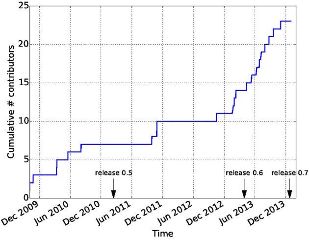
Diffusion Imaging in Python (Dipy) is a free and open source software project for the analysis of data from diffusion magnetic resonance imaging (dMRI) experiments. dMRI is an application of MRI that can be used to measure structural features of brain white matter. Many methods have been developed to use dMRI data to model the local configuration of white matter nerve fiber bundles and infer the trajectory of bundles connecting different parts of the brain. Dipy gathers implementations of many different methods in dMRI, including: diffusion signal pre-processing; reconstruction of diffusion distributions in individual voxels; fiber tractography and fiber track post-processing, analysis and visualization. Dipy aims to provide transparent implementations for all the different steps of dMRI analysis with a uniform programming interface. We have implemented classical signal reconstruction techniques, such as the diffusion tensor model and deterministic fiber tractography. In addition, cutting edge novel reconstruction techniques are implemented, such as constrained spherical deconvolution and diffusion spectrum imaging (DSI) with deconvolution, as well as methods for probabilistic tracking and original methods for tractography clustering. Many additional utility functions are provided to calculate various statistics, informative visualizations, as well as file-handling routines to assist in the development and use of novel techniques. In contrast to many other scientific software projects, Dipy is not being developed by a single research group. Rather, it is an open project that encourages contributions from any scientist/developer through GitHub and open discussions on the project mailing list. Consequently, Dipy today has an international team of contributors, spanning seven different academic institutions in five countries and three continents, which is still growing.
Overview
The claims and statistics of this paper haven’t been checked yet.
Run the check — free, no account needed. It reads the open-access text and checks the sources it cites.
Integrity signals
Registry
Cites
Statistics
Figures
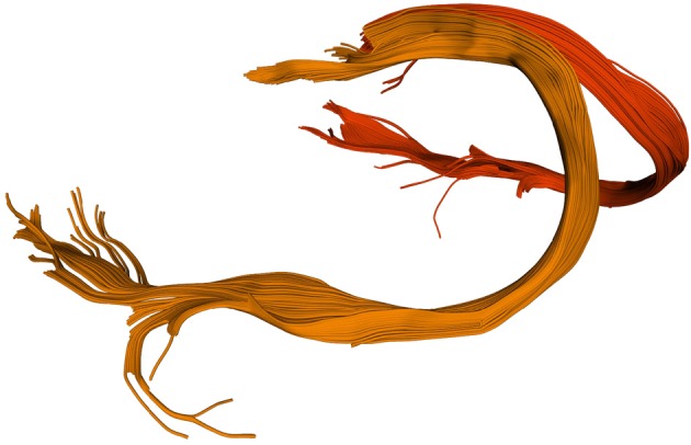
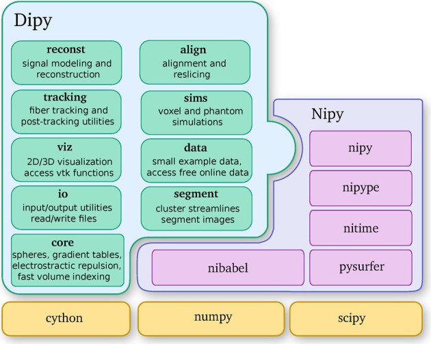
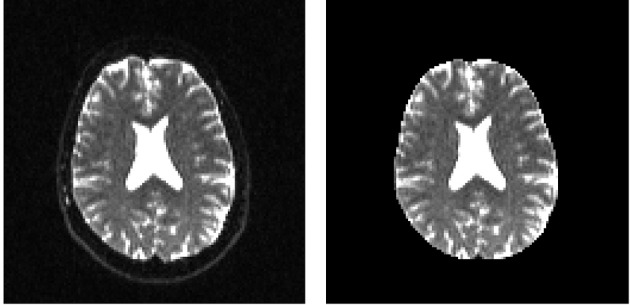
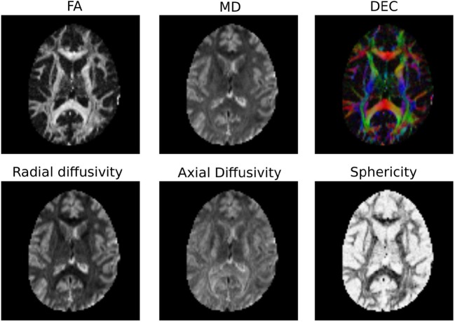
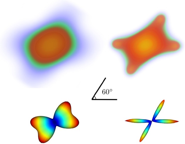
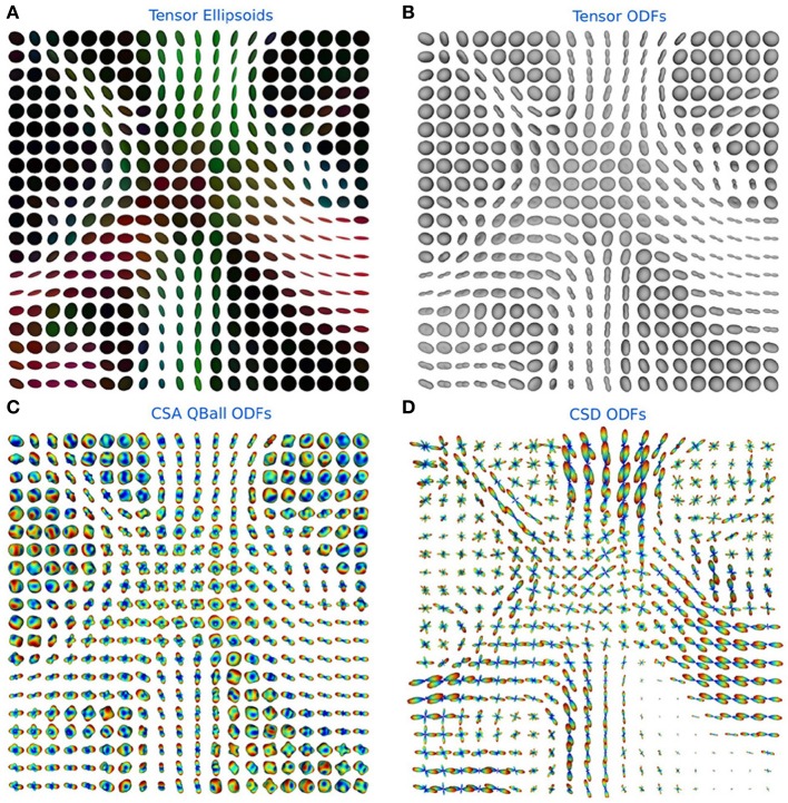
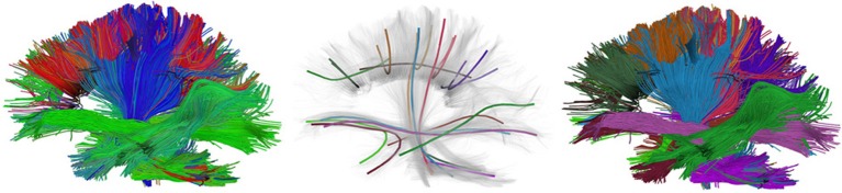
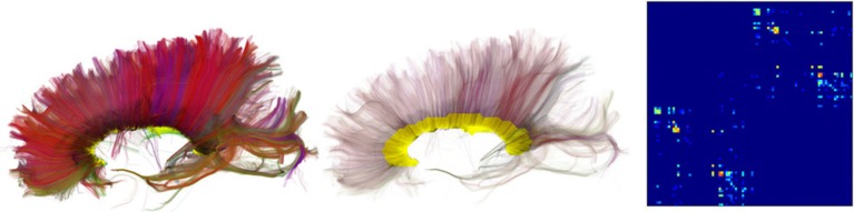
Verification
Claims the paper makes
The claims and statistics of this paper haven’t been checked yet.
Run the check — free, no account needed. It reads the open-access text and checks the sources it cites.
Claims other papers make about this one
Each is checked against this paper’s own text: does it say what the citing paper says it does?
The cited source says what the claim attributes to it.
In the citing paper
“In contrast to projects such as DiPy (Garyfallidis et al., 2014) , this library does not provide functionality to manipulate raw image data within Python itself, but provides functionality to invoke and manage external high-performance executables to perform the actual computations.”
From MRtrix3: A fast, flexible and open software framework for medical image processing and visualisation (2019) · cites this paper as Garyfallidis et al., 2014
Self-check
Percentages, sums and reported differences re-computed from the paper’s own numbers.
The self-check needs the paper’s full text.
Citations
Cites
A Threshold Selection Method from Gray-Level Histograms
Otsu, N. (1979) · IEEE Transactions on Systems, Man and Cybernetics · doi:10.1109/tsmc.1979.4310076 · Cited by 44,708 · cited 1×
“…median_otsu (see Figure 3 ). from dipy.segment.mask import median_otsu mask, S0_mask = median_otsu(data [:, :, :, 0]) median_otsu uses first a median filter to smooth the S0 and then Otsu’s method, an automated histogram method (Otsu, 1979) to separate the brain (foreground) from its background.”
MethodAdvances in functional and structural MR image analysis and implementation as FSL
Smith, S. M., Jenkinson, M., Woolrich, M. W., Beckmann, C. F., Behrens, T. E. J., Johansen-Berg, H., Bannister, P. R., De Luca, M., Drobnjak, I., Flitney, D. E., et al. (2004) · NeuroImage · doi:10.1016/j.neuroimage.2004.07.051 · Cited by 13,630
Fischl, B. (2012) · NeuroImage · doi:10.1016/j.neuroimage.2012.01.021 · Cited by 9,526 · cited 4×
“…al., 2006), AFNI (Cox, 2012), MITK (Fritzsche et al., 2012), BrainVoyager QX (Goebel, 2012), DTI-Query/Quench (Sherbondy et al., 2005), FreeSurfer (Fischl, 2012), FSL-FDT (Smith et al., 2004), MedInria (Toussaint et al., 2007), MRtrix (Tournier et al., 2012), Diffusion Toolkit/Trackvis (Wang et…”
MethodInfluentialThe NumPy Array: A Structure for Efficient Numerical Computation
van der Walt, S., Colbert, S. C., & Varoquaux, G. (2011) · Computing in Science & Engineering · doi:10.1109/mcse.2011.37 · Cited by 9,381
MR diffusion tensor spectroscopy and imaging
Basser, P. J., Mattiello, J., & LeBihan, D. (1994) · Biophysical Journal · doi:10.1016/s0006-3495(94)80775-1 · Cited by 5,970 · cited 2×
“Another alternative is to make assumptions about the distribution of the PDF such as a Gaussian assumption, which leads to the popular diffusion tensor imaging (DTI) technique (Basser et al., 1994).”
BackgroundMethodThe WU-Minn Human Connectome Project: An overview
Van Essen, D. C., Smith, S. M., Barch, D. M., Behrens, T. E. J., Yacoub, E., & Ugurbil, K. (2013) · NeuroImage · doi:10.1016/j.neuroimage.2013.05.041 · Cited by 5,651
The basis of anisotropic water diffusion in the nervous system—A technical review.
Beaulieu, C. (2002) · NMR in Biomedicine · doi:10.1002/nbm.782 · Cited by 4,628
Basser, P. J., & Pierpaoli, C. (1996) · Journal of Magnetic Resonance, Series B · doi:10.1006/jmrb.1996.0086 · Cited by 4,306 · cited 3×
“…of the myelin wrapping the axons in the voxel may affect both MD and FA, changes in other factors, such as the distribution of directions of crossing of fiber populations through the voxel may also affect these measures (Basser and Pierpaoli, 1996; Jones et al., 2013; Wandell and Yeatman, 2013).”
BackgroundMethodMedical image analysis: progress over two decades and the challenges ahead
Duncan, J. S., & Ayache, N. (2000) · IEEE Transactions on Pattern Analysis and Machine Intelligence · doi:10.1109/34.824822 · Cited by 4,167
IPython: A System for Interactive Scientific Computing
Pérez, F., & Granger, B. (2007) · Computing in Science & Engineering · doi:10.1109/mcse.2007.53 · Cited by 4,129 · cited 1×
“There has been large growth in the number of Python users, there are many Python tools for scientific computing (Pérez and Granger, 2007; Pérez et al., 2011; McKinney, 2012), and there are complementary neuroimaging packages in Python1.”
Background
Research works printed but not linked: 9
- Chamberland, 2013, Explore the brain white matter networks in real-time: multi-sticks fiber tracking
- Garyfallidis, 2012, Towards an Accurate Brain Tractography
- Garyfallidis, 2011, Dipy - a novel software library for diffusion MR and tractography
- LeBihan, 1985, Imagerie de diffusion in vivo par résonance magnétique nucléaire, C. R. Acad. Sci. Paris, 301, 1109
- Pajevic, 1999, Color schemes to represent the orientation of anisotropic tissues from diffusion tensor data: application to white matter fiber tract mapping in the human brain, Magn. Reson. Med, 42, 526, 10.1002/(SICI)1522-2594(199909)42:3526::AID-MRM153.3.CO;2-A · DOI
- Schroeder, 2001, The Visualization Toolkit-User's Guide
- Vaillancourt, 2011, A fiber navigator for neurosurgical planning
- Wang, 2007, Diffusion toolkit: a software package for diffusion imaging data processing and tractography, Proc. Int. Soc. Magn. Reson. Med, 15, 3720
- Westin, 1997, Geometrical diffusion measures for MRI from tensor basis analysis, Proc. ISMRM, 97, 1742
Cited by
Sorted by citations
Items 401–500 of 1,000
Tian, Q., Yang, G., Leuze, C. W., Rokem, A., Edlow, B. L., & McNab, J. A. (2019) · NeuroImage · doi:10.1016/j.neuroimage.2019.01.038 · Cited by 15
“Simulations were performed with a multi-tensor model using the “multi_tensor” function of the Diffusion Imaging in Python (DIPY) software (Garyfallidis et al., 2014) (http://nipy.”
Self-citationOptimization of q-space sampling for mean apparent propagator MRI metrics using a genetic algorithm
Olson, D. V., Arpinar, V. E., & Muftuler, L. T. (2019) · NeuroImage · doi:10.1016/j.neuroimage.2019.05.078 · Cited by 15
Han, X., Maharjan, S., Chen, J., Zhao, Y., Qi, Y., White, L. E., Johnson, G. A., & Wang, N. (2024) · NeuroImage · doi:10.1016/j.neuroimage.2024.120734 · Cited by 15
“Diffusion Imaging in Python (Dipy) (Garyfallidis et al., 2014) was used to compute DKI metrics.”
Chen, Y., Zhang, F., Zhang, C., Xue, T., Zekelman, L., He, J., Song, Y., Makris, N., Yogesh Rathi, A., Golby, A. J., et al. (2022) · Lecture notes in computer science · doi:10.1007/978-3-031-16431-6_17 · Cited by 14
Wang, X., Wang, Y., Gao, D., Zhao, Z., Wang, H., Wang, S., & Liu, S. (2023) · Japanese Journal of Radiology · doi:10.1007/s11604-023-01419-w · Cited by 14
Ghazi, N., Aarabi, M. H., & Hamid Soltanian-zadeh, A. (2023) · Neuroinformatics · doi:10.1007/s12021-023-09636-4 · Cited by 16
“PDD images were computed using Dipy (Garyfallidis et al., 2014) and subsequently re-sliced to match with T1w images.”
MethodCo-evolving dynamics and topology in a coupled oscillator model of resting brain function
Pope, M., Seguin, C., Varley, T. F., Faskowitz, J., & Sporns, O. (2023) · NeuroImage · doi:10.1016/j.neuroimage.2023.120266 · Cited by 14
Subject-Specific Automatic Reconstruction of White Matter Tracts
Meesters, S. P. L., Landers, M., Rutten, G., & Florack, L. (2023) · Journal of digital imaging · doi:10.1007/s10278-023-00883-0 · Cited by 11
“To produce white matter tracks that are more robust under the stochastic realizations of probabilistic tractography, we opted for a filtering based on the Fiber-To-Bundle Coher-ence (FBC) measures that are publicly available in the Diffusion Imaging in Python (DIPY) package [47, 48].”
MethodBaraković, M., Pizzolato, M., Tax, C. M. W., Rudrapatna, U. S., Magon, S., Dyrby, T. B., Granziera, C., Thiran, J., Jones, D. K., & Canales-Rodríguez, E. J. (2023) · Frontiers in Neuroscience · doi:10.3389/fnins.2023.1209521 · Cited by 17
“…method (Veraart et al., 2016) by using the matrix centering and patch-based aggregation options (Manjon et al., 2013), as implemented in dipy (Garyfallidis et al., 2014) 1 ; (2) attenuation of the Rician-noise dependent bias in the signal by implementing the postprocessing correction scheme…”
MethodThe representational geometry of emotional states in basolateral amygdala
O’Neill, P.-K., Posani, L., Mészáros, J., Warren, P., Schoonover, C. E., Fink, A. J. P., Fusi, S., & Salzman, C. D. (2026) · Nature Neuroscience · doi:10.1038/s41593-026-02315-y · Cited by 17
Ragone, E., Tanner, J., Jo, Y., Esfahlani, F. Z., Faskowitz, J., Pope, M., Coletta, L., Gozzi, A., & Betzel, R. F. (2024) · Communications Biology · doi:10.1038/s42003-024-05766-w · Cited by 16
Won, J., Maillard, P., Shan, K., Ashley, J., Cardim, D., Zhu, D. C., & Zhang, R. (2024) · Hypertension · doi:10.1161/hypertensionaha.123.22337 · Cited by 15
“28,29 Briefly, FW (ie, the amount of water molecules that are not restricted by their surroundings) was estimated by an isotropic tensor, and a voxel-wise FW map was calculated for each participant.”
MethodWhite Matter Microstructural Underpinnings of Mild Behavioral Impairment in Parkinson's Disease
Monchi, O., Pinilla-Monsalve, G. D., Almgren, H., Ghahremani, M., Kibreab, M., Maarouf, N., Kathol, I., Boré, A., Rheault, F., Descoteaux, M., et al. (2024) · Movement Disorders · doi:10.1002/mds.29804 · Cited by 14
Self-citationFeng, Y., Chandio, B. Q., Villalón‐Reina, J. E., Thomopoulos, S. I., Nir, T. M., Benavidez, S. M., Laltoo, E., Chattopadhyay, T., Joshi, H., Venkatasubramanian, G., et al. (2024) · Alzheimer s & Dementia · doi:10.1002/alz.14371 · Cited by 16
Real‐Time Tractography‐Assisted Neuronavigation for Transcranial Magnetic Stimulation
Aydogan, D. B., Souza, V. H., Matsuda, R. H., Lioumis, P., & Ilmoniemi, R. J. (2024) · Human Brain Mapping · doi:10.1002/hbm.70122 · Cited by 20
“dMRI was preprocessed using Marcenko-Pastur PCA denoising algorithm (Veraart et al. 2016; Garyfallidis et al. 2014), and corrected for eddy-current and motion artifacts (Andersson and Sotiropoulos 2016).”
Subject–Motion Correction in HARDI Acquisitions: Choices and Consequences
Elhabian, S., Gur, Y., Vachet, C., Piven, J., Styner, M. A., Leppert, I. R., Pike, G. B., & Gerig, G. (2014) · Frontiers in Neurology · doi:10.3389/fneur.2014.00240 · Cited by 14
“We employed the constrained spherical deconvolution (CSD) technique (57) to reconstruct fiber orientation distributions functions (fODFs) from the DWI data using the diffusion imaging Python (DiPy) library (58).”
MethodWhite Matter Tract Segmentation as Multiple Linear Assignment Problems
Sharmin, N., Olivetti, E., & Avesani, P. (2018) · Frontiers in Neuroscience · doi:10.3389/fnins.2017.00754 · Cited by 14
“The tractograms were obtained with the Euler Delta Crossing (EuDX) algorithm, see (Garyfallidis et al., 2014), implemented in DiPy 6 , with 10 6 seeds.”
MethodDiffusion Tensor Imaging in Contact and Non-Contact University-Level Sport Athletes
Lefebvre, G., Guay, S., Chamard, É., Theaud, G., de Guise, É., Bacon, B. A., Descoteaux, M., Beaumont, L. D., & Théoret, H. (2020) · Journal of Neurotrauma · doi:10.1089/neu.2020.7170 · Cited by 12
Self-citationEnciso-Olivera, C. O., Ordóñez-Rubiano, E. G., Libreros, R. C., Rivera, D., Zárate-Ardila, C., Rudas, J., Pulido, C., Gómez, F. A., Martínez, D., Guerrero, N., et al. (2021) · Scientific Reports · doi:10.1038/s41598-021-98506-7 · Cited by 14
Sleep and White Matter in Adults with Down Syndrome
Fleming, V., Piro‐Gambetti, B., Bazydlo, A., Zammit, M. D., Alexander, A. L., Christian, B. T., Handen, B. L., Plante, D. T., & Hartley, S. L. (2021) · Brain Sciences · doi:10.3390/brainsci11101322 · Cited by 12
Surówka, A. D., Czyżycki, M., Ziomber-Lisiak, A., Migliori, A., & Szczerbowska‐Boruchowska, M. (2021) · Ultramicroscopy · doi:10.1016/j.ultramic.2021.113408 · Cited by 14
“affine transform solutions, be it the DIPY package as an excellent option to be considered [51].”
BackgroundBecker, D., Neher, P., Jungk, C., Jesser, J., Pflüger, I., Brinster, R., Bendszus, M., Bruckner, T. M., Maier-Hein, K. H., Scherer, M., et al. (2021) · World Neurosurgery · doi:10.1016/j.wneu.2021.11.006 · Cited by 14
Intracranial Electrode Location and Analysis in MNE-Python
Rockhill, A. P., Larson, E., Stedelin, B., Mantovani, A., Raslan, A. M. T., Gramfort, A., & Swann, N. C. (2022) · The Journal of Open Source Software · doi:10.21105/joss.03897 · Cited by 14
“In MNE-Python, these are aligned using a mutual information histogrammatching approach using DIPY (Garyfallidis et al., 2014) that generally works without manual pre-alignment using the functions mne.transforms.compute_volume_registration and mne.transforms.apply_volume_registration.”
MethodInfluentialCarlos, A. F., Tosakulwong, N., Weigand, S. D., Buciuc, M., Ali, F., Clark, H. M., Botha, H., Utianski, R. L., Machulda, M. M., Schwarz, C. G., et al. (2022) · Brain Communications · doi:10.1093/braincomms/fcac108 · Cited by 14
“Diffusion tensors were estimated using non-linear least squares fitting and used to calculate fractional anisotropy (FA) and mean diffusivity (MD) images in dipy.(42)”
MethodLiu, X.-F., Prince, J. L., Xing, F.-X., Zhuo, J., Reese, T., Stone, M., Fakhri, G. E., & Woo, J. (2023) · Medical Image Anal. · doi:10.1016/j.media.2023.102851 · Cited by 15
Optimal DSI reconstruction parameter recommendations: Better ODFs and better connectivity
Paquette, M., Gilbert, G., & Descoteaux, M. (2016) · NeuroImage · doi:10.1016/j.neuroimage.2016.07.048 · Cited by 13
Self-citationVertex nomination: The canonical sampling and the extended spectral nomination schemes
Yoder, J., Chen, L., Pao, H., Bridgeford, E., Levin, K., Fishkind, D. E., Priebe, C. E., & Lyzinski, V. (2020) · Computational Statistics & Data Analysis · doi:10.1016/j.csda.2020.106916 · Cited by 14
“A deterministic tractography algorithm was applied using DiPy’s EuDX [19, 18] to obtain a fiber streamline from each voxel.”
MethodDiffusion MRI Spatial Super-Resolution Using Generative Adversarial Networks
Albay, E., Demir, U., & Ünal, G. (2018) · Lecture notes in computer science · doi:10.1007/978-3-030-00320-3_19 · Cited by 14
Cortical Surfaces Integration with Tractography for Structural Connectivity Analysis
St‐Onge, E., Al‐Sharif, N., Girard, G., Theaud, G., & Descoteaux, M. (2021) · Brain Connectivity · doi:10.1089/brain.2020.0930 · Cited by 15
“For all comparisons, streamlines were re-constructed inside the WM using PFT (Girard et al., 2014) available from DIPY (Garyfallidis et al., 2014).”
Self-citationBeaudoin, A., Rheault, F., Theaud, G., Laberge, F., Whittingstall, K., Lamontagne, A., & Descoteaux, M. (2021) · Frontiers in Neuroscience · doi:10.3389/fnins.2021.665017 · Cited by 18
Self-citationFast Streamline Search: An Exact Technique for Diffusion MRI Tractography
St‐Onge, E., Garyfallidis, E., & Collins, D. L. (2022) · Neuroinformatics · doi:10.1007/s12021-022-09590-7 · Cited by 13
“Tractography streamlines were reconstructed using probabilistic particle filtering tractography (Girard et al., 2014) implemented in Dipy (Garyfallidis et al., 2014).”
BackgroundMethodInfluentialSelf-citationA novel framework for in-vivo diffusion tensor distribution MRI of the human brain
Magdoom, K. N., Avram, A. V., Sarlls, J. E., Gasbarra, D., & Basser, P. J. (2023) · NeuroImage · doi:10.1016/j.neuroimage.2023.120003 · Cited by 13
Chen, Y., Zekelman, L., Zhang, C., Xue, T., Song, Y., Makris, N., Yogesh Rathi, A., Golby, A. J., Cai, W., Zhang, F., et al. (2024) · Medical Image Analysis · doi:10.1016/j.media.2024.103120 · Cited by 13
Cai, K., Tang, S., Qian, S., Shen, Z., & Yang, Y. (2024) · Chinese Journal of Aeronautics · doi:10.1016/j.cja.2024.03.003 · Cited by 13
Dhiman, S., Hickey, R. E., Thorn, K. E., Moss, H. G., McKinnon, E. T., Adisetiyo, V., Ades‐Aron, B., Jensen, J. H., & Benitez, A. M. (2024) · Journal of Visualized Experiments · doi:10.3791/66397 · Cited by 13
Pappas, C. A., Bauer, C. E., Zachariou, V., Maillard, P., Caprihan, A., Shao, X., Wang, D. J. J., & Gold, B. T. (2024) · Imaging Neuroscience · doi:10.1162/imag_a_00183 · Cited by 13
“…algorithm ( Bauer et al., 2022; Henriques et al., 2017) available through the Diffusion Imaging in Python (DIPY) open-source software library ( Garyfallidis et al., 2014) was used to calculate the fractional volume of FW per voxel in the motion-corrected diffusion data, resulting in FW…”
MethodTractometry of the Human Connectome Project: resources and insights
Kruper, J., Hagen, M. P., Rheault, F., Crane, I., Gilmore, A., Narayan, M., Motwani, K., Lila, E., Rorden, C., Yeatman, J. D., et al. (2024) · Frontiers in Neuroscience · doi:10.3389/fnins.2024.1389680 · Cited by 12
Self-citationNOSA, an Analytical Toolbox for Multicellular Optical Electrophysiology
Oltmanns, S., Abben, F. S., Ender, A., Aimon, S., Kovacs, R., Sigrist, S. J., Storace, D. A., Geiger, J. R. P., & Raccuglia, D. (2020) · Frontiers in Neuroscience · doi:10.3389/fnins.2020.00712 · Cited by 13
Guo, F., Tax, C. M. W., Luca, A. D., Viergever, M. A., Heemskerk, A. M., & Leemans, A. (2021) · Journal of Neuroimaging · doi:10.1111/jon.12901 · Cited by 11
Kerkelä, L., Nery, F., Callaghan, R., Zhou, F., Gyori, N. G., Szczepankiewicz, F., Palombo, M., Parker, G. J. M., Zhang, H. G., Hall, M. G., et al. (2021) · NeuroImage · doi:10.1016/j.neuroimage.2021.118445 · Cited by 13
To, M., Sarno, I. G., Chong, C., Jenkinson, M., & Carneiro, G. (2021) · Lecture notes in computer science · doi:10.1007/978-3-030-87234-2_63 · Cited by 13
Pedersen, W. S., Dean, D., Adluru, N., Gresham, L. K., Lee, S. D., Kelly, M. P., Mumford, J. A., Davidson, R. J., & Schaefer, S. M. (2021) · Emotion · doi:10.1037/emo0000996 · Cited by 12
“Weighted least squares was used to fit the tensors at each voxel using the Diffusion Imaging In Python package (v. .14.0; Garyfallidis et al., 2014).”
MethodInfluentialDiPiero, M. A., Surgent, O. J., Travers, B. G., Alexander, A. L., Lainhart, J. E., & Dean, D. (2022) · NeuroImage Clinical · doi:10.1016/j.nicl.2022.103306 · Cited by 13
“Diffusion tensors were estimated at each voxel using a weighted-least squares algorithm as part of the diffusion imaging in python (DIPY) opensource software package(65).”
Optic radiations representing different eccentricities age differently
Kruper, J., Benson, N. C., Caffarra, S., Owen, J. P., Wu, Y., Lee, A., Lee, C. S., Yeatman, J. D., & Rokem, A. (2023) · Human Brain Mapping · doi:10.1002/hbm.26267 · Cited by 11
“The dMRI signal in each voxel was modeled using the diffusional kurtosis model, implemented in DIPY (Garyfallidis et al., 2014; Henriques et al., 2021).”
MethodSelf-citationAssessing within‐subject rates of change of placental MRI diffusion metrics in normal pregnancy
Cromb, D., Slator, P. J., Fuente, M. D. L., Price, A. N., Rutherford, M., Egloff, A. M., Counsell, S. J., & Hutter, J. M. (2023) · Magnetic Resonance in Medicine · doi:10.1002/mrm.29665 · Cited by 13
“All subsequent analysis was performed on this masked and motion-corrected placental diffusion data using in-house python scripts and our extensions to the diffusion microstructure imaging in python library for diffusion models, 23,24 2).”
MethodQ‐space truncation and sampling in diffusion spectrum imaging
Tian, Q., Rokem, A., Folkerth, R. D., Nummenmaa, A. R., Fan, Q., Edlow, B. L., & McNab, J. A. (2016) · Magnetic Resonance in Medicine · doi:10.1002/mrm.26071 · Cited by 13
Self-citationCortical Thinning in Healthy Aging Correlates with Larger Motor-Evoked EEG Desynchronization
Provencher, D., Hennebelle, M., Cunnane, S. C., Bérubé-Lauzière, Y., & Whittingstall, K. (2016) · Frontiers in Aging Neuroscience · doi:10.3389/fnagi.2016.00063 · Cited by 16
Ioanas, H.-I., Saab, B. J., & Rudin, M. (2017) · Research Ideas and Outcomes · doi:10.3897/rio.3.e12095 · Cited by 13
“Neuroscientific software now usable under Gentoo includes but is not limited to: • Dipy (Garyfallidis et al. 2014) • FSL (Jenkinson et al. 2012) • Nipype (Gorgolewski et al. 2016) • Nilearn (Abraham et al. 2014) • PsychoPy (Peirce 2008) Herein we describe the implementation and current capabilities…”
MethodCloudknot: A Python Library to Run your Existing Code on AWS Batch
Richie-Halford, A., & Rokem, A. (2018) · Proceedings of the Python in Science Conferences · doi:10.25080/majora-4af1f417-001 · Cited by 10
Self-citationLefebvre, J., Delafontaine-Martel, P., Pouliot, P., Girouard, H., Descoteaux, M., & Lesage, F. (2018) · Neurophotonics · doi:10.1117/1.nph.5.4.045004 · Cited by 13
“First, inhouse implementations(38,39) of diffusion tensor imaging and HARDI reconstructions were performed using the Dipy library.(40) Fractional anisotropy (FA) was computed from the local diffusion tensors with a non-negative least square method.”
MethodSelf-citationTesting for Equivalence of Network Distribution Using Subgraph Counts
Maugis, P.-A., Olhede, S. C., Priebe, C., & Wolfe, P. (2020) · UCL Discovery (University College London) · Cited by 13
Gray and White Matter Correlates of Dysphagia in Progressive Supranuclear Palsy
Clark, H. M., Tosakulwong, N., Weigand, S. D., Ali, F., Botha, H., Pham, N. T. T., Schwarz, C. G., Reid, R. I., Senjem, M. L., Jack, C. R., et al. (2021) · Movement Disorders · doi:10.1002/mds.28731 · Cited by 13
“Diffusion tensors were estimated using nonlinear least squares fitting and used to calculate fractional anisotropy (FA) and mean diffusivity (MD) images in dipy.(29) The Johns Hopkins University “Eve” atlas was used to generate ROI-level data.”
MethodNeural dynamics and architecture of the heading direction circuit in a vertebrate brainPreprint
Petrucco, L., Lavian, H., Wu, Y. K., Svara, F., Štih, V., & Portugues, R. (2022) · bioRxiv (Cold Spring Harbor Laboratory) · doi:10.1101/2022.04.27.489672 · Cited by 13
Reduced myelin contributes to cognitive impairment in patients with monogenic small vessel disease
Denecke, J., Dewenter, A., Lee, J., Franzmeier, N., Valentim, C., Kopczak, A., Dichgans, M., Pirpamer, L., Gesierich, B. G., Duering, M., et al. (2025) · Alzheimer s & Dementia · doi:10.1002/alz.70127 · Cited by 13
Evaluating 35 Methods to Generate Structural Connectomes Using Pairwise Classification
Petrov, D., Ivanov, A., Faskowitz, J., Gutman, B. A., Moyer, D., Villalon, J. E., Jahanshad, N., & Thompson, P. M. (2017) · Lecture notes in computer science · doi:10.1007/978-3-319-66182-7_59 · Cited by 12
Weninger, L., Na, C.-H., Jütten, K., & Merhof, D. (2020) · PLoS ONE · doi:10.1371/journal.pone.0239475 · Cited by 12
Gothe, N. P., Erlenbach, E. D., García, V. M. G., Malani, R., Voss, S., Camacho, P., McAuley, E., Burd, N. A., Sutton, B. P., & Damoiseaux, J. S. (2023) · Contemporary Clinical Trials · doi:10.1016/j.cct.2023.107240 · Cited by 13
Mark, I. T., Wren-Jarvis, J., Xiao, J., Cai, L. T., Parekh, S. A., Bourla, I., Lazerwitz, M. C., Rowe, M. A., Marco, E. J., & Mukherjee, P. (2023) · Frontiers in Neuroscience · doi:10.3389/fnins.2023.1136424 · Cited by 12
“, 2016)] through the Dipy library (Garyfallidis et al., 2014).”
Bergamino, M., Keeling, E. G., McElvogue, M. M., Schaefer, S. Y., Burke, A. D., Prigatano, G. P., & Stokes, A. M. (2024) · Journal of Alzheimer s Disease · doi:10.3233/jad-230952 · Cited by 12
High‐resolution 3T to 7T ADC map synthesis with a hybrid CNN‐transformer model
Eidex, Z., Wang, J., Safari, M., Elder, E., Wynne, J. F., Wang, T., Shu, H. G., Mao, H. Z., & Yang, X. (2024) · Medical Physics · doi:10.1002/mp.17079 · Cited by 12
“15 Because the generation of ADC maps is computationally intensive and the skull produces a strong,unwanted signal in ADC maps,a brain mask is needed.”
InfluentialMoon, H. S., Mahzarnia, A., Stout, J. A., Anderson, R. J., Han, Z. Y., Tremblay, J. T., Badea, C. T., & Badea, A. (2024) · Imaging Neuroscience · doi:10.1162/imag_a_00245 · Cited by 14
“We calculated the diffusion-based microstructural parameters using MRtrix3 ( Tournier et al., 2019), and RTOP, RTAP, and RTPP using Dipy ( Garyfallidis et al., 2014).”
Individual Variability in the Structural Connectivity Architecture of the Human Brain
Huang, W., Chen, H., Liu, Z., Dong, X., Feng, G., Liu, G., Yang, A., Zhang, Z., Shmuel, A., Su, L., et al. (2024) · Journal of Neuroscience · doi:10.1523/jneurosci.2139-23.2024 · Cited by 16
Lifespan normative modeling of brain microstructure
Villalón‐Reina, J. E., Zhu, A. H., Nabulsi, L., Thomopoulos, S. I., Moreau, C. A., Feng, Y., Chattopadhyay, T., Benavidez, S. M., Kushan, L., John, J. P., et al. (2026) · Nature Communications · doi:10.1038/s41467-026-72875-x · Cited by 15
“DTI maps (FA, MD, RD, and AD) were computed with DiPy ’ s tensor fi tting function 72 for the following data-sets: AOMIC, CAMCAN, CHBMP, CHCP, HCP_A, HCP_D, HCP_YA, NIM-HANS, PNC, QTAB and UCLA.”
Training shortest-path tractography: Automatic learning of spatial priors
Kasenburg, N., Liptrot, M. G., Reislev, N. L., Ørting, S. N., Nielsen, M., Garde, E., & Feragen, A. (2016) · NeuroImage · doi:10.1016/j.neuroimage.2016.01.031 · Cited by 12
“, 2007) with order 8 was computed with the DiPy package (Garyfallidis et al., 2014); on the HCP DWIs using all three shells, and on the preprocessed DWIs from the standard dataset.”
MethodCieslak, M., Brennan, T., Meiring, W., Volz, L. J., Greene, C. A., Asturias, A., Suri, S., & Grafton, S. T. (2017) · NeuroImage · doi:10.1016/j.neuroimage.2017.12.039 · Cited by 12
“Effort is currently underway to also load results from reconstructing in DiPy (Garyfallidis et al., 2014) and MRTRIX (Tournier et al., 2010).”
BackgroundColl‐Font, J., Afacan, O., Hoge, W. S., Garg, H. K., Shashi, K., Marami, B., Gholipour, A., Chow, J. S., Warfield, S. K., & Kurugol, S. (2020) · Journal of Magnetic Resonance Imaging · doi:10.1002/jmri.27473 · Cited by 12
“The latter was done using in-house software for the IVIM fit with spatial regularization (C++) and the open-source software in DIPY for DTI.(30,31) The analysis of the results was done in Python and the statistical tests performed with the Wilcoxon test implementation in the SCIPY package (https://www.”
MethodEnsemble machine learning classification of daily living abilities among older people with HIV
Paul, R. H., Tsuei, T., Cho, K., Belden, A. C., Milanini, B., Bolzenius, J. D., Javandel, S., McBride, J. A., Cysique, L. A., Lesinski, S., et al. (2021) · EClinicalMedicine · doi:10.1016/j.eclinm.2021.100845 · Cited by 12
“Images were then corrected for eddy currents and fit to tensor eigenvalues using the Diffusion Imaging in Python Package [20].”
MethodTsuchida, A., Laurent, A., Crivello, F., Petit, L., Pepe, A., Beguedou, N., Debette, S., Tzourio, C., & Mazoyer, B. M. (2021) · Frontiers in Systems Neuroscience · doi:10.3389/fnsys.2021.692152 · Cited by 15
Grussu, F., Bernatowicz, K., Casanova‐Salas, I., Castro, N., Nuciforo, P. G., Mateo, J., Barba, I., & Pérez-López, R. (2022) · Magnetic Resonance in Medicine · doi:10.1002/mrm.29174 · Cited by 12
White matter tract strength correlates with therapy outcome in persistent developmental stuttering
Neef, N. E., Korzeczek, A., Primaßin, A., von Gudenberg, A. W., Dechent, P., Riedel, C. H., Paulus, W., & Sommer, M. (2022) · Human Brain Mapping · doi:10.1002/hbm.25853 · Cited by 12
“Wilcoxen test significant at ***p .001, **p = .01 diffusion kurtosis model, implemented in DIPY (https://dipy.org/; Garyfallidis et al., 2014), to the diffusion data separately for each session and projected the FA, MD, RD, and AD onto the segmented fiber tracts generated by pyAFQ.”
MethodRaikes, A. C., Hernandez, G. D., Mullins, V. A., Wang, Y., Lopez, C. M., Killgore, W. D. S., Chilton, F. H., & Brinton, R. D. (2022) · Frontiers in Neurology · doi:10.3389/fneur.2022.891531 · Cited by 12
“Details of the processing pipeline are described below and many internal operations of QSIPrep use Nilearn 0.6.2 (50) (RRID:SCR_001362) and Dipy (51).”
InfluentialArtificial intelligence-based locoregional markers of brain peritumoral microenvironment
Samani, Z. R., Parker, D., Akbari, H., Wolf, R. L., Brem, S. S., Bakas, S., & Verma, R. (2023) · Scientific Reports · doi:10.1038/s41598-022-26448-9 · Cited by 12
Wang, S., Peterson, D. J., Wang, Y., Wang, Q., Grabowski, T. J., Li, W., & Madhyastha, T. M. (2017) · Frontiers in Neurology · doi:10.3389/fneur.2017.00118 · Cited by 12
Brain status modeling with non-negative projective dictionary learning
Zhang, M., Desrosiers, C., Guo, Y., Khundrakpam, B. S., Al‐Sharif, N., Kiar, G., Valdes-Sosa, P. A., Poline, J., & Evans, A. C. (2019) · NeuroImage · doi:10.1016/j.neuroimage.2019.116226 · Cited by 12
“In the tensor estimation stage, the 6-component tensor model of the DIPY package (Garyfallidis et al., 2014) is then applied to get fractional anisotropy maps, which can reflect the structure and arrangement of the tensors.”
Steinhoff, M., Nehrke, K., Mertins, A., & Börnert, P. (2019) · NMR in Biomedicine · doi:10.1002/nbm.4185 · Cited by 12
PyQMRI: An accelerated Python based Quantitative MRI toolbox
Maier, O., Spann, S. M., Bödenler, M., & Stollberger, R. (2020) · The Journal of Open Source Software · doi:10.21105/joss.02727 · Cited by 12
“Another powerful Pyhton package, with a focus on neuroimaing, is DIPY (Garyfallidis et al., 2014), offering a multitude of ways to evaluated diffusion and perfusion MRI data.”
BackgroundFiberweb: Diffusion Visualization and Processing in the Browser
Ledoux, L.-P., Morency, F. C., Cousineau, M., Houde, J., Whittingstall, K., & Descoteaux, M. (2017) · Frontiers in Neuroinformatics · doi:10.3389/fninf.2017.00054 · Cited by 12
“fODFs were estimated with Dipy (Garyfallidis et al., 2014) using a maximal spherical harmonics order of 8.”
BackgroundMethodInfluentialSelf-citationApplying positivity constraints to q-space trajectory imaging: The QTI+ implementation
Boito, D., Herberthson, M., Haije, T. D., & Özarslan, E. (2022) · SoftwareX · doi:10.1016/j.softx.2022.101030 · Cited by 12
“The algorithm used for this purpose is a modified implementation of the median_otsu segmentation method from Dipy [19] • Computations are performed in parallel exploiting multicore processing, if available • 50 voxels are sent at a time to the SDP solver • All inserted diffusion volumes are used to…”
MethodNiPreps: enabling the division of labor in neuroimaging beyond fMRIPrepPreprint
Estéban, O., Wright, J., Markiewicz, C. J., Thompson, W. H., Goncalves, M., Ćirić, R., Blair, R. W., Feingold, F. W., Rokem, A., Ghosh, S., et al. (2019) · doi:10.31219/osf.io/ujxp6 · Cited by 12
Self-citationDeep Learning-Based Parameter Estimation for Neurophysiological Models of Neuroimaging DataPreprint
Griffiths, J. D., Wang, Z., Ather, S. H., Momi, D., Rich, S., Diaconescu, A. O., McIntosh, A. R., & Shen, K. (2022) · bioRxiv (Cold Spring Harbor Laboratory) · doi:10.1101/2022.05.19.492664 · Cited by 12
A software ecosystem for brain tractometry processing, analysis, and insight
Kruper, J., Richie-Halford, A., Qiao, J., Gilmore, A., Chang, K., Grotheer, M., Roy, E. A., Caffarra, S., Gomez, T., Chou, S., et al. (2025) · PLoS Computational Biology · doi:10.1371/journal.pcbi.1013323 · Cited by 13
“Tissue properties of the different tracts were characterized using the Diffusional Kurtosis Imaging model implemented in DIPY [15,32].”
Self-citationYang, C. W., Tian, S., Liu, M., Tan, F., Wang, W., Du, W., Gao, B., Song, Q., Liu, T., Zhang, H., et al. (2025) · Asian Journal of Psychiatry · doi:10.1016/j.ajp.2025.104667 · Cited by 11
Chen, Z., Bi, S., He, H., Qi, Z., Xu, X., Cui, B., Li, N., Hu, Y., Jin, Z., Yan, S., et al. (2025) · Neurobiology of Disease · doi:10.1016/j.nbd.2025.107064 · Cited by 11
Information theory for complex systems scientists: What, why, and how
Varley, T. F. (2025) · Physics Reports · doi:10.1016/j.physrep.2025.09.007 · Cited by 38
Raikes, A. C., Dyke, J. P., Nerattini, M., Boneu, C., Ajila, T., Fauci, F., Battista, M., Pahlajani, S. S., Williams, S., Brinton, R. D., et al. (2025) · Scientific Reports · doi:10.1038/s41598-025-24136-y · Cited by 12
“Many internal operations of QSIPrep use Nilearn 0.10.2 (RRID: SCR_001362) 87,88 and DIPY 89 .”
InfluentialFrontoparietal Anatomical Connectivity Predicts Second Language Learning Success
Sander, K., Barbeau, É. B., Chai, X. J., Kousaie, S., Petrides, M., Baum, S. R., & Klein, D. (2021) · Cerebral Cortex · doi:10.1093/cercor/bhab367 · Cited by 11
“The processing for the T1-weighted images in the pipeline consists of 8 steps, including denoising (Garyfallidis et al. 2014), N4 bias correction, brain mask extraction (Avants et al.”
MethodRegional Vulnerability of the Corpus Callosum in the Context of Cardiovascular Risk
VandeBunte, A. M., Fonseca, C. S., Paolillo, E. W., Gontrum, E. Q., Lee, S. Y., Kramer, J. H., & Casaletto, K. B. (2023) · Journal of Geriatric Psychiatry and Neurology · doi:10.1177/08919887231154931 · Cited by 11
Symptom-circuit mappings of the schizophrenia connectome
Wang, Y., Wang, J., Su, W., Hu, H., Xia, M., Zhang, T., Xu, L., Zhang, X., Taylor, H. M., Osipowicz, K. Z., et al. (2023) · Psychiatry Research · doi:10.1016/j.psychres.2023.115122 · Cited by 11
Lerma‐Usabiaga, G., Liu, M., Paz‐Alonso, P. M., & Wandell, B. A. (2023) · Scientific Reports · doi:10.1038/s41598-023-32924-7 · Cited by 11
Wu, Z., Hu, G., Cao, B., Liu, X., Zhang, Z., Dadario, N. B., Shi, Q., Fan, X., Tang, Y., Cheng, Z., et al. (2023) · Chinese Neurosurgical Journal · doi:10.1186/s41016-023-00325-4 · Cited by 11
Fislage, M., Winzeck, S., Woodrow, R., Lammers‐Lietz, F., Stamatakis, E. A., Correia, M., Preller, J., Feinkohl, I., Hendrikse, J., Pischon, T., et al. (2024) · Alzheimer s & Dementia · doi:10.1002/alz.13749 · Cited by 11
Pané, A., Videla, L., Calvet-Mirabent, À., Viaplana, J., Vaqué‐Alcázar, L., Ibarzábal, A., Aranha, M. R., Pegueroles, J., Moize, V. L., Vidal, J., et al. (2024) · Diabetes Care · doi:10.2337/dc23-2213 · Cited by 12
Berger, A., Cerra, M., Joris, V., Danthine, V., Macq, B. M. M., Dricot, L., Vandewalle, G., Delinte, N., & Tahry, R. E. (2024) · Neurotherapeutics · doi:10.1016/j.neurot.2024.e00422 · Cited by 11
“The preprocessing stepsincludedskullstripping(usingtheDiffusionImaginginPythonlibrary – DiPy, https://dipy.org/) [14], Rician denoising (Marchenko-Pastur Principal Component Analysis [15]), Eddy currents correction, susceptibility distortion correction and motion correction [16].”
Zimmermann, J., Reolon, B., Michels, L., Nemeth, B., Gorup, D., Barbagallo, M., Bellomo, J., van Niftrik, C. H. B., Sebök, M., Stumpo, V., et al. (2024) · Scientific Reports · doi:10.1038/s41598-024-81280-7 · Cited by 11
Karimi, F., Steiner, M., Newton, T., Lloyd, B., Cassara, A. M., de Fontenay, P., Farcito, S., Triebkorn, P., Beanato, E., Wang, H., et al. (2025) · Journal of Neural Engineering · doi:10.1088/1741-2552/adb88f · Cited by 13
“…inhomogeneous maps of anisotropic WM conductivity, which our pipeline generates in the following steps: First, diffusion tensor images (DTI) are reconstructed in the Python package DIPY [75, 76] using the pre-processed and registered DWI data, and saved in a Sim4Life-compatible format.”
Lavrova, A., Pham, N. T. T., Reid, R. I., Boeve, B. F., Knopman, D. S., Petersen, R. C., Nguyen, A. T., Reichard, R. R., Dickson, D. W., Jack, C. R., et al. (2025) · Neurobiology of Aging · doi:10.1016/j.neurobiolaging.2025.03.001 · Cited by 11
“Diffusion tensors were fit, and Fractional Anisotropy (FA) and Mean Diffusivity (MD) images were calculated using Dipy (Garyfallidis et al., 2014).”
Koppers, S., Friedrichs, M., & Merhof, D. (2017) · Mathematics and visualization · doi:10.1007/978-3-319-61358-1_17 · Cited by 11
Peripheral Innate Immune Activation Correlates With Disease Severity in GRN Haploinsufficiency
Ljubenkov, P. A., Miller, Z., Mumford, P., Zhang, J., Allen, I. E., Mitic, L. L., Staffaroni, A. M., Heuer, H. W., Rojas, J. C., Cobigo, Y., et al. (2019) · Frontiers in Neurology · doi:10.3389/fneur.2019.01004 · Cited by 10
“The Dipy nonlinear least-squares algorithm (22) was used to calculate diffusion tensors (DT), and a study specific template was created through iterative linear and non-linear registration of diffusion tensor images.”
MethodImaging correlates of visual function in multiple sclerosis
Caverzasi, E., Cordano, C., Zhu, A. H., Zhao, C., Bischof, A., Kirkish, G., Bennett, D. J., Devereux, M., Baker, N., Inman, J., et al. (2020) · PLoS ONE · doi:10.1371/journal.pone.0235615 · Cited by 11
“After correcting for distortions due to eddy current and head motion, maps of mean diffusivity (MD) were calculated by fitting the diffusion tensor model within each voxel, using dipy fit tensor [19].”
MethodFilipiak, P., Almairac, F., Papadopoulo, T., Fontaine, D., Mondot, L., Chanalet, S., Rachid Deriche, B., Clerc, M., & Wassermann, D. (2020) · NeuroImage · doi:10.1016/j.neuroimage.2020.117567 · Cited by 11
Metadata
- Authors
- Garyfallidis, E., Brett, M., Amirbekian, B., Rokem, A., van der Walt, S., Descoteaux, M., Nimmo-Smith, I., & Contributors, D.
- Affiliations
- Université de Sherbrooke (CA)
- University of California, Berkeley (US)
- University of California, San Francisco (US)
- Stanford University (US)
- Corresponding author
- Garyfallidis, Eleftherios
- Venue
- Frontiers in Neuroinformatics
- Publisher
- Frontiers Media SA
- Published
- 2014-02-21
- Volume
- 8
- Pages
- 8-8
- ISSN
- 1662-5196 (ISSN-L)
- PMC
- PMC3931231
- OpenAlex
- W2062791478
- Cited by
- 1,371
- References
- 98
- Citations by year
- 2026: 147 · 2025: 192 · 2024: 177 · 2023: 162 · 2022: 151 · 2021: 147 · 2020: 100 · 2019: 89 · 2018: 54 · 2017: 79 · 2016: 34 · 2015: 26 · 2014: 12
- FWCI
- 39.38
- Type
- journal article
- Language
- en
- Open access
- gold
- doi.org
- Licence
- cc-by
- Locations
- Frontiers in Neuroinformatics (publishedVersion, cc-by)
- Europe PMC (PubMed Central) (publishedVersion, cc-by)
- PubMed Central (publishedVersion, cc-by)
- PubMed (publishedVersion)
- DOAJ (DOAJ: Directory of Open Access Journals) (submittedVersion)
- University of Birmingham Research Portal (University of Birmingham) (submittedVersion, cc-by)
- Funders
- Fonds Québécois de la Recherche sur la Nature et les Technologies
- Natural Sciences and Engineering Research Council of Canada
- National Eye Institute (F32EY022294)
- Topics
- Health Sciences › Medicine › Radiology, Nuclear Medicine and Imaging › Advanced Neuroimaging Techniques and Applications
- Life Sciences › Neuroscience › Cognitive Neuroscience › Functional Brain Connectivity Studies
- Health Sciences › Medicine › Radiology, Nuclear Medicine and Imaging › Advanced MRI Techniques and Applications
- Keywords
- diffusion MRI, DIPY, white matter, fiber tractography, diffusion tensor model, constrained spherical deconvolution, diffusion spectrum imaging, probabilistic tracking, open source software, Python library
About these checks
Registry, retraction, Cites and statistics results are computed, not judged, with one exception: a printed reference that no index lists is read once by a model before it counts as a citation error. Claims are extracted by a model and kept only when their quote appears verbatim in the text; each cited source is then checked, with a model’s help, for whether it says what the claim relies on. Those verdicts can be wrong — read the claim’s quote and the cited source before relying on one.
Statistics and arithmetic are recomputed, not judged.
Confidence bands are shown only for fields calibrated on our annotated test set. None is calibrated yet, so no band is shown.
What counts as a claim ›How we measure confidence ›How we check this ›
Are you an author, or do you see a problem with this page? Request a review
