

The DOI matches its Crossref record; 98 of the 100 references it cites were found in a registry or an index, and 2 are not checked yet.
Dipy, a library for the analysis of diffusion MRI data
Open accessAbstract
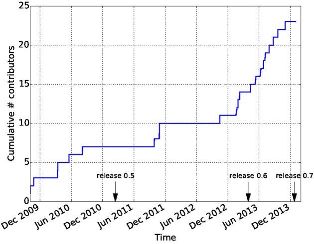
Diffusion Imaging in Python (Dipy) is a free and open source software project for the analysis of data from diffusion magnetic resonance imaging (dMRI) experiments. dMRI is an application of MRI that can be used to measure structural features of brain white matter. Many methods have been developed to use dMRI data to model the local configuration of white matter nerve fiber bundles and infer the trajectory of bundles connecting different parts of the brain. Dipy gathers implementations of many different methods in dMRI, including: diffusion signal pre-processing; reconstruction of diffusion distributions in individual voxels; fiber tractography and fiber track post-processing, analysis and visualization. Dipy aims to provide transparent implementations for all the different steps of dMRI analysis with a uniform programming interface. We have implemented classical signal reconstruction techniques, such as the diffusion tensor model and deterministic fiber tractography. In addition, cutting edge novel reconstruction techniques are implemented, such as constrained spherical deconvolution and diffusion spectrum imaging (DSI) with deconvolution, as well as methods for probabilistic tracking and original methods for tractography clustering. Many additional utility functions are provided to calculate various statistics, informative visualizations, as well as file-handling routines to assist in the development and use of novel techniques. In contrast to many other scientific software projects, Dipy is not being developed by a single research group. Rather, it is an open project that encourages contributions from any scientist/developer through GitHub and open discussions on the project mailing list. Consequently, Dipy today has an international team of contributors, spanning seven different academic institutions in five countries and three continents, which is still growing.
Overview
The claims and statistics of this paper haven’t been checked yet.
Run the check — free, no account needed. It reads the open-access text and checks the sources it cites.
Integrity signals
Registry
Cites
Statistics
Figures
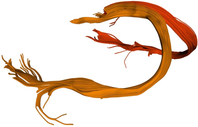
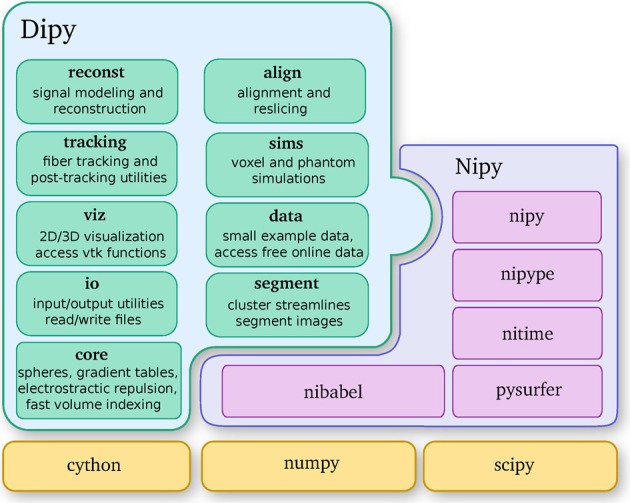
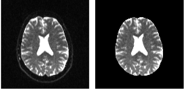
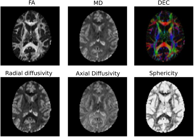
Verification
Claims the paper makes
The claims and statistics of this paper haven’t been checked yet.
Run the check — free, no account needed. It reads the open-access text and checks the sources it cites.
Claims other papers make about this one
Each is checked against this paper’s own text: does it say what the citing paper says it does?
The cited source says what the claim attributes to it.
In the citing paper
“In contrast to projects such as DiPy (Garyfallidis et al., 2014) , this library does not provide functionality to manipulate raw image data within Python itself, but provides functionality to invoke and manage external high-performance executables to perform the actual computations.”
From MRtrix3: A fast, flexible and open software framework for medical image processing and visualisation (2019) · cites this paper as Garyfallidis et al., 2014
Self-check
Percentages, sums and reported differences re-computed from the paper’s own numbers.
The self-check needs the paper’s full text.
Citations
Cites
A Threshold Selection Method from Gray-Level Histograms
Otsu, N. (1979) · IEEE Transactions on Systems, Man and Cybernetics · doi:10.1109/tsmc.1979.4310076 · Cited by 44,708 · cited 1×
“…median_otsu (see Figure 3 ). from dipy.segment.mask import median_otsu mask, S0_mask = median_otsu(data [:, :, :, 0]) median_otsu uses first a median filter to smooth the S0 and then Otsu’s method, an automated histogram method (Otsu, 1979) to separate the brain (foreground) from its background.”
MethodAdvances in functional and structural MR image analysis and implementation as FSL
Smith, S. M., Jenkinson, M., Woolrich, M. W., Beckmann, C. F., Behrens, T. E. J., Johansen-Berg, H., Bannister, P. R., De Luca, M., Drobnjak, I., Flitney, D. E., et al. (2004) · NeuroImage · doi:10.1016/j.neuroimage.2004.07.051 · Cited by 13,630
Fischl, B. (2012) · NeuroImage · doi:10.1016/j.neuroimage.2012.01.021 · Cited by 9,526 · cited 4×
“…al., 2006), AFNI (Cox, 2012), MITK (Fritzsche et al., 2012), BrainVoyager QX (Goebel, 2012), DTI-Query/Quench (Sherbondy et al., 2005), FreeSurfer (Fischl, 2012), FSL-FDT (Smith et al., 2004), MedInria (Toussaint et al., 2007), MRtrix (Tournier et al., 2012), Diffusion Toolkit/Trackvis (Wang et…”
MethodInfluentialThe NumPy Array: A Structure for Efficient Numerical Computation
van der Walt, S., Colbert, S. C., & Varoquaux, G. (2011) · Computing in Science & Engineering · doi:10.1109/mcse.2011.37 · Cited by 9,381
MR diffusion tensor spectroscopy and imaging
Basser, P. J., Mattiello, J., & LeBihan, D. (1994) · Biophysical Journal · doi:10.1016/s0006-3495(94)80775-1 · Cited by 5,970 · cited 2×
“Another alternative is to make assumptions about the distribution of the PDF such as a Gaussian assumption, which leads to the popular diffusion tensor imaging (DTI) technique (Basser et al., 1994).”
BackgroundMethodThe WU-Minn Human Connectome Project: An overview
Van Essen, D. C., Smith, S. M., Barch, D. M., Behrens, T. E. J., Yacoub, E., & Ugurbil, K. (2013) · NeuroImage · doi:10.1016/j.neuroimage.2013.05.041 · Cited by 5,651
The basis of anisotropic water diffusion in the nervous system—A technical review.
Beaulieu, C. (2002) · NMR in Biomedicine · doi:10.1002/nbm.782 · Cited by 4,628
Basser, P. J., & Pierpaoli, C. (1996) · Journal of Magnetic Resonance, Series B · doi:10.1006/jmrb.1996.0086 · Cited by 4,306 · cited 3×
“…of the myelin wrapping the axons in the voxel may affect both MD and FA, changes in other factors, such as the distribution of directions of crossing of fiber populations through the voxel may also affect these measures (Basser and Pierpaoli, 1996; Jones et al., 2013; Wandell and Yeatman, 2013).”
BackgroundMethodMedical image analysis: progress over two decades and the challenges ahead
Duncan, J. S., & Ayache, N. (2000) · IEEE Transactions on Pattern Analysis and Machine Intelligence · doi:10.1109/34.824822 · Cited by 4,167
IPython: A System for Interactive Scientific Computing
Pérez, F., & Granger, B. (2007) · Computing in Science & Engineering · doi:10.1109/mcse.2007.53 · Cited by 4,129 · cited 1×
“There has been large growth in the number of Python users, there are many Python tools for scientific computing (Pérez and Granger, 2007; Pérez et al., 2011; McKinney, 2012), and there are complementary neuroimaging packages in Python1.”
Background
Research works printed but not linked: 9
- Chamberland, 2013, Explore the brain white matter networks in real-time: multi-sticks fiber tracking
- Garyfallidis, 2012, Towards an Accurate Brain Tractography
- Garyfallidis, 2011, Dipy - a novel software library for diffusion MR and tractography
- LeBihan, 1985, Imagerie de diffusion in vivo par résonance magnétique nucléaire, C. R. Acad. Sci. Paris, 301, 1109
- Pajevic, 1999, Color schemes to represent the orientation of anisotropic tissues from diffusion tensor data: application to white matter fiber tract mapping in the human brain, Magn. Reson. Med, 42, 526, 10.1002/(SICI)1522-2594(199909)42:3526::AID-MRM153.3.CO;2-A · DOI
- Schroeder, 2001, The Visualization Toolkit-User's Guide
- Vaillancourt, 2011, A fiber navigator for neurosurgical planning
- Wang, 2007, Diffusion toolkit: a software package for diffusion imaging data processing and tractography, Proc. Int. Soc. Magn. Reson. Med, 15, 3720
- Westin, 1997, Geometrical diffusion measures for MRI from tensor basis analysis, Proc. ISMRM, 97, 1742
Cited by
Sorted by citations
Items 601–700 of 1,000
Fischer, J., Hancock, D. Y., Lowe, J. M., Turner, G., Snapp‐Childs, W., & Stewart, C. A. (2017) · ACM SIGUCCS Conference on User Services (SIGUCCS) · doi:10.1145/3123458.3123466 · Cited by 8
“Additional neuroscience-specific packages such as Dipy [11].”
Sun, J., Entezari, A., & Vemuri, B. C. (2019) · Medical Image Analysis · doi:10.1016/j.media.2019.02.014 · Cited by 9
Using the Wild Bootstrap to Quantify Uncertainty in Mean Apparent Propagator MRI
Gu, X., Eklund, A., Özarslan, E., & Knutsson, H. E. (2019) · Frontiers in Neuroinformatics · doi:10.3389/fninf.2019.00043 · Cited by 7
Discriminating VCID subgroups: A diffusion MRI multi-model fusion approach
Raja, R., Caprihan, A., Rosenberg, G., Rachakonda, S., & Calhoun, V. (2020) · Journal of Neuroscience Methods · doi:10.1016/j.jneumeth.2020.108598 · Cited by 6
“DTI and DKI model parameters were calculated using the DIPY software [49] resulting in 6 independent DTI tensor elements and 21 independent kurtosis parameters.”
MethodFree-Water Correction in Diffusion MRI: A Reliable and Robust Learning Approach
Weninger, L., Koppers, S., Na, C.-H., Jütten, K., & Merhof, D. (2020) · Mathematics and visualization · doi:10.1007/978-3-030-52893-5_8 · Cited by 8
Flow-based Generative Models for Learning Manifold to Manifold Mappings
Zhen, X., Chakraborty, R., Yang, L., & Singh, V. (2021) · Proceedings of the AAAI Conference on Artificial Intelligence · doi:10.1609/aaai.v35i12.17318 · Cited by 10
Li, Y., Yan, S., Zhang, G., Shen, N., Wu, D., Lu, J., Zhou, Y., Liu, Y., Zhu, H., Li, L., et al. (2021) · Journal of Magnetic Resonance Imaging · doi:10.1002/jmri.27911 · Cited by 8
Wang, Y., Deng, K., Sun, Y., Huang, X., Dai, Y., Chen, W., Hu, X., & Jiang, R. (2021) · Quantitative Imaging in Medicine and Surgery · doi:10.21037/qims-21-679 · Cited by 8
Axonal T2 estimation using the spherical variance of the strongly diffusion-weighted MRI signal
Pizzolato, M., Andersson, M., Canales-Rodríguez, E. J., Thiran, J., & Dyrby, T. B. (2021) · Magnetic Resonance Imaging · doi:10.1016/j.mri.2021.11.012 · Cited by 8
Elsheikh, S. S. M., Chimusa, E. R., Initiative, A. D. N., Mulder, N., & Crimi, A. (2021) · Frontiers in Human Neuroscience · doi:10.3389/fnhum.2021.761424 · Cited by 7
Connectomic disturbances underlying insomnia disorder and predictors of treatment response
Lu, Q., Zhang, W., Yan, H., Mansouri, N., Tanglay, O., Osipowicz, K. Z., Joyce, A. W., Young, I. M., Zhang, X., Doyen, S., et al. (2022) · Frontiers in Human Neuroscience · doi:10.3389/fnhum.2022.960350 · Cited by 9
“Diffusion weighted imaging (DWI) was processed using the Infinitome software (Omniscient Neurotechnology, 2020), which employs a standard processing steps in the Python language (Garyfallidis et al., 2014).”
MethodRegistration and quantification network (RQnet) for IVIM‐DKI analysis in MRI
Lee, W., Choi, G., Lee, J., & Park, H. (2022) · Magnetic Resonance in Medicine · doi:10.1002/mrm.29454 · Cited by 8
Early path dominance as a principle for neurodevelopment
Razban, R. M., Pachter, J. A., Dill, K. A., & Mujica-Parodi, L. R. (2023) · Proceedings of the National Academy of Sciences · doi:10.1073/pnas.2218007120 · Cited by 7
“Connectivity matrices are calculated using the Diffusion Imaging in Python (DIPY) software (54).”
MethodRebouças, S. C. L., Crivello, F., Tsuchida, A., Tzourio, C., Schweitzer, C., Korobelnik, J., Delcourt, C., & Helmer, C. (2023) · Human Brain Mapping · doi:10.1002/hbm.26412 · Cited by 7
“…DWI data and DTI fitting was performed using tools from the FMRIB software library (FSL) and the dipy package in Python (0.12.0, https://dipy.org) (Garyfallidis et al., 2014), and the AMICO (accelerated microstructure imaging via convex optimization) tool (Daducci et al., 2015) was used for NODDI…”
MethodInfluentialRobust Estimation of the Microstructure of the Early Developing Brain Using Deep Learning
Kebiri, H., Gholipour, A., Lin, R., Vasung, L., Karimi, D., & Cuadra, M. B. (2023) · Lecture notes in computer science · doi:10.1007/978-3-031-43990-2_28 · Cited by 8
Feng, Y., Chandio, B. Q., Thomopoulos, S. I., Chattopadhyay, T., & Thompson, P. M. (2023) · Annual International Conference of the IEEE Engineering in Medicine and Biology Society (EMBC) · doi:10.1109/embc40787.2023.10340009 · Cited by 8
“Auto-calibrated RecoBundles [8], [7] from DiPY [28] was used to extract 30 white matter tracts from all subjects in the MNI space.”
InfluentialTudela, R., Muñoz‐Moreno, E., López-Gil, X., & Sòria, G. (2017) · PLoS ONE · doi:10.1371/journal.pone.0170703 · Cited by 8
Bette, S. J., Barz, M., Nham, H., Huber, T. S., Berndt, M. T., Sales, A. H. A., Schmidt‐Graf, F., Meyer, H. S., Ryang, Y., Meyer, B., et al. (2020) · Cancers · doi:10.3390/cancers12040994 · Cited by 8
“[25], including affine registration of diffusion-weighted images to the b0 image and appropriate”
BackgroundWhite Matter Tract Segmentation with Self-supervised Learning
Lu, Q., Li, Y., & Ye, C. (2020) · Lecture notes in computer science · doi:10.1007/978-3-030-59728-3_27 · Cited by 8
Structural impacts on the timing and amplitude of the negative BOLD response
Provencher, D., Bizeau, A., Gilbert, G., Bérubé-Lauzière, Y., & Whittingstall, K. (2017) · Magnetic Resonance Imaging · doi:10.1016/j.mri.2017.09.007 · Cited by 7
“Preprocessing Slice-time correction, motion-correction, skull stripping, band-pass filtering and detrending were carried out in that order using AFNI [29] and non-local means denoising[30,31] applied in lieu of spatial smoothing using DIPY[32] on all images.”
MethodAn open, analysis-ready, and quality controlled resource for pediatric brain white-matter researchPreprint
Richie-Halford, A., Cieslak, M., Ai, L., Caffarra, S., Covitz, S. C., Franco, A. R., Karipidis, I. I., Kruper, J., Milham, M. P., Avelar‐Pereira, B., et al. (2022) · bioRxiv (Cold Spring Harbor Laboratory) · doi:10.1101/2022.02.24.481303 · Cited by 8
Self-citationPresurgical Executive Functioning in Low-Grade Glioma Patients Cannot Be Topographically Mapped
Landers, M. J. F., Smolders, L., Rutten, G.-J. M., Sitskoorn, M. M., Mandonnet, E., & Baene, W. D. (2023) · Cancers · doi:10.3390/cancers15030807 · Cited by 7
Brain signaling becomes less integrated and more segregated with age
Razban, R. M., Antal, B. B., Dill, K. A., & Mujica-Parodi, L. R. (2024) · Network Neuroscience · doi:10.1162/netn_a_00389 · Cited by 8
“For Figure S16, we also do reconstruction using diffusion tensor imaging (Garyfallidis et al., 2014 Zhang, Y., Wang, Y., Chen, N., Guo, M., Wang, X., Chen, G., ... others (2021).”
Mateu-Bosch, A., Segur‐Bailach, E., Muñoz‐Moreno, E., Barallobre, M. J., Arbonés, M. L., Gea‐Sorlí, S., Tort, F., Ribes, A., García‐Villoria, J., & Fillat, C. (2024) · Molecular Therapy — Methods & Clinical Development · doi:10.1016/j.omtm.2024.101276 · Cited by 7
“42 Processing includes eddy current correction, denoising and unbias, registration to T2 to correct for EPI distortion and fi tting of diffusion tensor (DT) model.”
Dissociation of White Matter Bundles in Different Recovery Measures in Poststroke Aphasia
García, A. O., Brambati, S. M., Brisebois, A., Houzé, B., Bedetti, C., Désautels, A., & Marcotte, K. (2024) · Stroke · doi:10.1161/strokeaha.124.047229 · Cited by 7
“35 We then performed diffusion tensor metric extraction using DIPY (Diffusion Imaging in Python), 36 and T1-weighted image registration using nonlinear symmetric normalization (SyN) through Advanced Normalization Tools (ANTs).”
Free Water MRI of White Matter in Wilson's Disease
Jing, X., Li, G., Wu, Y., Yuan, X., Yang, H., Chen, J., Wang, S., Wang, X., & Li, J. (2024) · Journal of Magnetic Resonance Imaging · doi:10.1002/jmri.29657 · Cited by 7
Longitudinal reproducibility of brain and spinal cord quantitative MRI biomarkers
Boudreau, M., Karakuzu, A., Boré, A., Pinsard, B., Zelenkovski, K., Alonso‐Ortiz, E., Boyle, J., Bellec, P., & Cohen‐Adad, J. (2024) · Imaging Neuroscience · doi:10.1162/imag_a_00409 · Cited by 7
“DTI metrics were computed using sct_compute_dti (powered by DIPY ( Garyfallidis et al., 2014)).”
InfluentialThe relationship of white matter tract orientation to vascular geometry in the human brain
Schilling, K. G., Newton, A. T., Tax, C. M. W., Chamberland, M., Remedios, S. W., Gao, Y., Li, M., Chang, C., Rheault, F., Sepherband, F., et al. (2025) · Scientific Reports · doi:10.1038/s41598-025-99724-z · Cited by 7
“Briefly, images were first filtered using the adaptive non-local mean filtering technique 32 (implemented in Dipy software 33 , with patch_radius = 1 voxel, block_radius = 5 voxels) to remove high frequency noise while preserving vascular signal intensities.”
Pillai, J. J., Reid, R. I., Weigand, S. D., Cogswell, P. M., Vemuri, P., Machulda, M. M., Knopman, D. S., Graff‐Radford, J., Petersen, R. C., & Jack, C. R. (2025) · Alzheimer s & Dementia · doi:10.1002/alz.70384 · Cited by 7
A Preliminary Study of MRI-derived Glymphatic System Indices in Early-Stage Normal Aging
Huang, M., Lyu, X., Wu, P., & Gao, B. (2025) · Academic Radiology · doi:10.1016/j.acra.2025.07.066 · Cited by 8
Lucchetti, F., Céléreau, E., Steullet, P., Alemán‐Gómez, Y., Hagmann, P., Klauser, A., & Klauser, P. (2025) · Nature Communications · doi:10.1038/s41467-025-66124-w · Cited by 7
“Brain visualizations were generated programmatically in Python using Nilearn 110 and FURY/DIPY 111 ; backgrounds were the MNI ICBM152 2009a template 112 and FreeSurfer fsaverage surfaces 113 .”
Xu, X., Yang, H., Cong, J., Xu, H., Kai, J., Zhao, S., Li, Y., Shou, H., Wang, K., Sydnor, V. J., et al. (2026) · Nature Communications · doi:10.1038/s41467-026-73072-6 · Cited by 12
“QSIPrep is an integrative platform for pre-processing dMRI and structuralimaging data, aswellasreconstructing white matter structural connectome 75 by incorporating tools from FSL 76 , DSI Studio (https://dsi-studio.labsolver.org/), DIPY 77 , ANTs (https://stnava.github.io/ANTs/), and MRtrix3 78 .”
Masson, H. L., Kang, H.-M., Petit, L., & Wallraven, C. (2017) · Brain Structure and Function · doi:10.1007/s00429-017-1510-3 · Cited by 7
“…correction using FSL from the FMRIB software library (Jenkinson et al. 2012), gradient table rotation (Leemans and Jones 2009), skull removal using FSL/BET, up-sampling the DWI data to 1 mm-isotropic resolution, and standard denoising using Dipy [(Garyfallidis et al. 2014), http://nipy.org/dipy].”
MethodDesign and characterization of a 3D‐printed axon‐mimetic phantom for diffusion MRI
Mushtaha, F. N., Kuehn, T. K., El‐Deeb, O., Rohani, S. A., Helpard, L. W., Moore, J. T., Ladak, H. M., Moehring, A., Baron, C. A., & KHAN, A. R. (2021) · Magnetic Resonance in Medicine · doi:10.1002/mrm.28886 · Cited by 7
White matter microstructure associations to amyloid burden in adults with Down syndrome
Bazydlo, A., Zammit, M. D., Wu, M., Lao, P. J., Dean, D., Johnson, S. C., Tudorascu, D., Cohen, A. D., Cody, K. A., Ances, B. M., et al. (2021) · NeuroImage Clinical · doi:10.1016/j.nicl.2021.102908 · Cited by 8
Jallais, M., Rodrigues, P. L. C., Gramfort, A., & Wassermann, D. (2022) · The Journal of Machine Learning for Biomedical Imaging · doi:10.59275/j.melba.2022-a964 · Cited by 9
Differential Impact of Brain Network Efficiency on Poststroke Motor and Attentional Deficits
Evangelista, G. G., Egger, P., Brügger, J., Beanato, E., Koch, P., Ceroni, M., Fleury, L., Cadic‐Melchior, A., Meyer, N. H., Rodríguez, D. D. L., et al. (2023) · Stroke · doi:10.1161/strokeaha.122.040001 · Cited by 8
“25 Every streamline was weighted fitting the underlying diffusion compartment model using a Stick-Ball-Zeppelin 26 model using Convex Optimization Modeling for Microstructure Informed Tractography (COMMIT), a practice designed to boost the anatomical accuracy of the tractography mainly by avoiding…”
MethodSuper‐resolved q‐space learning of diffusion MRI
Chen, Z., Peng, C., Li, Y., Zeng, Q., & Feng, Y. (2023) · Medical Physics · doi:10.1002/mp.16478 · Cited by 7
Wilkes, B. J., Adury, R. Z., Berryman, D., Concepcion, L. R., Liu, Y., Yokoi, F., Maugee, C., Li, Y., & Vaillancourt, D. E. (2023) · Experimental Neurology · doi:10.1016/j.expneurol.2023.114471 · Cited by 7
Supervised tractogram filtering using Geometric Deep Learning
Astolfi, P., Verhagen, R., Petit, L., Olivetti, E., Sarubbo, S., Masci, J., Boscaini, D., & Avesani, P. (2023) · Medical Image Analysis · doi:10.1016/j.media.2023.102893 · Cited by 9
Martinie, O., Karan, P., Traverse, É., Mercier, C., Descoteaux, M., & Robert, M. T. (2023) · Brain Sciences · doi:10.3390/brainsci13101386 · Cited by 9
Self-citationYu, H., Liu, Z., Pang, M., Luo, Q., Huang, C., He, W., Liu, B., & Rong, L. (2024) · Journal of Neurotrauma · doi:10.1089/neu.2023.0305 · Cited by 7
“31 Next, the DTI maps were registered to T2*-weighted images following a non-linear transformation (BSplineSyn algorithm).”
Impact of truncating diffusion MRI scans on diffusional kurtosis imaging
Fouto, A. R., Henriques, R. N., Golub, M., Freitas, A. C., Ruiz‐Tagle, A., Esteves, I., Gil‐Gouveia, R., da Silva, N. A., Vilela, P. F., Figueiredo, P., et al. (2024) · Magnetic Resonance Materials in Physics Biology and Medicine · doi:10.1007/s10334-024-01153-y · Cited by 7
“These simulations are implemented in Python using tools from diffusion imaging in Python (DIPY) package [30, 32].”
MethodGeometric deep learning for diffusion MRI signal reconstruction with continuous samplings (DISCUS)
Ewert, C., Kügler, D., Stirnberg, R., Koch, A., Yendiki, A., & Reuter, M. (2024) · Imaging Neuroscience · doi:10.1162/imag_a_00121 · Cited by 9
“We fit the DTI model with FSL dtifit ( Smith et al., 2004), the DKI and MAP-MRI models with Dipy ( Garyfallidis et al., 2014), and the NODDI model with the Microstructure Diffusion Toolbox ( Harms & Roebroeck, 2018; Harms et al., 2017).”
MethodHu, W., Qiu, Z., Huang, Q., Lin, Y., Mo, J., Wang, L., Wang, J., Deng, K., Feng, Y., Zhang, X., et al. (2024) · Arthritis Research & Therapy · doi:10.1186/s13075-024-03344-3 · Cited by 7
“Then, preprocessed multi-shell dMRI data were employed to assess DKI metrics (FA, MD, AD, RD, MK, AK and RK) with DIPY (https://dipy.org/) [18, 19] and NODDI metrics (NDI, ODI and VISO) with AMICO (https://github.com/ daducci/AMICO) [20].”
MethodKaran, P., Edde, M., Gilbert, G., Baraković, M., Magon, S., & Descoteaux, M. (2024) · Magnetic Resonance in Medicine · doi:10.1002/mrm.30195 · Cited by 7
“Tractoflow also generated the fODFs using constrained spherical deconvolution from the Python library DIPY, 32 and extracted the number of fiber orientation (NuFO), the fODF peaks and peak amplitudes (also called peak values) using the scil_compute_fodf_metrics.py script from the Python library Scilpy (https://github.…”
Self-citationHAITCH: A framework for distortion and motion correction in fetal multi-shell diffusion-weighted MRI
Snoussi, H., Karimi, D., Afacan, O., Ütkür, M., & Gholipour, A. (2025) · Imaging Neuroscience · doi:10.1162/imag_a_00490 · Cited by 7
“It is important to note that this basis differs from the one available in the Dipy library [52], resulting in more number of basis functions (72 in Merlet & Deriche vs. 50 in Dipy for radial order 6).”
Mazur, W., & Krzyżak, A. T. (2024) · Scientific Reports · doi:10.1038/s41598-024-66076-z · Cited by 9
“Preprocessing was carried out in Python DIPY software 48 and encompassed denoising and next, Gibbs ringing removal 49,50 .”
Hall, M., Suff, N., Slator, P. J., Rutherford, M., Shennan, A. H., Hutter, J. M., & Story, L. (2024) · BJOG An International Journal of Obstetrics & Gynaecology · doi:10.1111/1471-0528.17901 · Cited by 7
“24,25 Two combined diffusion-relaxometry models were fit to the data: a simple T2*- ADC model (Equation 1) and a two-compartment T2*- IVIM model that differentiates fast and slow diffusion, taken to represent perfusing and diffusing placental blood respectively (Equation 2): Where TE = echo time;…”
Brenlla, C., Sozzi, C., Girona, A., Muñoz‐Moreno, E., Laredo, C., Rodríguez-Vázquez, A., Amaro, S., Renú, A., Cubero, A. D.-M., Llull, L., et al. (2024) · American Journal of Neuroradiology · doi:10.3174/ajnr.a8591 · Cited by 7
Rethinking MRI as a measurement device through modular and portable pipelines
Karakuzu, A., Blostein, N., Caron, A. V., Boré, A., Rheault, F., Descoteaux, M., & Stikov, N. (2025) · Magnetic Resonance Materials in Physics Biology and Medicine · doi:10.1007/s10334-025-01245-3 · Cited by 7
“The Tractoflow container is based on scilpy [109], dipy [110], mrtrix3 [111], ANTS [112], FSL [113] and freesurfer [114].”
Self-citationPatch-Based DTI Grading: Application to Alzheimer’s Disease Classification
Initiative, A. D. N., Hett, K., Ta, V., Giraud, R., Mondino, M., Manjón, J. V., & Pierrick Coupé, A. (2016) · Lecture notes in computer science · doi:10.1007/978-3-319-47118-1_10 · Cited by 7
A Multi-Tissue Global Estimation Framework for Asymmetric Fiber Orientation Distributions
Wu, Y., Feng, Y., Shen, D. G., & Yap, P. (2018) · Lecture notes in computer science · doi:10.1007/978-3-030-00931-1_6 · Cited by 7
“Whole brain tractography was performed using deterministic and probabilistic algorithms [12] using the peak directions of the AFODFs and FODFs.”
MethodTractEM: Evaluation of protocols for deterministic tractography white matter atlas
Rheault, F., Bayrak, R. G., Wang, X., Schilling, K. G., Greer, J. M., Hansen, C. B., Kerley, C. I., Ramadass, K., Remedios, L. W., Blaber, J. A., et al. (2021) · Magnetic Resonance Imaging · doi:10.1016/j.mri.2021.10.017 · Cited by 7
Volumetric segmentation of the corpus callosum: training a deep learning model on diffusion MRI
Rodrigues, J., Pinheiro, G. R., do Carmo, D. S., & Rittner, L. (2021) · International Symposium on Medical Information Processing and Analysis (SIPAIM) · doi:10.1117/12.2606233 · Cited by 7
Quantitative MR Markers in Non-Myelopathic Spinal Cord Compression: A Narrative Review
Valošek, J., Bednařík, P., Keřkovský, M., Hluštı́k, P., Bednařík, J., & Svátková, A. (2022) · Journal of Clinical Medicine · doi:10.3390/jcm11092301 · Cited by 6
Karan, P., Reymbaut, A., Gilbert, G., & Descoteaux, M. (2021) · Medical Image Analysis · doi:10.1016/j.media.2022.102476 · Cited by 7
Self-citationContrastive semi-supervised harmonization of single-shell to multi-shell diffusion MRI
Hansen, C. B., Schilling, K. G., Rheault, F., Resnick, S. M., Shafer, A. T., Beason‐Held, L. L., & Landman, B. A. (2022) · Magnetic Resonance Imaging · doi:10.1016/j.mri.2022.06.004 · Cited by 7
Jiao, Y., Zhang, J., Yang, X., Zhan, T., Wu, Z., Li, Y., Zhao, S., Li, H.-G., Weng, J., Huo, R., et al. (2022) · American Journal of Neuroradiology · doi:10.3174/ajnr.a7735 · Cited by 7
Singh, N. A., Graff‐Radford, J., Machulda, M. M., Pham, N. T. T., Schwarz, C. G., Reid, R. I., Lowe, V. J., Petersen, R. C., Jack, C. R., Josephs, K. A., et al. (2023) · Journal of Alzheimer s Disease · doi:10.3233/jad-221217 · Cited by 7
“Diffusion tensors were estimated using nonlinear least squares fittingandusedtocalculatefractionalanisotropy(FA) and mean diffusivity (MD) images in dipy [30].”
Genç, B., Aslan, K., Özçağlayan, A., & İncesu, L. (2023) · Magnetic Resonance in Medical Sciences · doi:10.2463/mrms.mp.2023-0054 · Cited by 7
Gherardini, L., Zajdel, A., Pini, L., & Crimi, A. (2023) · Cerebral Cortex · doi:10.1093/cercor/bhad380 · Cited by 8
“For DWI, we performed brain extraction with BET2, proceeding with LPCA denoising and Gibbs correction from Dipy, then Eddy correction in FSL, and then co-registered to the previously registered T1-weighted image.”
InfluentialXiao, X., Yang, N., Gu, G., Wang, X., Jiang, Z., Li, T., Zhang, X., Ma, L., Zhang, P., Liao, H., et al. (2023) · European Radiology · doi:10.1007/s00330-023-10377-w · Cited by 7
“…imaging was performed in the sagittal plane with the following parameters: repetition time (TR) = The FMRIB Software Library (FSL, version 6.0) [21, 22] was used to analyze DWI data, including (1) bet command: brain tissue extraction, (2) topup command: estimating and correcting susceptibility…”
InfluentialNabulsi, L., Chandio, B. Q., McPhilemy, G., Martyn, F. M., Roberts, G. M. P., Hallahan, B. P., Dannlowski, U., Kircher, T. T. J., Haarman, B. C. M., Mitchell, P. B., et al. (2023) · International Symposium on Medical Information Processing and Analysis (SIPAIM) · doi:10.1109/sipaim56729.2023.10373494 · Cited by 9
Stout, J. A., Mahzarnia, A., Dai, R., Anderson, R. J., Cousins, S. W., Zhuang, J., Lad, E. M., Whitaker, D., Madden, D. J., Potter, G. G., et al. (2024) · Biomedicines · doi:10.3390/biomedicines12010147 · Cited by 7
“The diffusion images were preprocessed using a pipeline [50], modified to incorporate Principal Component Analysis denoising [51,52] and to use BET [53] for brain masking.”
MethodYao, T., Newlin, N. R., Kanakaraj, P., Nath, V., Cai, L. Y., Ramadass, K., Schilling, K. G., Landman, B. A., & Huo, Y. (2023) · Lecture notes in computer science · doi:10.1007/978-3-031-47292-3_2 · Cited by 7
“Ground-truth fODF maps were computed from MSMT-CSD using the DIPY library with the default settings [9].”
MethodCharacterizing Peritumoral Tissue Using DTI-Based Free Water Elimination
Ismail, A. A. O., Parker, D., Hernández-Fernández, M., Brem, S. S., Alexander, S., Pasternak, O., Caruyer, E., & Verma, R. (2019) · Lecture notes in computer science · doi:10.1007/978-3-030-11723-8_12 · Cited by 7
SHORE‐based detection and imputation of dropout in diffusion MRI
Koch, A., Zhukov, A., Stöcker, T., Groeschel, S., & Schultz, T. (2019) · Magnetic Resonance in Medicine · doi:10.1002/mrm.27893 · Cited by 7
Sleep spindles are resilient to extensive white matter deterioration
Sanchez, E., Arbour, C., El-Khatib, H., Marcotte, K., Blais, H., Baril, A., Bedetti, C., Descoteaux, M., Lina, J., Gilbert, D., et al. (2020) · Brain Communications · doi:10.1093/braincomms/fcaa071 · Cited by 10
“For probabilistic tractography, DTI and high angular resolution diffusion imaging metrics were computed with the DIPY software (https://dipy.org/; last accessed 10 June 2020) (Garyfallidis et al., 2014).”
MethodSelf-citationHafner, G., Guy, J., Witte, M., Truschow, P., Rüppel, A., Sirmpilatze, N., Dadarwal, R., Boretius, S., & Staiger, J. F. (2020) · Cerebral Cortex · doi:10.1093/cercor/bhaa280 · Cited by 10
“1994) was fitted to the preprocessed data and fractional anisotropy (FA) was derived (Garyfallidis et al. 2014).”
MethodWhi, W., Choi, H., Paeng, J. C., Cheon, G. J., Kang, K. W., & Lee, D. S. (2021) · EJNMMI Physics · doi:10.1186/s40658-021-00424-0 · Cited by 7
“The spatial normalization was performed by symmetric normalization (SyN) with the cross-correlation loss function implanted in the DIPY package [34].”
MethodAssessing the feasibility of estimating axon diameter using diffusion models and machine learning
Fick, R., Sepasian, N., Pizzolato, M., Ianuș, A., & Rachid Deriche, B. (2017) · IEEE International Symposium on Biomedical Imaging (ISBI) · doi:10.1109/isbi.2017.7950631 · Cited by 7
“We estimate the AAD using Laplacian-regularized MAPMRI [9, 10] on the multi-shell acquisition, and 1D-SHORE [7] on the ∆ = 30ms segment of the AxCaliber one to have the same ∆ between the two.”
MethodGroupwise track filtering via iterative message passing and pruning
Xia, Y., & Shi, Y. (2020) · NeuroImage · doi:10.1016/j.neuroimage.2020.117147 · Cited by 7
MVC-Net: A Convolutional Neural Network Architecture for Manifold-Valued Images With ApplicationsPreprint
Bouza, J., Yang, C.-H., Vaillancourt, D. E., & Vemuri, B. C. (2020) · arXiv (Cornell University) · doi:10.48550/arxiv.2003.01234 · Cited by 7
“We estimate the diffusion tensor images from the segmented dMRIs of the sensorimotor tracts using the DiPy software (Garyfallidis et al., 2014).”
MethodAnatomical and functional organization of the interpeduncular nucleus in larval zebrafishPreprint
Wu, Y. K., Petrucco, L., & Portugues, R. (2024) · bioRxiv (Cold Spring Harbor Laboratory) · doi:10.1101/2024.10.09.617353 · Cited by 7
“These stacks were aligned using the dipy library [72], focusing on the channel displaying the Tg(16715:Gal4) signal.”
MethodA Python Toolbox for Representational Similarity AnalysisPreprint
van den Bosch, J. J. F., Golan, T., Peters, B., Taylor, J., Shahbazi, M., Lin, B., Charest, I., Diedrichsen, J., Kriegeskorte, N., Mur, M., et al. (2025) · eLife · doi:10.7554/elife.107828.1 · Cited by 8
Caron, B., Bullock, D., Kitchell, L., McPherson, B., Kellar, D. A., Cheng, H., Newman, S. D., Port, N. L., & Pestilli, F. (2020) · doi:10.31234/osf.io/dxaqp · Cited by 9
Dong, X., Liu, B., Huang, W., Chen, H., Zhang, Y., Yao, Z., Shmuel, A., Yang, A., Dai, Z., Ma, G., et al. (2024) · Cerebral Cortex · doi:10.1093/cercor/bhae238 · Cited by 6
Muscle diffusion tensor imaging in facioscapulohumeral muscular dystrophy
Barzaghi, L., Paoletti, M., Monforte, M., Bortolani, S., Bonizzoni, C., Feiweier, T., Bergsland, N., Santini, F., Deligianni, X., Tasca, G. A., et al. (2024) · Muscle & Nerve · doi:10.1002/mus.28179 · Cited by 6
“22 In more detail, the DWI dataset was de-noised using a Joint Rician Linear Minimum Mean Square Error (LMMSE) estimator, 23 registered to the b0 image and then corrected for eddy currents prior to the tensor calculation and estimation of fractional anisotropy (FA), mean diffusivity (MD), radial…”
MethodWang, S.-M., Wen, H., Huang, F., Sun, C.-W., Huang, C., & Wang, S. (2025) · NeuroImage Clinical · doi:10.1016/j.nicl.2025.103758 · Cited by 7
Acosta‐Franco, J. A., Little, G. E., & Beaulieu, C. (2025) · Imaging Neuroscience · doi:10.1162/imag.a.115 · Cited by 6
“7.0 for tensor model fitting and DTI map generation ( Garyfallidis et al., 2014).”
InfluentialOptimizing cardiac diffusion tensor imaging in vivo: More directions or repetitions?
Coveney, S., Shelley, D., Foster, R. J., Afzali, M., Poenar, A., Sharrack, N., Plein, S., Dall’Armellina, E., Schneider, J. E., Nguyen, C. T., et al. (2025) · Journal of Cardiovascular Magnetic Resonance · doi:10.1016/j.jocmr.2025.101951 · Cited by 6
“The DTI signal representation was then fitted to the full dataset using robust weighted-least squares [41] implemented in DiPy [42], and the entire image series was predicted from this fit.”
Ajouz, A., Jansen, O., Frohwein, L. J., Seehafer, S., Larsen, N., & Hövener, J. (2025) · Magnetic Resonance in Medicine · doi:10.1002/mrm.70082 · Cited by 6
“The diffusion tensor and color-coded FA (RGB) maps (Dipy 26 ) were calculated on the masked diffusion data.”
Kim, G. S., Chandio, B. Q., Benavidez, S. M., Feng, Y., Thompson, P. M., & Lawrence, K. E. (2025) · Cerebral Cortex · doi:10.1093/cercor/bhaf291 · Cited by 8
“Next, a step known as RecoBundles was used to extract 9 individual white matter tracts for each subjectusingthewholebraintractogramandmodelbundlesfrom the HCP-842 template included in BUAN (Garyfallidis et al. 2014; Garyfallidis et al.2018; Yeh et al.2018; Chandio et al.2020).”
InfluentialLangley, J. A., Hwang, K., Hu, X. P., & Huddleston, D. E. (2022) · Frontiers in Neuroscience · doi:10.3389/fnins.2022.1048945 · Cited by 6
Boonsuth, R., Battiston, M., Grussu, F., Samlidou, C. M., Calvi, A., Samson, R. S., Wheeler‐Kingshott, C. A. M. G., & Yiannakas, M. C. (2023) · Scientific Reports · doi:10.1038/s41598-023-33618-w · Cited by 6
Multiple instance ensembling for paranasal anomaly classification in the maxillary sinus
Bhattacharya, D., Behrendt, F., Becker, B., Beyersdorff, D., Petersen, E. L., Petersen, M., Cheng, B., Eggert, D., Betz, C. S., Hoffmann, A. S., et al. (2023) · International Journal of Computer Assisted Radiology and Surgery · doi:10.1007/s11548-023-02990-3 · Cited by 6
“To ensure consistency across all of the head and neck MRI scans in our study, we apply a process of rigid registration using Dipy library [20].”
MethodДобрынина, Л. А., Kremneva, E. I., Витальевна, Ш. К., Geints, A. A., Filatov, A. S., Trubitsyna, V. V., Bitsieva, E. T., Byrochkina, A. A., Akhmetshina, Y. I., Maximov, I. I., et al. (2023) · S S Korsakov Journal of Neurology and Psychiatry · doi:10.17116/jnevro202312311195 · Cited by 6
Multi-shell diffusion MRI of the fornix as a biomarker for cognition in Alzheimer's disease
Sakaie, K., Koenig, K. A., Lerner, A. J., Appleby, B. S., Ogrocki, P. K., Pillai, J. A., Rao, S. M., Leverenz, J. B., & Lowe, M. J. (2024) · Magnetic Resonance Imaging · doi:10.1016/j.mri.2024.03.030 · Cited by 6
“…tool from FSL with the weighted least squares option [25], 2) a PVA-corrected diffusion tensor (DTIc), using an algorithm developed Hoy et al. [31] and implemented by Henriques et al. [32] with dipy [33] and 3) the NODDI model [22] implemented with the NODDI toolbox (http://mig.cs.ucl.ac.uk).”
Veldmann, M., Edwards, L., Pine, K., Ehses, P., Ferreira, M., Weiskopf, N., & Stoecker, T. (2024) · Magnetic Resonance in Medicine · doi:10.1002/mrm.30180 · Cited by 6
“The axon radius was additionally estimated along the left corticospinal tract (left CST) by applying along-fibre quantification 42 using Dipy 43 and pyAFQ.”
Kolluru, C., Joseph, N. M., Seckler, J. M., Fereidouni, F., Levenson, R. M., Shoffstall, A. J., Jenkins, M. W., & Wilson, D. L. (2024) · Journal of Biomedical Optics · doi:10.1117/1.jbo.29.7.076501 · Cited by 6
“36 However, these software tools are made to work with modality-specific datasets (e.g., diffusion-weighted images acquired at various gradient strengths).”
MethodGroisser, B. N., Thakur, A., Hillstrom, H. J., Adhiyaman, A., Zucker, C. P., Du, J. Y., Cunningham, M. E., Hresko, M. T., Haddas, R., Blanco, J., et al. (2024) · Journal of Robotic Surgery · doi:10.1007/s11701-024-02001-w · Cited by 6
“The optimization step was performed using the diffusion imaging in Python package; segmentation labels were used as inclusive masks for the preoperative volumes while screws were excluded from the postoperative volumes by an intensity threshold [37].”
MethodSpherical Harmonics-Based Deep Learning Achieves Generalized and Accurate Diffusion Tensor Imaging
Chen, Y., Li, J., Lu, Q., Wu, Y., Liu, X., Gao, Y., Feng, Y., Zhang, Z., & Zhang, X. (2024) · IEEE Journal of Biomedical and Health Informatics · doi:10.1109/jbhi.2024.3471769 · Cited by 6
“WLLS : Weighted linear least squares fitting approach, a widely used DTI parameter estimation method, implemented with the DIPY [43] library.”
Winther, S., Lundell, H., Rafael‐Patiño, J., Andersson, M., Thiran, J., & Dyrby, T. B. (2024) · Scientific Reports · doi:10.1038/s41598-024-79043-5 · Cited by 6
“First, Gibbs ringing effects and noise were suppressed for all dMRI scans using gibbs_removal() and mppca()from the DIPY library 74 .”
Cardiac diffusion kurtosis imaging in the human heart in vivo using 300 mT/m gradients
Afzali, M., Coveney, S., Mueller, L., Jones, S. F., Fasano, F., Evans, J., Teh, I., Dall’Armellina, E., Szczepankiewicz, F., Jones, D. K., et al. (2025) · Magnetic Resonance in Medicine · doi:10.1002/mrm.30626 · Cited by 6
“38,39 Real-valued diffusion-weighted images were first registered: For each slice, all low b-value images were registered to one user-specified low b-value image, and then all images were registered to the mean of the co-registered low b-value images.”
Chandio, B. Q., Olivetti, E., Romero-Bascones, D., Thomopoulos, S. I., Villalón‐Reina, J. E., Nir, T. M., Harezlak, J., Thompson, P. M., & Garyfallidis, E. (2026) · Medical Image Analysis · doi:10.1016/j.media.2026.104114 · Cited by 7
Self-citationMeasures of Tractography Convergence
Moyer, D., Thompson, P. M., & Steeg, G. V. (2019) · Mathematics and visualization · doi:10.1007/978-3-030-05831-9_23 · Cited by 6
inCCsight: A software for exploration and visualization of DT-MRI data of the Corpus Callosum
Caldeira, T., Júlio, P. R., Appenzeller, S., & Rittner, L. (2021) · Computers & Graphics · doi:10.1016/j.cag.2021.07.012 · Cited by 7
Snoussi, H., Cohen‐Adad, J., Combès, B., Bannier, É., Tounekti, S., Kerbrat, A., Barillot, C., & Caruyer, E. (2023) · Brain and Behavior · doi:10.1002/brb3.3159 · Cited by 6
“These DTI metrics were computed using the DIPY library (Garyfallidis et al., 2014).”
MethodPascual-Antón, R., Blasco‐Serra, A., Muñoz‐Moreno, E., Pilar-Cuéllar, F., Garro-Martínez, E., Florensa-Zanuy, E., López-Gil, X., Campa, V. M., Sòria, G., & Adell, A. (2021) · Brain Structure and Function · doi:10.1007/s00429-021-02354-0 · Cited by 6
Metadata
- Authors
- Garyfallidis, E., Brett, M., Amirbekian, B., Rokem, A., van der Walt, S., Descoteaux, M., Nimmo-Smith, I., & Contributors, D.
- Affiliations
- Université de Sherbrooke (CA)
- University of California, Berkeley (US)
- University of California, San Francisco (US)
- Stanford University (US)
- Corresponding author
- Garyfallidis, Eleftherios
- Venue
- Frontiers in Neuroinformatics
- Publisher
- Frontiers Media SA
- Published
- 2014-02-21
- Volume
- 8
- Pages
- 8-8
- ISSN
- 1662-5196 (ISSN-L)
- PMC
- PMC3931231
- OpenAlex
- W2062791478
- Cited by
- 1,371
- References
- 98
- Citations by year
- 2026: 147 · 2025: 192 · 2024: 177 · 2023: 162 · 2022: 151 · 2021: 147 · 2020: 100 · 2019: 89 · 2018: 54 · 2017: 79 · 2016: 34 · 2015: 26 · 2014: 12
- FWCI
- 39.38
- Type
- journal article
- Language
- en
- Open access
- gold
- doi.org
- Licence
- cc-by
- Locations
- Frontiers in Neuroinformatics (publishedVersion, cc-by)
- Europe PMC (PubMed Central) (publishedVersion, cc-by)
- PubMed Central (publishedVersion, cc-by)
- PubMed (publishedVersion)
- DOAJ (DOAJ: Directory of Open Access Journals) (submittedVersion)
- University of Birmingham Research Portal (University of Birmingham) (submittedVersion, cc-by)
- Funders
- Fonds Québécois de la Recherche sur la Nature et les Technologies
- Natural Sciences and Engineering Research Council of Canada
- National Eye Institute (F32EY022294)
- Topics
- Health Sciences › Medicine › Radiology, Nuclear Medicine and Imaging › Advanced Neuroimaging Techniques and Applications
- Life Sciences › Neuroscience › Cognitive Neuroscience › Functional Brain Connectivity Studies
- Health Sciences › Medicine › Radiology, Nuclear Medicine and Imaging › Advanced MRI Techniques and Applications
- Keywords
- diffusion MRI, DIPY, white matter, fiber tractography, diffusion tensor model, constrained spherical deconvolution, diffusion spectrum imaging, probabilistic tracking, open source software, Python library
About these checks
Registry, retraction, Cites and statistics results are computed, not judged, with one exception: a printed reference that no index lists is read once by a model before it counts as a citation error. Claims are extracted by a model and kept only when their quote appears verbatim in the text; each cited source is then checked, with a model’s help, for whether it says what the claim relies on. Those verdicts can be wrong — read the claim’s quote and the cited source before relying on one.
Statistics and arithmetic are recomputed, not judged.
Confidence bands are shown only for fields calibrated on our annotated test set. None is calibrated yet, so no band is shown.
What counts as a claim ›How we measure confidence ›How we check this ›
Are you an author, or do you see a problem with this page? Request a review
