

The DOI matches its Crossref record; 98 of the 100 references it cites were found in a registry or an index, and 2 are not checked yet.
Dipy, a library for the analysis of diffusion MRI data
Open accessAbstract
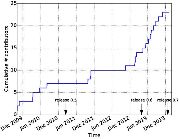
Diffusion Imaging in Python (Dipy) is a free and open source software project for the analysis of data from diffusion magnetic resonance imaging (dMRI) experiments. dMRI is an application of MRI that can be used to measure structural features of brain white matter. Many methods have been developed to use dMRI data to model the local configuration of white matter nerve fiber bundles and infer the trajectory of bundles connecting different parts of the brain. Dipy gathers implementations of many different methods in dMRI, including: diffusion signal pre-processing; reconstruction of diffusion distributions in individual voxels; fiber tractography and fiber track post-processing, analysis and visualization. Dipy aims to provide transparent implementations for all the different steps of dMRI analysis with a uniform programming interface. We have implemented classical signal reconstruction techniques, such as the diffusion tensor model and deterministic fiber tractography. In addition, cutting edge novel reconstruction techniques are implemented, such as constrained spherical deconvolution and diffusion spectrum imaging (DSI) with deconvolution, as well as methods for probabilistic tracking and original methods for tractography clustering. Many additional utility functions are provided to calculate various statistics, informative visualizations, as well as file-handling routines to assist in the development and use of novel techniques. In contrast to many other scientific software projects, Dipy is not being developed by a single research group. Rather, it is an open project that encourages contributions from any scientist/developer through GitHub and open discussions on the project mailing list. Consequently, Dipy today has an international team of contributors, spanning seven different academic institutions in five countries and three continents, which is still growing.
Overview
The claims and statistics of this paper haven’t been checked yet.
Run the check — free, no account needed. It reads the open-access text and checks the sources it cites.
Integrity signals
Registry
Cites
Statistics
Figures
Verification
Claims the paper makes
The claims and statistics of this paper haven’t been checked yet.
Run the check — free, no account needed. It reads the open-access text and checks the sources it cites.
Claims other papers make about this one
Each is checked against this paper’s own text: does it say what the citing paper says it does?
The cited source says what the claim attributes to it.
In the citing paper
“In contrast to projects such as DiPy (Garyfallidis et al., 2014) , this library does not provide functionality to manipulate raw image data within Python itself, but provides functionality to invoke and manage external high-performance executables to perform the actual computations.”
From MRtrix3: A fast, flexible and open software framework for medical image processing and visualisation (2019) · cites this paper as Garyfallidis et al., 2014
Self-check
Percentages, sums and reported differences re-computed from the paper’s own numbers.
The self-check needs the paper’s full text.
Citations
Cites
A Threshold Selection Method from Gray-Level Histograms
Otsu, N. (1979) · IEEE Transactions on Systems, Man and Cybernetics · doi:10.1109/tsmc.1979.4310076 · Cited by 44,708 · cited 1×
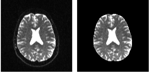
“…median_otsu (see Figure 3 ). from dipy.segment.mask import median_otsu mask, S0_mask = median_otsu(data [:, :, :, 0]) median_otsu uses first a median filter to smooth the S0 and then Otsu’s method, an automated histogram method (Otsu, 1979) to separate the brain (foreground) from its background.”
MethodAdvances in functional and structural MR image analysis and implementation as FSL
Smith, S. M., Jenkinson, M., Woolrich, M. W., Beckmann, C. F., Behrens, T. E. J., Johansen-Berg, H., Bannister, P. R., De Luca, M., Drobnjak, I., Flitney, D. E., et al. (2004) · NeuroImage · doi:10.1016/j.neuroimage.2004.07.051 · Cited by 13,630
Fischl, B. (2012) · NeuroImage · doi:10.1016/j.neuroimage.2012.01.021 · Cited by 9,526 · cited 4×
“…al., 2006), AFNI (Cox, 2012), MITK (Fritzsche et al., 2012), BrainVoyager QX (Goebel, 2012), DTI-Query/Quench (Sherbondy et al., 2005), FreeSurfer (Fischl, 2012), FSL-FDT (Smith et al., 2004), MedInria (Toussaint et al., 2007), MRtrix (Tournier et al., 2012), Diffusion Toolkit/Trackvis (Wang et…”
MethodInfluentialThe NumPy Array: A Structure for Efficient Numerical Computation
van der Walt, S., Colbert, S. C., & Varoquaux, G. (2011) · Computing in Science & Engineering · doi:10.1109/mcse.2011.37 · Cited by 9,381
MR diffusion tensor spectroscopy and imaging
Basser, P. J., Mattiello, J., & LeBihan, D. (1994) · Biophysical Journal · doi:10.1016/s0006-3495(94)80775-1 · Cited by 5,970 · cited 2×
“Another alternative is to make assumptions about the distribution of the PDF such as a Gaussian assumption, which leads to the popular diffusion tensor imaging (DTI) technique (Basser et al., 1994).”
BackgroundMethodThe WU-Minn Human Connectome Project: An overview
Van Essen, D. C., Smith, S. M., Barch, D. M., Behrens, T. E. J., Yacoub, E., & Ugurbil, K. (2013) · NeuroImage · doi:10.1016/j.neuroimage.2013.05.041 · Cited by 5,651
The basis of anisotropic water diffusion in the nervous system—A technical review.
Beaulieu, C. (2002) · NMR in Biomedicine · doi:10.1002/nbm.782 · Cited by 4,628
Basser, P. J., & Pierpaoli, C. (1996) · Journal of Magnetic Resonance, Series B · doi:10.1006/jmrb.1996.0086 · Cited by 4,306 · cited 3×
“…of the myelin wrapping the axons in the voxel may affect both MD and FA, changes in other factors, such as the distribution of directions of crossing of fiber populations through the voxel may also affect these measures (Basser and Pierpaoli, 1996; Jones et al., 2013; Wandell and Yeatman, 2013).”
BackgroundMethodMedical image analysis: progress over two decades and the challenges ahead
Duncan, J. S., & Ayache, N. (2000) · IEEE Transactions on Pattern Analysis and Machine Intelligence · doi:10.1109/34.824822 · Cited by 4,167
IPython: A System for Interactive Scientific Computing
Pérez, F., & Granger, B. (2007) · Computing in Science & Engineering · doi:10.1109/mcse.2007.53 · Cited by 4,129 · cited 1×
“There has been large growth in the number of Python users, there are many Python tools for scientific computing (Pérez and Granger, 2007; Pérez et al., 2011; McKinney, 2012), and there are complementary neuroimaging packages in Python1.”
Background
Research works printed but not linked: 9
- Chamberland, 2013, Explore the brain white matter networks in real-time: multi-sticks fiber tracking
- Garyfallidis, 2012, Towards an Accurate Brain Tractography
- Garyfallidis, 2011, Dipy - a novel software library for diffusion MR and tractography
- LeBihan, 1985, Imagerie de diffusion in vivo par résonance magnétique nucléaire, C. R. Acad. Sci. Paris, 301, 1109
- Pajevic, 1999, Color schemes to represent the orientation of anisotropic tissues from diffusion tensor data: application to white matter fiber tract mapping in the human brain, Magn. Reson. Med, 42, 526, 10.1002/(SICI)1522-2594(199909)42:3526::AID-MRM153.3.CO;2-A · DOI
- Schroeder, 2001, The Visualization Toolkit-User's Guide
- Vaillancourt, 2011, A fiber navigator for neurosurgical planning
- Wang, 2007, Diffusion toolkit: a software package for diffusion imaging data processing and tractography, Proc. Int. Soc. Magn. Reson. Med, 15, 3720
- Westin, 1997, Geometrical diffusion measures for MRI from tensor basis analysis, Proc. ISMRM, 97, 1742
Cited by
Sorted by citations
Items 501–600 of 1,000
Jiang, R., Hu, X., Deng, K., Jiang, S., Chen, W., & Zhang, Z. (2021) · European Journal of Radiology · doi:10.1016/j.ejrad.2021.109750 · Cited by 11
Fiber Clustering Acceleration With a Modified Kmeans++ Algorithm Using Data Parallelism
Goicovich, I., Olivares, P., Román, C., Vázquez, A., Poupon, C., Mangin, J., Guevara, P., & Hernández, C. (2021) · Frontiers in Neuroinformatics · doi:10.3389/fninf.2021.727859 · Cited by 11
Alshehri, A., Al‐iedani, O., Arm, J., Gholizadeh, N., Billiet, T., Lea, R. A., Lechner‐Scott, J., & Ramadan, S. A. (2022) · The Neuroradiology Journal · doi:10.1177/19714009211067400 · Cited by 11
“0.(25) to identify and reject outliers.”
Rheault, F., Schilling, K. G., Obaïd, S., Begnoche, J. P., Cutting, L. E., Descoteaux, M., Landman, B. A., & Petit, L. (2022) · Brain Structure and Function · doi:10.1007/s00429-022-02518-6 · Cited by 11
“The protocol is adapted for probabilistic tractography from HARDI local models (Garyfallidis et al. 2014; Tournier et al. 2019) and can be carried out in approximately 30 min without external processing apart from the tractography itself.”
MethodSelf-citationVillalón‐Reina, J. E., Moreau, C. A., Nir, T. M., Jahanshad, N., Simons Variation in Individuals Project Consortium, Maillard, A. M., Romascano, D., Draganski, B., Lippé, S., Bearden, C. E., et al. (2022) · Lecture notes in computer science · doi:10.1007/978-3-031-16431-6_20 · Cited by 11
Huang, C., Kritikos, M., Serrano‐Sosa, M., Hagan, T., Domkan, A., Meliker, J. R., Pellecchia, A. C., Santiago‐Michels, S., Carr, M. A., Kotov, R., et al. (2022) · Molecular Neurobiology · doi:10.1007/s12035-022-03059-z · Cited by 11
“While the DiPy toolbox [53] has previously been utilized for multi-shell FWF generation [54–56], single-shell FWF images were generated using the recent extension to DiPy proposed by Golub et al.”
Altered structural connectome of children with auditory processing disorder: a diffusion MRI study
Alvand, A., Kuruvilla-Mathew, A., Roberts, R. P., Pedersen, M., Kirk, I. J., & Purdy, S. C. (2023) · Cerebral Cortex · doi:10.1093/cercor/bhad075 · Cited by 13
“, 2014); RRID: SCR_001362) and Dipy (Garyfallidis et al., 2014).”
Griessmair, M., Delbridge, C., Ziegenfeuter, J., Bernhardt, D., Gempt, J., Schmidt‐Graf, F., Kertels, O., Thomas, M., Meyer, H. S., Zimmer, C., et al. (2023) · Cancers · doi:10.3390/cancers15082355 · Cited by 11
Pypes: Workflows for Processing Multimodal Neuroimaging Data
Savio, A. M., Schütte, M., Graña, M., & Yakushev, I. Y. (2017) · Frontiers in Neuroinformatics · doi:10.3389/fninf.2017.00025 · Cited by 11
“From Nipy, it’s possible to find projects to access different neuroimaging file formats 8 , diffusion brain MRI with Dipy (Garyfallidis et al., 2014), statistical learning and fMRI with Nilearn (Abraham et al., 2014) as well as building processing pipelines with Nipype, and others (Gramfort et al.,…”
MethodParra-Morales, A. M., Rudas, J., Vargas, J. A., Gómez, F. A., Enciso-Olivera, C. O., Trujillo‐Rodriguez, D., Martínez, D., Hernandez, J., Ordóñez-Rubiano, E. G., & Marín-Muñoz, J. H. (2019) · Medicine · doi:10.1097/md.0000000000015620 · Cited by 11
“2 cessed using FMRIB Software Library (FSL)([7]) and Diffusion Imaging in Python (DIPY) library.([8]) This process included automatic realignment, correction of eddy-current artifacts, reslicing to obtain isotropic voxel size, brain extraction, and improvement of signal-to-noise rate using a Non-Local mean algorithm.”
MethodLapa, P., Gonçalves, I., Rundo, L., & Castelli, M. (2019) · Genetic and Evolutionary Computation Conference Companion (GECCO Companion) · doi:10.1145/3319619.3326864 · Cited by 11
“The voxel normalization and the image registration were performed by using the Diffusion Imaging Python (DIPy) [10] package, and the Mutual Information metric was considered as the cost function [31].”
MethodTRAKO: Efficient Transmission of Tractography Data for Visualization
Haehn, D., Franke, L., Zhang, F., Karayumak, S. C., Pieper, S., O’Donnell, L. J., & Yogesh Rathi, A. (2020) · Lecture notes in computer science · doi:10.1007/978-3-030-59728-3_32 · Cited by 11
Bundle-specific fornix reconstruction for dual-tracer PET-tractometryPreprint
Rheault, F., Roy, M., Cunnane, S. C., & Descoteaux, M. (2018) · bioRxiv (Cold Spring Harbor Laboratory) · doi:10.1101/423459 · Cited by 11
Self-citationTractography analysis with the scilpy toolbox
Renauld, E., Boré, A., Poirier, C., Caron, A. V., Karan, P., Théberge, A., Theaud, G., Edde, M., Poulin, P., Girard, G., et al. (2026) · Aperture Neuro · doi:10.52294/001c.154022 · Cited by 10
Self-citationStructural Brain Imaging Predicts Individual-Level Task Activation Maps Using Deep Learning
Ellis, D. G., & Aizenberg, M. R. (2022) · Frontiers in Neuroimaging · doi:10.3389/fnimg.2022.834883 · Cited by 10
Gugger, J. J., Walter, A. E., Parker, D., Sinha, N., Morrison, J., Ware, J. B., Schneider, A. L. C., Petrov, D., Sandsmark, D. K., Verma, R., et al. (2022) · Journal of Neurotrauma · doi:10.1089/neu.2022.0259 · Cited by 10
“20 We used FERNET (Freewater EstimatoR using iNtErpo-lated iniTialization), 8 a recent method for free water correction in clinically acquired DTI data, to estimate the free water-volume fraction or VF and free water-corrected FA (fw-FA).”
Connectomics underlying motor functional outcomes in the acute period following stroke
Bian, R., Huo, M., Liu, W., Mansouri, N., Tanglay, O., Young, I. M., Osipowicz, K. Z., Hu, X., Zhang, X., Doyen, S., et al. (2023) · Frontiers in Aging Neuroscience · doi:10.3389/fnagi.2023.1131415 · Cited by 10
“Infinitome relies on standard processing steps using Python (Garyfallidis et al., 2014).”
MethodTiwari, A. S., Singh, R. K., & Shigwan, S. J. (2023) · Neural Computing and Applications · doi:10.1007/s00521-023-09206-4 · Cited by 10
Ten simple rules for scientific code review
Rokem, A. (2024) · PLoS Computational Biology · doi:10.1371/journal.pcbi.1012375 · Cited by 10
Self-citationA distributed privacy preserving model for the detection of Alzheimer’s disease
Mandal, P. K. (2024) · Neural Computing and Applications · doi:10.1007/s00521-024-10419-4 · Cited by 10
“MRI images were imported in the form of NIfTI (Neuroimaging Informatics Technology Imitative) images [5] and then converted into tensors with NiBabel [11].”
Unraveling the Diffusion MRI-Based Glymphatic System Alterations in Children with Rolandic Epilepsy
Yin, Y., Ma, M., Wang, F., Ju, C., Luo, X., Yu, H., Nie, L., Li, J., & Liu, H. (2024) · Academic Radiology · doi:10.1016/j.acra.2024.11.010 · Cited by 10
Outlier detection in cardiac diffusion tensor imaging: Shot rejection or robust fitting?
Coveney, S., Afzali, M., Mueller, L., Teh, I., Das, A., Dall’Armellina, E., Szczepankiewicz, F., Jones, D. K., & Schneider, J. E. (2024) · Medical Image Analysis · doi:10.1016/j.media.2024.103386 · Cited by 10
“We implemented our IRLS algorithms in DIPY (Garyfallidis et al., 2014).”
Ricchi, M., Campani, G., Nagmutdinova, A., Bortolotti, V., Greco, D., Golini, C., Grist, J. T., Brizi, L., & Testa, C. M. (2025) · European Radiology Experimental · doi:10.1186/s41747-025-00570-5 · Cited by 10
“The implementation of the Bingham-NODDI model was performed by using the Diffusion Microstructure Imaging in Python (Dmipy) package [39] to calculate the NODDI metrics: tissue volume fraction, β -fraction, neurite dispersion index (NDI), and orientation dispersion index (ODI) (see Supplementary…”
Fislage, M., Winzeck, S., Stamatakis, E. A., Correia, M., Preller, J., Feinkohl, I., Spies, C., Hendrikse, J., Slooter, A. J. C., Winterer, G., et al. (2022) · NeuroImage Clinical · doi:10.1016/j.nicl.2022.103208 · Cited by 10
“, 2005) were fitted with dipy (Garyfallidis et al., 2014) to compute FA, MD and MK maps.”
Bauer, C. E., Zachariou, V., Maillard, P., Caprihan, A., & Gold, B. T. (2022) · Frontiers in Aging Neuroscience · doi:10.3389/fnagi.2022.995425 · Cited by 10
“, 2017) from the open-source software package Diffusion Imaging in Python (DIPY; Garyfallidis et al., 2014).”
MethodSevere central nervous system demyelination in Sanfilippo disease
Taherzadeh, M., Zhang, E., Londoño, I., Song, S., Wang, S., Cooper, J. D., Kennedy, T. E., Morales, C. R., Chen, Z., Lodygensky, G. A., et al. (2023) · Frontiers in Molecular Neuroscience · doi:10.3389/fnmol.2023.1323449 · Cited by 10
Multi-scale V-net architecture with deep feature CRF layers for brain extraction
Park, J. S., Fadnavis, S., & Garyfallidis, E. (2024) · Communications Medicine · doi:10.1038/s43856-024-00452-8 · Cited by 9
“DIPY Horizon 57 was used for visualization.”
Self-citationDávid, G., Fricke, B., Oeschger, J. M., Ruthotto, L., Fritz, F. J., Ohana, O., Mordhorst, L., Sauvigny, T., Freund, P., Tabelow, K., et al. (2024) · Imaging Neuroscience · doi:10.1162/imag_a_00288 · Cited by 10
“…along with diffusion signal modeling capabilities, are widely available in open-source toolboxes such as FSL-FDT (Smith et al., 2004), DiPY (Garyfallidis et al., 2014), DESIGNER (Ades-Aron et al., 2018), ExploreDTI (Leemans et al., 2009), MRtrix3 (Tournier et al., 2019), TORTOISE…”
White Matter Correlates of Cognitive Performance on the UCSF Brain Health Assessment
Alioto, A. G., Mumford, P., Wolf, A. A., Casaletto, K. B., Erlhoff, S. J., Moskowitz, T., Kramer, J. H., Rankin, K. P., & Possin, K. L. (2019) · Journal of the International Neuropsychological Society · doi:10.1017/s1355617719000225 · Cited by 10
Kasa, L. W., Haast, R. A. M., Kuehn, T. K., Mushtaha, F. N., Baron, C. A., Peters, T. M., & KHAN, A. R. (2020) · Journal of Magnetic Resonance Imaging · doi:10.1002/jmri.27408 · Cited by 10
Caminiti, R., Girard, G., Battaglia‐Mayer, A., Borra, E., Schito, A., Innocenti, G. M., & Luppino, G. (2021) · eNeuro · doi:10.1523/eneuro.0102-21.2021 · Cited by 10
“, 2014) available in the DIPY software library (Garyfallidis et al., 2014).”
MethodNigral diffusivity, but not free water, correlates with iron content in Parkinson’s disease
Langley, J. A., Huddleston, D. E., & Hu, X. P. (2021) · Brain Communications · doi:10.1093/braincomms/fcab251 · Cited by 9
CTIMS: Automated Defect Detection Framework Using Computed Tomography
Gabbar, H. A., Chahid, A., Khan, M. J. A., Adegboro, O. G., & Samson, M. I. (2022) · Applied Sciences · doi:10.3390/app12042175 · Cited by 10
“The background subtraction function removes the unwanted background or regions of the input data by using an imaging library in Python called DIPY [38].”
MethodFilipiak, P., Shepherd, T. M., Lin, Y., Placantonakis, D. G., Boada, F. E., & Baete, S. H. (2022) · Magnetic Resonance in Medicine · doi:10.1002/mrm.29208 · Cited by 9
FiberNeat: Unsupervised White Matter Tract Filtering
Chandio, B. Q., Chattopadhyay, T., Owens‐Walton, C., Reina, J. E. V., Nabulsi, L., Thomopoulos, S. I., Garyfallidis, E., & Thompson, P. M. (2022) · Annual International Conference of the IEEE Engineering in Medicine and Biology Society (EMBC) · doi:10.1109/embc48229.2022.9870877 · Cited by 10
Self-citationKwan, S., Boudes, E., Benseler, A., Gilbert, G., Saint‐Martin, C., Shevell, M. I., & Wintermark, P. (2015) · Neural Plasticity · doi:10.1155/2015/653727 · Cited by 10
“Then, apparent diffusion coefficient (ADC) and fractional anisotropy (FA) maps were generated for the whole brain using the tools of the Diffusion Imaging in Python (DIPY) package (Sherbrooke Connectivity Imaging Lab, Sherbrooke University, Quebec, Canada) [41].”
MethodSensitive periods for white matter plasticity and reading interventionPreprint
Yeatman, J. D., & Huber, E. (2018) · bioRxiv (Cold Spring Harbor Laboratory) · doi:10.1101/346759 · Cited by 10
Data-science ready, multisite, human diffusion MRI white-matter-tract statistics
Lerma‐Usabiaga, G., Mukherjee, P., Perry, L. M., & Wandell, B. A. (2020) · Scientific Data · doi:10.1038/s41597-020-00760-3 · Cited by 9
Collegiate athlete brain data for white matter mapping and network neuroscience
Caron, B. A., Stuck, R., McPherson, B., Bullock, D. N., Kitchell, L. M., Faskowitz, J., Kellar, D., Cheng, H., Newman, S. D., Port, N., et al. (2021) · Scientific Data · doi:10.1038/s41597-021-00823-z · Cited by 11
Study protocol: Social Health Impact of Network Effects (SHINE) StudyPreprint
Cosme, D., Kang, Y., Tartak, J. C., Ahn, J., Corbani, F. E., Cooper, N., Doré, B., He, X., Helion, C., Jovanova, M., et al. (2022) · PsyArXiv (OSF Preprints) · doi:10.31234/osf.io/cj2nx · Cited by 9
Surgent, O., Dean, D. C., Alexander, A. L., Dadalko, O. I., Guerrero-Gonzalez, J., Taylor, D., Skaletski, E., & Travers, B. G. (2021) · Brain Communications · doi:10.1093/braincomms/fcab112 · Cited by 9
Saiote, C., Sutter, E. N., Xenopoulos-Oddsson, A., Rao, R., Georgieff, M., Rudser, K. D., Peyton, C., Dean, D., McAdams, R. M., & Gillick, B. T. (2022) · Pediatric Physical Therapy · doi:10.1097/pep.0000000000000886 · Cited by 9
The influence of anisotropy on the clinical target volume of brain tumor patients
Buti, G., Ajdari, A., Hochreuter, K., Shih, H. A., Bridge, C. P., Sharp, G. C., & Bortfeld, T. R. (2023) · Physics in Medicine and Biology · doi:10.1088/1361-6560/ad1997 · Cited by 9
DiffGEPCI: 3D MRI Synthesis from mGRE Signals using 2.5D Diffusion Model
Hu, Y., Kothapalli, S. V. V. N., Gan, W., Sukstanskii, A. L., Wu, G. F., Goyal, M. S., Yablonskiy, D. A., & Kamilov, U. S. (2024) · IEEE International Symposium on Biomedical Imaging (ISBI) · doi:10.1109/isbi56570.2024.10635694 · Cited by 15
“We co-registered the corresponding FLAIR and MPRAGE data to the mGRE sequence using a 3D Affine transformation from the Dipy package [18].”
MethodSpinal Cord Blood Perfusion Deficit is Associated with Clinical Impairment after Spinal Cord Injury
Lebret, A., Frese, S., Lévy, S., Curt, A. E. P., Callot, V., Freund, P., & Seif, M. (2024) · Journal of Neurotrauma · doi:10.1089/neu.2024.0267 · Cited by 9
Bundle Analytics based Data Harmonization for Multi-Site Diffusion MRI Tractometry
Chandio, B. Q., Villalón‐Reina, J. E., Nir, T. M., Thomopoulos, S. I., Feng, Y., Benavidez, S. M., Jahanshad, N., Harezlak, J., Garyfallidis, E., & Thompson, P. M. (2024) · Annual International Conference of the IEEE Engineering in Medicine and Biology Society (EMBC) · doi:10.1109/embc53108.2024.10782419 · Cited by 9
Self-citationMicroglia positron emission tomography and progression in multiple sclerosis: thalamus on fire
Zeydan, B., Neyal, N., Son, J., Schwarz, C. G., Thomas, J. C. K., Morrison, H. A., Bush, M. L., Reid, R. I., Przybelski, S. A., Fought, A. J., et al. (2025) · Brain Communications · doi:10.1093/braincomms/fcaf141 · Cited by 10
Zeng, B., Zeng, P., Xiang, Y., Peng, Y., Luo, D., Li, B., Chai, Y., Wang, D., Nie, L., & Li, Y. (2025) · Brain Research Bulletin · doi:10.1016/j.brainresbull.2025.111391 · Cited by 9
Mapping the microstructure of human cerebral cortex in vivo with diffusion MRI
Sadikov, A., Choi, H. L., Xiao, J., Cai, L. T., & Mukherjee, P. (2025) · Communications Biology · doi:10.1038/s42003-025-08523-9 · Cited by 12
“The DTI, DKI, and MAP-MRI fi tting was performed via dipy 68 ; the NODDI fi tting was performed via AMICO 69 .”
Fick, R., Zucchelli, M., Girard, G., Descoteaux, M., Menegaz, G., & Rachid Deriche, B. (2015) · IEEE International Symposium on Biomedical Imaging (ISBI) · doi:10.1109/isbi.2015.7163905 · Cited by 9
“In experiment (I) we generate fiber crossings with angles ranging from 0 0 to 90 0 using the multi-tensor model in Dipy [8].”
InfluentialSelf-citationA Continuous Model of Cortical Connectivity
Moyer, D., Gutman, B. A., Faskowitz, J., Jahanshad, N., & Thompson, P. M. (2016) · Lecture notes in computer science · doi:10.1007/978-3-319-46720-7_19 · Cited by 9
“Probabilistic streamline tractography was conducted using the DWI in 2mm isotropic MNI 152 space, using Dipy’s [7] implementation of constrained spherical deconvolution (CSD) [16] with a harmonic order of 6.”
MethodDense and structured representations of U-shape fiber connectivity in the central sulcus
Pron, A., Brun, L., Deruelle, C., & Coulon, O. (2018) · IEEE International Symposium on Biomedical Imaging (ISBI) · doi:10.1109/isbi.2018.8363670 · Cited by 9
“Diffuse toolbox(3), an in-house Brainvisa toolbox [7] wrapping FSL(4) and Dipy [8] functions.”
BackgroundGe, Y., Zheng, W., Li, Y., Dou, W., Ren, S., Chen, Z., & Wang, Z. (2022) · Frontiers in Aging Neuroscience · doi:10.3389/fnagi.2022.799251 · Cited by 9
Badea, A., Li, D., Niculescu, A. R., Anderson, R. J., Stout, J. A., Williams, C. L., Colton, C. A., Maeda, N., & Dunson, D. B. (2022) · Frontiers in Neuroscience · doi:10.3389/fnins.2022.848654 · Cited by 9
Chan, K., Chamberland, M., & Marques, J. P. (2022) · NeuroImage · doi:10.1016/j.neuroimage.2022.119824 · Cited by 9
“DWI data was pre-processed using an in-house pipeline consisting of the following procedures: (1) data denoising with Patch2self (Fadnavis et al., 2020; Garyfallidis et al., 2014), (2) Gibbs ringing removal (Kellner et al.”
MethodZhou, Z., Yang, Z., Jiang, S., Zhu, Τ., Ma, S., Li, Y., & Zhuo, J. (2023) · Visual Informatics · doi:10.1016/j.visinf.2023.05.003 · Cited by 9
PPA: Principal parcellation analysis for brain connectomes and multiple traits
Liu, R., Li, M., & Dunson, D. B. (2023) · NeuroImage · doi:10.1016/j.neuroimage.2023.120214 · Cited by 10
DiPiero, M. A., Cordash, H., Prigge, M. B. D., King, C. K., Morgan, J., Guerrero-Gonzalez, J. M., Adluru, N., King, J. B., LANGE, N. D., Bigler, E. D., et al. (2023) · Frontiers in Neuroscience · doi:10.3389/fnins.2023.1231719 · Cited by 9
“Diffusion tensors were estimated at each voxel using a weightedleast squares algorithm as part of the diffusion imaging in python (DIPY) open-source software package (Garyfallidis et al., 2014).”
MethodGoeckner, B. D., Brett, B. L., Mayer, A. R., España, L. Y., Banerjee, A., Muftuler, L. T., & Meier, T. B. (2023) · Human Brain Mapping · doi:10.1002/hbm.26556 · Cited by 8
“A positivity constraint was imposed to ensure a physically meaningful propagator estimate, and Laplacian regularization with a weighting of 0.05 was used to reduce spurious oscillations in the reconstruction (Fick et al., 2015; Garyfallidis et al., 2014).”
MethodInfluentialYao, T., Rheault, F., Nath, V., Asad, Z., Newlin, N. R., Cui, C., Deng, R., Ramadass, K., Shafer, A., Resnick, S., et al. (2024) · Journal of medical imaging · doi:10.1117/1.jmi.11.1.014005 · Cited by 9
“34 All diffusion signals are transformed to SH basis signal ODF as a unified input for DL models, using SHs with the “ tour-nier07 ” basis.”
MethodPrivacy preserving image registration
Taiello, R., Önen, M., Capano, F., Humbert, O., & Lorenzi, M. (2024) · Medical Image Analysis · doi:10.1016/j.media.2024.103129 · Cited by 15
E(3) × SO(3)-Equivariant Networks for Spherical Deconvolution in Diffusion MRI.
Elaldi, A., Gerig, G., & Dey, N. (2024) · PubMed · Cited by 9
DIMOND: DIffusion Model OptimizatioN with Deep Learning
Li, Z., Li, Z., Bilgiç, B., Lee, H., Ying, K., Huang, S. Y., Liao, H., & Tian, Q. (2024) · Advanced Science · doi:10.1002/advs.202307965 · Cited by 9
Non-parametric graphnet-regularized representation of dMRI in space and time
Fick, R., Petiet, A., Santin, M. D., Philippe, A.-C., Lehéricy, S., Rachid Deriche, B., & Wassermann, D. (2017) · Medical Image Analysis · doi:10.1016/j.media.2017.09.002 · Cited by 9
“We find optimal vales for regularization weights α and λ using cross-validation and mplemented the surrounding code infrastructure inside the DiPy ramework ( Garyfallidis et al., 2014 ).”
MethodInfluentialTobisch, A., Schultz, T., Stirnberg, R., Varela-Mattatall, G. E., Knutsson, H. E., Pablo Irarrázaval, M., & Stöcker, T. (2019) · NMR in Biomedicine · doi:10.1002/nbm.4055 · Cited by 10
Nonparametric Bayes Models of Fiber Curves Connecting Brain Regions
Zhang, Z., Descoteaux, M., & Dunson, D. B. (2019) · Journal of the American Statistical Association · doi:10.1080/01621459.2019.1574582 · Cited by 9
“First, high angular resolution diffusion imaging (HARDI) techniques are used to estimate the fiber orientation distribution function (ODF) at each location (Descoteaux et al. 2009) (implemented in dipy (Garyfallidis et al. 2014)).”
MethodSelf-citationKnowledge Transfer Between Datasets for Learning-Based Tissue Microstructure Estimation
Yu, Q., Li, Y., Liu, Z., & Ye, C. (2020) · IEEE International Symposium on Biomedical Imaging (ISBI) · doi:10.1109/isbi45749.2020.9098410 · Cited by 9
“The default parameters given by Dipy [10] are used for the SHORE representation and solving Eq.”
MethodInfluentialSpatiotemporal dynamics of nonhuman primate white matter development during the first year of life
Aggarwal, N., Moody, J. F., Dean, D., Tromp, D., Kecskemeti, S. R., Oler, J. A., Alexander, A. L., & Kalin, N. H. (2021) · NeuroImage · doi:10.1016/j.neuroimage.2021.117825 · Cited by 9
Riedel, M., Setsompop, K., Mertins, A., & Börnert, P. (2021) · Magnetic Resonance in Medicine · doi:10.1002/mrm.28813 · Cited by 9
Gagnon, A., Grenier, G., Bocti, C., Gillet, V., Lepage, J., Baccarelli, A., Posner, J. E., Descoteaux, M., & Takser, L. (2022) · Cerebral Cortex · doi:10.1093/cercor/bhac180 · Cited by 10
Self-citationMigneron-Foisy, V., Muckle, G., Jacobson, J. L., Ayotte, P., Jacobson, S. W., & Saint‐Amour, D. (2022) · NeuroToxicology · doi:10.1016/j.neuro.2022.08.010 · Cited by 10
Asymmetry Spectrum Imaging for Baby Diffusion Tractography
and the UNC/UMN Baby Connectome Project Consortium, Wu, Y., Lin, W., Shen, D. G., & Yap, P. (2019) · Lecture notes in computer science · doi:10.1007/978-3-030-20351-1_24 · Cited by 9
“We extended the direction getter mechanism described in [15], which originally caters to symmetric FODFs, to work with asymmetric FODFs and extracted five tracts-of-interest using RecoBundles [15] (see Figs.”
MethodSchilling, K. G., Tax, C. M. W., Rheault, F., Hansen, C. B., Yang, Q., Yeh, F.-C. F., Cai, L. Y., Anderson, A. W., & Landman, B. A. (2021) · bioRxiv (Cold Spring Harbor Laboratory) · doi:10.1101/2021.03.17.435872 · Cited by 10
A hybrid approach for optimal automatic segmentation of White Matter tracts in HARDI
Chekir, A., Descoteaux, M., Garyfallidis, E., Cote, M.-A., & Boumghar, F. O. (2014) · 2014 IEEE Conference on Biomedical Engineering and Sciences (IECBES) · doi:10.1109/iecbes.2014.7047481 · Cited by 12
Self-citationEffect of age on lateralized auditory processing
Stadler, J., Brechmann, A., & Angenstein, N. (2023) · Hearing Research · doi:10.1016/j.heares.2023.108791 · Cited by 8
Corriveau‐Lecavalier, N., Tosakulwong, N., Lesnick, T. G., Fought, A. J., Reid, R. I., Schwarz, C. G., Senjem, M. L., Jack, C. R., Jones, D. T., Vemuri, P., et al. (2023) · Neurobiology of Aging · doi:10.1016/j.neurobiolaging.2023.12.001 · Cited by 9
Barbeau, É. B., Badhwar, A., Kousaie, S., Bellec, P., Descoteaux, M., Klein, D., & Petrides, M. (2023) · eNeuro · doi:10.1523/eneuro.0363-23.2023 · Cited by 8
“Standard DTI metrics and fi ber orientation density functions of spherical harmonics order 8 were reconstructed with Dipy (Garyfallidis et al., 2014).”
Self-citationDCP : A pipeline toolbox for diffusion connectome
Huang, W., Dong, X., Zhao, T., Kucikova, L., Fu, A., & Shu, N. (2024) · Human Brain Mapping · doi:10.1002/hbm.26626 · Cited by 7
“The first set of tools, such as Camino (Cook et al., 2006), MRtrix3 (Tournier et al., 2019), Dipy (Garyfallidis et al., 2014), and QSIPrep (Cieslak et al., 2021), do not have GUI and require users to customize their pipelines by programming, which is challenging for scientific researchers without programming skills.”
MethodPhillips, J. S., Adluru, N., Chung, M. K., Radhakrishnan, H., Olm, C. A., Cook, P. A., Gee, J. C., Cousins, K. A. Q., Arezoumandan, S., Wolk, D. A., et al. (2024) · Frontiers in Neuroscience · doi:10.3389/fnins.2024.1353306 · Cited by 9
Orlandi, F., Carlos, A. F., Ali, F., Clark, H. M., Duffy, J. R., Utianski, R. L., Botha, H., Machulda, M. M., Stephens, Y. C., Schwarz, C. G., et al. (2024) · Brain Communications · doi:10.1093/braincomms/fcae113 · Cited by 8
Prescription Opioids and Brain Structure in Community-Dwelling Older Adults
Warner, N. S., Hanson, A. C., Schulte, P. J., Kara, F., Reid, R. I., Schwarz, C. G., Benarroch, E. E., Graff‐Radford, J., Vemuri, P., Jack, C. R., et al. (2024) · Mayo Clinic Proceedings · doi:10.1016/j.mayocp.2024.01.018 · Cited by 8
Consagra, W., Ning, L., & Yogesh Rathi, A. (2025) · Medical Image Analysis · doi:10.1016/j.media.2025.103537 · Cited by 8
“We use the implementations in the python library dipy (Garyfallidis et al., 2014) under the default suggested parameterizations.”
Добрынина, Л. А., Kremneva, E. I., Витальевна, Ш. К., Geints, A. A., Filatov, A. S., Gadzhieva, Z. S., Gnedovskaya, E. V., Кротенкова, М. В., & Maximov, I. I. (2024) · Diagnostics · doi:10.3390/diagnostics14161838 · Cited by 8
Nazeri, A., Hosseini, H., Dehkharghanian, T., Lindsay, K. E., LaMontagne, P., Shimony, J. S., Benzinger, T. L. S., & Sotiras, A. (2025) · Imaging Neuroscience · doi:10.1162/imag_a_00473 · Cited by 8
“5.0 (Garyfallidis et al., 2014).”
InfluentialCerebral hypoperfusion, brain structural integrity, and cognitive impairment in older APOE4 carriers
Pappas, I., Lohman, T., Dutt, S., Kapoor, A., Engstrom, A. C., Alitin, J. P. M., Barnes, S. R., Chakhoyan, A., Saca, L., Gaggar, R., et al. (2025) · GeroScience · doi:10.1007/s11357-025-01642-5 · Cited by 9
“White matter bundles were extracted using the RecoBundles framework as implemented in DIPY [45].”
Jang, K., & Back, M. (2025) · Brain Sciences · doi:10.3390/brainsci15060576 · Cited by 8
Anoosha Pai, S., Andrews, M. H., Gurchiek, R. D., Pincheira, P. A., Barbieri, M., Friedrich, T., Kogan, F., Gold, G. E., Mazzoli, V., Lichtwark, G. A., et al. (2025) · Journal of sport and health science/Journal of Sport and Health Science · doi:10.1016/j.jshs.2025.101070 · Cited by 8
“21 DTI data preprocessing comprised 3 steps: (a) denoising using non-local means, 22 (b) eddy current deformation and motion correction using affine registration of the diffusion-weighted images to the mean of non-weighted images, and (c) suscepti-bility-induced echo planer imaging (EPI) distortion…”
Glymphatic dysfunction links vascular pathology to Alzheimer’s biomarkers and cognitive decline
Kang, S. H., Kim, S., Kim, Y. J., Yoo, H., Lee, E. H., Jang, H. J., Shin, D., Yun, J., Kim, J. P., Kim, H., et al. (2026) · Alzheimer s Research & Therapy · doi:10.1186/s13195-026-01964-2 · Cited by 8
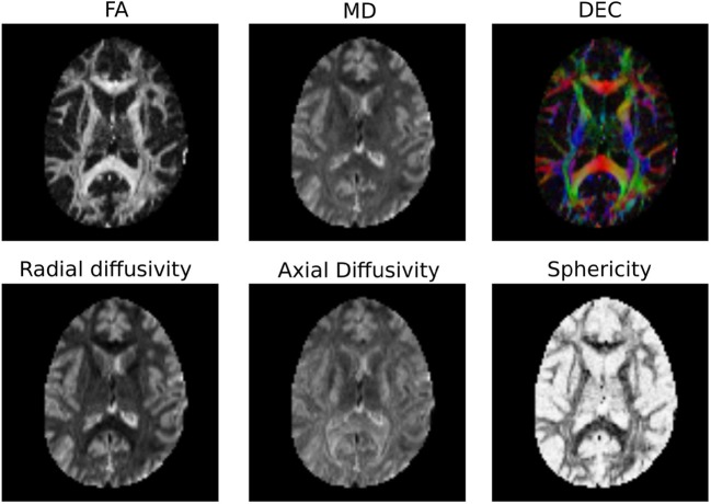
“DTI model was fitted to the preprocessed data using python library for the analysis of diffusion MRI (DIPY, h t t p s : / / d i p y . o r g / ) [31], and maps for fractional anisotropy (FA), mean diffusivity (MD), axial and radial diffusivity (AD and RD) were generated from the eigenvalues of the…”
A Unifying Framework for Spatial and Temporal Diffusion in Diffusion MRI
Fick, R., Wassermann, D., Pizzolato, M., & Rachid Deriche, B. (2015) · Lecture notes in computer science · doi:10.1007/978-3-319-19992-4_13 · Cited by 8
Bergamino, M., Burke, A. D., Baxter, L., Caselli, R., Sabbagh, M., Talboom, J., Huentelman, M., & Stokes, A. (2022) · Journal of Magnetic Resonance Imaging · doi:10.1002/jmri.28172 · Cited by 8
Valenzuela, I. J., Basurto, D., Regin, Y., Gie, A. G., der Veeken, L. V., Vergote, S., Muñoz‐Moreno, E., Leszczyński, B., Tielemans, B., Velde, G. V., et al. (2022) · Scientific Reports · doi:10.1038/s41598-022-22895-6 · Cited by 7
Gast, H., Horowitz, A., Krupnik, R., Barazany, D., Lifshits, S., Ben-Amitay, S., & Assaf, Y. (2023) · Neuroinformatics · doi:10.1007/s12021-023-09630-w · Cited by 8
“Tractography was done using the DiPy Library (Garyfallidis et al., 2014).”
MethodConnectomic insight into unique stroke patient recovery after rTMS treatment
Chen, R., Dadario, N. B., Cook, B., Sun, L., Wang, X., Li, Y., Hu, X., Zhang, X., & Sughrue, M. E. (2023) · Frontiers in Neurology · doi:10.3389/fneur.2023.1063408 · Cited by 8
“Diffusion tractography preprocessing includes standard processing steps (36), which include motion correction, elimination of excess movement, gradient distortion correction, eddy correction, and constrained spherical deconvolution-based deterministic tractography.”
MethodPracar, A. L., Ivanova, M. V., Richardson, A., & Dronkers, N. F. (2023) · Frontiers in Neurology · doi:10.3389/fneur.2023.1187399 · Cited by 9
“Further tractography data processing was done using algorithms from DIPY (45) as implemented in TractoFlow (46).”
MethodReeBundle: A Method for Topological Modeling of White Matter Pathways Using Diffusion MRI
Shailja, S., Bhagavatula, V., Cieslak, M., Vettel, J. M., Grafton, S. T., & Manjunath, B. S. (2023) · IEEE Transactions on Medical Imaging · doi:10.1109/tmi.2023.3306049 · Cited by 10
“In contrast, tract profiling techniques like DIPY [6], BUAN [7], and PyAFQ [8] quantify diffusion measures Fig.”
MethodDiffusion Tensor and Kurtosis MRI ‐Based Radiomics Analysis of Kidney Injury in Type 2 Diabetes
Yang, D., Tian, C., Liu, J., Peng, Y., Xiong, Z., Da, J., Yang, Y., Zha, Y., & Zeng, X. (2024) · Journal of Magnetic Resonance Imaging · doi:10.1002/jmri.29263 · Cited by 9
“14 All the b -values were utilized for fi tting both the DTI and DKI models.”
Planchuelo‐Gómez, Á., Descoteaux, M., Larochelle, H., Hutter, J. M., Jones, D. K., & Tax, C. M. W. (2024) · Medical Image Analysis · doi:10.1016/j.media.2024.103134 · Cited by 8
Self-citationGriessmair, M., Delbridge, C., Ziegenfeuter, J., Jung, K., Mueller, T., Schramm, S., Bernhardt, D., Schmidt‐Graf, F., Kertels, O., Thomas, M., et al. (2024) · Neuro-Oncology Advances · doi:10.1093/noajnl/vdae106 · Cited by 8
“15 To calculate leakage-corrected CBV, the algorithm of Arzanforoosh et al. 16 was used, which also requires a mask of normal-appearing white matter to normalize the resulting CBV. ANT’s Atropos was used for this purpose with the exclusion of tumor areas.”
MethodSequential Hierarchical Agglomerative Clustering of White Matter Fiber Pathways
Demir, A., & Çetingül, H. E. (2015) · IEEE Transactions on Biomedical Engineering · doi:10.1109/tbme.2015.2391913 · Cited by 8
Estéban, O., Caruyer, E., Daducci, A., Cuadra, M. B., Ledesma‐Carbayo, M. J., & Santos, A. (2016) · Frontiers in Neuroinformatics · doi:10.3389/fninf.2016.00004 · Cited by 8
“The software workflow released with this data report (the diffantomizer) ensures the reproducibility of Diffantom and enables the generation of custom diffantoms.”
BackgroundMethodInfluential
Metadata
- Authors
- Garyfallidis, E., Brett, M., Amirbekian, B., Rokem, A., van der Walt, S., Descoteaux, M., Nimmo-Smith, I., & Contributors, D.
- Affiliations
- Université de Sherbrooke (CA)
- University of California, Berkeley (US)
- University of California, San Francisco (US)
- Stanford University (US)
- Corresponding author
- Garyfallidis, Eleftherios
- Venue
- Frontiers in Neuroinformatics
- Publisher
- Frontiers Media SA
- Published
- 2014-02-21
- Volume
- 8
- Pages
- 8-8
- ISSN
- 1662-5196 (ISSN-L)
- PMC
- PMC3931231
- OpenAlex
- W2062791478
- Cited by
- 1,371
- References
- 98
- Citations by year
- 2026: 147 · 2025: 192 · 2024: 177 · 2023: 162 · 2022: 151 · 2021: 147 · 2020: 100 · 2019: 89 · 2018: 54 · 2017: 79 · 2016: 34 · 2015: 26 · 2014: 12
- FWCI
- 39.38
- Type
- journal article
- Language
- en
- Open access
- gold
- doi.org
- Licence
- cc-by
- Locations
- Frontiers in Neuroinformatics (publishedVersion, cc-by)
- Europe PMC (PubMed Central) (publishedVersion, cc-by)
- PubMed Central (publishedVersion, cc-by)
- PubMed (publishedVersion)
- DOAJ (DOAJ: Directory of Open Access Journals) (submittedVersion)
- University of Birmingham Research Portal (University of Birmingham) (submittedVersion, cc-by)
- Funders
- Fonds Québécois de la Recherche sur la Nature et les Technologies
- Natural Sciences and Engineering Research Council of Canada
- National Eye Institute (F32EY022294)
- Topics
- Health Sciences › Medicine › Radiology, Nuclear Medicine and Imaging › Advanced Neuroimaging Techniques and Applications
- Life Sciences › Neuroscience › Cognitive Neuroscience › Functional Brain Connectivity Studies
- Health Sciences › Medicine › Radiology, Nuclear Medicine and Imaging › Advanced MRI Techniques and Applications
- Keywords
- diffusion MRI, DIPY, white matter, fiber tractography, diffusion tensor model, constrained spherical deconvolution, diffusion spectrum imaging, probabilistic tracking, open source software, Python library
About these checks
Registry, retraction, Cites and statistics results are computed, not judged, with one exception: a printed reference that no index lists is read once by a model before it counts as a citation error. Claims are extracted by a model and kept only when their quote appears verbatim in the text; each cited source is then checked, with a model’s help, for whether it says what the claim relies on. Those verdicts can be wrong — read the claim’s quote and the cited source before relying on one.
Statistics and arithmetic are recomputed, not judged.
Confidence bands are shown only for fields calibrated on our annotated test set. None is calibrated yet, so no band is shown.
What counts as a claim ›How we measure confidence ›How we check this ›
Are you an author, or do you see a problem with this page? Request a review
