

The DOI matches its Crossref record; 10 of the 10 references it cites were found in a registry or an index.
Remyelination in multiple sclerosis from the miRNA perspective
Abstract
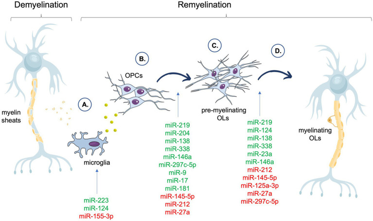
Remyelination relies on the repair of damaged myelin sheaths, involving microglia cells, oligodendrocyte precursor cells (OPCs), and mature oligodendrocytes. This process drives the pathophysiology of autoimmune chronic disease of the central nervous system (CNS), multiple sclerosis (MS), leading to nerve cell damage and progressive neurodegeneration. Stimulating the reconstruction of damaged myelin sheaths is one of the goals in terms of delaying the progression of MS symptoms and preventing neuronal damage. Short, noncoding RNA molecules, microRNAs (miRNAs), responsible for regulating gene expression, are believed to play a crucial role in the remyelination process. For example, studies showed that miR-223 promotes efficient activation and phagocytosis of myelin debris by microglia, which is necessary for the initiation of remyelination. Meanwhile, miR-124 promotes the return of activated microglia to the quiescent state, while miR-204 and miR-219 promote the differentiation of mature oligodendrocytes. Furthermore, miR-138, miR-145, and miR-338 have been shown to be involved in the synthesis and assembly of myelin proteins. Various delivery systems, including extracellular vesicles, hold promise as an efficient and non-invasive way for providing miRNAs to stimulate remyelination. This article summarizes the biology of remyelination as well as current challenges and strategies for miRNA molecules in potential diagnostic and therapeutic applications.
Overview
The claims and statistics of this paper haven’t been checked yet.
Run the check — free, no account needed. It reads the open-access text and checks the sources it cites.
Integrity signals
Registry
Cites
Statistics
Figures
Verification
Claims the paper makes
The claims and statistics of this paper haven’t been checked yet.
Run the check — free, no account needed. It reads the open-access text and checks the sources it cites.
Claims other papers make about this one
Each is checked against this paper’s own text: does it say what the citing paper says it does?
The cited source says what the claim attributes to it.
In the citing paper
“The miRNAs as regulators of the remyelination process can be considered as therapeutic strategies in demyelinated diseases such as MS (Maciak et al. 2023).”
From MiR-124 and MiR-155 as Therapeutic Targets in Microglia-Mediated Inflammation in Multiple Sclerosis (2025) · cites this paper as Maciak et al. 2023
Self-check
Percentages, sums and reported differences re-computed from the paper’s own numbers.
The self-check needs the paper’s full text.
Citations
Cites
Immunopathology of multiple sclerosis
Dendrou, C. A., Fugger, L., & Friese, M. A. (2015) · Nature Reviews Immunology · doi:10.1038/nri3871 · Cited by 2,215
Remyelination in the CNS: from biology to therapy
Franklin, R. J. M., & ffrench-Constant, C. (2008) · Nature Reviews Neuroscience · doi:10.1038/nrn2480 · Cited by 1,544
Lassmann, H. (2018) · Cold Spring Harbor Perspectives in Medicine · doi:10.1101/cshperspect.a028936 · Cited by 937
Regulatory T cells promote myelin regeneration in the central nervous system
Dombrowski, Y., O'Hagan, T., Dittmer, M., Penalva, R., Mayoral, S. R., Bankhead, P., Fleville, S., Eleftheriadis, G., Zhao, C., Naughton, M., et al. (2017) · Nature Neuroscience · doi:10.1038/nn.4528 · Cited by 533 · cited 1×
“These results indicated that Treg cells exhibit a new regenerative role in the CNS that is different from their immunomodulatory function (Dombrowski et al., 2017).”
BackgroundTargeting mi R‐155 restores abnormal microglia and attenuates disease in SOD 1 mice
Butovsky, O., Jedrychowski, M. P., Cialic, R., Krasemann, S., Murugaiyan, G., Fanek, Z., Greco, D. J., Wu, P. M., Doykan, C. E., Kiner, O., et al. (2015) · Annals of Neurology · doi:10.1002/ana.24304 · Cited by 358
Charles, P., Hernandez, M. P., Stankoff, B., Aigrot, M. S., Colin, C., Rougon, G., Zalc, B., & Lubetzki, C. (2000) · Proceedings of the National Academy of Sciences · doi:10.1073/pnas.100076197 · Cited by 306
miR-219 Cooperates with miR-338 in Myelination and Promotes Myelin Repair in the CNS
Wang, H., Moyano, A. L., Ma, Z., Deng, Y., Lin, Y., Zhao, C., Zhang, L., Jiang, M., He, X., Ma, Z., et al. (2017) · Developmental Cell · doi:10.1016/j.devcel.2017.03.001 · Cited by 174
MicroRNA-23a promotes myelination in the central nervous system
Lin, S.-T., Huang, Y., Zhang, L., Heng, M. Y., Ptáček, L. J., & Fu, Y.-H. (2013) · Proceedings of the National Academy of Sciences · doi:10.1073/pnas.1317182110 · Cited by 117
White matter integrity in mice requires continuous myelin synthesis at the inner tongue
Meschkat, M., Steyer, A. M., Weil, M.-T., Kusch, K., Jahn, O., Piepkorn, L., Agüi-Gonzalez, P., Phan, N. T. N., Ruhwedel, T., Sadowski, B., et al. (2022) · Nature Communications · doi:10.1038/s41467-022-28720-y · Cited by 112
Nuzziello, N., Vilardo, L., Pelucchi, P., Consiglio, A., Liuni, S., Trojano, M., & Liguori, M. (2018) · International Journal of Molecular Sciences · doi:10.3390/ijms19113652 · Cited by 46
Research works printed but not linked: 111
- Aparicio, 2013, The notch signaling pathway: its role in focal CNS demyelination and apotransferrin-induced remyelination, J. Neurochem., 127, 819, 10.1111/jnc.12440 · DOI
- Arranz, 2010, Environmental enrichment improves age-related immune system impairment: long-term exposure since adulthood increases life span in mice, Rejuvenation Res., 13, 415, 10.1089/rej.2009.0989 · DOI
- Billinghurst, 1998, Remyelination: cellular and gene therapy, Semin. Pediatr. Neurol., 5, 211, 10.1016/s1071-9091(98)80036-3 · DOI
- Birmpili, 2022, The translatability of multiple sclerosis animal models for biomarkers discovery and their clinical use, Int. J. Mol. Sci., 23, 11532, 10.3390/ijms231911532 · DOI
- Bottes, 2021, Live imaging of remyelination in the adult mouse corpus callosum, Proc. Natl. Acad. Sci., 118, e2025795118, 10.1073/pnas.2025795118 · DOI
- Brandi, 2021, Nerve growth factor neutralization promotes oligodendrogenesis by increasing miR-219a-5p levels, Cells, 10, 405, 10.3390/cells10020405 · DOI
- Bruinsma, 2017, Regulator of oligodendrocyte maturation, miR-219, a potential biomarker for MS, J. Neuroinflammation, 14, 235, 10.1186/s12974-017-1006-3 · DOI
- Chari, 2007, Remyelination in multiple sclerosis, Int. Rev. Neurobiol., 79, 589, 10.1016/S0074-7742(07)79026-8 · DOI
- Chen, 2021, Schwann cell remyelination of the central nervous system: why does it happen and what are the benefits?, Open Biol., 11, 200352, 10.1098/rsob.200352 · DOI
- Diao, 2015, Nanofiber-mediated microRNA delivery to enhance differentiation and maturation of oligodendroglial precursor cells, J. Control. Release, 208, 85, 10.1016/j.jconrel.2015.03.005 · DOI
- Dolati, 2018, Dysregulated network of miRNAs involved in the pathogenesis of multiple sclerosis, Biomed. Pharmacother., 104, 280, 10.1016/j.biopha.2018.05.050 · DOI · Paper page
- Duffy, 2020, The role of MicroRNAs in repair processes in multiple sclerosis, Cells, 9, 1711, 10.3390/cells9071711 · DOI
- Dugas, 2010, Dicer1 and miR-219 are required for normal oligodendrocyte differentiation and myelination, Neuron, 65, 597, 10.1016/j.neuron.2010.01.027 · DOI · Paper page
- Dutta, 2013, Hippocampal demyelination and memory dysfunction are associated with increased levels of the neuronal microRNA miR-124 and reduced AMPA receptors, Ann. Neurol., 73, 637, 10.1002/ana.23860 · DOI · Paper page
- Ebrahimkhani, 2017, Exosomal microRNA signatures in multiple sclerosis reflect disease status, Sci. Rep., 7, 14293, 10.1038/s41598-017-14301-3 · DOI
- Edgünlü, 2022, miR-181a-5p is a potential candidate epigenetic biomarker in multiple sclerosis, Genome, 65, 547, 10.1139/gen-2022-0040 · DOI · Paper page
- Engelhardt, 2005, The ins and outs of T-lymphocyte trafficking to the CNS: anatomical sites and molecular mechanisms, Trends Immunol., 26, 485, 10.1016/j.it.2005.07.004 · DOI
- Fancy, 2009, Dysregulation of the Wnt pathway inhibits timely myelination and remyelination in the mammalian CNS, Genes Dev., 23, 1571, 10.1101/gad.1806309 · DOI · Paper page
- Fang, 2023, A subset of OPCs do not express Olig2 during development which can be increased in the adult by brain injuries and complex motor learning, Glia, 71, 415, 10.1002/glia.24284 · DOI
- Fields, 2008, White matter in learning, cognition and psychiatric disorders, Trends Neurosci., 31, 361, 10.1016/j.tins.2008.04.001 · DOI · Paper page
- Filippi, 2018, Multiple sclerosis, Nat. Rev. Dis. Primers., 4, 1, 10.1038/s41572-018-0041-4 · DOI · Paper page
- Frischer, 2015, Clinical and pathological insights into the dynamic nature of the white matter multiple sclerosis plaque, Ann. Neurol., 78, 710, 10.1002/ana.24497 · DOI · Paper page
- Fu, 2014, Phagocytosis of microglia in the central nervous system diseases, Mol. Neurobiol., 49, 1422, 10.1007/s12035-013-8620-6 · DOI
- Fujiwara, 2022, microRNA-92a promotes CNS autoimmunity by modulating the regulatory and inflammatory T cell balance, J. Clin. Invest., 132, e155693, 10.1172/JCI155693 · DOI
- Gaesser, 2016, Intracellular signaling pathway regulation of myelination and remyelination in the CNS, Exp. Neurol., 283, 501, 10.1016/j.expneurol.2016.03.008 · DOI
- Galloway, 2019, miR-223 promotes regenerative myeloid cell phenotype and function in the demyelinated central nervous system, Glia, 67, 857, 10.1002/glia.23576 · DOI · Paper page
- Galloway, 2022, Investigating the NLRP3 inflammasome and its regulator miR-223-3p in multiple sclerosis and experimental demyelination, J. Neurochem., 163, 94, 10.1111/jnc.15650 · DOI
- Gebert, 2019, Regulation of microRNA function in animals, Nat. Rev. Mol. Cell Biol., 20, 21, 10.1038/s41580-018-0045-7 · DOI · Paper page
- Gholami, 2020, Apamin administration impact on miR-219 and miR-155-3p expression in cuprizone induced multiple sclerosis model, Mol. Biol. Rep., 47, 9013, 10.1007/s11033-020-05959-6 · DOI · Paper page
- Goldenberg, 2012, Multiple sclerosis review, P T, 37, 175
- Goldschmidt, 2009, Remyelination capacity of the MS brain decreases with disease chronicity, Neurology, 72, 1914, 10.1212/WNL.0b013e3181a8260a · DOI · Paper page
- Gruchot, 2019, The molecular basis for remyelination failure in multiple sclerosis, Cells, 8, 825, 10.3390/cells8080825 · DOI
- Haghikia, 2012, Regulated microRNAs in the CSF of patients with multiple sclerosis: a case-control study, Neurology, 79, 2166, 10.1212/WNL.0b013e3182759621 · DOI
- Hart, 2012, Age related changes in microglial phenotype vary between CNS regions: grey versus white matter differences, Brain Behav. Immun., 26, 754, 10.1016/j.bbi.2011.11.006 · DOI · Paper page
- Hemond, 2019, MRI phenotypes in MS: longitudinal changes and miRNA signatures, Neurol Neuroimmunol Neuroinflamm, 6, e530, 10.1212/NXI.0000000000000530 · DOI
- Hiebert, 2015, Traumatic brain injury and mitochondrial dysfunction, Am J Med Sci, 350, 132, 10.1097/MAJ.0000000000000506 · DOI
- Huang, 2012, Chapter 12 - Current status of myelin replacement therapies in multiple sclerosis, 219
- Jia, 2021, New insights into targeting mitochondria in ischemic injury, Apoptosis, 26, 163, 10.1007/s10495-021-01661-5 · DOI
- Jolanda Münzel, 2013, Promoting remyelination in multiple sclerosis—recent advances, Drugs, 73, 2017, 10.1007/s40265-013-0146-8 · DOI
- Kamma, 2022, Central nervous system macrophages in progressive multiple sclerosis: relationship to neurodegeneration and therapeutics, J. Neuroinflammation, 19, 45, 10.1186/s12974-022-02408-y · DOI
- Koch, 2015, Hydroxychloroquine reduces microglial activity and attenuates experimental autoimmune encephalomyelitis, J. Neurol. Sci., 358, 131, 10.1016/j.jns.2015.08.1525 · DOI
- Kremer, 2019, Current advancements in promoting remyelination in multiple sclerosis, Mult. Scler., 25, 7, 10.1177/1352458518800827 · DOI
- Kremer, 2015, Promoting remyelination in multiple sclerosis: current drugs and future prospects, Mult. Scler., 21, 541, 10.1177/1352458514566419 · DOI
- Kuhlmann, 2008, Differentiation block of oligodendroglial progenitor cells as a cause for remyelination failure in chronic multiple sclerosis, Brain, 131, 1749, 10.1093/brain/awn096 · DOI · Paper page
- Kuypers, 2016, Remyelinating oligodendrocyte precursor cell miRNAs from the Sfmbt2 cluster promote cell cycle arrest and differentiation, J. Neurosci., 36, 1698, 10.1523/JNEUROSCI.1240-15.2016 · DOI
- Ladak, 2019, A review of the molecular mechanisms of traumatic brain injury, World Neurosurg., 131, 126, 10.1016/j.wneu.2019.07.039 · DOI
- Lassmann, 2017, Multiple sclerosis: experimental models and reality, Acta Neuropathol., 133, 223, 10.1007/s00401-016-1631-4 · DOI · Paper page
- Lecca, 2016, MiR-125a-3p timely inhibits oligodendroglial maturation and is pathologically up-regulated in human multiple sclerosis, Sci. Rep., 6, 34503, 10.1038/srep34503 · DOI
- Letzen, 2010, MicroRNA expression profiling of oligodendrocyte differentiation from human embryonic stem cells, PLoS One, 5, e10480, 10.1371/journal.pone.0010480 · DOI · Paper page
- Lewkowicz, 2015, Dysregulated RNA-induced silencing complex (RISC) assembly within CNS corresponds with abnormal miRNA expression during autoimmune demyelination, J. Neurosci., 35, 7521, 10.1523/JNEUROSCI.4794-14.2015 · DOI
- Li, 2022, M2 microglia-derived extracellular vesicles promote white matter repair and functional recovery via miR-23a-5p after cerebral ischemia in mice, Theranostics, 12, 3553, 10.7150/thno.68895 · DOI · Paper page
- Lin, 2009, miR-23 regulation of lamin B1 is crucial for oligodendrocyte development and myelination, Dis. Model. Mech., 2, 178, 10.1242/dmm.001065 · DOI
- Lindner, 2008, Sequential myelin protein expression during remyelination reveals fast and efficient repair after central nervous system demyelination, Neuropathol. Appl. Neurobiol., 34, 105, 10.1111/j.1365-2990.2007.00879.x · DOI · Paper page
- Liu, 2017, MicroRNA-146a promotes oligodendrogenesis in stroke, Mol. Neurobiol., 54, 227, 10.1007/s12035-015-9655-7 · DOI · Paper page
- Liu, 2013, Targeting the shift from M1 to M2 macrophages in experimental autoimmune encephalomyelitis mice treated with fasudil, PLoS One, 8, e54841, 10.1371/journal.pone.0054841 · DOI
- Lopaisankrit, 2023, Brain and spinal cord MRI findings in thai multiple sclerosis patients, J. Imaging, 9, 27, 10.3390/jimaging9020027 · DOI
- Loulier, 2006, Increase of proliferating oligodendroglial progenitors in the adult mouse brain upon Sonic hedgehog delivery in the lateral ventricle, J. Neurochem., 98, 530, 10.1111/j.1471-4159.2006.03896.x · DOI
- Lucchinetti, 1999, A quantitative analysis of oligodendrocytes in multiple sclerosis lesions. A study of 113 cases, Brain, 122, 2279, 10.1093/brain/122.12.2279 · DOI · Paper page
- Marangon, 2020, In vivo silencing of miR-125a-3p promotes myelin repair in models of white matter demyelination, Glia, 68, 2001, 10.1002/glia.23819 · DOI
- Mason, 2000, Insulin-like growth Factor-1 inhibits mature oligodendrocyte apoptosis during primary demyelination, J. Neurosci., 20, 5703, 10.1523/JNEUROSCI.20-15-05703.2000 · DOI · Paper page
- Mazloumfard, 2020, Hydroxychloroquine effects on miR-155-3p and miR-219 expression changes in animal model of multiple sclerosis, Metab. Brain Dis., 35, 1299, 10.1007/s11011-020-00609-z · DOI · Paper page
- McCoy, 2017, miR-155 dysregulation and therapeutic intervention in multiple sclerosis, Adv. Exp. Med. Biol., 1024, 111, 10.1007/978-981-10-5987-2_5 · DOI · Paper page
- Medina-Rodríguez, 2017, Promoting in vivo remyelination with small molecules: a neuroreparative pharmacological treatment for multiple sclerosis, Sci. Rep., 7, 43545, 10.1038/srep43545 · DOI
- Mi, 2005, LINGO-1 negatively regulates myelination by oligodendrocytes, Nat. Neurosci., 8, 745, 10.1038/nn1460 · DOI
- Milbreta, 2019, Scaffold-mediated sustained, non-viral delivery of miR-219/miR-338 promotes CNS remyelination, Mol. Ther., 27, 411, 10.1016/j.ymthe.2018.11.016 · DOI
- Moody, 2017, Methods and novel technology for microRNA quantification in colorectal cancer screening, Clin. Epigenetics, 9, 119, 10.1186/s13148-017-0420-9 · DOI · Paper page
- Morris, 2015, Decrease in levels of the evolutionarily conserved microRNA miR-124 affects oligodendrocyte numbers in zebrafish, Danio rerio, Invert. Neurosci., 15, 4, 10.1007/s10158-015-0180-1 · DOI
- Muñoz-San Martín, 2019, Analysis of miRNA signatures in CSF identifies upregulation of miR-21 and miR-146a/b in patients with multiple sclerosis and active lesions, J. Neuroinflammation, 16, 220, 10.1186/s12974-019-1590-5 · DOI
- Neumann, 2017, Remyelination of regenerating axons, Acta Ophthalmol., 95, 10.1111/j.1755-3768.2017.03525 · DOI
- Nguyen, 2019, Effects of miR-219/miR-338 on microglia and astrocyte behaviors and astrocyte-oligodendrocyte precursor cell interactions, Neural Regen. Res., 15, 739, 10.4103/1673-5374.266922 · DOI
- Osorio-Querejeta, 2020, MiR-219a-5p enriched extracellular vesicles induce OPC differentiation and EAE improvement more efficiently than liposomes and polymeric nanoparticles, Pharmaceutics, 12, 186, 10.3390/pharmaceutics12020186 · DOI · Paper page
- Otero-Ortega, 2017, White matter repair after extracellular vesicles administration in an experimental animal model of subcortical stroke, Sci. Rep., 7, 44433, 10.1038/srep44433 · DOI
- Patrikios, 2006, Remyelination is extensive in a subset of multiple sclerosis patients, Brain, 129, 3165, 10.1093/brain/awl217 · DOI · Paper page
- Pedersen, 2000, Exercise and the immune system: regulation, integration, and adaptation, Physiol. Rev., 80, 1055, 10.1152/physrev.2000.80.3.1055 · DOI · Paper page
- Pennati, 2020, Regulatory B cells normalize CNS myeloid cell content in a mouse model of multiple sclerosis and promote oligodendrogenesis and remyelination, J. Neurosci., 40, 5105, 10.1523/JNEUROSCI.2840-19.2020 · DOI
- Piatek, 2019, MS CD49d+CD154+ lymphocytes reprogram oligodendrocytes into immune reactive cells affecting CNS regeneration, Cells, 8, 1508, 10.3390/cells8121508 · DOI
- Piatek, 2020, Multiple sclerosis CD49d+CD154+ as myelin-specific lymphocytes induced during remyelination, Cells, 9, 15, 10.3390/cells9010015 · DOI
- Piket, 2019, Small non-coding RNAs as important players, biomarkers and therapeutic targets in multiple sclerosis: a comprehensive overview, J. Autoimmun., 101, 17, 10.1016/j.jaut.2019.04.002 · DOI · Paper page
- Pusic, 2014, Youth and environmental enrichment generate serum exosomes containing miR-219 that promote CNS myelination, Glia, 62, 284, 10.1002/glia.22606 · DOI
- Pusic, 2016, Environmental enrichment stimulates immune cell secretion of exosomes that promote CNS myelination and may regulate inflammation, Cell. Mol. Neurobiol., 36, 313, 10.1007/s10571-015-0269-4 · DOI
- Qin, 2022, Signaling pathways involved in ischemic stroke: molecular mechanisms and therapeutic interventions, Sig. Transduct. Target Ther., 7, 215, 10.1038/s41392-022-01064-1 · DOI
- Ransohoff, 2012, Animal models of multiple sclerosis: the good, the bad and the bottom line, Nat. Neurosci., 15, 1074, 10.1038/nn.3168 · DOI · Paper page
- Ray, 2002, Molecular mechanisms in the pathogenesis of traumatic brain injury, Histol. Histopathol., 17, 1137, 10.14670/HH-17.1137 · DOI
- Regev, 2018, Identification of MS-specific serum miRNAs in an international multicenter study, Neurol Neuroimmunol. Neuroinflamm., 5, e491, 10.1212/NXI.0000000000000491 · DOI
- Regev, 2016, Comprehensive evaluation of serum microRNAs as biomarkers in multiple sclerosis, Neurol Neuroimmunol. Neuroinflamm., 3, e267, 10.1212/NXI.0000000000000267 · DOI
- Schmitz, 2008, Cytokines and myelination in the central nervous system, ScientificWorldJournal, 8, 1119, 10.1100/tsw.2008.140 · DOI
- Selmaj, 2017, Global exosome transcriptome profiling reveals biomarkers for multiple sclerosis, Ann. Neurol., 81, 703, 10.1002/ana.24931 · DOI
- Sheinerman, 2017, Circulating brain-enriched microRNAs as novel biomarkers for detection and differentiation of neurodegenerative diseases, Alzheimers Res. Ther., 9, 89, 10.1186/s13195-017-0316-0 · DOI · Paper page
- Shin, 2009, Dicer ablation in oligodendrocytes provokes neuronal impairment in mice, Ann. Neurol., 66, 843, 10.1002/ana.21927 · DOI
- Song, 2017, Biodegradable and biocompatible cationic polymer delivering microRNA-221/222 promotes nerve regeneration after sciatic nerve crush, Int. J. Nanomedicine, 12, 4195, 10.2147/IJN.S132190 · DOI
- Stolt, 2003, The Sox9 transcription factor determines glial fate choice in the developing spinal cord, Genes Dev., 17, 1677, 10.1101/gad.259003 · DOI · Paper page
- Stolt, 2002, Terminal differentiation of myelin-forming oligodendrocytes depends on the transcription factor Sox10, Genes Dev., 16, 165, 10.1101/gad.215802 · DOI
- Stolt, 2006, SoxD proteins influence multiple stages of oligodendrocyte development and modulate SoxE protein function, Dev. Cell, 11, 697, 10.1016/j.devcel.2006.08.011 · DOI · Paper page
- Swanson, 2019, The NLRP3 inflammasome: molecular activation and regulation to therapeutics, Nat. Rev. Immunol., 19, 477, 10.1038/s41577-019-0165-0 · DOI · Paper page
- Tepavčević, 2022, Oligodendrocyte progenitor cell recruitment and remyelination in multiple sclerosis: the more, the merrier?, Brain, 145, 4178, 10.1093/brain/awac307 · DOI
- Tripathi, 2019, Oligodendrocyte intrinsic miR-27a controls myelination and remyelination, Cell Rep., 29, 904, 10.1016/j.celrep.2019.09.020 · DOI
- Uchida, 2012, Human neural stem cells induce functional myelination in mice with severe dysmyelination, Sci. Transl. Med., 4, 155ra136, 10.1126/scitranslmed.3004371 · DOI
- Uyeda, 2020, Molecular mechanisms of central nervous system axonal regeneration and remyelination: a review, Int. J. Mol. Sci., 21, 8116, 10.3390/ijms21218116 · DOI
- Valério-Gomes, 2018, The absolute number of oligodendrocytes in the adult mouse brain, Front. Neuroanat., 12, 90, 10.3389/fnana.2018.00090 · DOI
- van Langelaar, 2020, B and T cells driving multiple sclerosis: identity, mechanisms and potential triggers, Front. Immunol., 11, 760, 10.3389/fimmu.2020.00760 · DOI
- Vistbakka, 2018, Evaluation of serum miR-191-5p, miR-24-3p, miR-128-3p, and miR-376c-3 in multiple sclerosis patients, Acta Neurol. Scand., 138, 130, 10.1111/ane.12921 · DOI
- Wang, 2017, MicroRNA-212 inhibits oligodendrocytes during maturation by down-regulation of differentiation-associated gene expression, J. Neurochem., 143, 112, 10.1111/jnc.14138 · DOI
- Wang, 1998, Notch receptor activation inhibits oligodendrocyte differentiation, Neuron, 21, 63, 10.1016/s0896-6273(00)80515-2 · DOI
- Wiggermann, 2023, Quantitative magnetic resonance imaging reflects different levels of histologically determined myelin densities in multiple sclerosis, including remyelination in inactive multiple sclerosis lesions, Brain Pathol., e13150, 10.1111/bpa.13150 · DOI
- Wittstatt, 2020, MicroRNA miR-204 regulates proliferation and differentiation of oligodendroglia in culture, Glia, 68, 2015, 10.1002/glia.23821 · DOI
- Wlodarczyk, 2017, A novel microglial subset plays a key role in myelinogenesis in developing brain, EMBO J., 36, 3292, 10.15252/embj.201696056 · DOI · Paper page
- Woodruff, 2004, Platelet-derived growth factor regulates oligodendrocyte progenitor numbers in adult CNS and their response following CNS demyelination, Mol. Cell. Neurosci., 25, 252, 10.1016/j.mcn.2003.10.014 · DOI · Paper page
- Xiao, 2019, Bone marrow-derived mesenchymal stem cells-derived exosomes prevent oligodendrocyte apoptosis through exosomal miR-134 by targeting caspase-8, J. Cell. Biochem., 120, 2109, 10.1002/jcb.27519 · DOI
- Zhao, 2010, MicroRNA-mediated control of oligodendrocyte differentiation, Neuron, 65, 612, 10.1016/j.neuron.2010.02.018 · DOI · Paper page
- Zheng, 2018, Propofol attenuates inflammatory response in LPS-activated microglia by regulating the miR-155/SOCS1 pathway, Inflammation, 41, 11, 10.1007/s10753-017-0658-6 · DOI
- Zurawska, 2019, Circular RNAs as a novel layer of regulatory mechanism in multiple sclerosis, J. Neuroimmunol., 334, 576971, 10.1016/j.jneuroim.2019.576971 · DOI
Cited by
Sorted by citations
MiR-124 and MiR-155 as Therapeutic Targets in Microglia-Mediated Inflammation in Multiple Sclerosis
Asl, E. R., Hosseini, S. E., Tahmasebi, F., Bolandi, N., & Barati, S. (2025) · Cellular and Molecular Neurobiology · doi:10.1007/s10571-025-01578-6 · Cited by 19
Metadata
- Authors
- Maciak, K., Dziedzic, A., & Saluk, J.
- Venue
- Frontiers in Molecular Neuroscience
- Publisher
- Frontiers Media SA
- Published
- 2023-06-01
- Volume
- 16
- ISSN
- 1662-5099
- PMC
- PMC10270307
- Cited by
- 28
- References
- 10
About these checks
Registry, retraction, Cites and statistics results are computed, not judged, with one exception: a printed reference that no index lists is read once by a model before it counts as a citation error. Claims are extracted by a model and kept only when their quote appears verbatim in the text; each cited source is then checked, with a model’s help, for whether it says what the claim relies on. Those verdicts can be wrong — read the claim’s quote and the cited source before relying on one.
Statistics and arithmetic are recomputed, not judged.
Confidence bands are shown only for fields calibrated on our annotated test set. None is calibrated yet, so no band is shown.
What counts as a claim ›How we measure confidence ›How we check this ›
Are you an author, or do you see a problem with this page? Request a review
