

Malvidin alleviates mitochondrial dysfunction and ROS accumulation through activating AMPK-α/UCP2 axis, thereby resisting inflammation and apoptosis in SAE mice
Open accessThe DOI matches its Crossref record; 92 of the 93 references it cites were found in a registry or an index.
Abstract
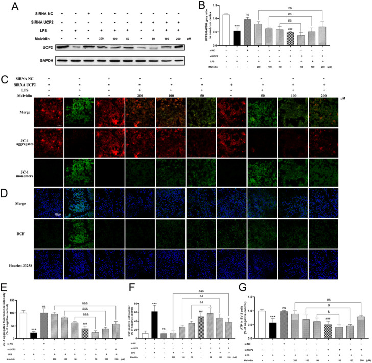
This study aimed to explore the protective roles of malvidin in life-threatened sepsis-associated encephalopathy (SAE) and illustrate the underlying mechanism. SAE mice models were developed and treated with malvidin for subsequently protective effects evaluation. Malvidin restored neurobehavioral retardation, declined serum S100β and NSE levels, sustained cerebrum morphological structure, improved blood-brain barrier integrity with elevated tight junction proteins, and decreased evans blue leakage, and finally protect SAE mice from brain injury. Mechanistically, malvidin prevented cerebrum from mitochondrial dysfunction with enhanced JC-1 aggregates and ATP levels, and ROS accumulation with decreased lipid peroxidation and increased antioxidant enzymes. UCP2 protein levels were found to be decreased after LPS stimulation in the cerebrum and BV-2 cells, and malvidin recovered its levels in a ROS dependent manner. In vivo inhibition of UCP2 with genipin or in vitro interference with siRNA UCP2 both disrupted the mitochondrial membrane potential, decreased ATP levels and intensified DCF signals, being a key target for malvidin. Moreover, dorsomorphin block assays verified that malvidin upregulated UCP2 expression through phosphorylating AMPK in SAE models. Also, malvidin alleviated SAE progression through inhibition of ROS-dependent NLRP3 inflammasome activation mediated serum pro-inflammatory cytokines secretion and mitochondrial pathway mediated apoptosis with weakened apoptosis body formation and tunel positive signals, and decreased Bax, cytochrome C, caspase-3 and increased Bcl-2 protein levels. Overall, this study illustrated that malvidin targeted AMPK-α/UCP2 axis to restore LPS-induced mitochondrial dysfunction and alleviate ROS accumulation, which further inhibits NLRP3 inflammasome activation and mitochondrial apoptosis in a ROS dependent way, and ultimately protected SAE mice, providing a reference for the targeted development of SAE prophylactic approach.
Overview
The claims and statistics of this paper haven’t been checked yet.
Free: it reads the open-access text and checks the first 5 claims that cite a source we can check. Create a free account to check every claim.
Integrity signals
Registry
Cites
Statistics
Figures




Verification
Claims the paper makes
The claims and statistics of this paper haven’t been checked yet.
Free: it reads the open-access text and checks the first 5 claims that cite a source we can check. Create a free account to check every claim.
Claims other papers make about this one
Each is checked against this paper’s own text: does it say what the citing paper says it does?
The source backs part of the claim, not all of it — or backs it for a different population, condition or comparison.
In the citing paper
“echinomycin improves mitochondrial structure and reduces inflammatory factors via the HIF-1α/BNIP3L axis [85]; ② Natural compounds (preclinical): Puerarin enhances BBB stability by blocking GSDMD cleavage; myricitrin alleviates neuroinflammation by modulating the Bax/Bcl-2 balance [86]; malvidin dually inhibits pyroptosis and apoptosis via the AMPKα/UCP2 axis [87].”
From Research Advances in the Pathogenesis of Sepsis-Associated Encephalopathy (2026) · cites this paper as [87]
Experimental — accuracy not yet measuredThe source backs part of the claim, not all of it — or backs it for a different population, condition or comparison.
In the citing paper
“echinomycin improves mitochondrial structure and reduces inflammatory factors via the HIF-1α/BNIP3L axis [85]; ② Natural compounds (preclinical): Puerarin enhances BBB stability by blocking GSDMD cleavage; myricitrin alleviates neuroinflammation by modulating the Bax/Bcl-2 balance [86]; malvidin dually inhibits pyroptosis and apoptosis via the AMPKα/UCP2 axis [87].”
From Research Advances in the Pathogenesis of Sepsis-Associated Encephalopathy (2026) · cites this paper as [87]
Experimental — accuracy not yet measured
Self-check
Percentages, sums and reported differences re-computed from the paper’s own numbers.
The self-check needs the paper’s full text.
Citations
Cites
The third international consensus definitions for sepsis and septic shock (Sepsis-3).
Singer, M., Deutschman, C. S., Seymour, C. W., Shankar-Hari, M., Annane, D., Bauer, M., Bellomo, R., Bernard, G. R., Chiche, J.-D., Coopersmith, C. M., et al. (2016) · JAMA · doi:10.1001/jama.2016.0287 · Cited by 28,250 · cited 2×
“Sepsis, a serious clinical syndrome in the field of critical care, is a life-threatening multi-organ dysfunction caused by an uncontrolled acute systemic inflammatory response of the host to bacterial toxins and has become a major public health problem worldwide (Singer et al., 2016).”
BackgroundThe NLRP3 Inflammasome: an overview of mechanisms of activation and regulation
Kelley, N., Jeltema, D., Duan, Y., & He, Y. (2019) · International Journal of Molecular Sciences · doi:10.3390/ijms20133328 · Cited by 3,539 · cited 1×
“ROS stimulation mediates NLRP3 oligomerization with ASC, recruitment of inflammatory caspases, assembly of inflammasomes to produce activated caspases, cleavage of IL1β precursors into mature IL-1β, and mediation of inflammatory responses (Kelley et al., 2019).”
BackgroundReactive oxygen species, cellular redox systems, and apoptosis
Circu, M. L., & Aw, T. Y. (2010) · Free Radical Biology and Medicine · doi:10.1016/j.freeradbiomed.2009.12.022 · Cited by 3,340 · cited 1×
“ROS accumulation promotes apoptosis and causes mitochondrial membrane potential changes, leading to the translocation of Bax and Bad across the mitochondrial membrane, and release of cytochrome c (Circu and Aw, 2010).”
BackgroundThe blood-brain barrier/neurovascular unit in health and disease
Hawkins, B. T., & Davis, T. P. (2005) · Pharmacological Reviews · doi:10.1124/pr.57.2.4 · Cited by 2,661 · cited 1×
“circulating blood into the CNS to maintain the dynamic balance of the internal environment (Hawkins and Davis, 2005).”
BackgroundOxidative Stress and Inflammation: What Polyphenols Can Do for Us?
Hussain, T., Tan, B., Yin, Y., Blachier, F., Tossou, M. C. B., & Rahu, N. (2016) · Oxidative Medicine and Cellular Longevity · doi:10.1155/2016/7432797 · Cited by 2,082 · cited 1×
“…are the most common active polyphenols, widely distributed in plants, with a variety of pharmacological effects, including anti-inflammatory (Hussain et al., 2016), anti-cancer (Kopustinskiene et al., 2020), anti-proliferative (Fernández et al., 2021), antioxidant, and inhibition of free…”
BackgroundEndonuclease G is an apoptotic DNase when released from mitochondria
Li, L. Y., Luo, X., & Wang, X. (2001) · Nature · doi:10.1038/35083620 · Cited by 1,787 · cited 1×
“Caspase-3 is downstream of the apoptotic cascade and is a common executor of several apoptotic pathways, which can directly cause apoptosis by shearing the structural proteins of the cell (Li et al., 2001).”
BackgroundMicroglia as a source and target of cytokines
Hanisch, U. (2002) · Glia · doi:10.1002/glia.10161 · Cited by 1,645 · cited 1×
“Microglia BV-2, an important line of defense for the CNS, are part of the mononuclear phagocyte system and are involved in innate defense mechanisms of the CNS, contributing to tissue repair and recovery (Hanisch, 2002).”
BackgroundNitric oxide in the central nervous system: neuroprotection versus neurotoxicity
Calabrese, V., Mancuso, C., Calvani, M., Rizzarelli, E., Butterfield, D. A., & Giuffrida Stella, A. M. (2007) · Nature Reviews Neuroscience · doi:10.1038/nrn2214 · Cited by 1,543
Molecular mechanisms regulating NLRP3 inflammasome activation
Jo, E.-K., Kim, J. K., Shin, D.-M., & Sasakawa, C. (2016) · Cellular & Molecular Immunology · doi:10.1038/cmi.2015.95 · Cited by 1,378 · cited 1×
“Activation of NLRP3 inflammasome will secrete pro-inflammatory factors IL-1β and IL-18, which regulate the inflammatory response, and ROS accumulation is thought to be an important pathway for NLRP3 inflammasome activation (Jo et al., 2016).”
BackgroundFlavonoids as Anticancer Agents
Kopustinskiene, D. M., Jakstas, V., Savickas, A., & Bernatoniene, J. (2020) · Nutrients · doi:10.3390/nu12020457 · Cited by 1,295 · cited 2×
“The mammalian genome encodes five UCP homologues, UCP1-5 (Krauss et al., 2005).”
BackgroundParhiz, H., Roohbakhsh, A., Soltani, F., Rezaee, R., & Iranshahi, M. (2015) · Phytotherapy Research · doi:10.1002/ptr.5256 · Cited by 908 · cited 1×
“, 2021), antioxidant, and inhibition of free radical-mediated processes (Parhiz et al., 2015).”
BackgroundSepsis-associated encephalopathy
Gofton, T. E., & Young, G. B. (2012) · Nature Reviews Neurology · doi:10.1038/nrneurol.2012.183 · Cited by 821
Mitochondrial Cristae: Where Beauty Meets Functionality
Cogliati, S., Enriquez, J. A., & Scorrano, L. (2016) · Trends in Biochemical Sciences · doi:10.1016/j.tibs.2016.01.001 · Cited by 816 · cited 1×
“(Annesley and Fisher, 2019), as well as maintaining calcium homeostasis and endogenous apoptosis and autophagy (Cogliati et al., 2016).”
BackgroundPhysiological functions of the mitochondrial uncoupling proteins UCP2 and UCP3
Brand, M. D., & Esteves, T. C. (2005) · Cell Metabolism · doi:10.1016/j.cmet.2005.06.002 · Cited by 785
Microglia and neurodegeneration: The role of systemic inflammation
Cunningham, C. (2013) · Glia · doi:10.1002/glia.22350 · Cited by 769 · cited 9×
“Microglia, a primary innate immune cell in the CNS, are the first defense line against infection and brain tissue injury.”
BackgroundInfluentialAMPK: a key regulator of energy balance in the single cell and the whole organism
Hardie, D. G. (2008) · International Journal of Obesity · doi:10.1038/ijo.2008.116 · Cited by 703
The mitochondrial uncoupling-protein homologues
Krauss, S., Zhang, C.-Y., & Lowell, B. B. (2005) · Nature Reviews Molecular Cell Biology · doi:10.1038/nrm1592 · Cited by 663
The mitochondrial pathways of apoptosis
Estaquier, J., Vallette, F., Vayssiere, J.-L., & Mignotte, B. (2012) · Advances in experimental medicine and biology · doi:10.1007/978-94-007-2869-1_7 · Cited by 637 · cited 1×
“The release of cytochrome c into the cytoplasm and caspase-9-mediated caspase-3 activation mediates the mitochondria pathway apoptosis (Estaquier et al., 2012).”
BackgroundMitochondria in Health and Disease
Annesley, S. J., & Fisher, P. R. (2019) · Cells · doi:10.3390/cells8070680 · Cited by 551
Rabinovitch, R. C., Samborska, B., Faubert, B., Ma, E. H., Gravel, S.-P., Andrzejewski, S., Raissi, T. C., Pause, A., St.-Pierre, J., & Jones, R. G. (2017) · Cell Reports · doi:10.1016/j.celrep.2017.09.026 · Cited by 543 · cited 1×
“AMPK is an evolutionarily conserved stress-sensing protein kinase, that is, activated to maintain cellular metabolic homeostasis by sensing a decrease in ATP (Rabinovitch et al., 2017).”
BackgroundRen, Q., Guo, F., Tao, S., Huang, R., Ma, L., & Fu, P. (2020) · Biomedicine & Pharmacotherapy · doi:10.1016/j.biopha.2019.109772 · Cited by 424 · cited 4×
“, 2008), kidney damage (Ren et al., 2020), and heart damage (Wu B.”
BackgroundInfluentialLong-term cerebral consequences of sepsis
Widmann, C. N., & Heneka, M. T. (2014) · The Lancet Neurology · doi:10.1016/s1474-4422(14)70017-1 · Cited by 361 · cited 1×
“SAE manifests as diffuse brain dysfunction and cognitive dysfunction, and is positively associated with mortality in sepsis patients (Widmann and Heneka, 2014; Helbing et al., 2018).”
BackgroundPathological alteration and therapeutic implications of sepsis-induced immune cell apoptosis
Cao, C., Yu, M., & Chai, Y. (2019) · Cell Death & Disease · doi:10.1038/s41419-019-2015-1 · Cited by 354 · cited 1×
“Apoptosis is a kind of programmed cell death and is involved in the development of sepsis and SAE (Cao et al., 2019; Zhou et al., 2020).”
BackgroundUnderstanding brain dysfunction in sepsis
Sonneville, R., Verdonk, F., Rauturier, C., Klein, I. F., Wolff, M., Annane, D., Chretien, F., & Sharshar, T. (2013) · Annals of Intensive Care · doi:10.1186/2110-5820-3-15 · Cited by 352 · cited 1×
“Patients with sepsis are often associated with CNS symptoms (Sonneville et al., 2013).”
BackgroundSharshar, T., Gray, F., de la Grandmaison, G. L., Hopklnson, N. S., Ross, E., Dorandeu, A., Orlikowski, D., Raphael, J.-C., Gajdos, P., & Annane, D. (2003) · The Lancet · doi:10.1016/s0140-6736(03)14899-4 · Cited by 341 · cited 1×
“main causes of neurological deficits and eventual development of SAE in patients with sepsis, as well as an important pathophysiological basis for SAE (Sharshar et al., 2003; Messaris et al., 2004).”
BackgroundCrosstalk between ER stress, NLRP3 inflammasome, and inflammation
Li, W., Cao, T., Luo, C., Cai, J., Zhou, X., Xiao, X., & Liu, S. (2020) · Applied Microbiology and Biotechnology · doi:10.1007/s00253-020-10614-y · Cited by 302 · cited 1×
“Host NLRP3 inflammasome, a multimeric protein complex, could sense various pathogen associated molecular patterns, endogenous danger-associated molecular patterns as well as ROS, and involves in regulating inflammation (Li et al., 2020).”
BackgroundCognitive decline after sepsis.
Annane, D., & Sharshar, T. (2015) · The Lancet Respiratory Medicine · doi:10.1016/s2213-2600(14)70246-2 · Cited by 290 · cited 1×
“It is reported that SAE patients have a mortality rate of 70%, while about 10%–20% of SAE survivors may develop long-term neurocognitive deficits, psychiatric disorders, and neurodegenerative diseases (Annane and Sharshar, 2015; Heming et al., 2017).”
BackgroundDeoxynivalenol induced apoptosis and inflammation of IPEC-J2 cells by promoting ROS production
Kang, R., Li, R., Dai, P., Li, Z., Li, Y., & Li, C. (2019) · Environmental Pollution · doi:10.1016/j.envpol.2019.05.026 · Cited by 289 · cited 1×
“Excess ROS accumulation promotes apoptosis (Wang et al., 2017; Kang et al., 2019).”
Blood-Brain Barrier Disruption by Lipopolysaccharide and Sepsis-Associated Encephalopathy
Peng, X., Luo, Z., He, S., Zhang, L., & Li, Y. (2021) · Frontiers in Cellular and Infection Microbiology · doi:10.3389/fcimb.2021.768108 · Cited by 267 · cited 1×
“Tight junction (TJ) proteins maintain BBB integrity (Peng et al., 2021).”
BackgroundAntiproliferative and palliative activity of flavonoids in colorectal cancer
Fernández, J., Silván, B., Entrialgo-Cadierno, R., Villar, C. J., Capasso, R., Uranga, J. A., Lombó, F., & Abalo, R. (2021) · Biomedicine & Pharmacotherapy · doi:10.1016/j.biopha.2021.112241 · Cited by 258 · cited 1×
“, 2020), anti-proliferative (Fernández et al., 2021), antioxidant, and inhibition of free radical-mediated processes (Parhiz et al.”
BackgroundSepsis-Associated Encephalopathy and Blood-Brain Barrier Dysfunction
Gao, Q., & Hernandes, M. S. (2021) · Inflammation · doi:10.1007/s10753-021-01501-3 · Cited by 257 · cited 1×
“However, the systemic inflammation caused by sepsis leads to activation of brain endothelial cells, which significantly disrupts the integrity of the BBB and causes acute brain dysfunction (Gao and Hernandes, 2021).”
BackgroundPeroxisome proliferator-activated receptor γ (PPARγ): A master gatekeeper in CNS injury and repair
Cai, W., Yang, T., Liu, H., Han, L., Zhang, K., Hu, X., Zhang, X., Yin, K.-J., Gao, Y., Bennett, M. V. L., et al. (2018) · Progress in Neurobiology · doi:10.1016/j.pneurobio.2017.10.002 · Cited by 236 · cited 1×
“Increased production of ROS attacks cardiolipin, an important component, and disrupts the activity of related enzymes (Cai et al., 2018).”
BackgroundBlood biomarkers for brain injury: What are we measuring?
Kawata, K., Liu, C. Y., Merkel, S. F., Ramirez, S. H., Tierney, R. T., & Langford, D. (2016) · Neuroscience & Biobehavioral Reviews · doi:10.1016/j.neubiorev.2016.05.009 · Cited by 235 · cited 1×
“S100β and NSE are widely recognized as biomarkers of central nervous system injury (Kawata et al., 2016).”
BackgroundManaenko, A., Chen, H., Kammer, J., Zhang, J. H., & Tang, J. (2011) · Journal of Neuroscience Methods · doi:10.1016/j.jneumeth.2010.12.013 · Cited by 232 · cited 1×
“The evans blue leakage into the cerebrum was quantified by the established standard curve (Manaenko et al., 2011).”
MethodFu, Q., Wu, J., Zhou, X.-Y., Ji, M.-H., Mao, Q.-H., Li, Q., Zong, M.-M., Zhou, Z.-Q., & Yang, J.-J. (2019) · Inflammation · doi:10.1007/s10753-018-0894-4 · Cited by 232
Sepsis-associated encephalopathy and septic encephalitis: an update
Tauber, S. C., Djukic, M., Gossner, J., Eiffert, H., Brück, W., & Nau, R. (2021) · Expert Review of Anti-infective Therapy · doi:10.1080/14787210.2020.1812384 · Cited by 228 · cited 1×
“Sepsis is accompanied with systemic inflammation, which could be transmitted into the central nervous system via destroyed blood-brain barrier (Tauber et al., 2021).”
BackgroundXu, X.-E., Liu, L., Wang, Y.-C., Wang, C.-T., Zheng, Q., Liu, Q.-X., Li, Z.-F., Bai, X.-J., & Liu, X.-H. (2019) · Brain, Behavior, and Immunity · doi:10.1016/j.bbi.2019.05.038 · Cited by 211
Haileselassie, B., Joshi, A. U., Minhas, P. S., Mukherjee, R., Andreasson, K. I., & Mochly-Rosen, D. (2020) · Journal of Neuroinflammation · doi:10.1186/s12974-019-1689-8 · Cited by 203 · cited 2×
“Frontiers in Pharmacology frontiersin.org07 Malvidin prevents cerebrum mitochondrial dysfunction and ROS accumulation in response to LPS stimulation Mitochondrial dysfunction is an important causative factor in sepsis-induced multiorgan failure, including SAE (Haileselassie et al., 2020).”
BackgroundAqil, F., Gupta, A., Munagala, R., Jeyabalan, J., Kausar, H., Sharma, R. J., Singh, I. P., & Gupta, R. C. (2012) · Nutrition and Cancer · doi:10.1080/01635581.2012.657766 · Cited by 188 · cited 1×
“Anthocyanins are a group of flavonoids with similar anti-inflammatory and antioxidant properties to other members of flavonoids family (Aqil et al., 2012).”
BackgroundThe Emerging Functions of UCP2 in Health, Disease, and Therapeutics
Mattiasson, G., & Sullivan, P. G. (2006) · Antioxidants & Redox Signaling · doi:10.1089/ars.2006.8.1 · Cited by 177 · cited 1×
“…energy metabolism disruption, reduced antioxidant production, and free radical accumulation are the main causes of neurological deficits and eventual development of SAE in patients with sepsis, as well as an important pathophysiological basis for SAE (Sharshar et al., 2003; Messaris et al., 2004).”
BackgroundInhibition of the NLRP3/IL‐1β axis protects against sepsis‐induced cardiomyopathy
Busch, K., Kny, M., Huang, N., Klassert, T. E., Stock, M., Hahn, A., Graeger, S., Todiras, M., Schmidt, S., Chamling, B., et al. (2021) · Journal of Cachexia, Sarcopenia and Muscle · doi:10.1002/jcsm.12763 · Cited by 167 · cited 1×
“Previous studies demonstrated that NLRP3 inflammasome and inflammatory caspase promoted septic heart injury and kidney injury (Deng et al., 2020; Busch et al., 2021).”
BackgroundNeuroanatomy of sepsis-associated encephalopathy
Heming, N., Mazeraud, A., Verdonk, F., Bozza, F. A., Chrétien, F., & Sharshar, T. (2017) · Critical Care · doi:10.1186/s13054-017-1643-z · Cited by 165 · cited 1×
“It is reported that SAE patients have a mortality rate of 70%, while about 10%–20% of SAE survivors may develop long-term neurocognitive deficits, psychiatric disorders, and neurodegenerative diseases (Annane and Sharshar, 2015; Heming et al., 2017).”
BackgroundQuoilin, C., Mouithys-Mickalad, A. A., Lécart, S., Fontaine‐Aupart, M., & Hoebeke, M. (2014) · Biochimica et Biophysica Acta (BBA) - Bioenergetics · doi:10.1016/j.bbabio.2014.07.005 · Cited by 163 · cited 1×
“However, the inflammatory cytokines that erupt during sepsis can also increase the expression of reduced nicotinamide purine dinucleotide phosphate oxidase and nitric oxide synthase, which in turn initiate oxidative stress and lead to increased ROS production (Quoilin et al., 2014).”
BackgroundYao, X., Carlson, D., Sun, Y., Ma, L., Wolf, S. E., Minei, J. P., & Zang, Q. S. (2015) · PLOS ONE · doi:10.1371/journal.pone.0139416 · Cited by 145 · cited 1×
“Previous studies indicated that inhibition of oxidative stress and protection of mitochondrial function could improve organ dysfunction caused by sepsis (Yao et al., 2015; Neri et al., 2016).”
BackgroundMitochondrial UCP2 in the central regulation of metabolism
Toda, C., & Diano, S. (2014) · Best Practice & Research Clinical Endocrinology & Metabolism · doi:10.1016/j.beem.2014.02.006 · Cited by 138 · cited 1×
“, 1999), regulating mitochondrial membrane potential and the production of ROS (Toda and Diano, 2014).”
BackgroundLuteolin attenuates sepsis‑induced myocardial�injury by enhancing autophagy in mice
Wu, B., Song, H., Fan, M., You, F., Zhang, L., Luo, J., Li, J., Wang, L., Li, C., & Yuan, M. (2020) · International Journal of Molecular Medicine · doi:10.3892/ijmm.2020.4536 · Cited by 135
Sepsis induces brain mitochondrial dysfunction.
d’Avila, J. D. C. P., Santiago, A. P. S. A., Amâncio, R. T., Galina, A., Oliveira, M. F., & Bozza, F. A. (2008) · Critical Care Medicine · doi:10.1097/ccm.0b013e3181760c4b · Cited by 134
Zhang, W.-X., He, B.-M., Wu, Y., Qiao, J.-F., & Peng, Z.-Y. (2019) · Life Sciences · doi:10.1016/j.lfs.2018.11.055 · Cited by 133 · cited 2×
“Apoptosis is a non-inflammatory cell death involved in sepsis-induced cardiac dysfunction (Zhang et al., 2019) and septic acute kidney injury (Koçkara and Kayataş, 2013).”
BackgroundSepsis-induced brain dysfunction
Adam, N., Kandelman, S., Mantz, J., Chrétien, F., & Sharshar, T. (2013) · Expert Review of Anti-infective Therapy · doi:10.1586/eri.12.159 · Cited by 132
Chen, S., Tang, C., Ding, H., Wang, Z., Liu, X., Chai, Y., Jiang, W., Han, Y., & Zeng, H. (2020) · Frontiers in Immunology · doi:10.3389/fimmu.2020.594071 · Cited by 132
Huang, J., Liu, W., Doycheva, D. M., Gamdzyk, M., Lu, W., Tang, J., & Zhang, J. H. (2019) · Free Radical Biology and Medicine · doi:10.1016/j.freeradbiomed.2019.07.001 · Cited by 116
Toklu, H. Z., Akbay, T. T., Velioğlu‐Öğünç, A., Ercan, F., Gedik, N., Keyer‐Uysal, M., & Şener, G. (2008) · Journal of Surgical Research · doi:10.1016/j.jss.2007.03.072 · Cited by 110 · cited 1×
“, 2019), cognitive dysfunction (Karimian, 2020), and sepsis-induced lung damage (Toklu et al., 2008), kidney damage (Ren et al.”
BackgroundYao, B., Zhang, L., Ai, Y., liu, Z.-Y., & Huang, L. (2014) · Neurochemical Research · doi:10.1007/s11064-014-1308-0 · Cited by 109
Betulinic Acid Induces Apoptosis in Differentiated PC12 Cells Via ROS-Mediated Mitochondrial Pathway
Wang, X., Lu, X., Zhu, R., Zhang, K., Li, S., Chen, Z., & Li, L. (2017) · Neurochemical Research · doi:10.1007/s11064-016-2147-y · Cited by 109 · cited 1×
“Excess ROS accumulation promotes apoptosis (Wang et al., 2017; Kang et al., 2019).”
Kim, Y. W., Lee, S. M., Shin, S. M., Hwang, S. J., Brooks, J. S., Kang, H. E., Lee, M. G., Kim, S. C., & Kim, S. G. (2009) · Free Radical Biology and Medicine · doi:10.1016/j.freeradbiomed.2009.07.018 · Cited by 106 · cited 1×
“Frontiers in Pharmacology frontiersin.org10 2008) and regulating oxidative stress (Kim et al., 2009).”
BackgroundMitochondrial Permeability Transition Pore and Calcium Handling
Wong, R., Steenbergen, C., & Murphy, E. (2012) · Methods in Molecular Biology · doi:10.1007/978-1-61779-382-0_15 · Cited by 97
Ding, Y., Zheng, Y., Huang, J., Peng, W., Chen, X., Kang, X., & Zeng, Q. (2019) · International Immunopharmacology · doi:10.1016/j.intimp.2019.03.043 · Cited by 84 · cited 2×
“Mice were gavaged with genipin (100 mg/kg BW, Selleck, Shanghai) once a day for 3 days before LPS injection (Ding et al., 2019), and only genipin treatment mice were used as”
BackgroundMethodTsukahara, S., Yamamoto, S., Shwe, T.-T.-W., Ahmed, S., Kunugita, N., Arashidani, K., & Fujimaki, H. (2006) · Neuroimmunomodulation · doi:10.1159/000094829 · Cited by 80 · cited 1×
“Under physiological conditions, the protein expression of the antiapoptotic gene Bcl-2 and the pro-apoptotic gene bax are in a relatively stable state, and overexpression of Bax can result in a significant increase in the number of bax/bax homodimers, and an increased cellular responsiveness to death signals, initiati…”
BackgroundUCP2 Inhibits ROS-Mediated Apoptosis in A549 under Hypoxic Conditions
Deng, S., Yang, Y., Han, Y., Li, X., Wang, X., Li, X., Zhang, Z., & Wang, Y. (2012) · PLoS ONE · doi:10.1371/journal.pone.0030714 · Cited by 79 · cited 1×
“inhibit the inflammatory response and the development of apoptosis (Deng et al., 2012; Zhong et al., 2019).”
BackgroundSelf-citationPrudente, A. S., Loddi, A. M. V., Duarte, M. D. R., Santos, A. R. S., Pochapski, M. T., Pizzolatti, M. G., Hayashi, S. S., Campos, F. R., Pontarolo, R., dos Santos, F. A., et al. (2013) · Food and Chemical Toxicology · doi:10.1016/j.fct.2013.04.042 · Cited by 77 · cited 1×
“Malvidin is the most common anthocyanin with antioxidant, anti-inflammatory, antibacterial, analgesic effects and anticholinesterase activity (Prudente et al., 2013).”
BackgroundTranscranial Doppler to assess sepsis-associated encephalopathy in critically ill patients
Pierrakos, C., Attou, R., Decorte, L., Kolyviras, A., Malinverni, S., Gottignies, P., Devriendt, J., & De Bels, D. (2014) · BMC Anesthesiology · doi:10.1186/1471-2253-14-45 · Cited by 75
Mitochondrial Dysfunction during Sepsis
Cesar Pontes Azevedo, L. (2010) · Endocrine, Metabolic & Immune Disorders - Drug Targets · doi:10.2174/187153010791936946 · Cited by 72 · cited 1×
“Meanwhile, several studies reported that the overproduction of TNF-α and IL-6 in SAE rats could further lead to other pathological changes such as oxidative stress, neuronal apoptosis, etc, (Calabrese et al., 2007; Azevedo, 2010; Towner et al., 2018).”
BackgroundNeri, M., Riezzo, I., Pomara, C., Schiavone, S., & Turillazzi, E. (2016) · Mediators of Inflammation · doi:10.1155/2016/3423450 · Cited by 70 · cited 1×
“Previous studies indicated that inhibition of oxidative stress and protection of mitochondrial function could improve organ dysfunction caused by sepsis (Yao et al., 2015; Neri et al., 2016).”
BackgroundRenal Cell Apoptosis and New Treatment Options in Sepsis-Induced Acute Kidney Injury
Koçkara, A., & Kayataş, M. (2013) · Renal Failure · doi:10.3109/0886022x.2012.744040 · Cited by 69 · cited 1×
“, 2019) and septic acute kidney injury (Koçkara and Kayataş, 2013).”
BackgroundTime-dependent mitochondrial-mediated programmed neuronal cell death prolongs survival in sepsis*
Messaris, E., Memos, N., Chatzigianni, E., Konstadoulakis, M. M., Menenakos, E., Katsaragakis, S., Voumvourakis, C., & Androulakis, G. (2004) · Critical Care Medicine · doi:10.1097/01.ccm.0000135744.30137.b4 · Cited by 68 · cited 1×
“main causes of neurological deficits and eventual development of SAE in patients with sepsis, as well as an important pathophysiological basis for SAE (Sharshar et al., 2003; Messaris et al., 2004).”
BackgroundDando, I., Pacchiana, R., Pozza, E. D., Cataldo, I., Bruno, S., Conti, P., Cordani, M., Grimaldi, A., Butera, G., Caraglia, M., et al. (2017) · Free Radical Biology and Medicine · doi:10.1016/j.freeradbiomed.2017.09.022 · Cited by 67 · cited 1×
“For inhibition of UCP2, BV-2 cells were transiently transfected with small interfering RNA targeting to UCP2 (siUCP2, 5′-GCUAAAGUCCGGUUACAGATT-3′, General Biol, Anhui, China) (Dando et al., 2017) using Lipofectamine 3000 transfection reagent (Invitrogen, United States) referring to the instructions.”
MethodLB100 ameliorates nonalcoholic fatty liver disease via the AMPK/Sirt1 pathway
Chen, X., Cai, C., Yu, M., Feng, Z., Zhang, Y.-W., Liu, P., Zeng, H., & Yu, C. (2019) · World Journal of Gastroenterology · doi:10.3748/wjg.v25.i45.6607 · Cited by 65 · cited 1×
“For inhibition of AMP-activated protein kinase α (AMPKα), BV-2 cells were pretreated with dorsomorphin (10 μM, MCE, Shanghai) for 2 h (Chen et al., 2019) and then incubated with malvidin followed by LPS stimulation.”
MethodZhu, Y., Wang, K., Ma, Z., Liu, D., Yang, Y., Sun, M., Wen, A., Hao, Y., Ma, S., Ren, F., et al. (2019) · Toxicology and Applied Pharmacology · doi:10.1016/j.taap.2018.10.013 · Cited by 60 · cited 2×
“Several studies have demonstrated that flavonoids alleviate sepsis brain injury (Zhu et al., 2019), memory impairment (Gong et al.”
BackgroundSepsis-associated encephalopathy
Helbing, D.-L., Böhm, L., & Witte, O. W. (2018) · Canadian Medical Association Journal · doi:10.1503/cmaj.180454 · Cited by 58 · cited 1×
“SAE manifests as diffuse brain dysfunction and cognitive dysfunction, and is positively associated with mortality in sepsis patients (Widmann and Heneka, 2014; Helbing et al., 2018).”
BackgroundTowner, R. A., Saunders, D., Smith, N., Towler, W., Cruz, M., Do, S., Maher, J. E., Whitaker, K., Lerner, M. R., & Morton, K. A. (2018) · GeroScience · doi:10.1007/s11357-018-0009-z · Cited by 56
Wu, L., Feng, Q., Ai, M.-L., Deng, S.-Y., Liu, Z.-Y., Huang, L., Ai, Y.-H., & Zhang, L. (2020) · Scientific Reports · doi:10.1038/s41598-020-64200-3 · Cited by 56
Zhong, X., He, J., Zhang, X., Li, C., Tian, X., Xia, W., Gan, H. T., & Xia, Y. (2019) · Biomedicine & Pharmacotherapy · doi:10.1016/j.biopha.2019.108914 · Cited by 48 · cited 1×
“inhibit the inflammatory response and the development of apoptosis (Deng et al., 2012; Zhong et al., 2019).”
BackgroundRole of Peroxynitrite in the Cardiovascular Dysfunction of Septic Shock
Liaudet, L., Rosenblatt‐Velin, N., & Pacher, P. (2013) · Current Vascular Pharmacology · doi:10.2174/1570161111311020009 · Cited by 44
Zhou, R., Li, Y., Qu, Y., Huang, Q., Sun, X., Mu, D., & Li, X. (2020) · CNS Neuroscience & Therapeutics · doi:10.1111/cns.13229 · Cited by 42 · cited 3×
“Malvidin inhibits apoptosis in LPS stimulated mice cerebrum and BV-2 cells in ROS dependent way Apoptosis is involved in several organs dysfunction in sepsisinduced injury (Zhang et al., 2019; Ren et al., 2020; Zhou et al., 2020).”
BackgroundSelf-citationBastin, A., Sadeghi, A., Nematollahi, M. H., Abolhassani, M., Mohammadi, A., & Akbari, H. (2020) · Journal of Cellular Physiology · doi:10.1002/jcp.30049 · Cited by 42 · cited 1×
“, 2020), human THP-1 cells (Bastin et al., 2021) and other cell types.”
BackgroundGuo, J., Zhang, K., Ji, Y., Jiang, X., & Zuo, S. (2008) · Journal of Huazhong University of Science and Technology [Medical Sciences] · doi:10.1007/s11596-008-0311-6 · Cited by 36 · cited 1×
“Reduced Bcl-2 can inhibit caspase-3 activation by promoting cytochrome c release and promoting apoptosis (Guo et al., 2008).”
BackgroundUCP2 protects hypothalamic cells from TNF‐α‐induced damage
Degasperi, G. R., Romanatto, T., Denis, R., ARAUJO, E. P. D., Moraes, J. C., Inada, N. M., Vercesi, A. E., & Velloso, L. A. (2008) · FEBS Letters · doi:10.1016/j.febslet.2008.08.006 · Cited by 35 · cited 1×
“UCP2 is abundantly expressed in neurons and immune cells (Mattiasson and Sullivan, 2006; Degasperi et al., 2008).”
BackgroundMitochondrial Permeability Transition Pore and Calcium Handling
Parks, R. J., Murphy, E., & Liu, J. C. (2018) · Methods in molecular biology · doi:10.1007/978-1-4939-7831-1_11 · Cited by 31 · cited 1×
“ATP is an important indicator of mitochondrial function, and mitochondrial dysfunction can lead to a decrease in mitochondrial ATP synthesis, cellular dysfunction, and a decrease in antioxidant synthetic components, leading to a further increase in ROS and ultimately a vicious cycle (Parks et al., 2018).”
BackgroundChung, C., Venet, F., Chen, Y., Jones, L. N., Wilson, D. C., Ayala, C. A., & Ayala, A. (2010) · Shock · doi:10.1097/shk.0b013e3181cf70fb · Cited by 30 · cited 1×
“It was reported that the protein expression of Bcl-2 in peripheral blood was significantly decreased and cells were abnormally apoptotic during sepsis (Chung et al., 2010).”
BackgroundCerebral blood flow and serum neuron-specific enolase in early-onset neonatal sepsis
Shimy, M. S. E., El-Raggal, N. M., El-Farrash, R. A., Shaaban, H. A., Mohamed, H. E., Barakat, N. M., Farag, A. S., Zohiery, A. K. E., Shaaban, M. A. A., & Salama, D. H. (2018) · Pediatric Research · doi:10.1038/s41390-018-0062-4 · Cited by 30
UCP2 regulates cholangiocarcinoma cell plasticity via mitochondria-to-AMPK signals
Yu, J., Shi, L., Shen, X., & Zhao, Y. (2019) · Biochemical Pharmacology · doi:10.1016/j.bcp.2019.05.017 · Cited by 30
Chen, W., Luo, S., Xie, P., Hou, T., Yu, T., & Fu, X. (2018) · Europe PMC (PubMed Central) · Cited by 29
Bastin, A. R., Sadeghi, A., Abolhassani, M., Doustimotlagh, A. H., & Mohammadi, A. (2020) · IUBMB Life · doi:10.1002/iub.2286 · Cited by 25 · cited 2×
“It has been shown that malvidin can alleviate oxidative stress and inflammation in LPS stimulated human peripheral blood mononuclear cells (Bastin et al., 2020), human THP-1 cells (Bastin et al.”
BackgroundMitochondrial uncoupler triclosan induces vasorelaxation of rat arteries
Zhang, X., Zhang, X., Zhang, Y., Liu, M., Jin, J., Yan, J., Shen, X., Hu, N., & Dong, D. (2017) · Acta Pharmaceutica Sinica B · doi:10.1016/j.apsb.2017.06.001 · Cited by 23
Joshi, L., Plastira, I., Bernhart, E. M., Reicher, H., Triebl, A., Köfeler, H. C., & Sattler, W. (2021) · International Journal of Molecular Sciences · doi:10.3390/ijms22168519 · Cited by 21 · cited 1×
“And BV2 cell line is an ideal in vitromodel for SAE study (Joshi et al., 2021).”
BackgroundDeng, H., Chen, F., Wang, Y., Jiang, H., Dong, Z., Yuan, B., & Zhao, X. (2020) · Saudi Journal of Biological Sciences · doi:10.1016/j.sjbs.2020.03.018 · Cited by 20 · cited 1×
“Previous studies demonstrated that NLRP3 inflammasome and inflammatory caspase promoted septic heart injury and kidney injury (Deng et al., 2020; Busch et al., 2021).”
BackgroundGong, J., Luo, S., Zhao, S., Yin, S.-L., Li, X., & Mou, T. (2019) · Folia Neuropathologica · doi:10.5114/fn.2019.89856 · Cited by 18 · cited 2×
“, 2019), memory impairment (Gong et al., 2019), cognitive dysfunction (Karimian, 2020) by exerting their antioxidant and anti-inflammatory functions.”
BackgroundSelf-citationLe, K. M., Kuhla, A., Abshagen, K., Minor, T., Stegemann, J., Ibrahim, S. M., Eipel, C., & Vollmar, B. (2008) · Critical Care Medicine · doi:10.1097/ccm.0b013e31819260ae · Cited by 16
Contribution to the Identification and Analysis of the Mitochondrial Uncoupling Proteins
Ricquier, D., Miroux, B., Cassard, A., Lévi-Meyrueis, C., Gelly, C., Raimbault, S., & Bouillaud, F. (1999) · Journal of Bioenergetics and Biomembranes · doi:10.1023/a:1005488105076 · Cited by 13 · cited 1×
“muscle, thymus and macrophages (Ricquier et al., 1999), regulating mitochondrial membrane potential and the production of ROS (Toda and Diano, 2014).”
BackgroundUCP2 silencing aggravates mitochondrial dysfunction in astrocytes under septic conditions
Peng, W., Huang, J., Zheng, Y., Ding, Y., Li, S., Zhang, J., Lyu, J., & Zeng, Q. (2019) · Molecular Medicine Reports · doi:10.3892/mmr.2019.10721 · Cited by 12
Cytoprotective activity of mitochondrial uncoupling protein‐2 in lung and spleen
Jabůrek, M., Ježek, J., & Ježek, P. (2018) · FEBS Open Bio · doi:10.1002/2211-5463.12410 · Cited by 6 · cited 1×
“, 2018), lung, spleen injury (Jabůrek et al., 2018), but adverse”
BackgroundKarimian, F. (2020) · International Journal of Surgery · doi:10.1016/j.ijsu.2020.03.074 · Cited by 1 · cited 4×
“, 2019), cognitive dysfunction (Karimian, 2020), and sepsis-induced lung damage (Toklu et al.”
BackgroundInfluential
Cited by
Merecz-Sadowska, A., Sitarek, P., Kowalczyk, T., Zajdel, K., Jęcek, M., Nowak, P. A., & Zajdel, R. (2023) · Nutrients · doi:10.3390/nu15133016 · Cited by 143
“, the effects of malvidin on a murine microglial cell line were investigated, showing that it prevented mitochondrial dysfunction and accumulation of ROS, decreased lipid peroxidation, and increased antioxidant enzyme activity in the cerebrum [81].”
BackgroundInfluentialPlant anthocyanins: Classification, biosynthesis, regulation, bioactivity, and health benefits
Lu, Z., Wang, X., Lin, X., Mostafa, S., Zou, H., Wang, L., & Jin, B. (2024) · Plant Physiology and Biochemistry · doi:10.1016/j.plaphy.2024.109268 · Cited by 127
Masyita, A., Hardinasinta, G., Astuti, A. D., Firdayani, F., Mayasari, D., Hori, A., Nisha, I. N. A., Nainu, F., & Kuraishi, T. (2025) · Frontiers in Pharmacology · doi:10.3389/fphar.2024.1507108 · Cited by 74
Microglial Activation: Key Players in Sepsis-Associated Encephalopathy
Hu, J., Xie, S., Zhang, H., Wang, X., Meng, B., & Zhang, L. (2023) · Brain Sciences · doi:10.3390/brainsci13101453 · Cited by 49
Self-citationConvergence of sepsis-associated encephalopathy pathogenesis onto microglia
Xiao, D., Wang, X., Liang, W., Yang, Y., Du, Y., Liu, C., Xu, F., Yang, Y., Wei, M., & Yang, G. (2025) · Journal of Translational Medicine · doi:10.1186/s12967-025-06635-8 · Cited by 35
Metadata
- Authors
- Zhao, P., Li, X., Yang, Q., Lu, Y., Wang, G., Yang, H., Dong, J., & Zhang, H.
- Affiliations
- The First People’s Hospital of Lianyungang (CN)
- Jiangsu Ocean University (CN)
- Corresponding author
- Zhang, Honggang
- Venue
- Frontiers in Pharmacology
- Publisher
- Frontiers Media SA
- Published
- 2023-01-09
- Volume
- 13
- Pages
- 1038802-1038802
- ISSN
- 1663-9812 (ISSN-L)
- PMC
- PMC9868257
- OpenAlex
- W4313891039
- Cited by
- 43
- References
- 93
- Citations by year
- 2026: 7 · 2025: 15 · 2024: 17 · 2023: 4
- FWCI
- 3.83
- Type
- journal article
- Language
- en
- Open access
- gold
- doi.org
- Licence
- Locations
- Frontiers in Pharmacology (publishedVersion, cc-by)
- PubMed Central (publishedVersion, cc-by)
- PubMed (publishedVersion)
- DOAJ (DOAJ: Directory of Open Access Journals) (submittedVersion, cc-by-sa)
- Funders
- Government of Jiangsu Province
- Priority Academic Program Development of Jiangsu Higher Education Institutions
- Topics
- Life Sciences › Biochemistry, Genetics and Molecular Biology › Molecular Biology › Mitochondrial Function and Pathology
- Life Sciences › Neuroscience › Neurology › Neuroprotection in Cerebral Ischemia
- Health Sciences › Medicine › Pharmacology (medical) › Flavonoids in Medical Research
- Keywords
- malvidin, sepsis-associated encephalopathy, mitochondrial dysfunction, reactive oxygen species, NLRP3 inflammasome, neuroinflammation, mitochondrial apoptosis, blood-brain barrier integrity, lipopolysaccharide, genipin, flavonoids, neuroprotection
About these checks
Registry, retraction, Cites and statistics results are computed, not judged, with one exception: a printed reference that no index lists is read once by a model before it counts as a citation error. Claims are extracted by a model and kept only when their quote appears verbatim in the text; each cited source is then checked, with a model’s help, for whether it says what the claim relies on. Those verdicts can be wrong — read the claim’s quote and the cited source before relying on one.
Statistics and arithmetic are recomputed, not judged.
Confidence bands are shown only for fields calibrated on our annotated test set. None is calibrated yet, so no band is shown.
What counts as a claim ›How we measure confidence ›How we check this ›
Are you an author, or do you see a problem with this page? Request a review
Data sources
Bibliographic data: Semantic Scholar, OpenAlex, Crossref.
Contains information from the Semantic Scholar Academic Graph Datasets, made available under the ODC Attribution License (ODC-BY 1.0).
Semantic Scholar’s data is described in: Kinney, R., et al. (2023). The Semantic Scholar Open Data Platform. arXiv:2301.10140.
