

The DOI matches its Crossref record; 19 of the 19 references it cites were found in a registry or an index.
Drug-Induced Liver Injury: Cascade of Events Leading to Cell Death, Apoptosis or Necrosis
Abstract
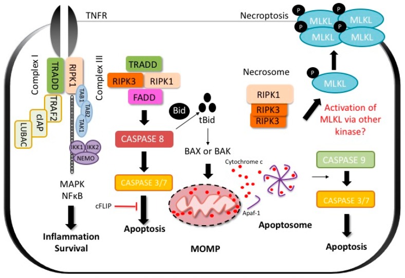
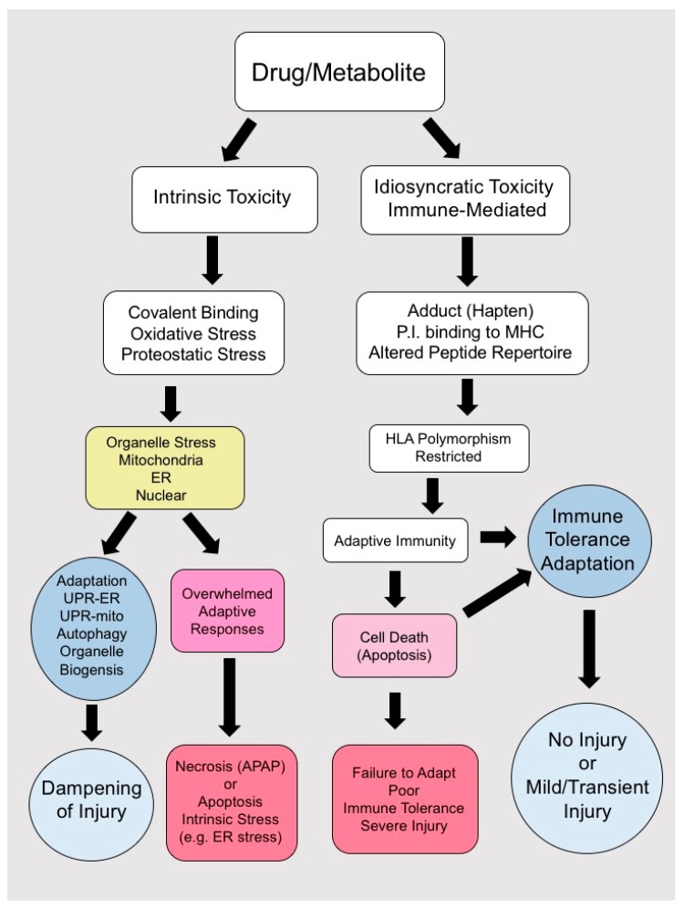
Drug-induced liver injury (DILI) can broadly be divided into predictable and dose dependent such as acetaminophen (APAP) and unpredictable or idiosyncratic DILI (IDILI). Liver injury from drug hepatotoxicity (whether idiosyncratic or predictable) results in hepatocyte cell death and inflammation. The cascade of events leading to DILI and the cell death subroutine (apoptosis or necrosis) of the cell depend largely on the culprit drug. Direct toxins to hepatocytes likely induce oxidative organelle stress (such as endoplasmic reticulum (ER) and mitochondrial stress) leading to necrosis or apoptosis, while cell death in idiosyncratic DILI (IDILI) is usually the result of engagement of the innate and adaptive immune system (likely apoptotic), involving death receptors (DR). Here, we review the hepatocyte cell death pathways both in direct hepatotoxicity such as in APAP DILI as well as in IDILI. We examine the known signaling pathways in APAP toxicity, a model of necrotic liver cell death. We also explore what is known about the genetic basis of IDILI and the molecular pathways leading to immune activation and how these events can trigger hepatotoxicity and cell death.
Overview
The claims and statistics of this paper haven’t been checked yet.
Run the check — free, no account needed. It reads the open-access text and checks the sources it cites.
Integrity signals
Registry
Cites
Statistics
Figures
Verification
Claims the paper makes
The claims and statistics of this paper haven’t been checked yet.
Run the check — free, no account needed. It reads the open-access text and checks the sources it cites.
Claims other papers make about this one
Each is checked against this paper’s own text: does it say what the citing paper says it does?
In the citing paper
“Despite these tools, most cases of DILI are diagnosed by excluding other causes of liver injury and by considering the temporal relationship of liver injury and initiation of the suspected drug.[8]”
From Drug-induced liver injury: Diagnosis, management and the role of liver transplantation (2025) · cites this paper as [8]
Self-check
Percentages, sums and reported differences re-computed from the paper’s own numbers.
The self-check needs the paper’s full text.
Citations
Cites
Interferon-γ: an overview of signals, mechanisms and functions
Schroder, K., Hertzog, P. J., Ravasi, T., & Hume, D. A. (2004) · Journal of Leukocyte Biology · doi:10.1189/jlb.0603252 · Cited by 4,120
Inactivation of the ferroptosis regulator Gpx4 triggers acute renal failure in mice
Friedmann Angeli, J. P., Schneider, M., Proneth, B., Tyurina, Y. Y., Tyurin, V. A., Hammond, V. J., Herbach, N., Aichler, M., Walch, A., Eggenhofer, E., et al. (2014) · Nature Cell Biology · doi:10.1038/ncb3064 · Cited by 3,732
Caspase-11 cleaves gasdermin D for non-canonical inflammasome signalling
Kayagaki, N., Stowe, I. B., Lee, B. L., O’Rourke, K., Anderson, K., Warming, S., Cuellar, T., Haley, B., Roose-Girma, M., Phung, Q. T., et al. (2015) · Nature · doi:10.1038/nature15541 · Cited by 3,686
Chemical inhibitor of nonapoptotic cell death with therapeutic potential for ischemic brain injury
Degterev, A., Huang, Z., Boyce, M., Li, Y., Jagtap, P., Mizushima, N., Cuny, G. D., Mitchison, T. J., Moskowitz, M. A., & Yuan, J. (2005) · Nature Chemical Biology · doi:10.1038/nchembio711 · Cited by 3,068
Induction of TNF receptor I-mediated apoptosis via two sequential signaling complexes
Micheau, O., & Tschopp, J. (2003) · Cell · doi:10.1016/s0092-8674(03)00521-x · Cited by 2,738
Galluzzi, L., Vitale, I., Abrams, J. M., Alnemri, E. S., Baehrecke, E. H., Blagosklonny, M. V., Dawson, T. M., Dawson, V. L., El-Deiry, W. S., Fulda, S., et al. (2012) · Cell Death & Differentiation · doi:10.1038/cdd.2011.96 · Cited by 2,421
Identification of RIP1 kinase as a specific cellular target of necrostatins
Degterev, A., Hitomi, J., Germscheid, M., Ch'en, I. L., Korkina, O., Teng, X., Abbott, D., Cuny, G. D., Yuan, C., Wagner, G., et al. (2008) · Nature Chemical Biology · doi:10.1038/nchembio.83 · Cited by 2,059
The Pseudokinase MLKL Mediates Necroptosis via a Molecular Switch Mechanism
Murphy, J. M., Czabotar, P. E., Hildebrand, J. M., Lucet, I. S., Zhang, J.-G., Alvarez-Diaz, S., Lewis, R., Lalaoui, N., Metcalf, D., Webb, A. I., et al. (2013) · Immunity · doi:10.1016/j.immuni.2013.06.018 · Cited by 1,220
Circulating microRNAs, potential biomarkers for drug-induced liver injury
Wang, K., Zhang, S., Marzolf, B., Troisch, P., Brightman, A., Hu, Z., Hood, L. E., & Galas, D. J. (2009) · Proceedings of the National Academy of Sciences · doi:10.1073/pnas.0813371106 · Cited by 1,149
Idiosyncratic drug hepatotoxicity
Kaplowitz, N. (2005) · Nature Reviews Drug Discovery · doi:10.1038/nrd1750 · Cited by 1,054
Self-citationBjörnsson, E. S., Bergmann, O. M., Björnsson, H. K., Kvaran, R. B., & Olafsson, S. (2013) · Gastroenterology · doi:10.1053/j.gastro.2013.02.006 · Cited by 865
Cell death modalities: classification and pathophysiological implications
Galluzzi, L., Maiuri, M. C., Vitale, I., Zischka, H., Castedo, M., Zitvogel, L., & Kroemer, G. (2007) · Cell Death & Differentiation · doi:10.1038/sj.cdd.4402148 · Cited by 832
Hanawa, N., Shinohara, M., Saberi, B., Gaarde, W. A., Han, D., & Kaplowitz, N. (2008) · Journal of Biological Chemistry · doi:10.1074/jbc.m708916200 · Cited by 527
Self-citationDaly, A. K., Aithal, G. P., Leathart, J. B. S., Swainsbury, R. A., Dang, T. S., & Day, C. P. (2007) · Gastroenterology · doi:10.1053/j.gastro.2006.11.023 · Cited by 348
Odin, J. A., Huebert, R. C., Casciola-Rosen, L., LaRusso, N. F., & Rosen, A. (2001) · Journal of Clinical Investigation · doi:10.1172/jci200110716 · Cited by 220
Ferroptosis is involved in acetaminophen induced cell death
Lőrincz, T., Jemnitz, K., Kardon, T., Mandl, J., & Szarka, A. (2015) · Pathology & Oncology Research · doi:10.1007/s12253-015-9946-3 · Cited by 219
Win, S., Than, T. A., Han, D., Petrovic, L. M., & Kaplowitz, N. (2011) · Journal of Biological Chemistry · doi:10.1074/jbc.m111.276089 · Cited by 197
Self-citationChakraborty, M., Fullerton, A. M., Semple, K., Chea, L. S., Proctor, W. R., Bourdi, M., Kleiner, D. E., Zeng, X., Ryan, P. M., Dagur, P. K., et al. (2015) · Hepatology · doi:10.1002/hep.27764 · Cited by 64
The role of microRNAs in liver injury at the crossroad between hepatic cell death and regeneration
Lauschke, V. M., Mkrtchian, S., & Ingelman-Sundberg, M. (2017) · Biochemical and Biophysical Research Communications · doi:10.1016/j.bbrc.2016.10.084 · Cited by 29
Research works printed but not linked: 164
- McQueen, C. (2017). Clinical considerations of drug-induced hepatotoxicity. Comprehensive Toxicology, Elsevier. [3rd ed.].
- Yuan, 2013, Mechanisms of drug-induced liver injury, Clin. Liver Dis., 17, 507, 10.1016/j.cld.2013.07.002 · DOI · Paper page
- Dara, L., Han, D., and Kaplowitz, N. (2012). Mechanisms of Cell Death and Relevance to Drug Toxicity, Academic Press (an imprint of Elsevier). [3rd ed.]. · DOI
- Larson, 2005, Acetaminophen-induced acute liver failure: Results of a united states multicenter, prospective study, Hepatology, 42, 1364, 10.1002/hep.20948 · DOI · Paper page
- Nathwani, 2006, Drug hepatotoxicity, Clin. Liver Dis., 10, 207, 10.1016/j.cld.2006.05.009 · DOI
- Senior, 2007, Drug hepatotoxicity from a regulatory perspective, Clin. Liver Dis., 11, 507, 10.1016/j.cld.2007.06.002 · DOI
- Kaplowitz, 2015, Targeting signal transduction pathways which regulate necrosis in acetaminophen hepatotoxicity, J. Hepatol., 63, 5, 10.1016/j.jhep.2015.02.050 · DOI
- Lucena, 2009, Phenotypic characterization of idiosyncratic drug-induced liver injury: The influence of age and sex, Hepatology, 49, 2001, 10.1002/hep.22895 · DOI · Paper page
- Denk, 2002, Drug-induced liver injury, Verh. Dtsch. Ges. Pathol., 86, 120
- Chen, 2013, High lipophilicity and high daily dose of oral medications are associated with significant risk for drug-induced liver injury, Hepatology, 58, 388, 10.1002/hep.26208 · DOI · Paper page
- Kaplowitz, 2013, Avoiding idiosyncratic DILI: Two is better than one, Hepatology, 58, 15, 10.1002/hep.26295 · DOI
- Bell, 2009, Epidemiology of idiosyncratic drug-induced liver injury, Semin. Liver Dis., 29, 337, 10.1055/s-0029-1240002 · DOI
- Ding, W.X., and Yin, X.M. (2017). Cell death in drug induced liver injury. Cell Death in Liver Disease, Springer. · DOI
- DeLeve, 1997, Sinusoidal endothelial cells as a target for acetaminophen toxicity: Direct action versus requirement for hepatocyte activation in different mouse strains, Biochem. Pharmacol., 53, 1339, 10.1016/S0006-2952(97)00048-8 · DOI
- DeLeve, 1996, Toxicity of azathioprine and monocrotaline in murine sinusoidal endothelial cells and hepatocytes: The role of glutathione and relevance to hepatic venoocclusive disease, Hepatology, 23, 589, 10.1002/hep.510230326 · DOI
- Guicciardi, 2009, Life and death by death receptors, FASEB J., 23, 1625, 10.1096/fj.08-111005 · DOI
- Tian, 2013, Natural killer cells in liver disease, Hepatology, 57, 1654, 10.1002/hep.26115 · DOI · Paper page
- Protzer, 2012, Living in the liver: Hepatic infections, Nat. Rev. Immunol., 12, 201, 10.1038/nri3169 · DOI · Paper page
- Ashkenazi, 2014, Regulated cell death: Signaling and mechanisms, Annu. Rev. Cell Dev. Biol., 30, 337, 10.1146/annurev-cellbio-100913-013226 · DOI · Paper page
- Karin, 2002, NFκB at the crossroads of life and death, Nat. Immunol., 3, 221, 10.1038/ni0302-221 · DOI · Paper page
- Micheau, 2001, NFκB signals induce the expression of c-flip, Mol. Cell. Biol., 21, 5299, 10.1128/MCB.21.16.5299-5305.2001 · DOI · Paper page
- Nagai, 2002, Reduced glutathione depletion causes necrosis and sensitization to tumor necrosis factor-α-induced apoptosis in cultured mouse hepatocytes, Hepatology, 36, 55, 10.1053/jhep.2002.33995 · DOI
- Pierce, 2000, Disruption of redox homeostasis in tumor necrosis factor-induced apoptosis in a murine hepatocyte cell line, Am. J. Pathol., 157, 221, 10.1016/S0002-9440(10)64533-6 · DOI
- Lou, 2007, Glutathione depletion down-regulates tumor necrosis factor α-induced NFκB activity via IκB kinase-dependent and -independent mechanisms, J. Biol. Chem., 282, 29470, 10.1074/jbc.M706145200 · DOI
- Han, 2006, Hydrogen peroxide and redox modulation sensitize primary mouse hepatocytes to TNF-induced apoptosis, Free Radic. Biol. Med., 41, 627, 10.1016/j.freeradbiomed.2006.05.002 · DOI
- Kim, 2005, Formation of apoptosome is initiated by cytochrome c-induced dATP hydrolysis and subsequent nucleotide exchange on APAF-1, Proc. Natl. Acad. Sci. USA, 102, 17545, 10.1073/pnas.0507900102 · DOI · Paper page
- Zou, 1997, APAF-1, a human protein homologous to C. elegans CED-4, participates in cytochrome c-dependent activation of caspase-3, Cell, 90, 405, 10.1016/S0092-8674(00)80501-2 · DOI · Paper page
- Acehan, 2002, Three-dimensional structure of the apoptosome: Implications for assembly, procaspase-9 binding, and activation, Mol. Cell, 9, 423, 10.1016/S1097-2765(02)00442-2 · DOI · Paper page
- Bratton, 2010, Regulation of the APAF-1-caspase-9 apoptosome, J. Cell Sci., 123, 3209, 10.1242/jcs.073643 · DOI · Paper page
- Newton, 2016, Necroptosis and inflammation, Annu. Rev. Biochem., 85, 743, 10.1146/annurev-biochem-060815-014830 · DOI
- Sun, 2012, Mixed lineage kinase domain-like protein mediates necrosis signaling downstream of RIP3 kinase, Cell, 148, 213, 10.1016/j.cell.2011.11.031 · DOI · Paper page
- Wang, 2014, Mixed lineage kinase domain-like protein MLKL causes necrotic membrane disruption upon phosphorylation by RIP3, Mol. Cell, 54, 133, 10.1016/j.molcel.2014.03.003 · DOI
- Gunther, 2016, The pseudokinase MLKL mediates programmed hepatocellular necrosis independently of RIPK3 during hepatitis, J. Clin. Investig., 126, 4346, 10.1172/JCI87545 · DOI · Paper page
- Afonso, 2015, Necroptosis is a key pathogenic event in human and experimental murine models of non-alcoholic steatohepatitis, Clin. Sci., 129, 721, 10.1042/CS20140732 · DOI
- Dara, 2016, Questions and controversies: The role of necroptosis in liver disease, Cell Death Discov., 2, 16089, 10.1038/cddiscovery.2016.89 · DOI
- Newton, 2014, Activity of protein kinase RIPK3 determines whether cells die by necroptosis or apoptosis, Science, 343, 1357, 10.1126/science.1249361 · DOI · Paper page
- Degterev, 2014, Assays for necroptosis and activity of RIP kinases, Methods Enzymol., 545, 1, 10.1016/B978-0-12-801430-1.00001-9 · DOI
- Sun, 1999, RIP3, a novel apoptosis-inducing kinase, J. Biol. Chem., 274, 16871, 10.1074/jbc.274.24.16871 · DOI
- Kasof, 2000, The RIP-like kinase, RIP3, induces apoptosis and NFκB nuclear translocation and localizes to mitochondria, FEBS Lett., 473, 285, 10.1016/S0014-5793(00)01473-3 · DOI
- Dara, 2015, Receptor interacting protein kinase 1 mediates murine acetaminophen toxicity independent of the necrosome and not through necroptosis, Hepatology, 62, 1847, 10.1002/hep.27939 · DOI
- An, 2013, ARC is a novel therapeutic approach against acetaminophen-induced hepatocellular necrosis, J. Hepatol., 58, 297, 10.1016/j.jhep.2012.10.002 · DOI
- Ramachandran, 2013, Receptor interacting protein kinase 3 is a critical early mediator of acetaminophen-induced hepatocyte necrosis in mice, Hepatology, 58, 2099, 10.1002/hep.26547 · DOI · Paper page
- Deutsch, 2015, Divergent effects of RIP1 or RIP3 blockade in murine models of acute liver injury, Cell Death Dis., 6, e1759, 10.1038/cddis.2015.126 · DOI
- Ni, 2012, Activation of autophagy protects against acetaminophen-induced hepatotoxicity, Hepatology, 55, 222, 10.1002/hep.24690 · DOI · Paper page
- Aglietti, R.A., and Dueber, E.C. (2017). Recent insights into the molecular mechanisms underlying pyroptosis and gasdermin family functions. Trends Immunol. · DOI
- Shi, 2015, Cleavage of GSDMD by inflammatory caspases determines pyroptotic cell death, Nature, 526, 660, 10.1038/nature15514 · DOI · Paper page
- Cao, 2016, Mechanisms of ferroptosis, Cell. Mol. Life Sci., 73, 2195, 10.1007/s00018-016-2194-1 · DOI · Paper page
- Dixon, 2012, Ferroptosis: An iron-dependent form of nonapoptotic cell death, Cell, 149, 1060, 10.1016/j.cell.2012.03.042 · DOI · Paper page
- Monshi, 2013, Human leukocyte antigen (HLA)-B*57:01-restricted activation of drug-specific T cells provides the immunological basis for flucloxacillin-induced liver injury, Hepatology, 57, 727, 10.1002/hep.26077 · DOI · Paper page
- Tsutsui, 1992, Drug-specific T cells derived from patients with drug-induced allergic hepatitis, J. Immunol., 149, 706, 10.4049/jimmunol.149.2.706 · DOI
- Dara, 2015, Mechanisms of adaptation and progression in idiosyncratic drug induced liver injury, clinical implications, Liver Int., 36, 158, 10.1111/liv.12988 · DOI
- Lauschke, V.M., and Ingelman-Sundberg, M. (2016). The importance of patient-specific factors for hepatic drug response and toxicity. Int. J. Mol. Sci., 17. · DOI
- Chalasani, 2015, Features and outcomes of 899 patients with drug-induced liver injury: The dilin prospective study, Gastroenterology, 148, 1340, 10.1053/j.gastro.2015.03.006 · DOI · Paper page
- Daly, 2009, HLA-B*5701 genotype is a major determinant of drug-induced liver injury due to flucloxacillin, Nat. Genet., 41, 816, 10.1038/ng.379 · DOI · Paper page
- Wuillemin, 2013, HLA haplotype determines hapten or p-i t cell reactivity to flucloxacillin, J. Immunol., 190, 4956, 10.4049/jimmunol.1202949 · DOI
- Bhogaraju, 1999, Diclofenac-associated hepatitis, South. Med. J., 92, 711, 10.1097/00007611-199907000-00011 · DOI
- Mallal, 2008, HLA-B*5701 screening for hypersensitivity to abacavir, N. Engl. J. Med., 358, 568, 10.1056/NEJMoa0706135 · DOI
- Hautekeete, 1999, HLA association of amoxicillin-clavulanate--induced hepatitis, Gastroenterology, 117, 1181, 10.1016/S0016-5085(99)70404-X · DOI · Paper page
- Kindmark, 2008, Genome-wide pharmacogenetic investigation of a hepatic adverse event without clinical signs of immunopathology suggests an underlying immune pathogenesis, Pharmacogenomics J., 8, 186, 10.1038/sj.tpj.6500458 · DOI
- Lucena, 2011, Susceptibility to amoxicillin-clavulanate-induced liver injury is influenced by multiple HLA class I and II alleles, Gastroenterology, 141, 338, 10.1053/j.gastro.2011.04.001 · DOI · Paper page
- Sharma, 2002, Evaluation of clinical and immunogenetic risk factors for the development of hepatotoxicity during antituberculosis treatment, Am. J. Respir. Crit. Care Med., 166, 916, 10.1164/rccm.2108091 · DOI · Paper page
- Chen, 2015, The association between HLA-DQB1 polymorphism and antituberculosis drug-induced liver injury: A case-control study, J. Clin. Pharm. Ther., 40, 110, 10.1111/jcpt.12211 · DOI
- Petros, 2017, HLA-B*57 allele is associated with concomitant anti-tuberculosis and antiretroviral drugs induced liver toxicity in ethiopians, Front. Pharmacol., 8, 90, 10.3389/fphar.2017.00090 · DOI
- Spraggs, 2011, HLA-DQA1*02:01 is a major risk factor for lapatinib-induced hepatotoxicity in women with advanced breast cancer, J. Clin. Oncol., 29, 667, 10.1200/JCO.2010.31.3197 · DOI
- Urban, T.J., Nicoletti, P., Chalasani, N., Serrano, J., Stolz, A., Daly, A., Aithal, G., Dillon, J., Navarro, V., and Odin, J. (2017). Minocycline hepatotoxicity: Clinical characterization and identification of HLA-B*35:02 as a risk factor. J. Hepatol. · DOI · Paper page
- Nicoletti, 2017, Association of liver injury from specific drugs, or groups of drugs, with polymorphisms in HLA and other genes in a genome-wide association study, Gastroenterology, 152, 1078, 10.1053/j.gastro.2016.12.016 · DOI · Paper page
- Phillips, 2013, Associations between HLA-DRB1*0102, HLA-B*5801, and hepatotoxicity during initiation of nevirapine-containing regimens in south africa, J. Acquir. Immune Defic. Syndr., 62, e55, 10.1097/QAI.0b013e31827ca50f · DOI
- Uetrecht, 2008, Idiosyncratic drug reactions: Past, present, and future, Chem. Res. Toxicol., 21, 84, 10.1021/tx700186p · DOI · Paper page
- Saito, 2015, Predictive genomic markers for severe adverse drug reactions, Yakugaku Zasshi, 135, 589, 10.1248/yakushi.14-00249-3 · DOI
- Nicoletti, 2016, HLA-DRB1*16:01-DQB1*05:02 is a novel genetic risk factor for flupirtine-induced liver injury, Pharmacogenet. Genom., 26, 218, 10.1097/FPC.0000000000000209 · DOI
- Berson, 1994, Possible role of HLA in hepatotoxicity: An exploratory study in 71 patients with drug-induced idiosyncratic hepatitis, J. Hepatol., 20, 336, 10.1016/S0168-8278(94)80004-9 · DOI
- Daly, 2009, Genetic association studies in drug-induced liver injury, Semin. Liver Dis., 29, 400, 10.1055/s-0029-1240009 · DOI
- Russmann, 2005, Risk of cholestatic liver disease associated with flucloxacillin and flucloxacillin prescribing habits in the UK: Cohort study using data from the UK general practice research database, Br. J. Clin. Pharmacol., 60, 76, 10.1111/j.1365-2125.2005.02370.x · DOI · Paper page
- Singer, 2010, A genome-wide study identifies HLA alleles associated with lumiracoxib-related liver injury, Nat. Genet., 42, 711, 10.1038/ng.632 · DOI · Paper page
- Xu, 2016, HLA-B*57:01 confers susceptibility to pazopanib-associated liver injury in patients with cancer, Clin. Cancer Res., 22, 1371, 10.1158/1078-0432.CCR-15-2044 · DOI
- Hirata, 2008, Ticlopidine-induced hepatotoxicity is associated with specific human leukocyte antigen genomic subtypes in japanese patients: A preliminary case-control study, Pharmacogenom. J., 8, 29, 10.1038/sj.tpj.6500442 · DOI
- Kurosaki, 2000, HLA-A33/B44/DR6 is highly related to intrahepatic cholestasis induced by tiopronin, Dig. Dis. Sci., 45, 1103, 10.1023/A:1005585515826 · DOI
- Andrade, 2005, Drug-induced liver injury: An analysis of 461 incidences submitted to the spanish registry over a 10-year period, Gastroenterology, 129, 512, 10.1016/j.gastro.2005.05.006 · DOI · Paper page
- Bjornsson, 2015, Drug-induced liver injury: An overview over the most critical compounds, Arch. Toxicol., 89, 327, 10.1007/s00204-015-1456-2 · DOI
- Bessone, F., Hernandez, N., Lucena, M.I., Andrade, R.J., Latin DILI Network, and Spanish DILI Registry (2016). The Latin American DILI registry experience: A successful ongoing collaborative strategic initiative. Int. J. Mol. Sci., 17. · DOI
- Takikawa, 2009, Drug-induced liver injury in japan: An analysis of 1676 cases between 1997 and 2006, Hepatol. Res., 39, 427, 10.1111/j.1872-034X.2008.00486.x · DOI · Paper page
- Roth, 2017, Idiosyncratic drug-induced liver injury: Is drug-cytokine interaction the linchpin?, J. Pharmacol. Exp. Ther., 360, 461, 10.1124/jpet.116.237578 · DOI
- Kusters, 1996, Interferon γ plays a critical role in T cell-dependent liver injury in mice initiated by concanavalin A, Gastroenterology, 111, 462, 10.1053/gast.1996.v111.pm8690213 · DOI
- Morita, 1995, Protective effect of hepatocyte growth factor on interferon-γ-induced cytotoxicity in mouse hepatocytes, Hepatology, 21, 1585
- Vodovotz, 2004, Inflammatory modulation of hepatocyte apoptosis by nitric oxide: In vivo, in vitro, and in silico studies, Curr. Mol. Med., 4, 753, 10.2174/1566524043359944 · DOI
- Nagaki, 1994, Hepatic injury and lethal shock in galactosamine-sensitized mice induced by the superantigen staphylococcal enterotoxin B, Gastroenterology, 106, 450, 10.1016/0016-5085(94)90604-1 · DOI
- Gantner, 1995, Tumor necrosis factor-induced hepatic DNA fragmentation as an early marker of T cell-dependent liver injury in mice, Gastroenterology, 109, 166, 10.1016/0016-5085(95)90282-1 · DOI
- Gantner, 1995, Concanavalin A-induced T-cell-mediated hepatic injury in mice: The role of tumor necrosis factor, Hepatology, 21, 190
- Tiegs, 1992, A T cell-dependent experimental liver injury in mice inducible by concanavalin a, J. Clin. Investig., 90, 196, 10.1172/JCI115836 · DOI · Paper page
- Leist, 1997, Tumor necrosis factor-induced apoptosis during the poisoning of mice with hepatotoxins, Gastroenterology, 112, 923, 10.1053/gast.1997.v112.pm9041255 · DOI
- Dara, 2016, A murder mystery in the liver: Who done it and how?, J. Clin. Investig., 126, 4068, 10.1172/JCI90830 · DOI
- Suda, 2016, Knockdown of RIPK1 markedly exacerbates murine immune-mediated liver injury through massive apoptosis of hepatocytes, independent of necroptosis and inhibition of nf-kappab, J. Immunol., 197, 3120, 10.4049/jimmunol.1600690 · DOI
- Filliol, 2016, RIPK1 protects from TNF-α-mediated liver damage during hepatitis, Cell Death Dis., 7, e2462, 10.1038/cddis.2016.362 · DOI
- Bultemann, 2001, Degeneracy and additional alloreactivity of drug-specific human αβ+ T cell clones, Int. Immunol., 13, 877, 10.1093/intimm/13.7.877 · DOI
- Zanni, 1999, Interaction of sulfonamide derivatives with the TCR of sulfamethoxazole-specific human αβ+ T cell clones, J. Immunol., 162, 595, 10.4049/jimmunol.162.1.595 · DOI
- Pichler, 2002, Pharmacological interaction of drugs with antigen-specific immune receptors: The p-i concept, Curr. Opin. Allergy Clin. Immunol., 2, 301, 10.1097/00130832-200208000-00003 · DOI
- Grove, 2015, Human leukocyte antigen genetic risk factors of drug-induced liver toxicology, Expert Opin. Drug Metab. Toxicol., 11, 395, 10.1517/17425255.2015.992414 · DOI
- Ostrov, 2012, Drug hypersensitivity caused by alteration of the MHC-presented self-peptide repertoire, Proc. Natl. Acad. Sci. USA, 109, 9959, 10.1073/pnas.1207934109 · DOI · Paper page
- Wei, 2012, Direct interaction between HLA-B and carbamazepine activates T cells in patients with stevens-johnson syndrome, J. Allergy Clin. Immunol., 129, 1562, 10.1016/j.jaci.2011.12.990 · DOI
- Li, 2002, A review of the common properties of drugs with idiosyncratic hepatotoxicity and the “multiple determinant hypothesis” for the manifestation of idiosyncratic drug toxicity, Chem. Biol. Interact., 142, 7, 10.1016/S0009-2797(02)00051-0 · DOI
- Ulrich, 2007, Idiosyncratic toxicity: A convergence of risk factors, Annu. Rev. Med., 58, 17, 10.1146/annurev.med.58.072905.160823 · DOI
- Ray, 1991, Halothane hepatitis, Br. J. Anaesth., 67, 84, 10.1093/bja/67.1.84 · DOI
- Dugan, 2010, A mouse model of severe halothane hepatitis based on human risk factors, J. Pharmacol. Exp. Ther., 333, 364, 10.1124/jpet.109.164541 · DOI
- Khouri, 1987, Hepatocanalicular injury associated with vitamin a derivative etretinate. An idiosyncratic hypersensitivity reaction, Dig. Dis. Sci., 32, 1207, 10.1007/BF01300208 · DOI
- Fukano, 2000, Subacute hepatic failure associated with a new antidiabetic agent, troglitazone: A case report with autopsy examination, Hum. Pathol., 31, 250, 10.1016/S0046-8177(00)80229-4 · DOI
- Deng, 2009, Inflammatory stress and idiosyncratic hepatotoxicity: Hints from animal models, Pharmacol. Rev., 61, 262, 10.1124/pr.109.001727 · DOI
- Shaw, 2010, Idiosyncratic drug-induced liver injury and the role of inflammatory stress with an emphasis on an animal model of trovafloxacin hepatotoxicity, Toxicol. Sci., 118, 7, 10.1093/toxsci/kfq168 · DOI · Paper page
- Mak, 2015, The role of CD8 T cells in amodiaquine-induced liver injury in PD1−/− mice cotreated with anti-CTLA-4, Chem. Res. Toxicol., 28, 1567, 10.1021/acs.chemrestox.5b00137 · DOI
- Metushi, 2015, Treatment of PD-1−/− mice with amodiaquine and anti-CTLA4 leads to liver injury similar to idiosyncratic liver injury in patients, Hepatology, 61, 1332, 10.1002/hep.27549 · DOI
- Calne, 1969, Induction of immunological tolerance by porcine liver allografts, Nature, 223, 472, 10.1038/223472a0 · DOI
- Knolle, 2000, Local control of the immune response in the liver, Immunol. Rev., 174, 21, 10.1034/j.1600-0528.2002.017408.x · DOI · Paper page
- Adams, 2015, From immunosuppression to tolerance, J. Hepatol., 62, S170, 10.1016/j.jhep.2015.02.042 · DOI
- Mitchell, 1975, Acetylation rates and monthly liver function tests during one year of isoniazid preventive therapy, Chest, 68, 181, 10.1378/chest.68.2.181 · DOI
- Uetrecht, 2015, Inhibition of immune tolerance unmasks drug-induced allergic hepatitis, Hepatology, 62, 346, 10.1002/hep.27824 · DOI
- Wisse, 1985, The liver sieve: Considerations concerning the structure and function of endothelial fenestrae, the sinusoidal wall and the space of disse, Hepatology, 5, 683, 10.1002/hep.1840050427 · DOI
- Fraser, 1995, Lipoproteins and the liver sieve: The role of the fenestrated sinusoidal endothelium in lipoprotein metabolism, atherosclerosis, and cirrhosis, Hepatology, 21, 863
- Lohse, 1996, Antigen-presenting function and B7 expression of murine sinusoidal endothelial cells and kupffer cells, Gastroenterology, 110, 1175, 10.1053/gast.1996.v110.pm8613007 · DOI · Paper page
- Knolle, 1999, Endotoxin down-regulates T cell activation by antigen-presenting liver sinusoidal endothelial cells, J. Immunol., 162, 1401, 10.4049/jimmunol.162.3.1401 · DOI
- Bissell, 1995, Cell-specific expression of transforming growth factor-β in rat liver. Evidence for autocrine regulation of hepatocyte proliferation, J. Clin. Investig., 96, 447, 10.1172/JCI118055 · DOI · Paper page
- Knolle, 1998, Interleukin-10 expression is autoregulated at the transcriptional level in human and murine kupffer cells, Hepatology, 27, 93, 10.1002/hep.510270116 · DOI
- Knolle, 1995, Human Kupffer cells secrete IL-10 in response to lipopolysaccharide (LPS) challenge, J. Hepatol., 22, 226, 10.1016/0168-8278(95)80433-1 · DOI · Paper page
- Rieder, 1990, Prostanoid release of cultured liver sinusoidal endothelial cells in response to endotoxin and tumor necrosis factor: Comparison with umbilical vein endothelial cells, J. Hepatol., 11, 359, 10.1016/0168-8278(90)90222-D · DOI
- Dieter, 1987, Prostaglandin release but not superoxide production by rat Kupffer cells stimulated in vitro depends on Na+/H+ exchange, Eur. J. Biochem., 170, 201, 10.1111/j.1432-1033.1987.tb13687.x · DOI
- Mak, 2017, Effects of immunization and checkpoint inhibition on amodiaquine-induced liver injury, J. Immunotoxicol., 14, 89, 10.1080/1547691X.2017.1290716 · DOI
- Lee, 2004, Acetaminophen and the U.S. acute liver failure study group: Lowering the risks of hepatic failure, Hepatology, 40, 6, 10.1002/hep.20293 · DOI · Paper page
- Major, 2016, Trends in rates of acetaminophen-related adverse events in the United States, Pharmacoepidemiol. Drug Saf., 25, 590, 10.1002/pds.3906 · DOI
- Nourjah, 2006, Estimates of acetaminophen (paracetomal)-associated overdoses in the United States, Pharmacoepidemiol. Drug Saf., 15, 398, 10.1002/pds.1191 · DOI · Paper page
- Rowden, 2005, Updates on acetaminophen toxicity, Med. Clin. N. Am., 89, 1145, 10.1016/j.mcna.2005.06.009 · DOI
- Prescott, 1974, Gastrointestinal absorption of drugs, Med. Clin. N. Am., 58, 907, 10.1016/S0025-7125(16)32088-0 · DOI
- Josting, 1976, Glucuronidation of paracetamol, morphine and 1-naphthol in the rat intestinal loop, Biochem. Pharmacol., 25, 613, 10.1016/0006-2952(76)90399-3 · DOI
- Mitchell, 1973, Acetaminophen-induced hepatic necrosis. I. Role of drug metabolism, J. Pharmacol. Exp. Ther., 187, 185, 10.1016/S0022-3565(25)29663-X · DOI · Paper page
- Mitchell, 1974, Acetaminophen-induced hepatic injury: Protective role of glutathione in man and rationale for therapy, Clin. Pharmacol. Ther., 16, 676, 10.1002/cpt1974164676 · DOI · Paper page
- Kulkens, 1986, Paracetamol, 3-monoalkyl- and 3,5-dialkyl derivatives: Comparison of their microsomal cytochrome P-450 dependent oxidation and toxicity in freshly isolated hepatocytes, Biochem. Pharmacol., 35, 3693, 10.1016/0006-2952(86)90653-2 · DOI
- Moldeus, 1978, Paracetamol metabolism and toxicity in isolated hepatocytes from rat and mouse, Biochem. Pharmacol., 27, 2859, 10.1016/0006-2952(78)90201-0 · DOI
- Gibson, 1996, Mechanism of acetaminophen-induced hepatotoxicity: Covalent binding versus oxidative stress, Chem. Res. Toxicol., 9, 580, 10.1021/tx950153d · DOI
- Pumford, 1990, Immunoblot analysis of protein containing 3-(cystein-S-yl) acetaminophen adducts in serum and subcellular liver fractions from acetaminophen-treated mice, Toxicol. Appl. Pharmacol., 104, 521, 10.1016/0041-008X(90)90174-S · DOI
- Szkolnicka, 2016, Reducing hepatocyte injury and necrosis in response to paracetamol using noncoding RNAs, Stem Cells Transl. Med., 5, 764, 10.5966/sctm.2015-0117 · DOI
- Jaeschke, 2011, Reactive oxygen and mechanisms of inflammatory liver injury: Present concepts, J. Gastroenterol. Hepatol., 26, 173, 10.1111/j.1440-1746.2010.06592.x · DOI
- Gujral, 2002, Mode of cell death after acetaminophen overdose in mice: Apoptosis or oncotic necrosis?, Toxicol. Sci., 67, 322, 10.1093/toxsci/67.2.322 · DOI · Paper page
- Jaeschke, 2004, Apoptosis and necrosis in liver disease, Liver Int., 24, 85, 10.1111/j.1478-3231.2004.0906.x · DOI
- Lawson, 1999, Inhibition of Fas receptor (CD95)-induced hepatic caspase activation and apoptosis by acetaminophen in mice, Toxicol. Appl. Pharmacol., 156, 179, 10.1006/taap.1999.8635 · DOI
- Bajt, 2006, Nuclear translocation of endonuclease G and apoptosis-inducing factor during acetaminophen-induced liver cell injury, Toxicol. Sci., 94, 217, 10.1093/toxsci/kfl077 · DOI
- Jaeschke, 2011, No evidence for caspase-dependent apoptosis in acetaminophen hepatotoxicity, Hepatology, 53, 718, 10.1002/hep.23940 · DOI
- Sharma, 2012, Critical role for mixed-lineage kinase 3 in acetaminophen-induced hepatotoxicity, Mol. Pharmacol., 82, 1001, 10.1124/mol.112.079863 · DOI
- Nakagawa, 2008, Deletion of apoptosis signal-regulating kinase 1 attenuates acetaminophen-induced liver injury by inhibiting c-Jun N-terminal kinase activation, Gastroenterology, 135, 1311, 10.1053/j.gastro.2008.07.006 · DOI
- Gunawan, 2006, c-Jun N-terminal kinase plays a major role in murine acetaminophen hepatotoxicity, Gastroenterology, 131, 165, 10.1053/j.gastro.2006.03.045 · DOI · Paper page
- Huo, 2017, Antcin h protects against acute liver injury through disruption of the interaction of c-Jun N-terminal kinase with mitochondria, Antioxid. Redox Signal., 26, 207, 10.1089/ars.2016.6833 · DOI
- Win, 2016, c-Jun N-terminal kinase mediates mouse liver injury through a novel Sab (SH3BP5)-dependent pathway leading to inactivation of intramitochondrial Src, Hepatology, 63, 1987, 10.1002/hep.28486 · DOI
- Uzi, 2013, Chop is a critical regulator of acetaminophen-induced hepatotoxicity, J. Hepatol., 59, 495, 10.1016/j.jhep.2013.04.024 · DOI
- Kon, 2004, Mitochondrial permeability transition in acetaminophen-induced necrosis and apoptosis of cultured mouse hepatocytes, Hepatology, 40, 1170, 10.1002/hep.20437 · DOI · Paper page
- Masubuchi, 2005, Involvement of mitochondrial permeability transition in acetaminophen-induced liver injury in mice, J. Hepatol., 42, 110, 10.1016/j.jhep.2004.09.015 · DOI · Paper page
- Ramachandran, 2011, Cyclophilin D deficiency protects against acetaminophen-induced oxidant stress and liver injury, Free Radic. Res., 45, 156, 10.3109/10715762.2010.520319 · DOI
- Loguidice, 2011, Acetaminophen overdose-induced liver injury in mice is mediated by peroxynitrite independently of the cyclophilin d-regulated permeability transition, Hepatology, 54, 969, 10.1002/hep.24464 · DOI
- Lin, 2016, Advances in engineered liver models for investigating drug-induced liver injury, BioMed Res. Int., 2016, 1829148, 10.1155/2016/1829148 · DOI · Paper page
- Porceddu, 2012, Prediction of liver injury induced by chemicals in human with a multiparametric assay on isolated mouse liver mitochondria, Toxicol. Sci., 129, 332, 10.1093/toxsci/KFS197 · DOI
- Khetani, 2008, Microscale culture of human liver cells for drug development, Nat. Biotechnol., 26, 120, 10.1038/nbt1361 · DOI · Paper page
- Funakoshi, 2014, Human induced pluripotent stem cells in hepatology: Beyond the proof of concept, Am. J. Pathol., 184, 332, 10.1016/j.ajpath.2013.09.026 · DOI
- Teschke, R., Schulze, J., Eickhoff, A., and Danan, G. (2017). Drug induced liver injury: Can biomarkers assist RUCAM in causality assessment?. Int. J. Mol. Sci., 18. · DOI
- Fontana, 2009, Drug-induced liver injury network (DILIN) prospective study: Rationale, design and conduct, Drug Saf., 32, 55, 10.2165/00002018-200932010-00005 · DOI · Paper page
- Lucena, 2001, Comparison of two clinical scales for causality assessment in hepatotoxicity, Hepatology, 33, 123, 10.1053/jhep.2001.20645 · DOI · Paper page
- Danan, G., and Teschke, R. (2016). Rucam in drug and herb induced liver injury: The update. Int. J. Mol. Sci., 17. · DOI · Paper page
- Kaplowitz, 2001, Causality assessment versus guilt-by-association in drug hepatotoxicity, Hepatology, 33, 308, 10.1053/jhep.2001.21083 · DOI
- Chen, 2015, Drug-induced liver injury: Interactions between drug properties and host factors, J. Hepatol., 63, 503, 10.1016/j.jhep.2015.04.016 · DOI · Paper page
- Russmann, 2009, Current concepts of mechanisms in drug-induced hepatotoxicity, Curr. Med. Chem., 16, 3041, 10.2174/092986709788803097 · DOI
Cited by
EASL Clinical Practice Guidelines: Drug-induced liver injury
Andrade, R. J., Aithal, G. P., Björnsson, E. S., Kaplowitz, N., Kullak-Ublick, G. A., Larrey, D., & Karlsen, T. H. (2019) · Journal of Hepatology · doi:10.1016/j.jhep.2019.02.014 · Cited by 1,276
Self-citationAndrade, R. J., Chalasani, N., Björnsson, E. S., Suzuki, A., Kullak-Ublick, G. A., Watkins, P. B., Devarbhavi, H., Merz, M., Lucena, M. I., Kaplowitz, N., et al. (2019) · Nature Reviews Disease Primers · doi:10.1038/s41572-019-0105-0 · Cited by 924
Self-citationAutophagy in liver diseases: Time for translation?
Allaire, M., Rautou, P.-E., Codogno, P., & Lotersztajn, S. (2019) · Journal of Hepatology · doi:10.1016/j.jhep.2019.01.026 · Cited by 378
Drug induced liver injury – a 2023 update
Allison, R., Guraka, A., Shawa, I. T., Tripathi, G., Moritz, W., & Kermanizadeh, A. (2023) · Journal of Toxicology and Environmental Health, Part B · doi:10.1080/10937404.2023.2261848 · Cited by 167
Mechanisms of immune checkpoint inhibitor-mediated liver injury
Shojaie, L., Ali, M., Iorga, A., & Dara, L. (2021) · Acta Pharmaceutica Sinica B · doi:10.1016/j.apsb.2021.10.003 · Cited by 98
Self-citation
Metadata
- Authors
- Iorga, A., Dara, L., & Kaplowitz, N.
- Affiliations
- Research Center for Liver Disease, Keck School of Medicine, University of Southern California, Los Angeles, CA 90033, USA
- ORCID
- Dara, Lily
- Venue
- International Journal of Molecular Sciences
- Publisher
- MDPI AG
- Published
- 2017-05-09
- Volume
- 18
- Issue
- 5
- Pages
- 1018
- ISSN
- 1422-0067
- PMC
- PMC5454931
- Cited by
- 268
- References
- 19
- Language
- en
- Funders
- National Institute of Diabetes and Digestive and Kidney Diseases (K08DK109141, R01DK067215, P30DK048522)
About these checks
Registry, retraction, Cites and statistics results are computed, not judged, with one exception: a printed reference that no index lists is read once by a model before it counts as a citation error. Claims are extracted by a model and kept only when their quote appears verbatim in the text; each cited source is then checked, with a model’s help, for whether it says what the claim relies on. Those verdicts can be wrong — read the claim’s quote and the cited source before relying on one.
Statistics and arithmetic are recomputed, not judged.
Confidence bands are shown only for fields calibrated on our annotated test set. None is calibrated yet, so no band is shown.
What counts as a claim ›How we measure confidence ›How we check this ›
Are you an author, or do you see a problem with this page? Request a review
Data sources
Bibliographic data: Semantic Scholar, OpenAlex, Crossref.
Contains information from the Semantic Scholar Academic Graph Datasets, made available under the ODC Attribution License (ODC-BY 1.0).
Semantic Scholar’s data is described in: Kinney, R., et al. (2023). The Semantic Scholar Open Data Platform. arXiv:2301.10140.
