

The DOI matches its Crossref record; 50 of the 50 references it cites were found in a registry or an index.
Comprehensive alpha, beta and delta cell transcriptomes reveal that ghrelin selectively activates delta cells and promotes somatostatin release from pancreatic islets
Open accessAbstract
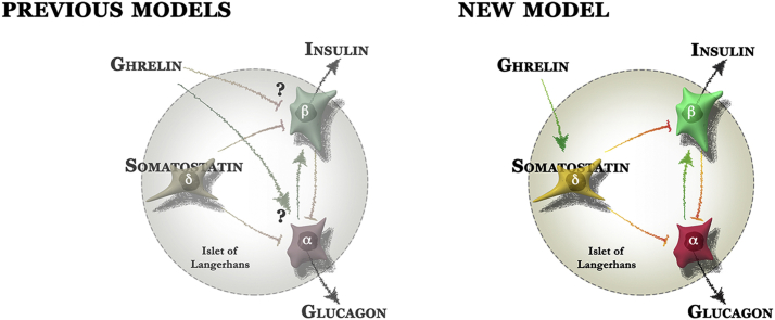
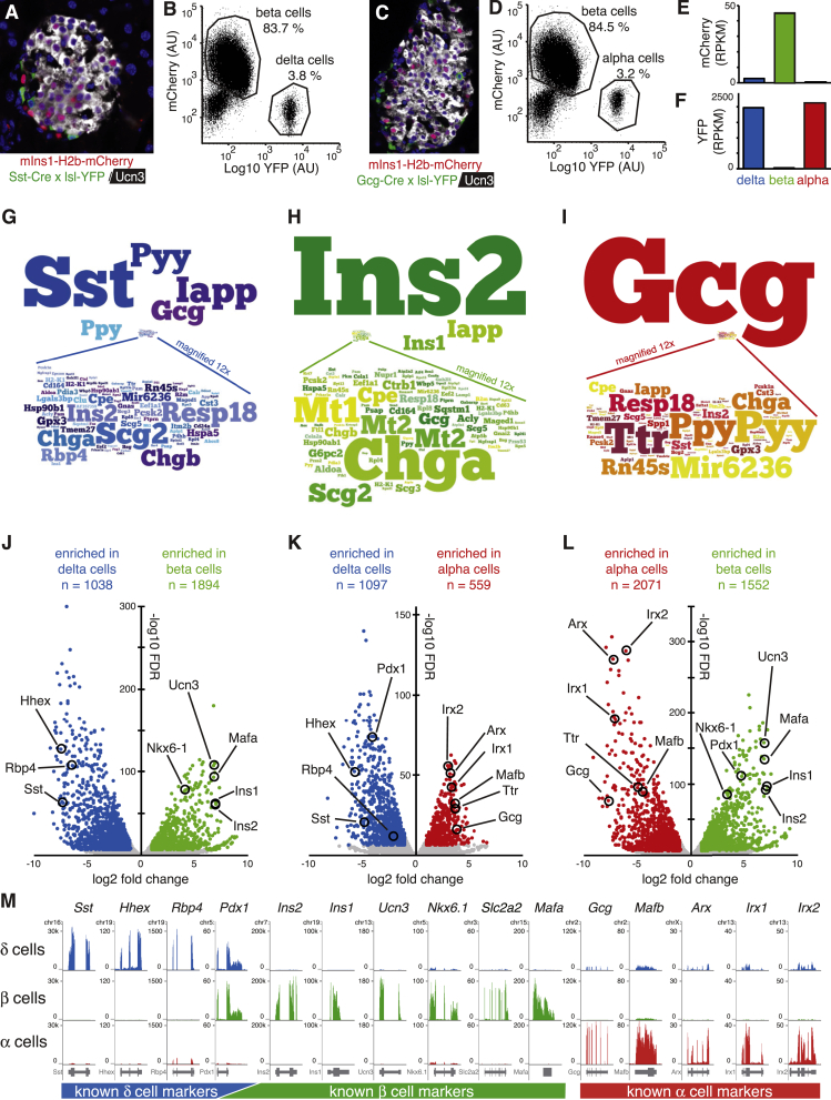
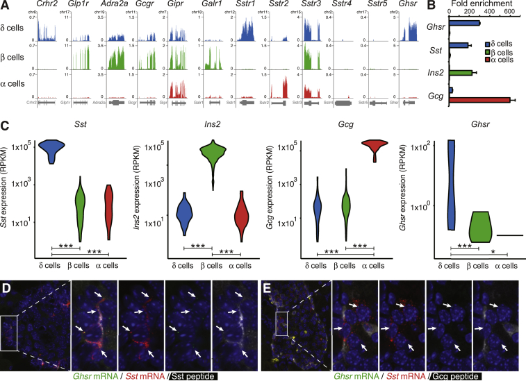
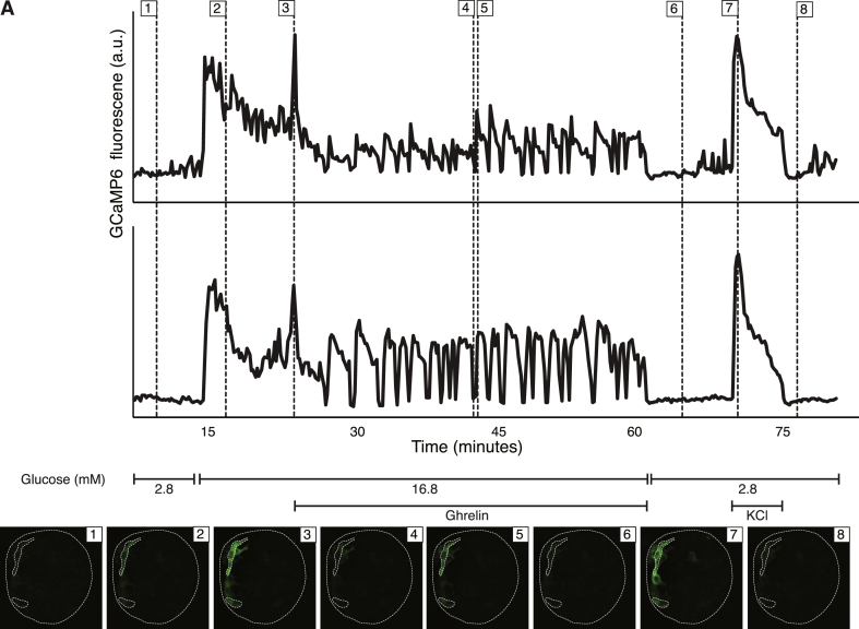
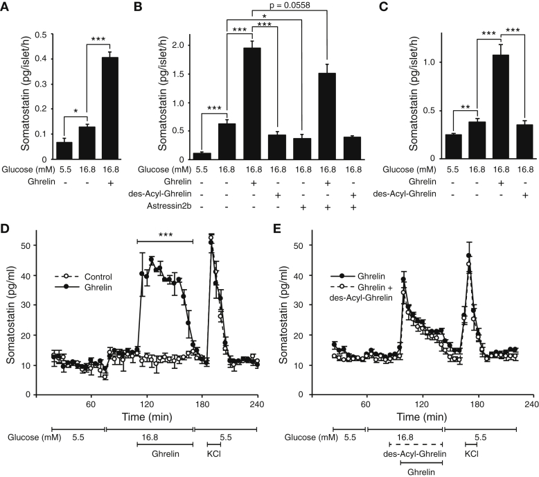
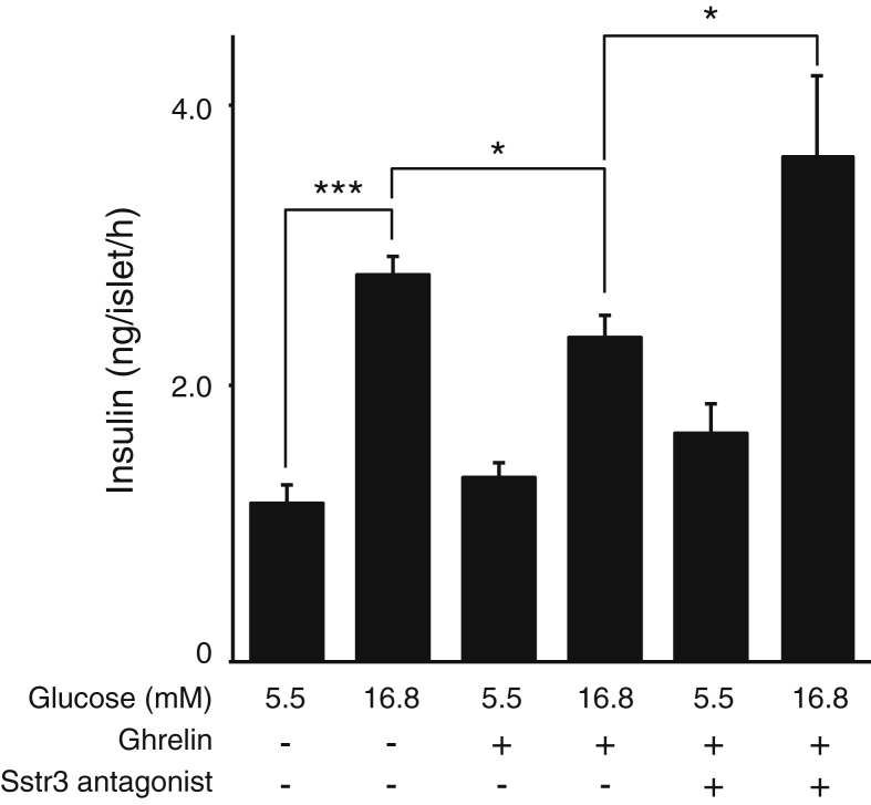
OBJECTIVE: Complex local crosstalk amongst endocrine cells within the islet ensures tight coordination of their endocrine output. This is illustrated by the recent demonstration that the negative feedback control by delta cells within pancreatic islets determines the homeostatic set-point for plasma glucose during mouse postnatal development. However, the close association of islet endocrine cells that facilitates paracrine crosstalk also complicates the distinction between effects mediated directly on beta cells from indirect effects mediated via local intermediates, such as somatostatin from delta cells. METHODS: To resolve this problem, we generated reporter mice that allow collection of pure pancreatic delta cells along with alpha and beta cells from the same islets and generated comprehensive transcriptomes for each islet endocrine cell type. These transcriptomes afford an unparalleled view of the receptors expressed by delta, alpha and beta cells, and allow the prediction of which signal targets which endocrine cell type with great accuracy. RESULTS: From these transcriptomes, we discovered that the ghrelin receptor is expressed exclusively by delta cells within the islet, which was confirmed by fluorescent in situ hybridization and qPCR. Indeed, ghrelin increases intracellular calcium in delta cells in intact mouse islets, measured by GCaMP6 and robustly potentiates glucose-stimulated somatostatin secretion on mouse and human islets in both static and perfusion assays. In contrast, des-acyl-ghrelin at the same dose had no effect on somatostatin secretion and did not block the actions of ghrelin. CONCLUSIONS: These results offer a straightforward explanation for the well-known insulinostatic actions of ghrelin. Rather than engaging beta cells directly, ghrelin engages delta cells to promote local inhibitory feedback that attenuates insulin release. These findings illustrate the power of our approach to resolve some of the long-standing conundrums with regard to the rich feedback that occurs within the islet that is integral to islet physiology and therefore highly relevant to diabetes.
Overview
The claims and statistics of this paper haven’t been checked yet.
Run the check — free, no account needed. It reads the open-access text and checks the sources it cites.
Integrity signals
Registry
Cites
Statistics
Figures
Verification
Claims the paper makes
The claims and statistics of this paper haven’t been checked yet.
Run the check — free, no account needed. It reads the open-access text and checks the sources it cites.
Claims other papers make about this one
Each is checked against this paper’s own text: does it say what the citing paper says it does?
The check wasn’t confident either way, so it gives no verdict — never disputed.
In the citing paper
“We estimate (Equation 3.2. in ref. 26) the gap junctional conductance between β-cells and δ-cells to be 23 pS.”
From Loss of electrical β-cell to δ-cell coupling underlies impaired hypoglycaemia-induced glucagon secretion in type-1 diabetes (2024) · cites this paper as [16]
Experimental — accuracy not yet measuredThe check wasn’t confident either way, so it gives no verdict — never disputed.
In the citing paper
“Both mouse (3, 146) and human (68, 475) β-cells express Hcn/HCN2-4.”
From Pancreatic β-Cell Electrical Activity and Insulin Secretion: Of Mice and Men (2018) · cites this paper as [146]
Experimental — accuracy not yet measuredThe check wasn’t confident either way, so it gives no verdict — never disputed.
In the citing paper
“which is expressed in mouse β-cells(3, 146).”
From Pancreatic β-Cell Electrical Activity and Insulin Secretion: Of Mice and Men (2018) · cites this paper as [146]
Experimental — accuracy not yet measuredThe check wasn’t confident either way, so it gives no verdict — never disputed.
In the citing paper
“However, these are only expressed at low levels in mouse and human β-cells alike (3, 68, 146, 475).”
From Pancreatic β-Cell Electrical Activity and Insulin Secretion: Of Mice and Men (2018) · cites this paper as [146]
Experimental — accuracy not yet measuredThe check wasn’t confident either way, so it gives no verdict — never disputed.
In the citing paper
“Both human and mouse β-cells also express Slc6a17/SLC6a17 (3, 68, 146, 475) that encodes a synaptic vesicle glycine transporter (502).”
From Pancreatic β-Cell Electrical Activity and Insulin Secretion: Of Mice and Men (2018) · cites this paper as [146]
Experimental — accuracy not yet measured
The check wasn’t confident either way, so it gives no verdict — never disputed.
In the citing paper
“These are both expressed at low levels in mouse and human β-cells (3, 68, 146, 475).”
From Pancreatic β-Cell Electrical Activity and Insulin Secretion: Of Mice and Men (2018) · cites this paper as [146]
Experimental — accuracy not yet measuredThe check wasn’t confident either way, so it gives no verdict — never disputed.
In the citing paper
“Mouse and human β-cells express CAPS1 (Cadps/CADPS) and low amounts of CAPS2 (Cadps2/CADPS2) (3, 68, 146, 475).”
From Pancreatic β-Cell Electrical Activity and Insulin Secretion: Of Mice and Men (2018) · cites this paper as [146]
Experimental — accuracy not yet measuredThe check wasn’t confident either way, so it gives no verdict — never disputed.
In the citing paper
“In human β-cells, only SYN1 is expressed, whereas mouse β-cells express both Syn1 and Syn2 (3, 68, 146, 475).”
From Pancreatic β-Cell Electrical Activity and Insulin Secretion: Of Mice and Men (2018) · cites this paper as [146]
Experimental — accuracy not yet measured
Self-check
Percentages, sums and reported differences re-computed from the paper’s own numbers.
The self-check needs the paper’s full text.
Citations
Cites
STAR: ultrafast universal RNA-seq aligner
Dobin, A., Davis, C. A., Schlesinger, F., Drenkow, J., Zaleski, C., Jha, S., Batut, P., Chaisson, M., & Gingeras, T. R. (2013) · Bioinformatics · doi:10.1093/bioinformatics/bts635 · Cited by 51,709
Robinson, M. D., McCarthy, D. J., & Smyth, G. K. (2010) · Bioinformatics · doi:10.1093/bioinformatics/btp616 · Cited by 41,364
featureCounts: an efficient general purpose program for assigning sequence reads to genomic features
Liao, Y., Smyth, G. K., & Shi, W. (2014) · Bioinformatics · doi:10.1093/bioinformatics/btt656 · Cited by 27,219
Ultrasensitive fluorescent proteins for imaging neuronal activity
Chen, T.-W., Wardill, T. J., Sun, Y., Pulver, S. R., Renninger, S. L., Baohan, A., Schreiter, E. R., Kerr, R. A., Orger, M. B., Jayaraman, V., et al. (2013) · Nature · doi:10.1038/nature12354 · Cited by 6,474
Hypothalamic Polypeptide That Inhibits the Secretion of Immunoreactive Pituitary Growth Hormone
Brazeau, P., Vale, W., Burgus, R. C., LING, N. C., Butcher, M., Rivier, J., & Guillemin, R. (1973) · Science · doi:10.1126/science.179.4068.77 · Cited by 3,536
Cre reporter strains produced by targeted insertion of EYFP and ECFP into the ROSA26 locus
Srinivas, S., Watanabe, T., Lin, C.-S., William, C. M., Tanabe, Y., Jessell, T. M., & Costantini, F. (2001) · BMC Developmental Biology · doi:10.1186/1471-213x-1-4 · Cited by 3,121
A Receptor in Pituitary and Hypothalamus That Functions in Growth Hormone Release
Howard, A., Feighner, S., Cully, D., Arena, J. P., Liberator, P., Rosenblum, C., Hamelin, M., Hreniuk, D. L., Palyha, O., Anderson, J., et al. (1996) · Science · doi:10.1126/science.273.5277.974 · Cited by 2,106
Guide to receptors and channels (GRAC), 5th edition
Alexander, S., Mathie, A., & Peters, J. (2011) · British Journal of Pharmacology · doi:10.1111/j.1476-5381.2011.01649_1.x · Cited by 1,947
A resource of Cre driver lines for genetic targeting of GABAergic neurons in cerebral cortex
Taniguchi, H., He, M., Wu, P., Kim, S., Paik, R., Sugino, K., Kvitsani, D., Fu, Y., Lu, J., Lin, Y., et al. (2011) · Neuron · doi:10.1016/j.neuron.2011.07.026 · Cited by 1,828
Müller, T. D., Nogueiras, R., Andermann, M. L., Andrews, Z. B., Anker, S. D., Argente, J., Batterham, R. L., Benoit, S. C., Bowers, C. Y., Broglio, F., et al. (2015) · Molecular Metabolism · doi:10.1016/j.molmet.2015.03.005 · Cited by 1,034
Cited by
Sorted by citations
A Single-Cell Transcriptome Atlas of the Human Pancreas
Muraro, M. J., Dharmadhikari, G., Grün, D., Gröen, N., Dielen, T., Jansen, E., van Gurp, L., Engelse, M. A., Carlotti, F., de Koning, E. J. P., et al. (2016) · Cell Systems · doi:10.1016/j.cels.2016.09.002 · Cited by 1,307
Pancreatic β-Cell Electrical Activity and Insulin Secretion: Of Mice and Men
Rorsman, P., & Ashcroft, F. M. (2018) · Physiological Reviews · doi:10.1152/physrev.00008.2017 · Cited by 793
Mechanisms controlling pancreatic islet cell function in insulin secretion
Campbell, J. E., & Newgard, C. B. (2021) · Nature Reviews Molecular Cell Biology · doi:10.1038/s41580-020-00317-7 · Cited by 682
Lawlor, N. T., George, J., Bolisetty, M. T., Kursawe, R., Sun, L., Vijayakumar, S., Kycia, I., Robson, P., & Stitzel, M. L. (2016) · Genome Research · doi:10.1101/gr.212720.116 · Cited by 543
The Homeostatic Force of Ghrelin
Yanagi, S., Sato, T., Kangawa, K., & Nakazato, M. (2018) · Cell Metabolism · doi:10.1016/j.cmet.2018.02.008 · Cited by 332
Revisiting the Complexity of GLP-1 Action from Sites of Synthesis to Receptor Activation
McLean, B. A., Wong, C. K., Campbell, J. E., Hodson, D. J., Trapp, S., & Drucker, D. J. (2021) · Endocrine Reviews · doi:10.1210/endrev/bnaa032 · Cited by 289
The somatostatin-secreting pancreatic δ-cell in health and disease
Rorsman, P., & Huising, M. O. (2018) · Nature Reviews Endocrinology · doi:10.1038/s41574-018-0020-6 · Cited by 259
Self-citationSingle-nucleus and in situ RNA-sequencing reveal cell topographies in the human pancreas
Tosti, L., Hang, Y., Debnath, O., Tiesmeyer, S., Trefzer, T., Steiger, K., Ten, F. W., Lukassen, S., Ballke, S., Kühl, A. A., et al. (2021) · Gastroenterology · doi:10.1053/j.gastro.2020.11.010 · Cited by 228
Adriaenssens, A. E., Svendsen, B., Lam, B. Y. H., Yeo, G. S. H., Holst, J. J., Reimann, F., & Gribble, F. M. (2016) · Diabetologia · doi:10.1007/s00125-016-4033-1 · Cited by 223
Virgin Beta Cells Persist throughout Life at a Neogenic Niche within Pancreatic Islets
van der Meulen, T., Mawla, A. M., DiGruccio, M. R., Adams, M. W., Nies, V. J. M., Dólleman, S., Liu, S., Ackermann, A. M., Cáceres, E. L., Hunter, A. E., et al. (2017) · Cell Metabolism · doi:10.1016/j.cmet.2017.03.017 · Cited by 231
Self-citationVitamin D Switches BAF Complexes to Protect β Cells
Wei, Z., Yoshihara, E., He, N., Hah, N., Fan, W., Pinto, A. F. M., Huddy, T. F., Wang, Y., Ross, B., Estepa, G., et al. (2018) · Cell · doi:10.1016/j.cell.2018.04.013 · Cited by 207
M’Kadmi, C., Cabral, A., Barrile, F., Giribaldi, J., Cantel, S., Damian, M., Mary, S., Denoyelle, S., Dutertre, S., Péraldi‐Roux, S., et al. (2018) · Journal of Medicinal Chemistry · doi:10.1021/acs.jmedchem.8b01644 · Cited by 193
Chiou, J., Zeng, C., Zhang, C., Han, J. Y., Schlichting, M., Miller, M., Mendez, R., Huang, S., Wang, J., Sui, Y., et al. (2021) · Nature Genetics · doi:10.1038/s41588-021-00823-0 · Cited by 186
A tool kit of highly selective and sensitive genetically encoded neuropeptide sensors
Wang, H., Qian, T., Zhao, Y., Zhuo, Y., Wu, C., Osakada, T., Chen, P., Chen, Z., Ren, H., Yan, Y., et al. (2023) · Science · doi:10.1126/science.abq8173 · Cited by 166
δ‐cells and β‐cells are electrically coupled and regulate α‐cell activity via somatostatin
Briant, L. J. B., Reinbothe, T. M., Spiliotis, I., Miranda, C., Rodriguez, B., & Rorsman, P. (2018) · The Journal of Physiology · doi:10.1113/jp274581 · Cited by 155
PAX6 maintains β cell identity by repressing genes of alternative islet cell types
Swisa, A., Avrahami, D., Eden, N., Zhang, J., Feleke, E., Dahan, T., Cohen‐Tayar, Y., Stolovich-Rain, M., Kaestner, K. H., Gläser, B., et al. (2016) · Journal of Clinical Investigation · doi:10.1172/jci88015 · Cited by 155
Mechanisms of the amplifying pathway of insulin secretion in the β cell
Kalwat, M. A., & Cobb, M. H. (2017) · Pharmacology & Therapeutics · doi:10.1016/j.pharmthera.2017.05.003 · Cited by 151
Pyruvate Kinase Controls Signal Strength in the Insulin Secretory Pathway
Lewandowski, S. L., CARDONE, R. L., Foster, H. R., Ho, T., Potapenko, E. S., Poudel, C., VanDeusen, H. R., Sdao, S. M., Alves, T. C., Zhao, X., et al. (2020) · Cell Metabolism · doi:10.1016/j.cmet.2020.10.007 · Cited by 147
Recent Advances in Incretin-Based Pharmacotherapies for the Treatment of Obesity and Diabetes
Tan, Q., Akindehin, S. E., Orsso, C. E., Waldner, R. C., DiMarchi, R. D., Müller, T. D., & Haqq, A. M. (2022) · Frontiers in Endocrinology · doi:10.3389/fendo.2022.838410 · Cited by 146
The α-cell in diabetes mellitus
Gromada, J., Chabosseau, P., & Rutter, G. A. (2018) · Nature Reviews Endocrinology · doi:10.1038/s41574-018-0097-y · Cited by 143
Ast, J., Arvaniti, A., Fine, N. H. F., Nasteska, D., Ashford, F. B., Stamataki, Z., Kőszegi, Z., Bacon, A., Jones, B. J., Lucey, M., et al. (2020) · Nature Communications · doi:10.1038/s41467-020-14309-w · Cited by 141
Mani, B. K., & Zigman, J. M. (2017) · Trends in Endocrinology and Metabolism · doi:10.1016/j.tem.2017.10.001 · Cited by 140
Integrating the inputs that shape pancreatic islet hormone release
Noguchi, G. M., & Huising, M. O. (2019) · Nature Metabolism · doi:10.1038/s42255-019-0148-2 · Cited by 138
Self-citationTargeting the GIPR for obesity: To agonize or antagonize? Potential mechanisms
Campbell, J. E. (2021) · Molecular Metabolism · doi:10.1016/j.molmet.2020.101139 · Cited by 133
GIP mediates the incretin effect and glucose tolerance by dual actions on α cells and β cells
El, K., Gray, S. M., Capozzi, M. E., Knuth, E. R., Jin, E., Svendsen, B., Clifford, A., Brown, J. L., Encisco, S. E., Chazotte, B. M., et al. (2021) · Science Advances · doi:10.1126/sciadv.abf1948 · Cited by 132
Ramracheya, R., Chapman, C., Chibalina, M., Dou, H., Miranda, C., González, A., Moritoh, Y., Shigeto, M., Zhang, Q., Braun, M., et al. (2018) · Physiological Reports · doi:10.14814/phy2.13852 · Cited by 128
Structural basis for delta cell paracrine regulation in pancreatic islets
Drigo, R. A. E., Jacob, S., García‐Prieto, C. F., Zheng, X., Fukuda, M., Nhu, H. T. T., Stelmashenko, O., Peçanha, F. L. M., Rodriguez‐Diaz, R., Bushong, E. A., et al. (2019) · Nature Communications · doi:10.1038/s41467-019-11517-x · Cited by 129
Glucose transporters in pancreatic islets
Berger, C., & Zdzieblo, D. (2020) · Pflügers Archiv - European Journal of Physiology · doi:10.1007/s00424-020-02383-4 · Cited by 133
Navigating the Depths and Avoiding the Shallows of Pancreatic Islet Cell Transcriptomes
Mawla, A. M., & Huising, M. O. (2019) · Diabetes · doi:10.2337/dbi18-0019 · Cited by 119
Self-citationTritschler, S., Theis, F. J., Lickert, H., & Böttcher, A. (2017) · Molecular Metabolism · doi:10.1016/j.molmet.2017.06.021 · Cited by 112
Paracrine regulation of insulin secretion
Huising, M. O. (2020) · Diabetologia · doi:10.1007/s00125-020-05213-5 · Cited by 116
Self-citationThe past, present, and future physiology and pharmacology of glucagon
Capozzi, M. E., D’Alessio, D. A., & Campbell, J. E. (2022) · Cell Metabolism · doi:10.1016/j.cmet.2022.10.001 · Cited by 114
Targeting lipid GPCRs to treat type 2 diabetes mellitus — progress and challenges
Ghislain, J. J., & Poitout, V. (2021) · Nature Reviews Endocrinology · doi:10.1038/s41574-020-00459-w · Cited by 110
Petrenko, V., Saini, C., Giovannoni, L., Gobet, C., Sage, D., Unser, M., Heddad-Masson, M., Gu, G., Bosco, D., Gachon, F., et al. (2017) · Genes & Development · doi:10.1101/gad.290379.116 · Cited by 108
Aslanoglou, D., Bertera, S., Sánchez‐Soto, M., Free, R. B., Lee, J., Zong, W., Xue, X., Shrestha, S., Briššová, M., Logan, R. W., et al. (2021) · Translational Psychiatry · doi:10.1038/s41398-020-01171-z · Cited by 102
Ghrelin’s Relationship to Blood Glucose
Mani, B. K., Shankar, K., & Zigman, J. M. (2019) · Endocrinology · doi:10.1210/en.2019-00074 · Cited by 101
The role of GIP in α-cells and glucagon secretion
EL, K. M., & Campbell, J. E. (2019) · Peptides · doi:10.1016/j.peptides.2019.170213 · Cited by 98
Modelling the endocrine pancreas in health and disease
Bakhti, M., Böttcher, A., & Lickert, H. (2018) · Nature Reviews Endocrinology · doi:10.1038/s41574-018-0132-z · Cited by 103
Hamilton, A., Zhang, Q., Salehi, A. A., Willems, M., Knudsen, J. G., Ringgaard, A. K., Chapman, C., González, A., Surdo, N. C., Zaccolo, M., et al. (2018) · Diabetes · doi:10.2337/db17-1102 · Cited by 100
The Role of α-Cells in Islet Function and Glucose Homeostasis in Health and Type 2 Diabetes
Gilon, P. (2020) · Journal of Molecular Biology · doi:10.1016/j.jmb.2020.01.004 · Cited by 93
Ye, Y., Barghouth, M., Dou, H., Luan, C., Wang, Y., Karagiannopoulos, A., Jiang, X., Krus, U., Fex, M., Zhang, Q., et al. (2022) · Nature Communications · doi:10.1038/s41467-022-31103-y · Cited by 91
Delineating mouse β-cell identity during lifetime and in diabetes with a single cell atlas
Hrovatin, K., Bastidas-Ponce, A., Bakhti, M., Zappia, L., Büttner, M., Salinno, C., Sterr, M., Böttcher, A., Migliorini, A., Lickert, H., et al. (2023) · Nature Metabolism · doi:10.1038/s42255-023-00876-x · Cited by 89
Chen, Z., King, W. C., Hwang, A., Gerstein, M., & Zhang, J. (2022) · Science Advances · doi:10.1126/sciadv.abq3745 · Cited by 80
Regulatory Mechanisms of Somatostatin Expression
Ampofo, E., Nalbach, L., Menger, M. D., & Laschke, M. W. (2020) · International Journal of Molecular Sciences · doi:10.3390/ijms21114170 · Cited by 85
LEAP2 reduces postprandial glucose excursions and ad libitum food intake in healthy men
Hagemann, C. A., Jensen, M. S., Holm, S., Gasbjerg, L. S., Byberg, S., Skov-Jeppesen, K., Hartmann, B., Holst, J. J., Dela, F., Vilsbøll, T., et al. (2022) · Cell Reports Medicine · doi:10.1016/j.xcrm.2022.100582 · Cited by 81
The Human Islet: Mini-Organ With Mega-Impact
Walker, J. T., Saunders, D. C., Briššová, M., & Powers, A. C. (2021) · Endocrine Reviews · doi:10.1210/endrev/bnab010 · Cited by 88
Zaïmia, N., Obeid, J., Varrault, A., Sabatier, J., Broca, C., Gilon, P., Costes, S., Bertrand, G., & Ravier, M. A. (2023) · Cell Reports · doi:10.1016/j.celrep.2023.113326 · Cited by 82
Knudsen, J. G., Hamilton, A., Ramracheya, R. D., Tarasov, A. I., Brereton, M. F., Haythorne, E., Chibalina, M. V., Spégel, P., Mulder, H., Zhang, Q., et al. (2018) · Cell Metabolism · doi:10.1016/j.cmet.2018.10.003 · Cited by 76
Ghrelin Regulation of Glucose Metabolism
Gray, S., Page, L. C., & Tong, J. (2019) · Journal of neuroendocrinology · doi:10.1111/jne.12705 · Cited by 45
Regulation of islet glucagon secretion: Beyond calcium
Hughes, J. W., Ustione, A., Lavagnino, Z., & Piston, D. W. (2018) · Diabetes Obesity and Metabolism · doi:10.1111/dom.13381 · Cited by 78
Genetic Drivers of Pancreatic Islet Function
Keller, M. P., Gatti, D. M., Schueler, K. L., Rabaglia, M. E., Stapleton, D. S., Šimeček, P., Vincent, M. J., Allen, S. A., Broman, A. T., Bacher, R. L., et al. (2018) · Genetics · doi:10.1534/genetics.118.300864 · Cited by 64
Involvement of the Adhesion GPCRs Latrophilins in the Regulation of Insulin Release
Röthe, J., Thor, D., Winkler, J., Knierim, A. B., Binder, C. R., Huth, S., Kraft, R., Rothemund, S., Schöneberg, T., & Prömel, S. (2019) · Cell Reports · doi:10.1016/j.celrep.2019.01.040 · Cited by 67
Genomics of Islet (Dys)function and Type 2 Diabetes
Lawlor, N. T., Khetan, S., Ucar, D., & Stitzel, M. L. (2017) · Trends in Genetics · doi:10.1016/j.tig.2017.01.010 · Cited by 64
Reduced somatostatin signalling leads to hypersecretion of glucagon in mice fed a high-fat diet
Kellard, J. A., Rorsman, N. J. G., Hill, T. G., Armour, S. L., van de Bunt, M., Rorsman, P., Knudsen, J. G., & Briant, L. J. B. (2020) · Molecular Metabolism · doi:10.1016/j.molmet.2020.101021 · Cited by 63
Functional identification of islet cell types by electrophysiological fingerprinting
Briant, L. J. B., Zhang, Q., Vergari, E., Kellard, J. A., Rodriguez, B., Ashcroft, F. M., & Rorsman, P. (2017) · Journal of The Royal Society Interface · doi:10.1098/rsif.2016.0999 · Cited by 59
Pullen, T. J., Huising, M. O., & Rutter, G. A. (2017) · Frontiers in Genetics · doi:10.3389/fgene.2017.00041 · Cited by 57
Self-citationThe Difference δ-Cells Make in Glucose Control
Huising, M. O., van der Meulen, T., Huang, J. L., Pourhosseinzadeh, M. S., & Noguchi, G. M. (2018) · Physiology · doi:10.1152/physiol.00029.2018 · Cited by 65
Self-citationIon Channels of the Islets in Type 2 Diabetes
Jacobson, D. A., & Shyng, S. (2019) · Journal of Molecular Biology · doi:10.1016/j.jmb.2019.08.014 · Cited by 59
Singer, R. A., Arnes, L., Cui, Y., Wang, J., Gao, Y., Guney, M. A., Burnum-Johnson, K., Rabadán, R., Ansong, C. K., Orr, G., et al. (2019) · Cell Metabolism · doi:10.1016/j.cmet.2019.09.013 · Cited by 58
Walker, J. T., Haliyur, R., Nelson, H. A., Ishahak, M., Poffenberger, G., Aramandla, R., Reihsmann, C., Luchsinger, J. R., Saunders, D. C., Wang, P., et al. (2020) · JCI Insight · doi:10.1172/jci.insight.137017 · Cited by 65
GLP-1 receptor signaling increases PCSK1 and β cell features in human α cells
Saikia, M., Holter, M. M., Donahue, L. R., Lee, I. S., Zheng, Q. C., Wise, J. L., Todero, J. E., Phuong, D. J., Garibay, D., Coch, R. W., et al. (2021) · JCI Insight · doi:10.1172/jci.insight.141851 · Cited by 58
Ghrelin function in human obesity and type 2 diabetes: a concise review
Churm, R., Davies, J. S., Stephens, J. W., & Prior, S. (2016) · Obesity Reviews · doi:10.1111/obr.12474 · Cited by 59
Tarifeño-Saldivia, E., Lavergne, A., Bernard, A., Padamata, K., Bergemann, D., Voz, M. L., Manfroid, I., & Peers, B. (2017) · BMC Biology · doi:10.1186/s12915-017-0362-x · Cited by 57
Mulvihill, E. E. (2018) · Peptides · doi:10.1016/j.peptides.2017.11.023 · Cited by 57
Intra-islet endothelial cell and β-cell crosstalk: Implication for islet cell transplantation
Narayanan, S., Loganathan, G., Dhanasekaran, M., Tucker, W., Patel, A., Venugopal, S., Mokshagundam, S. L., Hughes, M. G., Williams, S. K., & Balamurugan, A. N. (2017) · World Journal of Transplantation · doi:10.5500/wjt.v7.i2.117 · Cited by 55
Paracrine signaling in islet function and survival
Hartig, S. M., & Cox, A. R. (2020) · Journal of Molecular Medicine · doi:10.1007/s00109-020-01887-x · Cited by 65
Paracrine regulation of somatostatin secretion by insulin and glucagon in mouse pancreatic islets
Svendsen, B., & Holst, J. J. (2020) · Diabetologia · doi:10.1007/s00125-020-05288-0 · Cited by 60
Overton, D. L., & Mastracci, T. L. (2022) · Frontiers in Endocrinology · doi:10.3389/fendo.2022.904004 · Cited by 55
Mani, B. K., Osborne-Lawrence, S. L., Vijayaraghavan, P., Hepler, C., & Zigman, J. M. (2016) · Journal of Clinical Investigation · doi:10.1172/jci86270 · Cited by 52
Opposite Regulation of Ghrelin and Glucagon-like Peptide-1 by Metabolite G-Protein-Coupled Receptors
Engelstoft, M. S., & Schwartz, T. W. (2016) · Trends in Endocrinology and Metabolism · doi:10.1016/j.tem.2016.07.001 · Cited by 53
GLP-1 receptor agonist-based therapies and cardiovascular risk: a review of mechanisms
Mullur, N., Morissette, A., Morrow, N. M., & Mulvihill, E. E. (2024) · Journal of Endocrinology · doi:10.1530/joe-24-0046 · Cited by 51
“A LEAP 2 conclusions? Targeting the ghrelin system to treat obesity and diabetes”
Gupta, D., Ogden, S. B., Shankar, K., Varshney, S., & Zigman, J. M. (2020) · Molecular Metabolism · doi:10.1016/j.molmet.2020.101128 · Cited by 55
Foster, H. R., Ho, T., Potapenko, E. S., Sdao, S. M., Huang, S. M., Lewandowski, S. L., VanDeusen, H. R., Davidson, S. M., CARDONE, R. L., Prentki, M., et al. (2022) · eLife · doi:10.7554/elife.79422 · Cited by 54
Interrogating islets in health and disease with single-cell technologies
Carrano, A. C., Mulas, F., Zeng, C., & Sander, M. (2017) · Molecular Metabolism · doi:10.1016/j.molmet.2017.04.012 · Cited by 49
Purification of pancreatic endocrine subsets reveals increased iron metabolism in beta cells
Berthault, C., Staels, W., & Scharfmann, R. (2020) · Molecular Metabolism · doi:10.1016/j.molmet.2020.101060 · Cited by 50
Dickerson, M. T., Dadi, P. K., Zaborska, K. E., Nakhe, A. Y., Schaub, C. M., Dobson, J. R., Wright, N. M., Lynch, J. C., Scott, C. F., Robinson, L. D., et al. (2022) · Nature Communications · doi:10.1038/s41467-022-34166-z · Cited by 47
Enhanced Endosomal Signaling and Desensitization of GLP-1R vs GIPR in Pancreatic Beta Cells
Manchanda, Y., Bitsi, S., Chen, S., Broichhagen, J., de la Serna, J. B., Jones, B. J., & Tomás, A. (2023) · Endocrinology · doi:10.1210/endocr/bqad028 · Cited by 46
From “Hunger Hormone” to “It’s Complicated”: Ghrelin Beyond Feeding Control
Deschaine, S. L., & Leggio, L. (2021) · Physiology · doi:10.1152/physiol.00024.2021 · Cited by 48
Keller, M. P., Paul, P. K., Rabaglia, M. E., Stapleton, D. S., Schueler, K. L., Broman, A. T., Ye, S., Leng, N., Brandon, C. J., Neto, E. C., et al. (2016) · PLoS Genetics · doi:10.1371/journal.pgen.1006466 · Cited by 43
Paracrine and autocrine control of insulin secretion in human islets: evidence and pending questions
Henquin, J. (2020) · American Journal of Physiology-Endocrinology and Metabolism · doi:10.1152/ajpendo.00485.2020 · Cited by 48
Croze, M. L., Flisher, M. F., Guillaume, A., Tremblay, C. C., Noguchi, G. M., Granziera, S., Vivot, K., Castillo, V., Campbell, S. A., Ghislain, J. J., et al. (2021) · Molecular Metabolism · doi:10.1016/j.molmet.2021.101166 · Cited by 47
Self-citationKim, A., Knudsen, J. G., Madara, J. C., Benrick, A., Hill, T., Kadir, L. A., Kellard, J. A., Mellander, L., Miranda, C., Lin, H., et al. (2021) · eLife · doi:10.7554/elife.72919 · Cited by 52
Xu, S. F. S., Andersen, D. B., Izarzugaza, J. M. G., Kuhre, R. E., & Holst, J. J. (2020) · Acta Physiologica · doi:10.1111/apha.13464 · Cited by 44
Identification and Metabolic Profiling of a Novel Human Gut-derived LEAP2 Fragment
Hagemann, C. A., Zhang, C., Hansen, H. H., Jorsal, T., Rigbolt, K. T. G., Madsen, M. R., Bergmann, N. C., Heimbürger, S. M. N., Falkenhahn, M., Theis, S., et al. (2020) · The Journal of Clinical Endocrinology & Metabolism · doi:10.1210/clinem/dgaa803 · Cited by 45
Pathological Mechanisms in Diabetes of the Exocrine Pancreas: What’s Known and What’s to Know
Wei, Q., Qi, L., Lin, H., Liu, D., Zhu, X., Dai, Y., Waldron, R. T., Lugea, A., Goodarzi, M. O., Pandol, S. J., et al. (2020) · Frontiers in Physiology · doi:10.3389/fphys.2020.570276 · Cited by 49
Glucagon in type 2 diabetes: Friend or foe?
Caruso, I., Marrano, N., Biondi, G., Genchi, V. A., D'Oria, R., Sorice, G. P., Perrini, S., Cignarelli, A., Natalicchio, A., Laviola, L., et al. (2023) · Diabetes/Metabolism Research and Reviews · doi:10.1002/dmrr.3609 · Cited by 35
Effects of Arachidonic Acid and Its Metabolites on Functional Beta-Cell Mass
Bosma, K. J., Kaiser, C. E., Kimple, M. E., & GANNON, M. A. (2022) · Metabolites · doi:10.3390/metabo12040342 · Cited by 43
Characterization of stem-cell-derived islets during differentiation and after implantation
Cuesta-Gomez, N., Verhoeff, K., Jasra, I. T., Pawlick, R., Dadheech, N., & Shapiro, A. M. J. (2022) · Cell Reports · doi:10.1016/j.celrep.2022.111238 · Cited by 41
Lai, B. K., Chae, H., Gómez-Ruiz, A., Cheng, P., Gallo, P., Antoine, N., Beauloye, C., Jonas, J., Seghers, V. J., Seino, S., et al. (2018) · Diabetes · doi:10.2337/db17-0880 · Cited by 40
Reagents and models for detecting endogenous GLP1R and GIPR
Ast, J., Broichhagen, J., & Hodson, D. J. (2021) · EBioMedicine · doi:10.1016/j.ebiom.2021.103739 · Cited by 40
Islet Long Noncoding RNAs: A Playbook for Discovery and Characterization
Singer, R. A., & Sussel, L. (2018) · Diabetes · doi:10.2337/dbi18-0001 · Cited by 40
Sleeve gastrectomy rapidly enhances islet function independently of body weight
Douros, J. D., Niu, J., Sdao, S. M., Gregg, T., Fisher‐Wellman, K. H., Bharadwaj, M. S., Molina, A. J. A., Arumugam, R., Martin, M. D., Petretto, E. G., et al. (2019) · JCI Insight · doi:10.1172/jci.insight.126688 · Cited by 38
Development and Characteristics of Pancreatic Epsilon Cells
Sakata, N., Yoshimatsu, G., & Kodama, S. (2019) · International Journal of Molecular Sciences · doi:10.3390/ijms20081867 · Cited by 40
Molecular and genetic regulation of pig pancreatic islet cell development
Kim, S., Whitener, R. L., Peiris, H. S., Gu, X., Chang, C., Lam, J. Y., Camuñas-Soler, J., Park, I., Bevacqua, R. J., Tellez, K., et al. (2020) · Development · doi:10.1242/dev.186213 · Cited by 41
GLP1R and GIPR expression and signaling in pancreatic alpha cells, beta cells and delta cells
Shilleh, A. H., Viloria, K., Broichhagen, J., Campbell, J. E., & Hodson, D. J. (2024) · Peptides · doi:10.1016/j.peptides.2024.171179 · Cited by 39
Denwood, G., Tarasov, A. I., Salehi, A. A., Vergari, E., Ramracheya, R. D., Takahashi, H., Nikolaev, V. O., Seino, S., Gribble, F. M., Reimann, F. M., et al. (2019) · The Journal of General Physiology · doi:10.1085/jgp.201912351 · Cited by 38
Discordance between GLP-1R gene and protein expression in mouse pancreatic islet cells
Gray, S. M., Xin, Y., Ross, E. C., Chazotte, B. M., Capozzi, M. E., EL, K. M., Svendsen, B., Ravn, P., Sloop, K. W., Tong, J., et al. (2020) · Journal of Biological Chemistry · doi:10.1074/jbc.ra120.014368 · Cited by 41
Ghrelin receptor signaling in health and disease: a biased view
Gross, J., Zhou, Y., Barak, L. S., & Caron, M. G. (2022) · Trends in Endocrinology and Metabolism · doi:10.1016/j.tem.2022.12.001 · Cited by 42
Vierra, N. C., Dickerson, M. T., Jordan, K. L., Dadi, P. K., Katdare, K. A., Altman, M. K., Milian, S. C., & Jacobson, D. A. (2018) · Molecular Metabolism · doi:10.1016/j.molmet.2018.01.016 · Cited by 32
Li, N., Yang, Z., Li, Q., Yu, Z., Chen, X., Li, J., Li, B., Ning, S., Cui, M., Sun, J., et al. (2018) · Cell Death and Disease · doi:10.1038/s41419-018-0741-4 · Cited by 41
Metadata
- Authors
- DiGruccio, M. R., Mawla, A. M., Donaldson, C. J., Noguchi, G. M., Vaughan, J., Cowing-Zitron, C., van der Meulen, T., & Huising, M. O.
- Affiliations
- University of California, Davis (US)
- Salk Institute for Biological Studies (US)
- Corresponding author
- Huising, Mark O.
- Venue
- Molecular Metabolism
- Publisher
- Elsevier BV
- Published
- 2016-07
- Volume
- 5
- Issue
- 7
- Pages
- 449-458
- ISSN
- 2212-8778 (ISSN-L)
- PMC
- PMC4921781
- OpenAlex
- W2346764160
- Cited by
- 353
- References
- 50
- Citations by year
- 2026: 25 · 2025: 23 · 2024: 41 · 2023: 45 · 2022: 41 · 2021: 47 · 2020: 40 · 2019: 29 · 2018: 27 · 2017: 23 · 2016: 12
- FWCI
- 33.60
- Type
- journal article
- Language
- en
- Open access
- gold
- doi.org
- Licence
- Locations
- Molecular Metabolism (publishedVersion, cc-by-nc-nd)
- Europe PMC (PubMed Central) (publishedVersion, cc-by-nc-nd)
- PubMed Central (publishedVersion, cc-by-nc-nd)
- PubMed (publishedVersion)
- DOAJ (DOAJ: Directory of Open Access Journals) (submittedVersion, cc-by-sa)
- eScholarship (California Digital Library) (submittedVersion, cc-by-nc-nd)
- Funders
- Juvenile Diabetes Research Foundation (2-2013-54)
- Hartwell Foundation (201500731)
- Topics
- Health Sciences › Medicine › Endocrinology, Diabetes and Metabolism › Pancreatic function and diabetes
- Health Sciences › Medicine › Endocrinology, Diabetes and Metabolism › Diabetes Treatment and Management
- Life Sciences › Neuroscience › Cellular and Molecular Neuroscience › Neuropeptides and Animal Physiology
- Keywords
- ghrelin, pancreatic delta cells, somatostatin secretion, growth hormone secretagogue receptor, pancreatic islets, islet endocrine cells, alpha cells, pancreatic beta cells, intracellular calcium, des-acyl ghrelin, paracrine signaling, glucose-stimulated insulin secretion, islet transcriptome, reporter mice, diabetes mellitus
About these checks
Registry, retraction, Cites and statistics results are computed, not judged, with one exception: a printed reference that no index lists is read once by a model before it counts as a citation error. Claims are extracted by a model and kept only when their quote appears verbatim in the text; each cited source is then checked, with a model’s help, for whether it says what the claim relies on. Those verdicts can be wrong — read the claim’s quote and the cited source before relying on one.
Statistics and arithmetic are recomputed, not judged.
Confidence bands are shown only for fields calibrated on our annotated test set. None is calibrated yet, so no band is shown.
What counts as a claim ›How we measure confidence ›How we check this ›
Are you an author, or do you see a problem with this page? Request a review
