

The DOI matches its Crossref record; 209 of the 225 references it cites were found in a registry or an index; 2 of the 282 sources its claims cite were checked for support.
Systems-based proteomics to resolve the biology of Alzheimer’s disease beyond amyloid and tau
Open accessAbstract
The repeated failures of amyloid-targeting therapies have challenged our narrow understanding of Alzheimer’s disease (AD) pathogenesis and inspired wide-ranging investigations into the underlying mechanisms of disease. Increasing evidence indicates that AD develops from an intricate web of biochemical and cellular processes that extend far beyond amyloid and tau accumulation. This growing recognition surrounding the diversity of AD pathophysiology underscores the need for holistic systems-based approaches to explore AD pathogenesis. Here we describe how network-based proteomics has emerged as a powerful tool and how its application to the AD brain has provided an informative framework for the complex protein pathophysiology underlying the disease. Furthermore, we outline how the AD brain network proteome can be leveraged to advance additional scientific and translational efforts, including the discovery of novel protein biomarkers of disease.
Overview
AI snapshot
Written from the paper’s full text.
TL;DR
This review argues that Alzheimer’s disease pathophysiology extends beyond amyloid and tau and that network-based proteomics of human brain, CSF, and plasma can resolve conserved protein modules reflecting inflammatory, myelination, RNA-binding/splicing, synaptic, mitochondrial, and cytoskeletal changes. It synthesizes evidence that these systems-level proteomic signatures can guide biomarker discovery and cell-type-specific investigation.
Key findings
- Six core, highly conserved modules are reported across AsymAD and AD cohorts: three consistently increased in AD brain network proteome (inflammatory, myelination, RNA binding/splicing) and three consistently decreased (synaptic, mitochondrial, cytoskeleton).
- Protein and transcriptomic networks are only partly overlapping: only 30–40% of AD brain network proteome modules overlap with transcriptomic modules, differential protein expression in overlapping modules is reasonably concordant (R~0.5), and only half of disease-related variance in the proteome is reflected at the transcriptome level.
- TMT-MS quantified 6,533 proteins across 47 brain tissues compared with 2,736 proteins by LFQ-MS on the same samples; a deep DLPFC proteome quantified >8,000 proteins and yielded 44 modules, and a consensus AD brain network was derived from hundreds of brain samples.
- In biomarker translation, 15 of 44 brain modules strongly overlapped the CSF proteome, collectively harboring nearly 300 proteins altered in AD CSF; these were segregated into five systems-based panels and validated in >500 CSF samples, narrowing to 60 proteins, while ~20 proteins from the MSN module showed significant elevations in AD CSF across two cohorts totaling nearly 400 spinal fluid samples.
Methods
- Narrative review, not a primary research study; it synthesizes network-based proteomic analyses of postmortem AD brain, CSF, and plasma and discusses their translational applications.
- Describes bottom-up mass spectrometry workflows including DDA/LFQ, isobaric labeling (TMT/iTRAQ), DIA/SWATH, and targeted SRM/MRM/PRM, plus network construction with WGCNA and module enrichment for cell types, pathways, and GWAS risk factors.
- Data discussed include AMP-AD efforts leveraging over 2,000 brain tissues and multiple AD cohorts spanning control, AsymAD, and AD cases across regions such as DLPFC, precuneus, and temporal cortex, with integration to CSF and plasma proteomes.
Limitations
- Paper-stated: bulk human brain proteomics is limited by inter- and intra-regional variability, tissue heterogeneity, averaging of cellular microenvironments, reference cell-type data derived from healthy brain that may not reflect disease, and an inability to directly probe disease mechanisms.
- Paper-stated: local/cell-type proteomic approaches have limitations summarized in Table 2, including lower proteome coverage and possible contamination for LCM, inability to isolate intact neurons for MACS/FACS, and potential cell activation or shear stress during sorting.
- Evident from the text: as a narrative review, it presents no new primary data or systematic meta-analysis, so its conclusions depend on the cited studies and the authors’ consensus interpretation.
Model-written summary. Check it against the paper before relying on it.
Integrity signals
Registry
Cites
Statistics
Figures
Verification
Claims the paper makes
157 Not checked yet · 43 Own interpretation · 18 No basis given · 4 Own result · 1 Supported so far · 1 Own method
The sources with a verdict support this claim; the rest weren’t checked yet, have no full text we could read, or their verdicts aren’t rated yet.
In the paper
“Clinical trials of methylene blue-derived tau inhibitors in AD have generally yielded disappointing results [47–49].”
§ Systems-based proteomics to resolve the biology of Alzheimer’s disease beyond amyloid and tau > Expanding AD pathogenesis beyond amyloid and tau > The evolving role of tau
What the sources say
- Supportedcites [47] Gauthier S, Feldman HH, Schneider LS, Wilcock GK, Frisoni GB, et al. Efficacy and safety of tau-aggregation inhibitor therapy in patients with mild or moderate Alzheimer’s disease: a randomised, controlled, double-blind, parallel-arm, phase 3 trial. Lancet. 2016;388:2873–84. source
The Discussion (page not provided) states that the trial’s primary analysis was negative and found no benefit of LMTM as an add-on treatment for mild to moderate Alzheimer’s disease. That supports citing this trial as an example of a disappointing result, although the paper also reports a possible benefit in a monotherapy subgroup. (LLM, conf=0.90)
- Not checked yetcites [48] Seripa D, Solfrizzi V, Imbimbo BP, Daniele A, Santamato A, Lozupone M, et al. Tau-directed approaches for the treatment of Alzheimer’s disease: focus on leuco-methylthioninium. Expert Rev Neurother. 2016;16:259–77. source
- Supportedcites [49] Congdon EE, Sigurdsson EM. Tau-targeting therapies for Alzheimer disease. Nat Rev Neurol. 2018;14:399–415. source
The Conclusions state that clinical data on methylene blue derivatives are “not very promising.” The Tau aggregation inhibitors section reports no significant benefit in two phase III LMTX trials in patients with AD, supporting the characterization of generally disappointing results. (LLM, conf=0.94)
plain- Asserted: passes — The authors put it forward as true.
- Checkable: passes — It can be checked against evidence.
- Load-bearing: unclear — It is unclear whether the paper’s conclusions depend on it.
- Based: passes — The paper gives a reason why it holds.
- Supported
None of the sources cited for this claim have been checked yet.
In the paper
“While differential protein expression within these overlapping modules is reasonably concordant (R~0.5), we have repeatedly observed targets among these modules with highly discordant changes at the protein and RNA levels [62–64].”
§ Systems-based proteomics to resolve the biology of Alzheimer’s disease beyond amyloid and tau > A global network proteomic approach to the AD brain
What the sources say
- Not checked yetcites [62] Seyfried NT, Dammer EB, Swarup V, Nandakumar D, Duong DM, Yin L, et al. A multi-network approach identifies protein-specific co-expression in asymptomatic and symptomatic Alzheimer’s disease. Cell Syst. 2017;4:60–72. source
- Not checked yetcites [63] Higginbotham L, Ping L, Dammer EB, Duong DM, Zhou M, Gearing M, et al. Integrated proteomics reveals brain-based cerebrospinal fluid biomarkers in asymptomatic and symptomatic Alzheimer’s disease. bioRxiv. 2019, https://doi.org/10.1101/806752. source
- Not checked yetcites [64] Swarup V, Chang TS, Duong DM, Dammer EB, Dai J, Lah JJ, et al. Identification of conserved proteomic networks in neurodegenerative dementia. Cell Rep. 2020;31:107807. source
boostedcorrelational- Asserted: passes — The authors put it forward as true.
- Checkable: passes — It can be checked against evidence.
- Load-bearing: passes — The paper’s conclusions depend on it.
- Based: passes — The paper gives a reason why it holds.
In the paper
“Furthermore, only half of the disease-related variance observed in the AD network proteome is reflected in gene expression at the transcriptome level, the remainder representing post-transcriptional and post-translational effects [64].”
§ Systems-based proteomics to resolve the biology of Alzheimer’s disease beyond amyloid and tau > A global network proteomic approach to the AD brain
What the sources say
- Not checked yetcites [64] Swarup V, Chang TS, Duong DM, Dammer EB, Dai J, Lah JJ, et al. Identification of conserved proteomic networks in neurodegenerative dementia. Cell Rep. 2020;31:107807. source
plaincorrelational- Asserted: passes — The authors put it forward as true.
- Checkable: passes — It can be checked against evidence.
- Load-bearing: passes — The paper’s conclusions depend on it.
- Based: passes — The paper gives a reason why it holds.
A result the paper reports from its own data — it cites no other source for it, so there is none to check. The quote shows it is in the paper.
In the paper
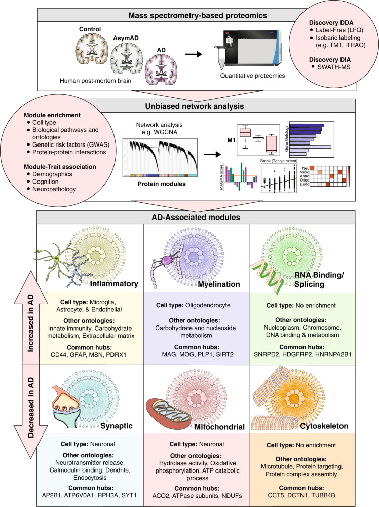
“Three of the modules are consistently increased in the AD brain network proteome: inflammatory, myelination, and RNA binding/splicing, while the remaining three are consistently decreased: synaptic, mitochondrial, and cytoskeleton.”
§ Systems-based proteomics to resolve the biology of Alzheimer’s disease beyond amyloid and tau > A global network proteomic approach to the AD brain > Strategies for mass spectrometry-based quantification
plaincorrelational- Asserted: passes — The authors put it forward as true.
- Checkable: passes — It can be checked against evidence.
- Load-bearing: passes — The paper’s conclusions depend on it.
- Based: passes — The paper gives a reason why it holds.
- Rests on an assumption: Protein co-expression modules reflect various molecular, cellular, and circuit-level phenotypes.
A result the paper reports from its own data — it cites no other source for it, so there is none to check. The quote shows it is in the paper.
In the paper
“Using this framework, six core and highly conserved modules across AsymAD and AD cohorts with reproducible links to specific cell types, organelles, and biological functions have been identified.”
§ Systems-based proteomics to resolve the biology of Alzheimer’s disease beyond amyloid and tau > A global network proteomic approach to the AD brain > Strategies for mass spectrometry-based quantification
plaincorrelational- Asserted: passes — The authors put it forward as true.
- Checkable: passes — It can be checked against evidence.
- Load-bearing: passes — The paper’s conclusions depend on it.
- Based: passes — The paper gives a reason why it holds.
- Rests on an assumption: Protein co-expression modules reflect various molecular, cellular, and circuit-level phenotypes.
In the paper
“Johnson et al. demonstrated this advantage in one of the first TMT-MS network proteomes of the AD brain [74], which quantified 6533 proteins across 47 brain tissues compared to just 2736 proteins quantified by LFQ-MS when applied to the same samples.”
§ Systems-based proteomics to resolve the biology of Alzheimer’s disease beyond amyloid and tau > A global network proteomic approach to the AD brain > Strategies for mass spectrometry-based quantification
What the sources say
- Not checked yetcites [74] Johnson ECB, Dammer EB, Duong DM, Yin L, Thambisetty M, Troncoso JC, et al. Deep proteomic network analysis of Alzheimer’s disease brain reveals alterations in RNA binding proteins and RNA splicing associated with disease. Mol Neurodegener. 2018;13:52. source
boosteddirectn = 47- Asserted: passes — The authors put it forward as true.
- Checkable: passes — It can be checked against evidence.
- Load-bearing: passes — The paper’s conclusions depend on it.
- Based: passes — The paper gives a reason why it holds.
None of the sources cited for this claim have been checked yet.
In the paper
“Only 30–40% of modules in the AD brain network proteome overlap with those of the network transcriptome [62, 63].”
§ Systems-based proteomics to resolve the biology of Alzheimer’s disease beyond amyloid and tau > A global network proteomic approach to the AD brain
What the sources say
- Not checked yetcites [62] Seyfried NT, Dammer EB, Swarup V, Nandakumar D, Duong DM, Yin L, et al. A multi-network approach identifies protein-specific co-expression in asymptomatic and symptomatic Alzheimer’s disease. Cell Syst. 2017;4:60–72. source
- Not checked yetcites [63] Higginbotham L, Ping L, Dammer EB, Duong DM, Zhou M, Gearing M, et al. Integrated proteomics reveals brain-based cerebrospinal fluid biomarkers in asymptomatic and symptomatic Alzheimer’s disease. bioRxiv. 2019, https://doi.org/10.1101/806752. source
plaincorrelational- Asserted: passes — The authors put it forward as true.
- Checkable: passes — It can be checked against evidence.
- Load-bearing: passes — The paper’s conclusions depend on it.
- Based: passes — The paper gives a reason why it holds.
None of the sources cited for this claim have been checked yet.
In the paper
“While differential protein expression within these overlapping modules is reasonably concordant (R~0.5), we have repeatedly observed targets among these modules with highly discordant changes at the protein and RNA levels [62–64].”
§ Systems-based proteomics to resolve the biology of Alzheimer’s disease beyond amyloid and tau > A global network proteomic approach to the AD brain
What the sources say
- Not checked yetcites [62] Seyfried NT, Dammer EB, Swarup V, Nandakumar D, Duong DM, Yin L, et al. A multi-network approach identifies protein-specific co-expression in asymptomatic and symptomatic Alzheimer’s disease. Cell Syst. 2017;4:60–72. source
- Not checked yetcites [63] Higginbotham L, Ping L, Dammer EB, Duong DM, Zhou M, Gearing M, et al. Integrated proteomics reveals brain-based cerebrospinal fluid biomarkers in asymptomatic and symptomatic Alzheimer’s disease. bioRxiv. 2019, https://doi.org/10.1101/806752. source
- Not checked yetcites [64] Swarup V, Chang TS, Duong DM, Dammer EB, Dai J, Lah JJ, et al. Identification of conserved proteomic networks in neurodegenerative dementia. Cell Rep. 2020;31:107807. source
plaincorrelational- Asserted: passes — The authors put it forward as true.
- Checkable: passes — It can be checked against evidence.
- Load-bearing: unclear — It is unclear whether the paper’s conclusions depend on it.
- Based: passes — The paper gives a reason why it holds.
None of the sources cited for this claim have been checked yet.
In the paper
“Among the resulting 44 modules, we resolved an ECM-associated module with highly significant endothelial enrichment and relatively weak glial co-expression.”
§ Systems-based proteomics to resolve the biology of Alzheimer’s disease beyond amyloid and tau > Organization of the AD brain network proteome > An early anti-inflammatory glial response
What the sources say
- Not checked yetcites [63] Higginbotham L, Ping L, Dammer EB, Duong DM, Zhou M, Gearing M, et al. Integrated proteomics reveals brain-based cerebrospinal fluid biomarkers in asymptomatic and symptomatic Alzheimer’s disease. bioRxiv. 2019, https://doi.org/10.1101/806752. source
- Not checked yetcites [75] Ping L, Duong DM, Yin L, Gearing M, Lah JJ, Levey AI, et al. Global quantitative analysis of the human brain proteome in Alzheimer’s and Parkinson’s disease. Sci Data. 2018;5:180036. source
plaincorrelational- Asserted: passes — The authors put it forward as true.
- Checkable: passes — It can be checked against evidence.
- Load-bearing: passes — The paper’s conclusions depend on it.
- Based: passes — The paper gives a reason why it holds.
None of the sources cited for this claim have been checked yet.
In the paper
“Antemortem diagnosis also relies heavily on the presence of Aβ and tau, either as measured in the spinal fluid or imaged using positron emission tomography (PET) [7–10].”
§ Systems-based proteomics to resolve the biology of Alzheimer’s disease beyond amyloid and tau > Introduction
What the sources say
- Not checked yetcites [7] Jack CR Jr, Wiste HJ, Schwarz CG, Lowe VJ, Senjem ML, Vemuri P, et al. Longitudinal tau PET in ageing and Alzheimer’s disease. Brain. 2018;141:1517–28. source
- Not checked yetcites [8] Lowe VJ, Lundt ES, Albertson SM, Przybelski SA, Senjem ML, Parisi JE, et al. Neuroimaging correlates with neuropathologic schemes in neurodegenerative disease. Alzheimers Dement. 2019;15:927–39. source
- Not checked yetcites [9] Fagan AM, Mintun MA, Mach RH, Lee SY, Dence CS, Shah AR, et al. Inverse relation between in vivo amyloid imaging load and cerebrospinal fluid Abeta42 in humans. Ann Neurol. 2006;59:512–9. source
- Not checked yetcites [10] Fagan AM, Roe CM, Xiong C, Mintun MA, Morris JC, Holtzman DM. Cerebrospinal fluid tau/beta-amyloid(42) ratio as a prediction of cognitive decline in nondemented older adults. Arch Neurol. 2007;64:343–9. source
plaincorrelational- Asserted: passes — The authors put it forward as true.
- Checkable: passes — It can be checked against evidence.
- Load-bearing: passes — The paper’s conclusions depend on it.
- Based: passes — The paper gives a reason why it holds.
- It may state two separate claims as one. 57
- Probably not a substantial claim. 39
None of the sources cited for this claim have been checked yet.
In the paper
“Network analyses of AD brain tissue demonstrate the most reproducibility at the module level, followed by the hub protein level, and then finally at the level of precise protein connectivity rankings and module protein membership [61, 89–94].”
§ Systems-based proteomics to resolve the biology of Alzheimer’s disease beyond amyloid and tau > A global network proteomic approach to the AD brain > Principles of network building and module identification
What the sources say
- Not checked yetcites [61] Parikshak NN, Gandal MJ, Geschwind DH. Systems biology and gene networks in neurodevelopmental and neurodegenerative disorders. Nat Rev Genet. 2015;16:441–58. source
- Not checked yetcites [89] Langfelder P, Luo R, Oldham MC, Horvath S. Is my network module preserved and reproducible? PLoS Comput Biol. 2011;7:e1001057. source
- Not checked yetcites [90] Song L, Langfelder P, Horvath S. Comparison of co-expression measures: mutual information, correlation, and model based indices. BMC Bioinforma. 2012;13:328.
- Not checked yetcites [91] Allen JD, Xie Y, Chen M, Girard L, Xiao G. Comparing statistical methods for constructing large scale gene networks. PLoS ONE. 2012;7:e29348. source
- Not checked yetcites [92] Gaiteri C, Ding Y, French B, Tseng GC, Sibille E. Beyond modules and hubs: the potential of gene coexpression networks for investigating molecular mechanisms of complex brain disorders. Genes Brain Behav. 2014;13:13–24. source
- Not checked yetcites [93] Ballouz S, Verleyen W, Gillis J. Guidance for RNA-seq co-expression network construction and analysis: safety in numbers. Bioinformatics. 2015;31:2123–30. source
- Not checked yetcites [94] Marbach D, Costello JC, Kuffner R, et al. Wisdom of crowds for robust gene network inference. Nat Methods. 2012;9:796–804. source
boostedcorrelational- Asserted: passes — The authors put it forward as true.
- Checkable: passes — It can be checked against evidence.
- Load-bearing: passes — The paper’s conclusions depend on it.
- Based: passes — The paper gives a reason why it holds.
It may state two separate claims as one.
A result the paper reports from its own data — it cites no other source for it, so there is none to check. The quote shows it is in the paper.
In the paper
“Three of the modules are consistently increased in the AD brain network proteome: inflammatory, myelination, and RNA binding/splicing, while the remaining three are consistently decreased: synaptic, mitochondrial, and cytoskeleton.”
§ Systems-based proteomics to resolve the biology of Alzheimer’s disease beyond amyloid and tau > A global network proteomic approach to the AD brain > Strategies for mass spectrometry-based quantification
plaincorrelational- Asserted: passes — The authors put it forward as true.
- Checkable: passes — It can be checked against evidence.
- Load-bearing: passes — The paper’s conclusions depend on it.
- Based: passes — The paper gives a reason why it holds.
It may state two separate claims as one.
- Rests on an assumption: Protein co-expression modules reflect various molecular, cellular, and circuit-level phenotypes.
A result the paper reports from its own data — it cites no other source for it, so there is none to check. The quote shows it is in the paper.
In the paper
“This module was strongly associated with wound healing and other aspects of humoral immunity and featured modest upregulation in AD dementia.”
§ Systems-based proteomics to resolve the biology of Alzheimer’s disease beyond amyloid and tau > Organization of the AD brain network proteome > An early anti-inflammatory glial response
boostedcorrelational- Asserted: passes — The authors put it forward as true.
- Checkable: passes — It can be checked against evidence.
- Load-bearing: unclear — It is unclear whether the paper’s conclusions depend on it.
- Based: passes — The paper gives a reason why it holds.
It may state two separate claims as one.
None of the sources cited for this claim have been checked yet.
In the paper
“A wealth of evidence suggests these proteins play a pathogenic role in disease.”
§ Systems-based proteomics to resolve the biology of Alzheimer’s disease beyond amyloid and tau > Introduction
What the sources say
- Not checked yetcites [3] Duyckaerts C, Delatour B, Potier MC. Classification and basic pathology of Alzheimer disease. Acta Neuropathol. 2009;118:5–36. source
- Not checked yetcites [4] Julia TCW, Goate AM. Genetics of beta-amyloid precursor protein in Alzheimer’s disease. Cold Spring Harb Perspect Med. 2017;7:a024539. source
- Not checked yetcites [5] Hardy JA, Higgins GA. Alzheimer’s disease: the amyloid cascade hypothesis. Science. 1992;256:184–5. source
hedgeddirect- Asserted: passes — The authors put it forward as true.
- Checkable: passes — It can be checked against evidence.
- Load-bearing: passes — The paper’s conclusions depend on it.
- Based: passes — The paper gives a reason why it holds.
It may state two separate claims as one.
None of the sources cited for this claim have been checked yet.
In the paper
“In addition, gene mutations that directly enhance Aβ aggregation underlie rare familial forms of the disease [4, 5].”
§ Systems-based proteomics to resolve the biology of Alzheimer’s disease beyond amyloid and tau > Introduction
What the sources say
plaindirect- Asserted: passes — The authors put it forward as true.
- Checkable: passes — It can be checked against evidence.
- Load-bearing: fails — The paper’s conclusions may not depend on it.
- Based: passes — The paper gives a reason why it holds.
Probably not a substantial claim. The paper’s conclusions may not depend on it.
None of the sources cited for this claim have been checked yet.
In the paper
“This hypothesis gained favor in the 1990s after several gene mutations directly responsible for aberrant Aβ accumulation (APP, PSEN1, PSEN2) were found to underlie rare familial forms of early-onset AD [20–22].”
§ Systems-based proteomics to resolve the biology of Alzheimer’s disease beyond amyloid and tau > Expanding AD pathogenesis beyond amyloid and tau > The amyloid hypothesis controversy
What the sources say
- Not checked yetcites [20] Masters CL, Bateman R, Blennow K, Rowe CC, Sperling RA, Cummings JL. Alzheimer’s disease. Nat Rev Dis Prim. 2015;1:15056. source
- Not checked yetcites [21] Goate A, Chartier-Harlin MC, Mullan M, Brown J, Crawford F, Fidani L, et al. Segregation of a missense mutation in the amyloid precursor protein gene with familial Alzheimer’s disease. Nature. 1991;349:704–6. source
- Not checked yetcites [22] Scheuner D, Eckman C, Jensen M, Song X, Citron M, Suzuki N, et al. Secreted amyloid beta-protein similar to that in the senile plaques of Alzheimer’s disease is increased in vivo by the presenilin 1 and 2 and APP mutations linked to familial Alzheimer’s disease. Nat Med. 1996;2:864–70. source
plaindirect- Asserted: passes — The authors put it forward as true.
- Checkable: passes — It can be checked against evidence.
- Load-bearing: fails — The paper’s conclusions may not depend on it.
- Based: passes — The paper gives a reason why it holds.
Probably not a substantial claim. The paper’s conclusions may not depend on it.
In the paper
“Aβ peptide is the main component of extracellular plaque pathology in the AD brain [15].”
§ Systems-based proteomics to resolve the biology of Alzheimer’s disease beyond amyloid and tau > Expanding AD pathogenesis beyond amyloid and tau > The amyloid hypothesis controversy
What the sources say
- Not checked yetcites [15] Glenner GG, Wong CW. Alzheimer’s disease: initial report of the purification and characterization of a novel cerebrovascular amyloid protein. Biochem Biophys Res Commun. 1984;120:885–90. source
plain- Asserted: passes — The authors put it forward as true.
- Checkable: passes — It can be checked against evidence.
- Load-bearing: fails — The paper’s conclusions may not depend on it.
- Based: passes — The paper gives a reason why it holds.
Probably not a substantial claim. The paper’s conclusions may not depend on it.
In the paper
“Aβ, which begins accumulating over a decade prior to clinical symptoms, comprises the cortical extracellular plaques characteristic of AD [3].”
§ Systems-based proteomics to resolve the biology of Alzheimer’s disease beyond amyloid and tau > Introduction
What the sources say
- Not checked yetcites [3] Duyckaerts C, Delatour B, Potier MC. Classification and basic pathology of Alzheimer disease. Acta Neuropathol. 2009;118:5–36. source
plaincorrelational- Asserted: passes — The authors put it forward as true.
- Checkable: passes — It can be checked against evidence.
- Load-bearing: fails — The paper’s conclusions may not depend on it.
- Based: passes — The paper gives a reason why it holds.
Probably not a substantial claim. The paper’s conclusions may not depend on it. It may state two separate claims as one.
In the paper
“Large clinical trials of Aβ-targeting therapies have repeatedly failed, suggesting that amyloid-centric therapy is insufficient to quell disease [11].”
§ Systems-based proteomics to resolve the biology of Alzheimer’s disease beyond amyloid and tau > Introduction
What the sources say
- Not checked yetcites [11] Long JM, Holtzman DM. Alzheimer disease: an update on pathobiology and treatment strategies. Cell. 2019;179:312–39. source
hedgeddirect- Asserted: passes — The authors put it forward as true.
- Checkable: passes — It can be checked against evidence.
- Load-bearing: passes — The paper’s conclusions depend on it.
- Based: passes — The paper gives a reason why it holds.
It may state two separate claims as one.
In the paper
“This pathogenic framework argues that the dynamic interaction of both phases is critical for disease progression and cognitive decline.”
§ Systems-based proteomics to resolve the biology of Alzheimer’s disease beyond amyloid and tau > Introduction
What the sources say
- Not checked yetcites [12] De Strooper B, Karran E. The cellular phase of Alzheimer’s disease. Cell. 2016;164:603–15. source
plaindirect- Asserted: passes — The authors put it forward as true.
- Checkable: passes — It can be checked against evidence.
- Load-bearing: passes — The paper’s conclusions depend on it.
- Based: passes — The paper gives a reason why it holds.
Probably not a substantial claim.
None of the sources cited for this claim have been checked yet.
In the paper
“Aberrant Aβ deposition begins over a decade before the onset of clinical symptoms and spreads diffusely throughout the cerebral cortex [16, 17].”
§ Systems-based proteomics to resolve the biology of Alzheimer’s disease beyond amyloid and tau > Expanding AD pathogenesis beyond amyloid and tau > The amyloid hypothesis controversy
What the sources say
- Not checked yetcites [16] Price JL, Morris JC. Tangles and plaques in nondemented aging and “preclinical” Alzheimer’s disease. Ann Neurol. 1999;45:358–68. source
- Not checked yetcites [17] Vermunt L, Sikkes SAM, van den Hout A, et al. Duration of preclinical, prodromal, and dementia stages of Alzheimer’s disease in relation to age, sex, and APOE genotype. Alzheimers Dement. 2019;15:888–98. source
plain- Asserted: passes — The authors put it forward as true.
- Checkable: passes — It can be checked against evidence.
- Load-bearing: unclear — It is unclear whether the paper’s conclusions depend on it.
- Based: passes — The paper gives a reason why it holds.
It may state two separate claims as one.
In the paper
“Thus, some have postulated that AD maintains two pathogenic phases, including a proteopathic “biochemical” phase characterized by the direct toxic effects of Aβ and tau aggregation and a “cellular” phase encompassing a web of complex feedback and feedforward cell-mediated mechanisms that promote irreversible degeneration [12].”
§ Systems-based proteomics to resolve the biology of Alzheimer’s disease beyond amyloid and tau > Introduction
What the sources say
- Not checked yetcites [12] De Strooper B, Karran E. The cellular phase of Alzheimer’s disease. Cell. 2016;164:603–15. source
hedgeddirect- Asserted: fails — The authors may not put it forward as true.
- Checkable: passes — It can be checked against evidence.
- Load-bearing: passes — The paper’s conclusions depend on it.
- Based: passes — The paper gives a reason why it holds.
Probably not a substantial claim. The authors may not put it forward as true. It may state two separate claims as one.
In the paper
“Meanwhile, tau accumulation as intracellular neurofibrillary tangles (NFTs) is another hallmark feature of AD and strongly associated with progressive neuronal loss and cognitive decline [3].”
§ Systems-based proteomics to resolve the biology of Alzheimer’s disease beyond amyloid and tau > Introduction
What the sources say
- Not checked yetcites [3] Duyckaerts C, Delatour B, Potier MC. Classification and basic pathology of Alzheimer disease. Acta Neuropathol. 2009;118:5–36. source
boostedcorrelational- Asserted: passes — The authors put it forward as true.
- Checkable: passes — It can be checked against evidence.
- Load-bearing: unclear — It is unclear whether the paper’s conclusions depend on it.
- Based: passes — The paper gives a reason why it holds.
It may state two separate claims as one.
In the paper
“However, the repeated failures of Aβ-targeting drug trials have recently bolstered critics of this hypothesis and broadened investigations of AD pathophysiology.”
§ Systems-based proteomics to resolve the biology of Alzheimer’s disease beyond amyloid and tau > Expanding AD pathogenesis beyond amyloid and tau
What the sources say
- Not checked yetcites [11] Long JM, Holtzman DM. Alzheimer disease: an update on pathobiology and treatment strategies. Cell. 2019;179:312–39. source
plaindirect- Asserted: passes — The authors put it forward as true.
- Checkable: passes — It can be checked against evidence.
- Load-bearing: passes — The paper’s conclusions depend on it.
- Based: passes — The paper gives a reason why it holds.
It may state two separate claims as one.
In the paper
“For these reasons, a definitive AD diagnosis requires the postmortem presence of both Aβ and tau pathologies in the cerebral cortex [6].”
§ Systems-based proteomics to resolve the biology of Alzheimer’s disease beyond amyloid and tau > Introduction
What the sources say
- Not checked yetcites [6] Jack CR Jr, Bennett DA, Blennow K, Carrillo MC, Dunn B, Haeberlein SB, et al. NIA-AA research framework: toward a biological definition of Alzheimer’s disease. Alzheimers Dement. 2018;14:535–62. source
plaincorrelational- Asserted: passes — The authors put it forward as true.
- Checkable: passes — It can be checked against evidence.
- Load-bearing: fails — The paper’s conclusions may not depend on it.
- Based: passes — The paper gives a reason why it holds.
Probably not a substantial claim. The paper’s conclusions may not depend on it.
In the paper
“Hyperphosphorylated tau accumulates as intraneuronal NFTs, a hallmark feature of AD pathology [3].”
§ Systems-based proteomics to resolve the biology of Alzheimer’s disease beyond amyloid and tau > Expanding AD pathogenesis beyond amyloid and tau > The evolving role of tau
What the sources say
- Not checked yetcites [3] Duyckaerts C, Delatour B, Potier MC. Classification and basic pathology of Alzheimer disease. Acta Neuropathol. 2009;118:5–36. source
plaincorrelational- Asserted: passes — The authors put it forward as true.
- Checkable: passes — It can be checked against evidence.
- Load-bearing: fails — The paper’s conclusions may not depend on it.
- Based: passes — The paper gives a reason why it holds.
Probably not a substantial claim. The paper’s conclusions may not depend on it.
In the paper
“Thus, the hallmark proteinopathy of AD does not directly cause symptomatology but instead serves as an upstream trigger for destructive cellular processes.”
§ Systems-based proteomics to resolve the biology of Alzheimer’s disease beyond amyloid and tau > Expanding AD pathogenesis beyond amyloid and tau > The cellular phase of AD
What the sources say
- Not checked yetcites [12] De Strooper B, Karran E. The cellular phase of Alzheimer’s disease. Cell. 2016;164:603–15. source
plaindirect- Asserted: passes — The authors put it forward as true.
- Checkable: passes — It can be checked against evidence.
- Load-bearing: passes — The paper’s conclusions depend on it.
- Based: passes — The paper gives a reason why it holds.
It may state two separate claims as one.
None of the sources cited for this claim have been checked yet.
In the paper
“Likewise, genome-wide association studies (GWAS) have identified several single-nucleotide polymorphisms (SNPs) in immune genes as independent risk factors for LOAD [52–55], directly implicating microglia-mediated mechanisms in sporadic AD progression.”
§ Systems-based proteomics to resolve the biology of Alzheimer’s disease beyond amyloid and tau > Expanding AD pathogenesis beyond amyloid and tau > The cellular phase of AD
What the sources say
- Not checked yetcites [52] Lambert JC, Ibrahim-Verbaas CA, Harold D, Naj AC, Sims R, Bellenguez C, et al. Meta-analysis of 74,046 individuals identifies 11 new susceptibility loci for Alzheimer’s disease. Nat Genet. 2013;45:1452–8. source
- Not checked yetcites [53] Lambert JC, Grenier-Boley B, Chouraki V, Heath S, Zelenika D, Fievet N, et al. Implication of the immune system in Alzheimer’s disease: evidence from genome-wide pathway analysis. J Alzheimer’s Dis: JAD. 2010;20:1107–18. source
- Not checked yetcites [54] Heneka MT, Golenbock DT, Latz E. Innate immunity in Alzheimer’s disease. Nat Immunol. 2015;16:229–36. source
- Not checked yetcites [55] Karch CM, Goate AM. Alzheimer’s disease risk genes and mechanisms of disease pathogenesis. Biol Psychiatry. 2015;77:43–51. source
plaindirect- Asserted: passes — The authors put it forward as true.
- Checkable: passes — It can be checked against evidence.
- Load-bearing: passes — The paper’s conclusions depend on it.
- Based: passes — The paper gives a reason why it holds.
It may state two separate claims as one.
None of the sources cited for this claim have been checked yet.
In the paper
“This was followed by evidence indicating that soluble oligomeric Aβ mediates synaptic dysfunction and neuronal toxicity [31–33].”
§ Systems-based proteomics to resolve the biology of Alzheimer’s disease beyond amyloid and tau > Expanding AD pathogenesis beyond amyloid and tau > The amyloid hypothesis controversy
What the sources say
- Not checked yetcites [31] Benilova I, Karran E, De, Strooper B. The toxic Abeta oligomer and Alzheimer’s disease: an emperor in need of clothes. Nat Neurosci. 2012;15:349–57. source
- Not checked yetcites [32] Walsh DM, Klyubin I, Fadeeva JV, Cullen WK, Anwyl R, Wolfe MS, et al. Naturally secreted oligomers of amyloid beta protein potently inhibit hippocampal long-term potentiation in vivo. Nature. 2002;416:535–9. source
- Not checked yetcites [33] Glabe CG. Common mechanisms of amyloid oligomer pathogenesis in degenerative disease. Neurobiol Aging. 2006;27:570–5. source
hedgeddirect- Asserted: passes — The authors put it forward as true.
- Checkable: passes — It can be checked against evidence.
- Load-bearing: fails — The paper’s conclusions may not depend on it.
- Based: passes — The paper gives a reason why it holds.
Probably not a substantial claim. The paper’s conclusions may not depend on it. It may state two separate claims as one.
In the paper
“It is this cellular transition, argue De Strooper and Karran, that propels the onset of cognitive symptoms.”
§ Systems-based proteomics to resolve the biology of Alzheimer’s disease beyond amyloid and tau > Expanding AD pathogenesis beyond amyloid and tau > The cellular phase of AD
What the sources say
- Not checked yetcites [12] De Strooper B, Karran E. The cellular phase of Alzheimer’s disease. Cell. 2016;164:603–15. source
plaindirect- Asserted: passes — The authors put it forward as true.
- Checkable: passes — It can be checked against evidence.
- Load-bearing: passes — The paper’s conclusions depend on it.
- Based: passes — The paper gives a reason why it holds.
Probably not a substantial claim.
None of the sources cited for this claim have been checked yet.
In the paper
“Subsequent studies identified the locus coeruleus as the first site of pathology in AD subjects, with NFTs preceding cortical tau or Aβ deposits [41–43].”
§ Systems-based proteomics to resolve the biology of Alzheimer’s disease beyond amyloid and tau > Expanding AD pathogenesis beyond amyloid and tau > The evolving role of tau
What the sources say
- Not checked yetcites [41] Kelly SC, He B, Perez SE, Ginsberg SD, Mufson EJ, Counts SE. Locus coeruleus cellular and molecular pathology during the progression of Alzheimer’s disease. Acta Neuropathologica Commun. 2017;5:8. source
- Not checked yetcites [42] Grudzien A, Shaw P, Weintraub S, Bigio E, Mash DC, Mesulam MM. Locus coeruleus neurofibrillary degeneration in aging, mild cognitive impairment and early Alzheimer’s disease. Neurobiol Aging. 2007;28:327–35. source
- Not checked yetcites [43] Bondareff W, Mountjoy CQ, Roth M, Rossor MN, Iversen LL, Reynolds GP, et al. Neuronal degeneration in locus ceruleus and cortical correlates of Alzheimer disease. Alzheimer Dis Assoc Disord. 1987;1:256–62. source
plaincorrelational- Asserted: passes — The authors put it forward as true.
- Checkable: passes — It can be checked against evidence.
- Load-bearing: unclear — It is unclear whether the paper’s conclusions depend on it.
- Based: passes — The paper gives a reason why it holds.
It may state two separate claims as one.
A conclusion the paper draws from its own results — it cites no other source for it, so there is none to check. The quote shows it is in the paper.
In the paper
“Though the amyloid hypothesis places NFTs downstream of Aβ deposition, increasing evidence suggests that aberrant tau accumulation is a complex multifactorial process that can occur independent of amyloid.”
§ Systems-based proteomics to resolve the biology of Alzheimer’s disease beyond amyloid and tau > Expanding AD pathogenesis beyond amyloid and tau > The evolving role of tau
hedged- Asserted: passes — The authors put it forward as true.
- Checkable: passes — It can be checked against evidence.
- Load-bearing: passes — The paper’s conclusions depend on it.
- Based: passes — The paper gives a reason why it holds.
It may state two separate claims as one.
In the paper
“Dysfunction of the astrocytes and specialized endothelium comprising this clearance system not only contributes to increasing amyloid accumulation but also promotes irrevocable disruption of other neurovascular and glioneuronal functions, including lipid metabolism, myelin turnover, and immune regulation.”
§ Systems-based proteomics to resolve the biology of Alzheimer’s disease beyond amyloid and tau > Expanding AD pathogenesis beyond amyloid and tau > The cellular phase of AD
What the sources say
- Not checked yetcites [12] De Strooper B, Karran E. The cellular phase of Alzheimer’s disease. Cell. 2016;164:603–15. source
plaindirect- Asserted: passes — The authors put it forward as true.
- Checkable: passes — It can be checked against evidence.
- Load-bearing: passes — The paper’s conclusions depend on it.
- Based: passes — The paper gives a reason why it holds.
It may state two separate claims as one.
In the paper
“Cell-mediated feedback and feedforward mechanisms allow this dysfunction to thrive independently of accumulating amyloid and tau, precipitating chronic inflammation and circuitry imbalances that result in cell failure and death.”
§ Systems-based proteomics to resolve the biology of Alzheimer’s disease beyond amyloid and tau > Expanding AD pathogenesis beyond amyloid and tau > The cellular phase of AD
What the sources say
- Not checked yetcites [12] De Strooper B, Karran E. The cellular phase of Alzheimer’s disease. Cell. 2016;164:603–15. source
plaindirect- Asserted: passes — The authors put it forward as true.
- Checkable: passes — It can be checked against evidence.
- Load-bearing: passes — The paper’s conclusions depend on it.
- Based: passes — The paper gives a reason why it holds.
It may state two separate claims as one.
In the paper
“Compared to amyloid burden, tau levels correlate much more strongly to cognitive symptoms, suggesting a more direct link to disease progression [46].”
§ Systems-based proteomics to resolve the biology of Alzheimer’s disease beyond amyloid and tau > Expanding AD pathogenesis beyond amyloid and tau > The evolving role of tau
What the sources say
- Not checked yetcites [46] Hanseeuw BJ, Betensky RA, Jacobs HIL, Schultz AP, Sepulcre J, Becker JA, et al. Association of amyloid and tau with cognition in preclinical Alzheimer disease: a longitudinal study. JAMA Neurol. 2019;76:915–24. source
hedgedcorrelational- Asserted: passes — The authors put it forward as true.
- Checkable: passes — It can be checked against evidence.
- Load-bearing: passes — The paper’s conclusions depend on it.
- Based: passes — The paper gives a reason why it holds.
It may state two separate claims as one.
None of the sources cited for this claim have been checked yet.
In the paper
“These genetic findings led to several transgenic mouse models that successfully recapitulated Aβ plaque formation, increasing support for the hypothesis (Table 1) [23–29].”
§ Systems-based proteomics to resolve the biology of Alzheimer’s disease beyond amyloid and tau > Expanding AD pathogenesis beyond amyloid and tau > The amyloid hypothesis controversy
What the sources say
- Not checked yetcites [23] Lamb BT, Call LM, Slunt HH, Bardel KA, Lawler AM, Eckman CB, et al. Altered metabolism of familial Alzheimer’s disease-linked amyloid precursor protein variants in yeast artificial chromosome transgenic mice. Hum Mol Genet. 1997;6:1535–41. source
- Not checked yetcites [24] Borchelt DR, Ratovitski T, van Lare J, Lee MK, Gonzales V, Jenkins NA, et al. Accelerated amyloid deposition in the brains of transgenic mice coexpressing mutant presenilin 1 and amyloid precursor proteins. Neuron. 1997;19:939–45. source
- Not checked yetcites [25] Games D, Adams D, Alessandrini R, Barbour R, Berthelette P, Blackwell C, et al. Alzheimer-type neuropathology in transgenic mice overexpressing V717F beta-amyloid precursor protein. Nature. 1995;373:523–7. source
- Not checked yetcites [26] Sturchler-Pierrat C, Abramowski D, Duke M, Wiederhold KH, Mistl C, Rothacher S, et al. Two amyloid precursor protein transgenic mouse models with Alzheimer disease-like pathology. Proc Natl Acad Sci USA. 1997;94:13287–92. source
- Not checked yetcites [27] Moechars D, Dewachter I, Lorent K, Reverse D, Baekelandt V, Naidu A, et al. Early phenotypic changes in transgenic mice that overexpress different mutants of amyloid precursor protein in brain. J Biol Chem. 1999;274:6483–92. source
- Not checked yetcites [28] Reaume AG, Howland DS, Trusko SP, Savage MJ, Lang DM, Greenberg BD, et al. Enhanced amyloidogenic processing of the beta-amyloid precursor protein in gene-targeted mice bearing the Swedish familial Alzheimer’s disease mutations and a “humanized” Abeta sequence. The. J Biol Chem. 1996;271:23380–8. source
- Not checked yetcites [29] Mucke L, Masliah E, Yu GQ, Mallory M, Rockenstein EM, Tatsuno G, et al. High-level neuronal expression of abeta 1-42 in wild-type human amyloid protein precursor transgenic mice: synaptotoxicity without plaque formation. J Neurosci. 2000;20:4050–8. source
plaindirect- Asserted: passes — The authors put it forward as true.
- Checkable: passes — It can be checked against evidence.
- Load-bearing: unclear — It is unclear whether the paper’s conclusions depend on it.
- Based: passes — The paper gives a reason why it holds.
It may state two separate claims as one.
In the paper
“These findings, consistent with previous comparisons of protein and mRNA data [65], strongly support the utility of protein profiling in AD and its complementarity with transcriptomic studies.”
§ Systems-based proteomics to resolve the biology of Alzheimer’s disease beyond amyloid and tau > A global network proteomic approach to the AD brain
What the sources say
- Not checked yetcites [65] de Sousa Abreu R, Penalva LO, Marcotte EM, Vogel C. Global signatures of protein and mRNA expression levels. Mol Biosyst. 2009;5:1512–26.
boostedcorrelational- Asserted: passes — The authors put it forward as true.
- Checkable: passes — It can be checked against evidence.
- Load-bearing: passes — The paper’s conclusions depend on it.
- Based: passes — The paper gives a reason why it holds.
It may state two separate claims as one.
A conclusion the paper draws from its own results — it cites no other source for it, so there is none to check. The quote shows it is in the paper.
In the paper
“As discussed in the next section, collaborative efforts, such as AMP-AD, and advances in high-throughput proteomics have allowed the field to overcome many of these challenges and identify a systems-based proteomic organization conserved across brain samples from a variety of different AD cohorts and cortical regions.”
§ Systems-based proteomics to resolve the biology of Alzheimer’s disease beyond amyloid and tau > A global network proteomic approach to the AD brain > Limitations of proteomic analysis in human brain tissue
plaindirect- Asserted: passes — The authors put it forward as true.
- Checkable: passes — It can be checked against evidence.
- Load-bearing: passes — The paper’s conclusions depend on it.
- Based: fails — The paper may give no reason why it holds.
Probably not a substantial claim. The paper may give no reason why it holds. It may state two separate claims as one.
A conclusion the paper draws from its own results — it cites no other source for it, so there is none to check. The quote shows it is in the paper.
In the paper
“Cell type-specific perturbations appear to drive many of these disease-associated network changes with glia-enriched modules most increased in AD and neuronal modules most decreased in disease (Fig. 1).”
§ Systems-based proteomics to resolve the biology of Alzheimer’s disease beyond amyloid and tau > Organization of the AD brain network proteome
hedgedconditional- Asserted: passes — The authors put it forward as true.
- Checkable: passes — It can be checked against evidence.
- Load-bearing: passes — The paper’s conclusions depend on it.
- Based: passes — The paper gives a reason why it holds.
It may state two separate claims as one.
None of the sources cited for this claim have been checked yet.
In the paper
“Variants of the microglial TREM2 gene have been found to increase the risk of sporadic AD by approximately 3- to 5-fold [56, 57].”
§ Systems-based proteomics to resolve the biology of Alzheimer’s disease beyond amyloid and tau > Organization of the AD brain network proteome > An early anti-inflammatory glial response
What the sources say
- Not checked yetcites [56] Guerreiro R, Wojtas A, Bras J, Carrasquillo M, Rogaeva E, et al. TREM2 variants in Alzheimer’s disease. N. Engl J Med. 2013;368:117–27. source
- Not checked yetcites [57] Jonsson T, Stefansson H, Steinberg S, Jonsdottir I, Jonsson PV, Snaedal J, et al. Variant of TREM2 associated with the risk of Alzheimer’s disease. N. Engl J Med. 2013;368:107–16. source
plaindirect- Asserted: passes — The authors put it forward as true.
- Checkable: passes — It can be checked against evidence.
- Load-bearing: fails — The paper’s conclusions may not depend on it.
- Based: passes — The paper gives a reason why it holds.
Probably not a substantial claim. The paper’s conclusions may not depend on it.
In the paper
“Alterations in these disease-associated modules are strongly conserved across cohorts and cortical regions, allowing for the recent construction of a large consensus AD brain network derived from hundreds of brain samples [96].”
§ Systems-based proteomics to resolve the biology of Alzheimer’s disease beyond amyloid and tau > Organization of the AD brain network proteome
What the sources say
- Not checked yetcites [96] Johnson ECB, Dammer EB, Duong DM, Ping L, Zhou M, Yin L, et al. Large-scale proteomic analysis of Alzheimer's disease brain and cerebrospinal fluid reveals early changes in energy metabolism associated with microglia and astrocyte activation. Nat Med. 2020;26:769–80. source
boostedcorrelational- Asserted: passes — The authors put it forward as true.
- Checkable: passes — It can be checked against evidence.
- Load-bearing: passes — The paper’s conclusions depend on it.
- Based: passes — The paper gives a reason why it holds.
It may state two separate claims as one.
The paper states this without a source or data behind it here, so there is nothing to check it against. The quote shows it is in the paper.
In the paper
“Cell populations display a wide dynamic range of protein concentrations driven by their immediate surroundings.”
§ Systems-based proteomics to resolve the biology of Alzheimer’s disease beyond amyloid and tau > A global network proteomic approach to the AD brain > Limitations of proteomic analysis in human brain tissue
plaindirect- Asserted: passes — The authors put it forward as true.
- Checkable: passes — It can be checked against evidence.
- Load-bearing: passes — The paper’s conclusions depend on it.
- Based: fails — The paper may give no reason why it holds.
Probably not a substantial claim. The paper may give no reason why it holds.
A conclusion the paper draws from its own results — it cites no other source for it, so there is none to check. The quote shows it is in the paper.
In the paper
“Furthermore, as discussed below, the application of network approaches to protein interactomes has helped characterize key aggregation mechanisms underlying the biochemical phase of AD.”
§ Systems-based proteomics to resolve the biology of Alzheimer’s disease beyond amyloid and tau > A global network proteomic approach to the AD brain > Limitations of proteomic analysis in human brain tissue
plaindirect- Asserted: passes — The authors put it forward as true.
- Checkable: passes — It can be checked against evidence.
- Load-bearing: fails — The paper’s conclusions may not depend on it.
- Based: fails — The paper may give no reason why it holds.
Probably not a substantial claim. The paper’s conclusions may not depend on it. The paper may give no reason why it holds.
A conclusion the paper draws from its own results — it cites no other source for it, so there is none to check. The quote shows it is in the paper.
In the paper
“Despite a variety of technological advancements, proteomic analysis of bulk human brain tissue has its limitations. Perhaps the biggest challenge is the inter- and intra-regional variability found throughout brain tissue.”
§ Systems-based proteomics to resolve the biology of Alzheimer’s disease beyond amyloid and tau > A global network proteomic approach to the AD brain > Limitations of proteomic analysis in human brain tissue
hedged- Asserted: passes — The authors put it forward as true.
- Checkable: passes — It can be checked against evidence.
- Load-bearing: passes — The paper’s conclusions depend on it.
- Based: passes — The paper gives a reason why it holds.
It may state two separate claims as one.
A conclusion the paper draws from its own results — it cites no other source for it, so there is none to check. The quote shows it is in the paper.
In the paper
“While the network proteomic analysis of bulk tissue can provide an invaluable view of global cell type-specific changes in disease, this approach is limited in examination of the cellular microenvironment and its effects on protein levels.”
§ Systems-based proteomics to resolve the biology of Alzheimer’s disease beyond amyloid and tau > A global network proteomic approach to the AD brain > Limitations of proteomic analysis in human brain tissue
plain- Asserted: passes — The authors put it forward as true.
- Checkable: passes — It can be checked against evidence.
- Load-bearing: passes — The paper’s conclusions depend on it.
- Based: passes — The paper gives a reason why it holds.
It may state two separate claims as one.
A conclusion the paper draws from its own results — it cites no other source for it, so there is none to check. The quote shows it is in the paper.
In the paper
“The identification of modules altered in AsymAD also provides a window into the earliest drivers of AD and may help guide the mechanistic evaluation of preclinical disease.”
§ Systems-based proteomics to resolve the biology of Alzheimer’s disease beyond amyloid and tau > A global network proteomic approach to the AD brain > Limitations of proteomic analysis in human brain tissue
hedgedconditional- Asserted: passes — The authors put it forward as true.
- Checkable: fails — It may be too vague to check against evidence.
- Load-bearing: passes — The paper’s conclusions depend on it.
- Based: passes — The paper gives a reason why it holds.
Probably not a substantial claim. It may be too vague to check against evidence. It may state two separate claims as one.
A conclusion the paper draws from its own results — it cites no other source for it, so there is none to check. The quote shows it is in the paper.
In the paper
“For example, genetic risk factor enrichment can identify those modules more likely to have causative, rather than reactive, roles in disease and support further investigations into the potential disease-related mechanisms associated with these protein communities.”
§ Systems-based proteomics to resolve the biology of Alzheimer’s disease beyond amyloid and tau > A global network proteomic approach to the AD brain > Limitations of proteomic analysis in human brain tissue
plainconditional- Asserted: passes — The authors put it forward as true.
- Checkable: passes — It can be checked against evidence.
- Load-bearing: fails — The paper’s conclusions may not depend on it.
- Based: fails — The paper may give no reason why it holds.
Probably not a substantial claim. The paper’s conclusions may not depend on it. The paper may give no reason why it holds.
- Rests on an assumption: Modules enriched with AD risk factors reflect causal, rather than reactive, disease mechanisms.
None of the sources cited for this claim have been checked yet.
In the paper
“Therefore, DIA results in the identification of nearly all detectable peptides within a selected mass range, allowing for comprehensive and accurate quantification of identified proteins in the sample with minimal missing values [81, 82].”
§ Systems-based proteomics to resolve the biology of Alzheimer’s disease beyond amyloid and tau > A global network proteomic approach to the AD brain > Strategies for mass spectrometry-based quantification
What the sources say
- Not checked yetcites [81] Meyer JG, Schilling B. Clinical applications of quantitative proteomics using targeted and untargeted data-independent acquisition techniques. Expert Rev Proteom. 2017;14:419–29. source
- Not checked yetcites [82] Crowgey EL, Matlock A, Venkatraman V, Fert-Bober J, Van Eyk JE. Mapping biological networks from quantitative data-independent acquisition mass spectrometry: data to knowledge pipelines. Methods Mol Biol. 2017;1558:395–413. source
plaindirect- Asserted: passes — The authors put it forward as true.
- Checkable: passes — It can be checked against evidence.
- Load-bearing: passes — The paper’s conclusions depend on it.
- Based: passes — The paper gives a reason why it holds.
It may state two separate claims as one.
None of the sources cited for this claim have been checked yet.
In the paper
“Using high-throughput MS techniques, these studies have revealed highly reproducible modules across the AD proteome, several of which consistently demonstrate strong correlations to AD diagnosis, Aβ burden, and other phenotypic traits of disease.”
§ Systems-based proteomics to resolve the biology of Alzheimer’s disease beyond amyloid and tau > Organization of the AD brain network proteome
boostedcorrelational- Asserted: passes — The authors put it forward as true.
- Checkable: passes — It can be checked against evidence.
- Load-bearing: passes — The paper’s conclusions depend on it.
- Based: passes — The paper gives a reason why it holds.
It may state two separate claims as one.
A conclusion the paper draws from its own results — it cites no other source for it, so there is none to check. The quote shows it is in the paper.
In the paper
“Yet, these network studies have also yielded AD-associated modules with no apparent links to specific cell types, suggesting these protein changes reflect the biochemical phase of AD.”
§ Systems-based proteomics to resolve the biology of Alzheimer’s disease beyond amyloid and tau > Organization of the AD brain network proteome
hedgedcorrelational- Asserted: passes — The authors put it forward as true.
- Checkable: passes — It can be checked against evidence.
- Load-bearing: unclear — It is unclear whether the paper’s conclusions depend on it.
- Based: passes — The paper gives a reason why it holds.
It may state two separate claims as one.
A conclusion the paper draws from its own results — it cites no other source for it, so there is none to check. The quote shows it is in the paper.
In the paper
“The wealth of evidence indicating the causative role of these cellular processes in AD pathogenesis strongly supports a more holistic approach to AD investigation and biomarker discovery.”
§ Systems-based proteomics to resolve the biology of Alzheimer’s disease beyond amyloid and tau > Expanding AD pathogenesis beyond amyloid and tau > The cellular phase of AD
boosteddirect- Asserted: passes — The authors put it forward as true.
- Checkable: fails — It may be too vague to check against evidence.
- Load-bearing: passes — The paper’s conclusions depend on it.
- Based: passes — The paper gives a reason why it holds.
Probably not a substantial claim. It may be too vague to check against evidence.
In the paper
“Peroxiredoxin 1 (PRDX1), another hub of this module, is a family member of antioxidant enzymes also implicated in protective cellular responses [125].”
§ Systems-based proteomics to resolve the biology of Alzheimer’s disease beyond amyloid and tau > Organization of the AD brain network proteome > An early anti-inflammatory glial response
What the sources say
- Not checked yetcites [125] Ding C, Fan X, Wu G. Peroxiredoxin 1—an antioxidant enzyme in cancer. J Cell Mol Med. 2017;21:193–202. source
plaincorrelational- Asserted: passes — The authors put it forward as true.
- Checkable: passes — It can be checked against evidence.
- Load-bearing: unclear — It is unclear whether the paper’s conclusions depend on it.
- Based: passes — The paper gives a reason why it holds.
It may state two separate claims as one.
In the paper
“The authors subsequently concluded that RNA-binding proteins may drive the proteopathic “biochemical” phase of AD.”
§ Systems-based proteomics to resolve the biology of Alzheimer’s disease beyond amyloid and tau > Organization of the AD brain network proteome > A role for RNA binding in the biochemical phase
What the sources say
- Not checked yetcites [74] Johnson ECB, Dammer EB, Duong DM, Yin L, Thambisetty M, Troncoso JC, et al. Deep proteomic network analysis of Alzheimer’s disease brain reveals alterations in RNA binding proteins and RNA splicing associated with disease. Mol Neurodegener. 2018;13:52. source
hedgedconditional- Asserted: passes — The authors put it forward as true.
- Checkable: passes — It can be checked against evidence.
- Load-bearing: passes — The paper’s conclusions depend on it.
- Based: passes — The paper gives a reason why it holds.
Probably not a substantial claim.
None of the sources cited for this claim have been checked yet.
In the paper
“network proteomics has revealed one other highly conserved AD-associated module with possible links to this phase of disease. Unlike the RNA-binding/splicing modules, this module is consistently downregulated throughout AD and strongly associated with the cytoskeleton (Fig. 1) [62, 63, 96].”
§ Systems-based proteomics to resolve the biology of Alzheimer’s disease beyond amyloid and tau > Organization of the AD brain network proteome > A role for RNA binding in the biochemical phase
What the sources say
- Not checked yetcites [62] Seyfried NT, Dammer EB, Swarup V, Nandakumar D, Duong DM, Yin L, et al. A multi-network approach identifies protein-specific co-expression in asymptomatic and symptomatic Alzheimer’s disease. Cell Syst. 2017;4:60–72. source
- Not checked yetcites [63] Higginbotham L, Ping L, Dammer EB, Duong DM, Zhou M, Gearing M, et al. Integrated proteomics reveals brain-based cerebrospinal fluid biomarkers in asymptomatic and symptomatic Alzheimer’s disease. bioRxiv. 2019, https://doi.org/10.1101/806752. source
- Not checked yetcites [96] Johnson ECB, Dammer EB, Duong DM, Ping L, Zhou M, Yin L, et al. Large-scale proteomic analysis of Alzheimer's disease brain and cerebrospinal fluid reveals early changes in energy metabolism associated with microglia and astrocyte activation. Nat Med. 2020;26:769–80. source
hedgedcorrelational- Asserted: passes — The authors put it forward as true.
- Checkable: passes — It can be checked against evidence.
- Load-bearing: passes — The paper’s conclusions depend on it.
- Based: passes — The paper gives a reason why it holds.
It may state two separate claims as one.
In the paper
“This analysis successfully organized the U1-70K interactome into seven biologically meaningful modules, two of which demonstrated a strong association with pathogenic U1-70K domains.”
§ Systems-based proteomics to resolve the biology of Alzheimer’s disease beyond amyloid and tau > Organization of the AD brain network proteome > A role for RNA binding in the biochemical phase
What the sources say
- Not checked yetcites [138] Bishof I, Dammer EB, Duong DM, Kundinger SR, Gearing M, Lah JJ, et al. RNA-binding proteins with basic-acidic dipeptide (BAD) domains self-assemble and aggregate in Alzheimer’s disease. J Biol Chem. 2018;293:11047–66. source
boostedcorrelational- Asserted: passes — The authors put it forward as true.
- Checkable: passes — It can be checked against evidence.
- Load-bearing: passes — The paper’s conclusions depend on it.
- Based: passes — The paper gives a reason why it holds.
It may state two separate claims as one.
In the paper
“These ribonucleoproteins, constituents of the spliceosome complex responsible for RNA processing, are mislocalized in AD to the cytoplasm of neuronal cell bodies where they aggregate with NFTs and contribute to widespread alterations in RNA processing [134].”
§ Systems-based proteomics to resolve the biology of Alzheimer’s disease beyond amyloid and tau > Organization of the AD brain network proteome > A role for RNA binding in the biochemical phase
What the sources say
- Not checked yetcites [134] Bai B, Hales CM, Chen PC, Gozal Y, Dammer EB, Fritz JJ, et al. U1 small nuclear ribonucleoprotein complex and RNA splicing alterations in Alzheimer’s disease. Proc Natl Acad Sci USA. 2013;110:16562–7. source
plaindirect- Asserted: passes — The authors put it forward as true.
- Checkable: passes — It can be checked against evidence.
- Load-bearing: passes — The paper’s conclusions depend on it.
- Based: passes — The paper gives a reason why it holds.
It may state two separate claims as one.
A conclusion the paper draws from its own results — it cites no other source for it, so there is none to check. The quote shows it is in the paper.
In the paper
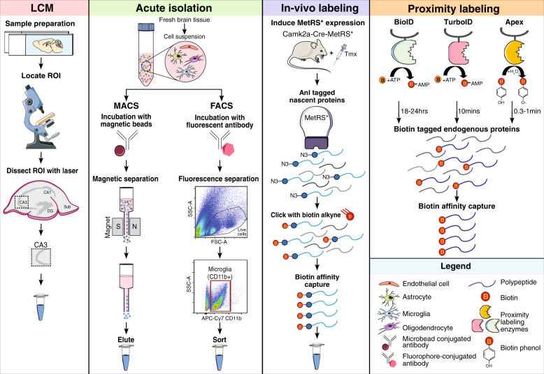
“This FACS pipeline can be easily optimized for proteomic analyses of multiple cell types with >95% purity by confidently excluding debris.”
§ Systems-based proteomics to resolve the biology of Alzheimer’s disease beyond amyloid and tau > Emerging technologies for cell type-specific proteomics > Acute isolation of brain cell types
plaindirect- Asserted: passes — The authors put it forward as true.
- Checkable: passes — It can be checked against evidence.
- Load-bearing: fails — The paper’s conclusions may not depend on it.
- Based: fails — The paper may give no reason why it holds.
Probably not a substantial claim. The paper’s conclusions may not depend on it. The paper may give no reason why it holds.
None of the sources cited for this claim have been checked yet.
In the paper
“BioID has been used extensively in vitro [162–166] to define interacting protein partners and proteins in subcellular compartments, but its application in vivo is limited.”
§ Systems-based proteomics to resolve the biology of Alzheimer’s disease beyond amyloid and tau > Emerging technologies for cell type-specific proteomics > In vitro and in vivo protein labeling strategies
What the sources say
- Not checked yetcites [162] Chen AL, Kim EW, Toh JY, Vashisht AA, Rashoff AQ, Van C, et al. Novel components of the Toxoplasma inner membrane complex revealed by BioID. mBio. 2015;6:e02357–14.
- Not checked yetcites [163] Gupta GD, Coyaud E, Goncalves J, Mojarad BA, Liu Y, Wu Q, et al. A dynamic protein interaction landscape of the human centrosome-cilium interface. Cell. 2015;163:1484–99. source
- Not checked yetcites [164] Morriswood B, Havlicek K, Demmel L, Yavuz S, Sealey-Cardona M, Vidilaseris K, et al. Novel bilobe components in Trypanosoma brucei identified using proximity-dependent biotinylation. Eukaryot Cell. 2013;12:356–67. source
- Not checked yetcites [165] Dingar D, Kalkat M, Chan PK, Srikumar T, Bailey SD, Tu WB, et al. BioID identifies novel c-MYC interacting partners in cultured cells and xenograft tumors. J Proteom. 2015;118:95–111. source
- Not checked yetcites [166] Kehrer J, Frischknecht F, Mair GR. Proteomic analysis of the plasmodium berghei gametocyte egressome and vesicular bioID of osmiophilic body proteins identifies merozoite TRAP-like protein (MTRAP) as an essential factor for parasite transmission. Mol Cell Proteom. 2016;15:2852–62. source
plain- Asserted: passes — The authors put it forward as true.
- Checkable: passes — It can be checked against evidence.
- Load-bearing: fails — The paper’s conclusions may not depend on it.
- Based: passes — The paper gives a reason why it holds.
Probably not a substantial claim. The paper’s conclusions may not depend on it. It may state two separate claims as one.
The paper states this without a source or data behind it here, so there is nothing to check it against. The quote shows it is in the paper.
In the paper
“Cell type-specific proteomics in the human brain is currently in its infancy, primarily due to the inability to isolate live, pure cell populations from frozen brain and limited access to fresh postmortem tissue.”
§ Systems-based proteomics to resolve the biology of Alzheimer’s disease beyond amyloid and tau > Emerging technologies for cell type-specific proteomics
plaindirect- Asserted: passes — The authors put it forward as true.
- Checkable: passes — It can be checked against evidence.
- Load-bearing: passes — The paper’s conclusions depend on it.
- Based: fails — The paper may give no reason why it holds.
Probably not a substantial claim. The paper may give no reason why it holds.
None of the sources cited for this claim have been checked yet.
In the paper
“By fusing BirA* to a bait protein with a synaptic localization signal or nuclear pore signal, BirA* expression was specifically guided to the synapse or nuclear pore complex to achieve biotinylation of the respective proteomes [167, 168].”
§ Systems-based proteomics to resolve the biology of Alzheimer’s disease beyond amyloid and tau > Emerging technologies for cell type-specific proteomics > In vitro and in vivo protein labeling strategies
What the sources say
- Not checked yetcites [167] Kim DI, Birendra KC, Zhu W, Motamedchaboki K, Doye V, Roux KJ. Probing nuclear pore complex architecture with proximity-dependent biotinylation. Proc Natl Acad Sci USA. 2014;111:E2453–61. source
- Not checked yetcites [168] Uezu A, Kanak DJ, Bradshaw TW, Soderblom EJ, Catavero CM, Burette AC, et al. Identification of an elaborate complex mediating postsynaptic inhibition. Science. 2016;353:1123–9. source
plaindirect- Asserted: passes — The authors put it forward as true.
- Checkable: passes — It can be checked against evidence.
- Load-bearing: fails — The paper’s conclusions may not depend on it.
- Based: passes — The paper gives a reason why it holds.
Probably not a substantial claim. The paper’s conclusions may not depend on it.
In the paper
“Cotl1 was identified as a novel microglia-specific marker with increased expression and strong association with AD neuropathology.”
§ Systems-based proteomics to resolve the biology of Alzheimer’s disease beyond amyloid and tau > Emerging technologies for cell type-specific proteomics > Acute isolation of brain cell types
What the sources say
- Not checked yetcites [146] Rangaraju S, Dammer EB, Raza SA, Gao T, Xiao H, Betarbet R, et al. Quantitative proteomics of acutely-isolated mouse microglia identifies novel immune Alzheimer’s disease-related proteins. Mol Neurodegeneration. 2018;13:34.
boostedcorrelational- Asserted: passes — The authors put it forward as true.
- Checkable: passes — It can be checked against evidence.
- Load-bearing: unclear — It is unclear whether the paper’s conclusions depend on it.
- Based: passes — The paper gives a reason why it holds.
It may state two separate claims as one.
In the paper
“One inherent limitation of MACS and FACS is that live neurons cannot be sampled from adult brain, tremendously limiting our ability to understand neuron-specific proteomic changes occurring in vivo, as described below.”
§ Systems-based proteomics to resolve the biology of Alzheimer’s disease beyond amyloid and tau > Emerging technologies for cell type-specific proteomics > Acute isolation of brain cell types
What the sources say
- Not checked yetcites Table 2
plaindirect- Asserted: passes — The authors put it forward as true.
- Checkable: passes — It can be checked against evidence.
- Load-bearing: passes — The paper’s conclusions depend on it.
- Based: passes — The paper gives a reason why it holds.
It may state two separate claims as one.
What the paper did — it cites no source for this, so there is none to check. The quote shows it is in the paper.
In the paper
“LCM-based MS results in a lower proteome coverage compared to bulk brain proteomics, and the inability to confidently exclude contamination by cells that are not of interest poses a challenge (Table 2).”
§ Systems-based proteomics to resolve the biology of Alzheimer’s disease beyond amyloid and tau > Emerging technologies for cell type-specific proteomics > MS-coupled laser capture microdissection
plaindirect- Asserted: passes — The authors put it forward as true.
- Checkable: passes — It can be checked against evidence.
- Load-bearing: unclear — It is unclear whether the paper’s conclusions depend on it.
- Based: passes — The paper gives a reason why it holds.
It may state two separate claims as one.
In the paper
“Proximity labeling with enzymes is another strategy to achieve cell type-specific global proteomic labeling [160].”
§ Systems-based proteomics to resolve the biology of Alzheimer’s disease beyond amyloid and tau > Emerging technologies for cell type-specific proteomics > In vitro and in vivo protein labeling strategies
What the sources say
- Not checked yetcites [160] Chen CL, Perrimon N. Proximity-dependent labeling methods for proteomic profiling in living cells. Wiley Interdiscip Rev Dev Biol. 2017;6:10.1002/wdev.272.
plaindirect- Asserted: passes — The authors put it forward as true.
- Checkable: passes — It can be checked against evidence.
- Load-bearing: fails — The paper’s conclusions may not depend on it.
- Based: passes — The paper gives a reason why it holds.
Probably not a substantial claim. The paper’s conclusions may not depend on it.
In the paper
“Apoe protein was also detected within Aβ plaque-associated microglia, suggesting a role for Apoe in phagocytic clearance of Aβ.”
§ Systems-based proteomics to resolve the biology of Alzheimer’s disease beyond amyloid and tau > Emerging technologies for cell type-specific proteomics > Acute isolation of brain cell types
What the sources say
- Not checked yetcites [146] Rangaraju S, Dammer EB, Raza SA, Gao T, Xiao H, Betarbet R, et al. Quantitative proteomics of acutely-isolated mouse microglia identifies novel immune Alzheimer’s disease-related proteins. Mol Neurodegeneration. 2018;13:34.
hedgedconditional- Asserted: passes — The authors put it forward as true.
- Checkable: passes — It can be checked against evidence.
- Load-bearing: unclear — It is unclear whether the paper’s conclusions depend on it.
- Based: passes — The paper gives a reason why it holds.
It may state two separate claims as one.
None of the sources cited for this claim have been checked yet.
In the paper
“This is reflected in the global AD network proteome, which consistently demonstrates the progressive downregulation of synaptic modules in asymptomatic and symptomatic AD brains, a finding that appears to be preserved across various cortical brain regions [62, 96, 105].”
§ Systems-based proteomics to resolve the biology of Alzheimer’s disease beyond amyloid and tau > Organization of the AD brain network proteome > A link between synaptic and mitochondrial dysfunction
What the sources say
- Not checked yetcites [62] Seyfried NT, Dammer EB, Swarup V, Nandakumar D, Duong DM, Yin L, et al. A multi-network approach identifies protein-specific co-expression in asymptomatic and symptomatic Alzheimer’s disease. Cell Syst. 2017;4:60–72. source
- Not checked yetcites [96] Johnson ECB, Dammer EB, Duong DM, Ping L, Zhou M, Yin L, et al. Large-scale proteomic analysis of Alzheimer's disease brain and cerebrospinal fluid reveals early changes in energy metabolism associated with microglia and astrocyte activation. Nat Med. 2020;26:769–80. source
- Not checked yetcites [105] Higginbotham L, Dammer EB, Duong DM, Modeste E, Montine TJ, Lah JJ, et al. Network analysis of a membrane-enriched brain proteome across stages of Alzheimer’s disease. Proteomes. 2019;7:30. source
hedgedcorrelational- Asserted: passes — The authors put it forward as true.
- Checkable: passes — It can be checked against evidence.
- Load-bearing: passes — The paper’s conclusions depend on it.
- Based: passes — The paper gives a reason why it holds.
It may state two separate claims as one.
In the paper
“Emerging evidence in AD mouse models indicates this homeostatic, microglia-mediated synaptic phagocytosis may be one of the earliest events in AD progression and contribute to synaptic losses during asymptomatic stages of disease [143].”
§ Systems-based proteomics to resolve the biology of Alzheimer’s disease beyond amyloid and tau > Organization of the AD brain network proteome > A link between synaptic and mitochondrial dysfunction
What the sources say
- Not checked yetcites [143] Hong S, Beja-Glasser VF, Nfonoyim BM, Frouin A, Li S, Ramakrishnan S, et al. Complement and microglia mediate early synapse loss in Alzheimer mouse models. Science. 2016;352:712–6. source
hedgedconditional- Asserted: passes — The authors put it forward as true.
- Checkable: passes — It can be checked against evidence.
- Load-bearing: passes — The paper’s conclusions depend on it.
- Based: passes — The paper gives a reason why it holds.
It may state two separate claims as one.
In the paper
“A recent study compared the proteomic profiles of MACS-enriched microglia and FACS-isolated microglia from adult mouse brain and found that the FACS-isolated microglia proteome was significantly enriched in canonical microglial proteins (e.g., Ctsd) and contained much lower levels of non-microglia proteins (e.g., Gfap) [153].”
§ Systems-based proteomics to resolve the biology of Alzheimer’s disease beyond amyloid and tau > Emerging technologies for cell type-specific proteomics > Acute isolation of brain cell types
What the sources say
- Not checked yetcites [153] Rayaprolu S, Gao T, Xiao H, Ramesha S, Duong DM, Dammer EB, et al. Quantitative multiplexed proteomics of mouse microglia by flow-cytometric sorting reveals a core set of highly-abundant microglial proteins. bioRxiv. https://doi.org/10.1101/802694. source
plaincorrelational- Asserted: passes — The authors put it forward as true.
- Checkable: passes — It can be checked against evidence.
- Load-bearing: unclear — It is unclear whether the paper’s conclusions depend on it.
- Based: passes — The paper gives a reason why it holds.
It may state two separate claims as one.
The paper states this without a source or data behind it here, so there is nothing to check it against. The quote shows it is in the paper.
In the paper
“The fidelity of in vivo BONCAT has thus far only been shown in mouse excitatory neurons”
§ Systems-based proteomics to resolve the biology of Alzheimer’s disease beyond amyloid and tau > Emerging technologies for cell type-specific proteomics > In vitro and in vivo protein labeling strategies
plain- Asserted: passes — The authors put it forward as true.
- Checkable: passes — It can be checked against evidence.
- Load-bearing: fails — The paper’s conclusions may not depend on it.
- Based: fails — The paper may give no reason why it holds.
Probably not a substantial claim. The paper’s conclusions may not depend on it. The paper may give no reason why it holds.
A conclusion the paper draws from its own results — it cites no other source for it, so there is none to check. The quote shows it is in the paper.
In the paper
“These early CSF protein derangements suggest the aberrant exocytosis of neuronal components into the extracellular space, which could be explained by synaptic pruning.”
§ Systems-based proteomics to resolve the biology of Alzheimer’s disease beyond amyloid and tau > Organization of the AD brain network proteome > A link between synaptic and mitochondrial dysfunction
hedgedconditional- Asserted: passes — The authors put it forward as true.
- Checkable: passes — It can be checked against evidence.
- Load-bearing: passes — The paper’s conclusions depend on it.
- Based: passes — The paper gives a reason why it holds.
It may state two separate claims as one.
In the paper
“Of 4133 proteins identified, 187 microglial proteins were differentially expressed in 5xFAD mice, including proteins with known (e.g., Apoe) and novel (e.g., Cotl1) relevance to AD biology.”
§ Systems-based proteomics to resolve the biology of Alzheimer’s disease beyond amyloid and tau > Emerging technologies for cell type-specific proteomics > Acute isolation of brain cell types
What the sources say
- Not checked yetcites [146] Rangaraju S, Dammer EB, Raza SA, Gao T, Xiao H, Betarbet R, et al. Quantitative proteomics of acutely-isolated mouse microglia identifies novel immune Alzheimer’s disease-related proteins. Mol Neurodegeneration. 2018;13:34.
plaincorrelational- Asserted: passes — The authors put it forward as true.
- Checkable: passes — It can be checked against evidence.
- Load-bearing: passes — The paper’s conclusions depend on it.
- Based: passes — The paper gives a reason why it holds.
It may state two separate claims as one.
The paper states this without a source or data behind it here, so there is nothing to check it against. The quote shows it is in the paper.
In the paper
“Both validation methods offer highly sensitive and accurate quantitation, though MS may offer certain advantages, such as highly selective target detection using unique peptides and the ability to cost-effectively analyze large panels of proteins in an initial verification phase prior to more expensive validation efforts.”
§ Systems-based proteomics to resolve the biology of Alzheimer’s disease beyond amyloid and tau > Translating the brain network proteome into AD biomarkers > Integration of brain and biofluid proteomes
plain- Asserted: passes — The authors put it forward as true.
- Checkable: passes — It can be checked against evidence.
- Load-bearing: fails — The paper’s conclusions may not depend on it.
- Based: fails — The paper may give no reason why it holds.
Probably not a substantial claim. The paper’s conclusions may not depend on it. The paper may give no reason why it holds. It may state two separate claims as one.
In the paper
“More recently, BioID was used to effectively label proteins within flies (Drosophila melanogaster) or worms (Caenorhabditis elegans) [169].”
§ Systems-based proteomics to resolve the biology of Alzheimer’s disease beyond amyloid and tau > Emerging technologies for cell type-specific proteomics > In vitro and in vivo protein labeling strategies
What the sources say
- Not checked yetcites [169] Branon TC, Bosch JA, Sanchez AD, Udeshi ND, Svinkina T, Carr SA, et al. Efficient proximity labeling in living cells and organisms with TurboID. Nat Biotechnol. 2018;36:880–7. source
plaindirect- Asserted: passes — The authors put it forward as true.
- Checkable: passes — It can be checked against evidence.
- Load-bearing: fails — The paper’s conclusions may not depend on it.
- Based: passes — The paper gives a reason why it holds.
Probably not a substantial claim. The paper’s conclusions may not depend on it.
The paper states this without a source or data behind it here, so there is nothing to check it against. The quote shows it is in the paper.
In the paper
“MS may offer certain advantages, such as highly selective target detection using unique peptides and the ability to cost-effectively analyze large panels of proteins in an initial verification phase prior to more expensive validation efforts.”
§ Systems-based proteomics to resolve the biology of Alzheimer’s disease beyond amyloid and tau > Translating the brain network proteome into AD biomarkers > Integration of brain and biofluid proteomes
hedgedconditional- Asserted: passes — The authors put it forward as true.
- Checkable: passes — It can be checked against evidence.
- Load-bearing: fails — The paper’s conclusions may not depend on it.
- Based: fails — The paper may give no reason why it holds.
Probably not a substantial claim. The paper’s conclusions may not depend on it. The paper may give no reason why it holds. It may state two separate claims as one.
A conclusion the paper draws from its own results — it cites no other source for it, so there is none to check. The quote shows it is in the paper.
In the paper
“These high rates of overlapping pathologies combined with additional biological heterogeneity introduced by individual genetic and environmental factors make for an overwhelmingly complex pathophysiological landscape among the over 40 million individuals thought to be living with AD worldwide.”
§ Systems-based proteomics to resolve the biology of Alzheimer’s disease beyond amyloid and tau > Translating the brain network proteome into AD biomarkers
plaindirect- Asserted: passes — The authors put it forward as true.
- Checkable: passes — It can be checked against evidence.
- Load-bearing: unclear — It is unclear whether the paper’s conclusions depend on it.
- Based: passes — The paper gives a reason why it holds.
It may state two separate claims as one.
None of the sources cited for this claim have been checked yet.
In the paper
“These assays are capable of high-fidelity validation, versatile in application, and easier to execute than MS-based assays.”
§ Systems-based proteomics to resolve the biology of Alzheimer’s disease beyond amyloid and tau > Translating the brain network proteome into AD biomarkers > Targeted validation and assay development
What the sources say
- Not checked yetcites [182] Kim CH, Tworoger SS, Stampfer MJ, Dillon ST, Gu X, Sawyer SJ, et al. Stability and reproducibility of proteomic profiles measured with an aptamer-based platform. Sci Rep. 2018;8:8382.
- Not checked yetcites [183] Giudice V, Biancotto A, Wu Z, Cheung F, Candia J, Fantoni G, et al. Aptamer-based proteomics of serum and plasma in acquired aplastic anemia. Exp Hematol. 2018;68:38–50. source
plain- Asserted: passes — The authors put it forward as true.
- Checkable: passes — It can be checked against evidence.
- Load-bearing: unclear — It is unclear whether the paper’s conclusions depend on it.
- Based: passes — The paper gives a reason why it holds.
It may state two separate claims as one.
A conclusion the paper draws from its own results — it cites no other source for it, so there is none to check. The quote shows it is in the paper.
In the paper
“Such a reductionist approach to AD stands to hinder advancements in diagnostic subtyping, disease monitoring, and therapeutic development.”
§ Systems-based proteomics to resolve the biology of Alzheimer’s disease beyond amyloid and tau > Translating the brain network proteome into AD biomarkers
hedgedconditional- Asserted: passes — The authors put it forward as true.
- Checkable: fails — It may be too vague to check against evidence.
- Load-bearing: passes — The paper’s conclusions depend on it.
- Based: passes — The paper gives a reason why it holds.
Probably not a substantial claim. It may be too vague to check against evidence.
A conclusion the paper draws from its own results — it cites no other source for it, so there is none to check. The quote shows it is in the paper.
In the paper
“Plasma biomarker discovery represents another potential extension of AD brain network analysis.”
§ Systems-based proteomics to resolve the biology of Alzheimer’s disease beyond amyloid and tau > Translating the brain network proteome into AD biomarkers > Integration of brain and biofluid proteomes
hedged- Asserted: passes — The authors put it forward as true.
- Checkable: fails — It may be too vague to check against evidence.
- Load-bearing: fails — The paper’s conclusions may not depend on it.
- Based: passes — The paper gives a reason why it holds.
Probably not a substantial claim. It may be too vague to check against evidence. The paper’s conclusions may not depend on it.
The paper states this without a source or data behind it here, so there is nothing to check it against. The quote shows it is in the paper.
In the paper
“As discussed previously, the anti-inflammatory microglia-enriched MSN module is elevated in the AsymAD brain and thought to play a causative role in disease pathogenesis.”
§ Systems-based proteomics to resolve the biology of Alzheimer’s disease beyond amyloid and tau > Translating the brain network proteome into AD biomarkers > Integration of brain and biofluid proteomes
hedgedconditional- Asserted: passes — The authors put it forward as true.
- Checkable: passes — It can be checked against evidence.
- Load-bearing: passes — The paper’s conclusions depend on it.
- Based: fails — The paper may give no reason why it holds.
Probably not a substantial claim. The paper may give no reason why it holds. It may state two separate claims as one.
The paper states this without a source or data behind it here, so there is nothing to check it against. The quote shows it is in the paper.
In the paper
“Second, in the absence of high-quality, commercially available antibodies or aptamers, assays must be generated de novo, optimized, and validated separately, a process that can also be costly and time-consuming.”
§ Systems-based proteomics to resolve the biology of Alzheimer’s disease beyond amyloid and tau > Translating the brain network proteome into AD biomarkers > Targeted validation and assay development
plaindirect- Asserted: passes — The authors put it forward as true.
- Checkable: passes — It can be checked against evidence.
- Load-bearing: fails — The paper’s conclusions may not depend on it.
- Based: passes — The paper gives a reason why it holds.
Probably not a substantial claim. The paper’s conclusions may not depend on it. It may state two separate claims as one.
The paper states this without a source or data behind it here, so there is nothing to check it against. The quote shows it is in the paper.
In the paper
“A major advantage of APEX over BioID, is the rapid kinetics and high efficiency of labeling”
§ Systems-based proteomics to resolve the biology of Alzheimer’s disease beyond amyloid and tau > Emerging technologies for cell type-specific proteomics > In vitro and in vivo protein labeling strategies
plaindirect- Asserted: passes — The authors put it forward as true.
- Checkable: passes — It can be checked against evidence.
- Load-bearing: fails — The paper’s conclusions may not depend on it.
- Based: passes — The paper gives a reason why it holds.
Probably not a substantial claim. The paper’s conclusions may not depend on it. It may state two separate claims as one.
None of the sources cited for this claim have been checked yet.
In the paper
“In two separate smaller analyses of AD CSF, many of the same AD-associated synaptic and metabolic biomarkers were identified, further supporting the reproducibility these systems-based CSF marker panels [78, 172].”
§ Systems-based proteomics to resolve the biology of Alzheimer’s disease beyond amyloid and tau > Translating the brain network proteome into AD biomarkers > Integration of brain and biofluid proteomes
What the sources say
- Not checked yetcites [78] Bai B, Wang X, Li Y, Chen PC, Yu K, Dey KK, et al. Deep multilayer brain proteomics identifies molecular networks in Alzheimer’s disease progression. Neuron 2020;105:975–91. source
- Not checked yetcites [172] Bader JM, Geyer PE, Muller JB, Strauss MT, Koch M, Leypoldt F, et al. Proteome profiling in cerebrospinal fluid reveals novel biomarkers of Alzheimer’s disease. Mol Syst Biol. 2020;16:e9356. source
plaincorrelational- Asserted: passes — The authors put it forward as true.
- Checkable: passes — It can be checked against evidence.
- Load-bearing: passes — The paper’s conclusions depend on it.
- Based: passes — The paper gives a reason why it holds.
It may state two separate claims as one.
The paper states this without a source or data behind it here, so there is nothing to check it against. The quote shows it is in the paper.
In the paper
“TurboID retains the promiscuous biotinylation property of BioID, but rapidly labels proteins in 10 minutes compared to the 18–24 hours required by BioID (Fig. 2).”
§ Systems-based proteomics to resolve the biology of Alzheimer’s disease beyond amyloid and tau > Emerging technologies for cell type-specific proteomics > In vitro and in vivo protein labeling strategies
plaindirect- Asserted: passes — The authors put it forward as true.
- Checkable: passes — It can be checked against evidence.
- Load-bearing: fails — The paper’s conclusions may not depend on it.
- Based: passes — The paper gives a reason why it holds.
Probably not a substantial claim. The paper’s conclusions may not depend on it. It may state two separate claims as one.
In the paper
“These 15 brain modules were also high-yield sources of markers differentially expressed in AD CSF, collectively harboring nearly 300 proteins with significantly altered levels in AD spinal fluid compared to controls.”
§ Systems-based proteomics to resolve the biology of Alzheimer’s disease beyond amyloid and tau > Translating the brain network proteome into AD biomarkers > Integration of brain and biofluid proteomes
What the sources say
- Not checked yetcites [63] Higginbotham L, Ping L, Dammer EB, Duong DM, Zhou M, Gearing M, et al. Integrated proteomics reveals brain-based cerebrospinal fluid biomarkers in asymptomatic and symptomatic Alzheimer’s disease. bioRxiv. 2019, https://doi.org/10.1101/806752. source
plaincorrelational- Asserted: passes — The authors put it forward as true.
- Checkable: passes — It can be checked against evidence.
- Load-bearing: passes — The paper’s conclusions depend on it.
- Based: passes — The paper gives a reason why it holds.
It may state two separate claims as one.
The paper states this without a source or data behind it here, so there is nothing to check it against. The quote shows it is in the paper.
In the paper
“The direct proximity of CSF to the brain presents a strong rationale for discovery-driven integration of these two proteomes.”
§ Systems-based proteomics to resolve the biology of Alzheimer’s disease beyond amyloid and tau > Translating the brain network proteome into AD biomarkers > Integration of brain and biofluid proteomes
boostedcorrelational- Asserted: passes — The authors put it forward as true.
- Checkable: passes — It can be checked against evidence.
- Load-bearing: passes — The paper’s conclusions depend on it.
- Based: passes — The paper gives a reason why it holds.
Probably not a substantial claim.
In the paper
“Interestingly, the final CSF marker panels demonstrated AD-specificity and altered levels in AsymAD spinal fluid, indicating a potential role for these panels in preclinical disease stages.”
§ Systems-based proteomics to resolve the biology of Alzheimer’s disease beyond amyloid and tau > Translating the brain network proteome into AD biomarkers > Integration of brain and biofluid proteomes
What the sources say
- Not checked yetcites [63] Higginbotham L, Ping L, Dammer EB, Duong DM, Zhou M, Gearing M, et al. Integrated proteomics reveals brain-based cerebrospinal fluid biomarkers in asymptomatic and symptomatic Alzheimer’s disease. bioRxiv. 2019, https://doi.org/10.1101/806752. source
hedgedcorrelational- Asserted: passes — The authors put it forward as true.
- Checkable: passes — It can be checked against evidence.
- Load-bearing: passes — The paper’s conclusions depend on it.
- Based: passes — The paper gives a reason why it holds.
It may state two separate claims as one.
The paper states this without a source or data behind it here, so there is nothing to check it against. The quote shows it is in the paper.
In the paper
“however, APEX is not suitable for in vivo studies due to the toxicity of biotin-phenol.”
§ Systems-based proteomics to resolve the biology of Alzheimer’s disease beyond amyloid and tau > Emerging technologies for cell type-specific proteomics > In vitro and in vivo protein labeling strategies
plaindirect- Asserted: passes — The authors put it forward as true.
- Checkable: passes — It can be checked against evidence.
- Load-bearing: fails — The paper’s conclusions may not depend on it.
- Based: passes — The paper gives a reason why it holds.
Probably not a substantial claim. The paper’s conclusions may not depend on it.
The paper states this without a source or data behind it here, so there is nothing to check it against. The quote shows it is in the paper.
In the paper
“First, the costs of commercially available antibody or aptamer reagents necessary for large-scale studies of hundreds or thousands of proteins in sizeable clinical research studies can be prohibitive, limiting validation and translation efforts.”
§ Systems-based proteomics to resolve the biology of Alzheimer’s disease beyond amyloid and tau > Translating the brain network proteome into AD biomarkers > Targeted validation and assay development
plaindirect- Asserted: passes — The authors put it forward as true.
- Checkable: passes — It can be checked against evidence.
- Load-bearing: fails — The paper’s conclusions may not depend on it.
- Based: passes — The paper gives a reason why it holds.
Probably not a substantial claim. The paper’s conclusions may not depend on it.
In the paper
“TurboID-based proteome labeling has been successfully applied in flies and worms without much toxicity and with high labeling efficiency [169].”
§ Systems-based proteomics to resolve the biology of Alzheimer’s disease beyond amyloid and tau > Emerging technologies for cell type-specific proteomics > In vitro and in vivo protein labeling strategies
What the sources say
- Not checked yetcites [169] Branon TC, Bosch JA, Sanchez AD, Udeshi ND, Svinkina T, Carr SA, et al. Efficient proximity labeling in living cells and organisms with TurboID. Nat Biotechnol. 2018;36:880–7. source
plain- Asserted: passes — The authors put it forward as true.
- Checkable: passes — It can be checked against evidence.
- Load-bearing: fails — The paper’s conclusions may not depend on it.
- Based: passes — The paper gives a reason why it holds.
Probably not a substantial claim. The paper’s conclusions may not depend on it. It may state two separate claims as one.
In the paper
“SMOC1, YWHAZ, ALDOA, and MAP1B emerged as biomarkers that could best discriminate between individuals with AD and non-AD cognitive impairment, correlating well with Aβ and tau levels [192].”
§ Systems-based proteomics to resolve the biology of Alzheimer’s disease beyond amyloid and tau > Translating the brain network proteome into AD biomarkers > Targeted validation and assay development
What the sources say
- Not checked yetcites [192] Zhou M, Haque RU, Dammer EB, Duong DM, Ping L, Johnson ECB, et al. Targeted mass spectrometry to quantify brain-derived cerebrospinal fluid biomarkers in Alzheimer’s disease. Clin Proteom. 2020;17:19. source
hedgedcorrelational- Asserted: passes — The authors put it forward as true.
- Checkable: passes — It can be checked against evidence.
- Load-bearing: passes — The paper’s conclusions depend on it.
- Based: passes — The paper gives a reason why it holds.
It may state two separate claims as one.
In the paper
“As Cilento et al. argue in a recent review of targeted MS in AD biomarker discovery, these approaches provide a cost-effective means of systematically verifying large panels of protein biomarker candidates prior to more expensive and large-scale validation testing [187].”
§ Systems-based proteomics to resolve the biology of Alzheimer’s disease beyond amyloid and tau > Translating the brain network proteome into AD biomarkers > Targeted validation and assay development
What the sources say
- Not checked yetcites [187] Cilento EM, Jin L, Stewart T, Shi M, Sheng L, Zhang J. Mass spectrometry: a platform for biomarker discovery and validation for Alzheimer’s and Parkinson’s diseases. J Neurochem. 2019;151:397–416. source
plaindirect- Asserted: passes — The authors put it forward as true.
- Checkable: passes — It can be checked against evidence.
- Load-bearing: fails — The paper’s conclusions may not depend on it.
- Based: passes — The paper gives a reason why it holds.
Probably not a substantial claim. The paper’s conclusions may not depend on it.
In the paper
“Several proteins, including tau and neurofilaments, were significantly increased in AD CSF and capable of distinguishing AD cases from controls and non-AD dementia.”
§ Systems-based proteomics to resolve the biology of Alzheimer’s disease beyond amyloid and tau > Translating the brain network proteome into AD biomarkers > Targeted validation and assay development
What the sources say
- Not checked yetcites [192] Zhou M, Haque RU, Dammer EB, Duong DM, Ping L, Johnson ECB, et al. Targeted mass spectrometry to quantify brain-derived cerebrospinal fluid biomarkers in Alzheimer’s disease. Clin Proteom. 2020;17:19. source
plaincorrelational- Asserted: passes — The authors put it forward as true.
- Checkable: passes — It can be checked against evidence.
- Load-bearing: passes — The paper’s conclusions depend on it.
- Based: passes — The paper gives a reason why it holds.
It may state two separate claims as one.
In the paper
“Targeted MS approaches have been increasingly employed for the detection and quantification of protein biomarkers from biofluids [187].”
§ Systems-based proteomics to resolve the biology of Alzheimer’s disease beyond amyloid and tau > Translating the brain network proteome into AD biomarkers > Targeted validation and assay development
What the sources say
- Not checked yetcites [187] Cilento EM, Jin L, Stewart T, Shi M, Sheng L, Zhang J. Mass spectrometry: a platform for biomarker discovery and validation for Alzheimer’s and Parkinson’s diseases. J Neurochem. 2019;151:397–416. source
plain- Asserted: passes — The authors put it forward as true.
- Checkable: passes — It can be checked against evidence.
- Load-bearing: passes — The paper’s conclusions depend on it.
- Based: passes — The paper gives a reason why it holds.
Probably not a substantial claim.
The paper relies on this without support.
Protein co-expression modules reflect various molecular, cellular, and circuit-level phenotypes.
“This approach organizes complex proteomic data into unbiased groups or “modules” of protein co-expression that reflect various molecular, cellular, and circuit-level phenotypes.”
The paper relies on this without support.
Modules enriched with AD risk factors reflect causal, rather than reactive, disease mechanisms.
“Modules enriched with AD risk factors are considered reflective of causal, rather than reactive, disease mechanisms.”
Claims other papers make about this one
Each is checked against this paper’s own text: does it say what the citing paper says it does?
In the citing paper
“Alterations in these disease-associated modules are strongly conserved across AD cohorts [151].”
From Role of Oxidative Damage in Alzheimer’s Disease and Neurodegeneration: From Pathogenic Mechanisms to Biomarker Discovery (2021) · cites this paper as [151]
Self-check
Percentages, sums and reported differences re-computed from the paper’s own numbers.
Each contradiction between two passages listed here was confirmed by an independent check.
Rated minor: it would not touch a result — a value no result rests on, a p-value that stays on the same side of the significance level, or a conflict that is clerical, resolved elsewhere in the paper, or outside its own findings.
In the paper
§ Systems-based proteomics to resolve the biology of Alzheimer’s disease beyond amyloid and tau > Emerging technologies for cell type-specific proteomics
“MetRS* harbors a mutation (L247G) in the amino acid binding site”
§ Systems-based proteomics to resolve the biology of Alzheimer’s disease beyond amyloid and tau > Emerging technologies for cell type-specific proteomics > In vitro and in vivo protein labeling strategies
“BONCAT leverages the L274G mutant of mouse methionine-tRNA synthetase (MetRS*)”
Citations
Cites
Items 201–209 of 209
Han, S.-H., Kim, J. S., Lee, Y., Choi, H., Kim, J.-W., Na, D. L., Yang, E. G., Yu, M.-H., Hwang, D., Lee, C., et al. (2014) · Molecular & Cellular Proteomics · doi:10.1074/mcp.m113.028639 · Cited by 29
Aptamer-based proteomics of serum and plasma in acquired aplastic anemia
Giudice, V., Biancotto, A., Wu, Z., Cheung, F., Candia, J., Fantoni, G., Kajigaya, S., Rios, O., Townsley, D., Feng, X., et al. (2018) · Experimental Hematology · doi:10.1016/j.exphem.2018.09.008 · Cited by 25
Assessment of network module identification across complex diseases
Choobdar, S., Ahsen, M. E., Crawford, J., et al. (2019) · PubMed · doi:10.18154/rwth-2020-02980 · Cited by 17
Global quantitative analysis of the human brain proteome and phosphoproteome in Alzheimer’s diseasePreprint
Ping, L., Kundinger, S. R., Duong, D. M., et al. (2020) · bioRxiv (Cold Spring Harbor Laboratory) · doi:10.1101/2020.05.19.105197 · Cited by 15
Self-citationAhmed, M. E., Selvakumar, G. P., Kempuraj, D., Raikwar, S. P., Thangavel, R., Bazley, K., Wu, K., Khan, O., Kukulka, K., Bussinger, B., et al. (2020) · Journal of Neurotrauma · doi:10.1089/neu.2019.6932 · Cited by 13
Crowgey, E. L., Matlock, A., Venkatraman, V., Fert-Bober, J., & Van Eyk, J. E. (2017) · Methods in Molecular Biology · doi:10.1007/978-1-4939-6783-4_19 · Cited by 9
Funk, A. J., Labilloy, G., Reigle, J. K., Alnafisah, R. S., Heaven, M. R., Roberts, R. C., Shamsaei, B., Greis, K. D., Meller, J., & McCullumsmith, R. E. (2020) · bioRxiv (Cold Spring Harbor Laboratory) · doi:10.1101/2020.05.04.076844 · Cited by 1
Trimble, I. R. (1971) · PubMed · Cited by 1
Price (1999) · doi:10.1002/1531-8249(199903)45:3
Research works printed but not linked: 1
- Rayaprolu S, Gao T, Xiao H, Ramesha S, Duong DM, Dammer EB, et al. Quantitative multiplexed proteomics of mouse microglia by flow-cytometric sorting reveals a core set of highly-abundant microglial proteins. bioRxiv. https://doi.org/10.1101/802694. · DOI · Paper page
Cited by
Sorted by citations
Proteomic landscape of Alzheimer’s Disease: novel insights into pathogenesis and biomarker discovery
Bai, B., Vanderwall, D., Li, Y., Wang, X., Poudel, S., Wang, H., Dey, K. K., Chen, P., Yang, K., & Peng, J. (2021) · Molecular Neurodegeneration · doi:10.1186/s13024-021-00474-z · Cited by 267
Role of JAK/STAT in the Neuroinflammation and its Association with Neurological Disorders
Jain, M., Singh, M. K., Shyam, H., Mishra, A., Kumar, S., Kumar, A., & Kushwaha, J. K. (2021) · Annals of Neurosciences · doi:10.1177/09727531211070532 · Cited by 196
Buccellato, F. R., D’Anca, M., Fenoglio, C., Scarpini, E., & Galimberti, D. (2021) · Antioxidants · doi:10.3390/antiox10091353 · Cited by 179
Compilation of reported protein changes in the brain in Alzheimer's disease
Askenazi, M., Kavanagh, T., Pires, G., Ueberheide, B., Wisniewski, T., & Drummond, E. (2023) · Nature Communications · doi:10.1038/s41467-023-40208-x · Cited by 174
“…synapse proteins, which broadly clustered into three groups; those associated with the clathrin vesicle coat (most notably strong enrichment of subunits of the AP-2 adaptor complex), those associated with synaptic vesicles, and those involved in actin fi lament organisation (Supplementary Data 6).”
InfluentialExploring ER stress response in cellular aging and neuroinflammation in Alzheimer's disease
Uddin, M. S., Yu, W. S., & Lim, L. W. (2021) · Ageing Research Reviews · doi:10.1016/j.arr.2021.101417 · Cited by 123
Metadata
- Authors
- Rayaprolu, S., Higginbotham, L., Bagchi, P., Watson, C. M., Zhang, T., Levey, A. I., Rangaraju, S., & Seyfried, N. T.
- Affiliations
- Emory University (US)
- Corresponding author
- Seyfried, Nicholas T.
- ORCID
- Venue
- Neuropsychopharmacology
- Published
- 2020-09-08
- Volume
- 46
- Issue
- 1
- Pages
- 98-115
- ISSN
- 0893-133X (ISSN-L)
- 1740-634X
- PMC
- PMC7689445
- OpenAlex
- W3084203681
- Cited by
- 118
- References
- 225
- Citations by year
- 2026: 9 · 2025: 17 · 2024: 30 · 2023: 25 · 2022: 22 · 2021: 13 · 2020: 2
- FWCI
- 2.01
- Type
- journal article
- Language
- en
- Open access
- hybrid
- doi.org
- Locations
- Neuropsychopharmacology (publishedVersion, cc-by)
- PubMed Central (publishedVersion, cc-by)
- PubMed (publishedVersion)
- Funders
- Alzheimer's Association
- Weston Brain Institute
- Versus Arthritis (11060)
- National Institute on Aging (RF1AG057470, R01AG057911, U01AG061357, RF1AG062181, U01AG046161, F32AG064862, RF1AG057471, P50AG025688, R01AG053960, R01AG061800, P30AG066511)
- National Institute of Neurological Disorders and Stroke (K08NS099474, R01NS114130, P30NS055077)
- Topics
- Life Sciences › Neuroscience › Cellular and Molecular Neuroscience › Alzheimer's disease research and treatments
- Health Sciences › Medicine › Psychiatry and Mental health › Dementia and Cognitive Impairment Research
- Physical Sciences › Chemistry › Spectroscopy › Advanced Proteomics Techniques and Applications
- Keywords
- Alzheimer's disease, amyloid, tau protein, protein biomarkers, brain proteome, systems biology
- MeSH terms
- Alzheimer Disease, Brain, Humans, Biomarkers, Amyloid beta-Peptides, tau Proteins, Proteomics
About these checks
Registry, retraction, Cites and statistics results are computed, not judged, with one exception: a printed reference that no index lists is read once by a model before it counts as a citation error. Claims are extracted by a model and kept only when their quote appears verbatim in the text; each cited source is then checked, with a model’s help, for whether it says what the claim relies on. Those verdicts can be wrong — read the claim’s quote and the cited source before relying on one.
A language model drew the claims from the paper on 2026-10-10, under version 3 of the claim definition (model deepseek-v4.1-flash, prompt extract-v3).
A second model labelled each claim on 2026-10-10 (model jev-1.13-20260917, definition revision gold-guideline-v0.2).
Cited sources are checked with a language model’s help (gpt-6-sol).
Statistics and arithmetic are recomputed, not judged.
Confidence bands are shown only for fields calibrated on our annotated test set. None is calibrated yet, so no band is shown.
What counts as a claim ›How we measure confidence ›How we check this ›
Are you an author, or do you see a problem with this page? Request a review
