

The DOI matches its Crossref record; 79 of the 79 references it cites were found in a registry or an index; 43 of the 94 sources its claims cite were checked for support.
Multivalent cross-linking of actin filaments and microtubules through the microtubule-associated protein Tau
Open accessAbstract
Microtubule-associated proteins regulate microtubule dynamics, bundle actin filaments, and cross-link actin filaments with microtubules. In addition, aberrant interaction of the microtubule-associated protein Tau with filamentous actin is connected to synaptic impairment in Alzheimer’s disease. Here we provide insight into the nature of interaction between Tau and actin filaments. We show that Tau uses several short helical segments to bind in a dynamic, multivalent process to the hydrophobic pocket between subdomains 1 and 3 of actin. Although a single Tau helix is sufficient to bind to filamentous actin, at least two, flexibly linked helices are required for actin bundling. In agreement with a structural model of Tau repeat sequences in complex with actin filaments, phosphorylation at serine 262 attenuates binding of Tau to filamentous actin. Taken together the data demonstrate that bundling of filamentous actin and cross-linking of the cellular cytoskeleton depend on the metamorphic and multivalent nature of microtubule-associated proteins.
Overview
AI snapshot
Written from the paper’s full text.
TL;DR
Tau binds filamentous actin through multiple short helical segments in its repeat/proline-rich regions, using a dynamic multivalent mechanism to bundle F-actin and cross-link actin with microtubules. Phosphorylation by MARK2, including at S262, weakens this interaction and blocks bundling.
Key findings
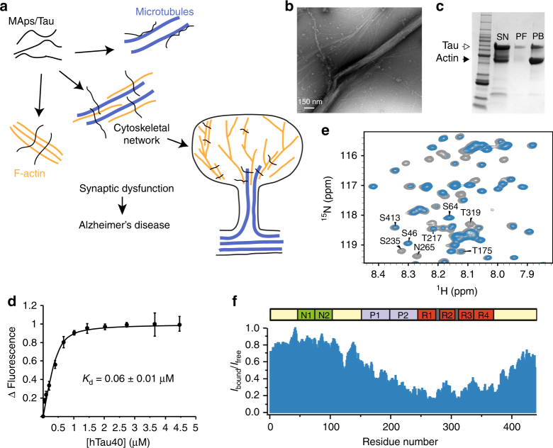
- Tau bundles F-actin; co-sedimentation showed ~63% of Tau remained in the supernatant, ~8% bound to single filaments, and ~29% associated with actin bundles. NBD-labeled F-actin binding gave a dissociation constant of 60 ± 10 nM; the Tau fragment K18 bound with a Kd of 110 ± 1 nM.
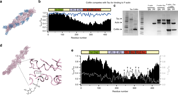
- Tau binds the hydrophobic pocket between actin subdomains 1 and 3: cofilin displaced Tau from F-actin, and paramagnetic broadening with MTSSL-labeled C374 identified Tau repeat-domain residues near this pocket.
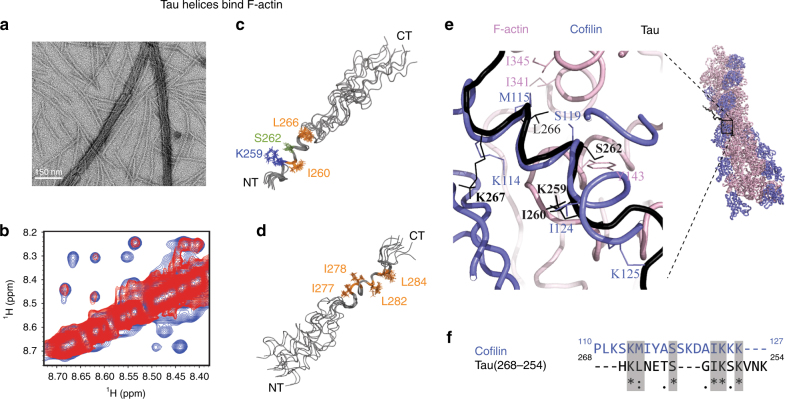
- Transfer NOE experiments showed Tau(254–290) forms helical regions around residues ~261–268 and ~277–280 upon F-actin binding. Tau(254–268), containing only one helix, bound F-actin but did not bundle it, indicating at least two flexibly linked helices are required for bundling.
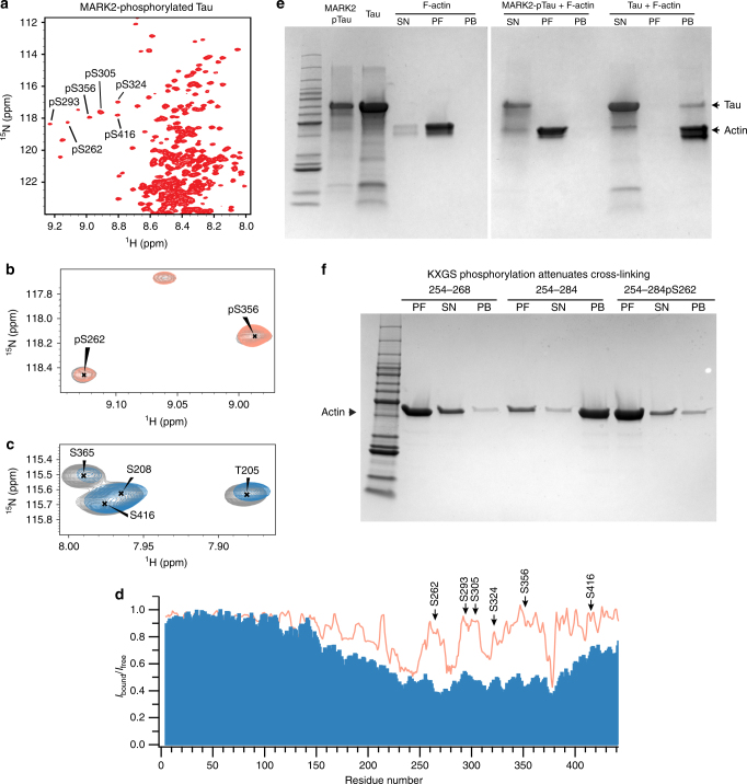
- MARK2 phosphorylation at S262, S293, S305, S324, S356, and S416 attenuated Tau binding to F-actin and prevented F-actin bundling; phosphorylation of S262 in Tau(254–284) decreased affinity and bundling.
Methods
- In vitro biophysics using human Tau 2N4R, synthetic Tau peptides, non-muscle human actin, and human cofilin: electron microscopy, co-sedimentation, and NBD-labeled F-actin fluorescence binding assays.
- NMR spectroscopy: 2D 1H-15N HSQC, cofilin competition, paramagnetic relaxation enhancement with MTSSL-tagged F-actin, and transfer NOESY/TOCSY for peptide structure; structure calculations used CYANA/XPLOR-NIH, and docking used HADDOCK.
- NMR samples included 20 µM 15N-labeled Tau with 30 µM F-actin for HSQC competition and 27 µM F-actin with 800 µM Tau peptide for NOESY experiments.
Limitations
- Stated: the single-binding-site fit cannot exclude other stoichiometries; NOE restraints were insufficient to define a unique peptide conformation or the relative orientation of the two helical regions; ensemble NMR cannot distinguish simultaneous folding from individual site binding.
- Stated: localization of phosphorylated Tau to actin-containing structures may involve additional actin-binding proteins, and future studies are needed to identify them.
- Evident from the text: the structural and bundling conclusions come from in vitro reconstituted proteins and peptides, not cellular assays; some NMR experiments use high peptide concentrations and excess F-actin/cofilin, so direct physiological relevance is not established.
Model-written summary. Check it against the paper before relying on it.
Integrity signals
Registry
Cites
Statistics
Figures
Verification
Claims the paper makes
42 Own result · 38 Own interpretation · 18 No full text of the sources · 15 Partly supported · 12 Supported · 4 Supported so far · 2 Couldn’t confirm · 2 Own method · 1 Not supported · 1 No basis given · accuracy not yet measured
At least one cited source doesn’t support this claim or points to a different paper, and none confirmed it — see the verdicts below.
In the paper
“This is likely due of differences in ionic strength, because previous studies used phosphate buffer7, while we chose general actin assembly buffer.”
§ Multivalent cross-linking of actin filaments and microtubules through the microtubule-associated protein Tau > Results > Tau interacts with and bundles actin filaments
What the sources say
- Disputedcites [7] Elie, A. et al. Tau co-organizes dynamic microtubule and actin networks. Sci. Rep. 5, 9964 (2015). sourceExperimental — accuracy not yet measured
The Methods section ('Tau interaction with microtubules or F-actin'; page unavailable) says the cited study measured Tau/F-actin binding in AP (actin-polymerization) buffer, not phosphate buffer. The Results section ('Tau simultaneously binds to microtubules and actin filaments'; page unavailable) reports the dissociation constant but does not attribute a difference from a previously reported value to ionic strength. (LLM, conf=0.85)
Checked by a reasoning model — may be wrong.
hedgedconditional- Asserted: passes — The authors put it forward as true.
- Checkable: passes — It can be checked against evidence.
- Load-bearing: unclear — It is unclear whether the paper’s conclusions depend on it.
- Based: passes — The paper gives a reason why it holds.
- Disputed
Every source the paper cites for this claim was checked and supports it.
In the paper
“According to the resonance assignment of Tau27, cross-peaks from residues such as S262, N265, and T319 were strongly attenuated.”
§ Multivalent cross-linking of actin filaments and microtubules through the microtubule-associated protein Tau > Results > Tau interacts with and bundles actin filaments
What the sources say
- Supportedcites [27] Mukrasch, M. D. et al. Structural polymorphism of 441-residue tau at single residue resolution. PLoS Biol. 7, e34 (2009). source
The citation in the printed sentence identifies the source of Tau’s resonance assignments; it does not attribute the F-actin attenuation experiment to that source. In Results, “Backbone Resonance Assignment of 441-Residue Tau” (page unavailable), the paper reports assigning more than 98% of non-proline backbone resonances and lists the exceptions, which do not include S262, N265, or T319. (LLM, conf=0.89)
boosteddirect- Asserted: passes — The authors put it forward as true.
- Checkable: passes — It can be checked against evidence.
- Load-bearing: passes — The paper’s conclusions depend on it.
- Based: passes — The paper gives a reason why it holds.
- Supported
Every source the paper cites for this claim was checked and supports it.
In the paper
“The derived dissociation constant is lower than a previously reported value of 241 ± 43 nM7.”
§ Multivalent cross-linking of actin filaments and microtubules through the microtubule-associated protein Tau > Results > Tau interacts with and bundles actin filaments
What the sources say
- Supportedcites [7] Elie, A. et al. Tau co-organizes dynamic microtubule and actin networks. Sci. Rep. 5, 9964 (2015). source
The Results section ('Tau simultaneously binds to microtubules and actin filaments') reports a Tau/F-actin apparent dissociation constant of 241 ± 43 nM. This supports the cited benchmark; the lower derived value belongs to the citing study, not to this source. (LLM, conf=0.93)
plain- Asserted: passes — The authors put it forward as true.
- Checkable: passes — It can be checked against evidence.
- Load-bearing: unclear — It is unclear whether the paper’s conclusions depend on it.
- Based: passes — The paper gives a reason why it holds.
- Supported
The cited sources that could be checked couldn’t confirm or dispute this claim.
In the paper
“Binding of Tau and MAP2 to filamentous actin results in actin cross-linking and filament bundling5, 6, 14, 22, 40–43.”
§ Multivalent cross-linking of actin filaments and microtubules through the microtubule-associated protein Tau > Discussion
What the sources say
- Not checked — no full text of the sourcecites [5] Sattilaro, R. F. Interaction of microtubule-associated protein 2 with actin filaments. Biochemistry 25, 2003–2009 (1986). source
- Not checked — no full text of the sourcecites [6] Selden, S. C. & Pollard, T. D. Phosphorylation of microtubule-associated proteins regulates their interaction with actin filaments. J. Biol. Chem. 258, 7064–7071 (1983). source
- Could not verifycites [14] Butner, K. A. & Kirschner, M. W. Tau protein binds to microtubules through a flexible array of distributed weak sites. J. Cell. Biol. 115, 717–730 (1991). sourceExperimental — accuracy not yet measured
The Introduction (p. 717) discusses possible tau-mediated cross-linking and bundling of microtubules, not actin filaments. Searches for filamentous actin, actin binding, and actin bundling found no passage in our copy addressing the claimed effects of tau and MAP2 on actin. (LLM, conf=0.76)
- Not checked — no full text of the sourcecites [22] Fulga, T. A. et al. Abnormal bundling and accumulation of F-actin mediates tau-induced neuronal degeneration in vivo. Nat. Cell. Biol. 9, 139–148 (2007). source
- Not checked — no full text of the sourcecites [40] Thomas, D. D., Seidel, J. C. & Gergely, J. Rotational dynamics of spin-labeled F-actin in the sub-millisecond time range. J. Mol. Biol. 132, 257–273 (1979). source
- Not checked — no full text of the sourcecites [41] Yoshimura, H. et al. Torsional motion of eosin-labeled F-actin as detected in the time-resolved anisotropy decay of the probe in the sub-millisecond time range. J. Mol. Biol. 179, 453–467 (1984). source
- Could not verifycites [42] Gamblin, T. C. et al. Caspase cleavage of tau: linking amyloid and neurofibrillary tangles in Alzheimer’s disease. Proc. Natl Acad. Sci. USA 100, 10032–10037 (2003). sourceExperimental — accuracy not yet measured
The Discussion describes caspase-cleaved tau assembling into tau filaments and mentions MAP2 only as a related protein. Searches for actin cross-linking, filament bundling, and binding to filamentous actin found no passage in our copy addressing the claimed Tau–MAP2 interaction with actin. (LLM, conf=0.76)
- Could not verifycites [43] Zhang, Z. et al. Cleavage of tau by asparagine endopeptidase mediates the neurofibrillary pathology in Alzheimer’s disease. Nat. Med. 20, 1254–1262 (2014). sourceExperimental — accuracy not yet measured
The Introduction and Results discuss tau’s effects on microtubules and the formation of tau filaments, not binding to filamentous actin. Searches for actin, MAP2, cross-linking, and bundling found no passage in our copy addressing the claimed effect. (LLM, conf=0.75)
plaindirect- Asserted: passes — The authors put it forward as true.
- Checkable: passes — It can be checked against evidence.
- Load-bearing: passes — The paper’s conclusions depend on it.
- Based: passes — The paper gives a reason why it holds.
The sources with a verdict support this claim; the rest weren’t checked yet, have no full text we could read, or their verdicts aren’t rated yet.
In the paper
“At the same time, binding to F-actin can be retained, but the cross-linking activity disrupted.”
§ Multivalent cross-linking of actin filaments and microtubules through the microtubule-associated protein Tau > Discussion
What the sources say
- Not checked — no full text of the sourcecites [5] Sattilaro, R. F. Interaction of microtubule-associated protein 2 with actin filaments. Biochemistry 25, 2003–2009 (1986). source
- Supportedcites [17] Correas, I., Padilla, R. & Avila, J. The tubulin-binding sequence of brain microtubule-associated proteins, tau and MAP-2, is also involved in actin binding. Biochem. J. 269, 61–64 (1990). source
The Results report that the synthetic tau peptide binds F-actin but cannot bundle actin filaments (Fig. 4). The paper contrasts this with whole tau, which does bundle filaments, supporting the distinction between retained binding and disrupted cross-linking activity in the peptide. (LLM, conf=0.91)
plain- Asserted: passes — The authors put it forward as true.
- Checkable: passes — It can be checked against evidence.
- Load-bearing: passes — The paper’s conclusions depend on it.
- Based: passes — The paper gives a reason why it holds.
The cited sources support only part of this claim — read each source’s verdict below.
In the paper
“cellular studies showed that phosphorylation of the KXGS motifs cause MAP2 and Tau to localize to the actin cytoskeleton9.”
§ Multivalent cross-linking of actin filaments and microtubules through the microtubule-associated protein Tau > Discussion
What the sources say
- Partly supportedcites [9] Biernat, J. et al. Protein kinase MARK/PAR-1 is required for neurite outgrowth and establishment of neuronal polarity. Mol. Biol. Cell. 13, 4013–4028 (2002). sourceExperimental — accuracy not yet measured
The Discussion reports a cellular study in which phosphorylation of MAP2’s KXGS motifs led to its translocation to the actin network. For Tau, however, the paper says it “hypothesize[s]” such translocation; the cellular observations described there show MARK2, not phosphorylated Tau, colocalizing with actin. The claim overstates the Tau finding as demonstrated. (LLM, conf=0.90)
plaindirect- Asserted: passes — The authors put it forward as true.
- Checkable: passes — It can be checked against evidence.
- Load-bearing: unclear — It is unclear whether the paper’s conclusions depend on it.
- Based: passes — The paper gives a reason why it holds.
- Partly supported
Every source the paper cites for this claim was checked and supports it.
In the paper
“in agreement with experimentally observed cross-linking of microtubules and actin filaments by Tau7.”
§ Multivalent cross-linking of actin filaments and microtubules through the microtubule-associated protein Tau > Discussion
What the sources say
- Supportedcites [7] Elie, A. et al. Tau co-organizes dynamic microtubule and actin networks. Sci. Rep. 5, 9964 (2015). source
The Results section (‘Tau simultaneously binds to microtubules and actin filaments’) reports that hybrid bundles of co-aligned microtubules and actin filaments appeared in the presence of Tau. The Discussion identifies Tau as a molecular linker between the two in an in vitro system, supporting the cited description of experimentally observed cross-linking. (LLM, conf=0.94)
plaindirect- Asserted: passes — The authors put it forward as true.
- Checkable: passes — It can be checked against evidence.
- Load-bearing: passes — The paper’s conclusions depend on it.
- Based: passes — The paper gives a reason why it holds.
- Supported
The cited sources support only part of this claim — read each source’s verdict below.
In the paper
“Interaction with the proline-rich region is primarily of electrostatic nature58.”
§ Multivalent cross-linking of actin filaments and microtubules through the microtubule-associated protein Tau > Discussion
What the sources say
- Partly supportedcites [58] He, H. J. et al. The proline-rich domain of tau plays a role in interactions with actin. BMC Cell Biol. 10, 81 (2009). sourceExperimental — accuracy not yet measured
The Discussion hypothesizes that electrostatic force underlies the interaction between tau’s proline-rich domain and actin, and reports that increasing ionic strength weakens the interaction. The claim presents this proposed mechanism as established and primarily electrostatic, which the paper does not demonstrate. (LLM, conf=0.85)
plaincorrelational- Asserted: passes — The authors put it forward as true.
- Checkable: passes — It can be checked against evidence.
- Load-bearing: unclear — It is unclear whether the paper’s conclusions depend on it.
- Based: passes — The paper gives a reason why it holds.
- Partly supported
Every source the paper cites for this claim was checked and supports it.
In the paper
“Tau has also been found on dynamic microtubules in the actin rich growth cone areas of developing neurons71.”
§ Multivalent cross-linking of actin filaments and microtubules through the microtubule-associated protein Tau > Discussion
What the sources say
- Supportedcites [71] Black, M. M., Slaughter, T., Moshiach, S., Obrocka, M. & Fischer, I. Tau is enriched on dynamic microtubules in the distal region of growing axons. J. Neurosci. 16, 3601–3619 (1996). source
The Results report tau on microtubules in growth cones. The Discussion identifies microtubules in the distal axon and growth cone as among the most dynamic, supporting the citing sentence’s central claim. (LLM, conf=0.89)
plaincorrelational- Asserted: passes — The authors put it forward as true.
- Checkable: passes — It can be checked against evidence.
- Load-bearing: unclear — It is unclear whether the paper’s conclusions depend on it.
- Based: passes — The paper gives a reason why it holds.
- Supported
Every source the paper cites for this claim was checked and supports it.
In the paper
“Consistent with these findings in cells, Tau binds in vitro simultaneously to microtubules and actin filaments7.”
§ Multivalent cross-linking of actin filaments and microtubules through the microtubule-associated protein Tau > Discussion
What the sources say
- Supportedcites [7] Elie, A. et al. Tau co-organizes dynamic microtubule and actin networks. Sci. Rep. 5, 9964 (2015). source
In the Results section, under “Tau simultaneously binds to microtubules and actin filaments” (page not provided), an in vitro co-sedimentation assay found that F-actin sedimented with microtubules only when tau was present. The authors interpret this as evidence that tau simultaneously interacts with both filament networks. (LLM, conf=0.94)
plaincorrelational- Asserted: passes — The authors put it forward as true.
- Checkable: passes — It can be checked against evidence.
- Load-bearing: passes — The paper’s conclusions depend on it.
- Based: passes — The paper gives a reason why it holds.
- Supported
- Probably not a substantial claim. 20
- It may state two separate claims as one. 29
Every source the paper cites for this claim was checked and supports it.
In the paper
“This is particularly the case for the interaction with filamentous actin, which plays an important role in dendritic spine morphology and postsynaptic reorganizations23.”
§ Multivalent cross-linking of actin filaments and microtubules through the microtubule-associated protein Tau > Introduction
What the sources say
- Supportedcites [23] Hotulainen, P. & Hoogenraad, C. C. Actin in dendritic spines: connecting dynamics to function. J. Cell. Biol. 189, 619–629 (2010). source
The “Actin organization in dendritic spines” section says that F-actin levels affect spine morphology and that actin stabilizes postsynaptic proteins and modulates spine-head structure in response to postsynaptic signaling. The section on actin dynamics also links synaptic plasticity to reorganization of the spine actin cytoskeleton. Page numbers were not provided in our copy. (LLM, conf=0.92)
plaindirect- Asserted: passes — The authors put it forward as true.
- Checkable: passes — It can be checked against evidence.
- Load-bearing: passes — The paper’s conclusions depend on it.
- Based: passes — The paper gives a reason why it holds.
Probably not a substantial claim.
- Supported
Every source the paper cites for this claim was checked and supports it.
In the paper
“In addition, acetylation of Tau modulates actin polymerization and enhances synaptic dysfunction26.”
§ Multivalent cross-linking of actin filaments and microtubules through the microtubule-associated protein Tau > Introduction
What the sources say
- Supportedcites [26] Tracy, T. E. et al. Acetylated Tau obstructs KIBRA-mediated signaling in synaptic plasticity and promotes tauopathy-related memory loss. Neuron 90, 245–260 (2016). source
The Discussion states that acetylated tau blocks activity-induced F-actin assembly and inhibits synaptic potentiation. The Results section likewise reports disrupted activity-dependent actin polymerization and impaired synaptic plasticity in mice expressing acetylation-mimicking tau. No page numbers are provided in the supplied copy. (LLM, conf=0.94)
plaindirect- Asserted: passes — The authors put it forward as true.
- Checkable: passes — It can be checked against evidence.
- Load-bearing: fails — The paper’s conclusions may not depend on it.
- Based: passes — The paper gives a reason why it holds.
Probably not a substantial claim. The paper’s conclusions may not depend on it. It may state two separate claims as one.
- Supported
The sources with a verdict support this claim; the rest weren’t checked yet, have no full text we could read, or their verdicts aren’t rated yet.
In the paper
“Misfolding and aberrant accumulation of Tau is a pathological hallmark of Alzheimer’s disease (AD)18, 19.”
§ Multivalent cross-linking of actin filaments and microtubules through the microtubule-associated protein Tau > Introduction
What the sources say
- Not checked — no full text of the sourcecites [18] Williams, D. R. Tauopathies: classification and clinical update on neurodegenerative diseases associated with microtubule-associated protein tau. Intern. Med. J. 36, 652–660 (2006). source
- Supportedcites [19] Fitzpatrick, A. W. P. et al. Cryo-EM structures of tau filaments from Alzheimer’s disease. Nature 547, 185–190 (2017). source
The Abstract states that Alzheimer’s disease is defined by abundant neurofibrillary lesions and that those lesions are made of Tau filaments. The Neuropathological characteristics section also reports abundant tangles and pathological, aggregated Tau in AD brain, supporting the paraphrase. (LLM, conf=0.92)
plaincorrelational- Asserted: passes — The authors put it forward as true.
- Checkable: passes — It can be checked against evidence.
- Load-bearing: passes — The paper’s conclusions depend on it.
- Based: passes — The paper gives a reason why it holds.
Probably not a substantial claim.
The cited sources support only part of this claim — read each source’s verdict below.
In the paper
“Tau occurs in six different isoforms in the human central nervous system13, 14.”
§ Multivalent cross-linking of actin filaments and microtubules through the microtubule-associated protein Tau > Introduction
What the sources say
- Not checked — no full text of the sourcecites [13] Gustke, N., Trinczek, B., Biernat, J., Mandelkow, E. M. & Mandelkow, E. Domains of tau protein and interactions with microtubules. Biochemistry 33, 9511–9522 (1994). source
- Partly supportedcites [14] Butner, K. A. & Kirschner, M. W. Tau protein binds to microtubules through a flexible array of distributed weak sites. J. Cell. Biol. 115, 717–730 (1991). sourceExperimental — accuracy not yet measured
The Introduction (p. 717) describes tau isolated from adult brain as six related phosphopeptides and discusses several tau isoforms. It does not establish that six distinct isoforms occur in the human central nervous system; it says phosphatase treatment reduces the number of resolvable forms to four. (LLM, conf=0.82)
plain- Asserted: passes — The authors put it forward as true.
- Checkable: passes — It can be checked against evidence.
- Load-bearing: fails — The paper’s conclusions may not depend on it.
- Based: passes — The paper gives a reason why it holds.
Probably not a substantial claim. The paper’s conclusions may not depend on it.
The cited sources that could be checked couldn’t confirm or dispute this claim.
In the paper
“The Tau isoforms differ in the number of N-terminal inserts and have either three or four imperfect repeats in their C-terminal half15.”
§ Multivalent cross-linking of actin filaments and microtubules through the microtubule-associated protein Tau > Introduction
What the sources say
- Could not verifycites [15] Weingarten, M. D., Lockwood, A. H., Hwo, S. Y. & Kirschner, M. W. A protein factor essential for microtubule assembly. Proc. Natl Acad. Sci. USA 72, 1858–1862 (1975). sourceExperimental — accuracy not yet measured
The Results and Discussion passages describe tau’s role in microtubule assembly, not the structures of tau isoforms. Searches for N-terminal inserts, three or four C-terminal repeats, and alternative splicing found no passage addressing the claim. Page numbers were not available in our copy. (LLM, conf=0.76)
plain- Asserted: passes — The authors put it forward as true.
- Checkable: passes — It can be checked against evidence.
- Load-bearing: fails — The paper’s conclusions may not depend on it.
- Based: passes — The paper gives a reason why it holds.
Probably not a substantial claim. The paper’s conclusions may not depend on it. It may state two separate claims as one.
- Could not verify
The cited sources support only part of this claim — read each source’s verdict below.
In the paper
“The interaction of MAPs with actin is important for neurite outgrowth11, 12.”
§ Multivalent cross-linking of actin filaments and microtubules through the microtubule-associated protein Tau > Introduction
What the sources say
- Could not verifycites [11] Knops, J. et al. Overexpression of tau in a nonneuronal cell induces long cellular processes. J. Cell. Biol. 114, 725–733 (1991). sourceExperimental — accuracy not yet measured
The paper reports that tau expression induces long processes in nonneuronal Sf9 cells and, separately, that cytochalasin treatment produces short retraction processes by disrupting the actin network. The passages on process formation and cytochalasin do not address whether MAPs interact with actin or whether such an interaction is important for neurite outgrowth (searched: actin interaction, tau actin cytoskeleton). (LLM, conf=0.72)
- Partly supportedcites [12] Biernat, J. & Mandelkow, E. M. The development of cell processes induced by tau protein requires phosphorylation of serine 262 and 356 in the repeat domain and is inhibited by phosphorylation in the proline-rich domains. Mol. Biol. Cell. 10, 727–740 (1999). sourceExperimental — accuracy not yet measured
The Introduction says MAPs regulate microtubules involved in neurite growth. In “A Model Relating tau Phosphorylation to Process Formation” (page unavailable), the paper says MAP interactions with cytoskeletal elements are important in process formation and separately mentions softening of the actin cortex. It does not specifically say that MAP–actin interactions are important for neurite outgrowth. (LLM, conf=0.81)
plaincorrelational- Asserted: passes — The authors put it forward as true.
- Checkable: passes — It can be checked against evidence.
- Load-bearing: fails — The paper’s conclusions may not depend on it.
- Based: passes — The paper gives a reason why it holds.
Probably not a substantial claim. The paper’s conclusions may not depend on it.
- Could not verify
The cited sources support only part of this claim — read each source’s verdict below.
In the paper
“Because a single repeat interacts with both monomeric and filamentous actin, but does not bundle actin filaments, more than one microtubule-binding repeat is believed to be required for bundling of filamentous actin (F-actin)17.”
§ Multivalent cross-linking of actin filaments and microtubules through the microtubule-associated protein Tau > Introduction
What the sources say
- Partly supportedcites [17] Correas, I., Padilla, R. & Avila, J. The tubulin-binding sequence of brain microtubule-associated proteins, tau and MAP-2, is also involved in actin binding. Biochem. J. 269, 61–64 (1990). sourceExperimental — accuracy not yet measured
The Results (page not provided) show that a peptide containing one tubulin-binding repeat binds G-actin and F-actin but does not bundle filaments. The authors suggest that bundling may require more than one actin-binding site; they do not establish that those sites must be multiple microtubule-binding repeats. (LLM, conf=0.85)
hedgedconditional- Asserted: passes — The authors put it forward as true.
- Checkable: passes — It can be checked against evidence.
- Load-bearing: passes — The paper’s conclusions depend on it.
- Based: passes — The paper gives a reason why it holds.
Probably not a substantial claim.
- Partly supported
Every source the paper cites for this claim was checked and supports it.
In the paper
“Because a single repeat interacts with both monomeric and filamentous actin, but does not bundle actin filaments, more than one microtubule-binding repeat is believed to be required for bundling of filamentous actin (F-actin)17.”
§ Multivalent cross-linking of actin filaments and microtubules through the microtubule-associated protein Tau > Introduction
What the sources say
- Supportedcites [17] Correas, I., Padilla, R. & Avila, J. The tubulin-binding sequence of brain microtubule-associated proteins, tau and MAP-2, is also involved in actin binding. Biochem. J. 269, 61–64 (1990). source
The Introduction identifies the synthetic tau peptide as one tubulin-binding-site repeat. The Results state that it binds both G-actin and F-actin but cannot bundle actin filaments, and suggest that bundling may require more than one actin-binding site. (LLM, conf=0.94)
plain- Asserted: passes — The authors put it forward as true.
- Checkable: passes — It can be checked against evidence.
- Load-bearing: passes — The paper’s conclusions depend on it.
- Based: passes — The paper gives a reason why it holds.
It may state two separate claims as one.
- Supported
The cited sources support only part of this claim — read each source’s verdict below.
In the paper
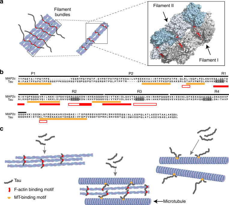
“In addition, MAPs bundle actin filaments and cross-link the cellular cytoskeleton formed by microtubules and actin filaments (Fig. 1a)3–10.”
§ Multivalent cross-linking of actin filaments and microtubules through the microtubule-associated protein Tau > Introduction
What the sources say
- Not checked — no full text of the sourcecites [3] Yamauchi, P. S. & Purich, D. L. Microtubule-associated protein interactions with actin filaments: evidence for differential behavior of neuronal MAP-2 and tau in the presence of phosphatidyl-inositol. Biochem. Biophys. Res. Commun. 190, 710–715 (1993). source
- Partly supportedcites [4] He, H. J. et al. The proline-rich domain of tau plays a role in interactions with actin. BMC Cell. Biol. 10, 81 (2009). sourceExperimental — accuracy not yet measured
The Results and Discussion show that tau, a microtubule-associated protein, bundles F-actin. The Discussion describes tau and MAP2c as potential cross-linkers between microtubules and actin, but does not establish the citing sentence’s categorical claim that MAPs cross-link those networks in cells. (LLM, conf=0.83)
- Not checked — no full text of the sourcecites [5] Sattilaro, R. F. Interaction of microtubule-associated protein 2 with actin filaments. Biochemistry 25, 2003–2009 (1986). source
- Not checked — no full text of the sourcecites [6] Selden, S. C. & Pollard, T. D. Phosphorylation of microtubule-associated proteins regulates their interaction with actin filaments. J. Biol. Chem. 258, 7064–7071 (1983). source
- Supportedcites [7] Elie, A. et al. Tau co-organizes dynamic microtubule and actin networks. Sci. Rep. 5, 9964 (2015). source
The Results section (“Tau simultaneously binds to microtubules and actin filaments”; page unavailable) reports that tau, a microtubule-associated protein, forms F-actin bundles. The Results section (“Tau coordinates dynamic microtubules and actin filaments”; page unavailable) also measures tau’s microtubule–actin cross-linking activity. These in vitro findings support the claim as an example of what a MAP can do. (LLM, conf=0.90)
- Not checked — no full text of the sourcecites [8] Griffith, L. M. & Pollard, T. D. The interaction of actin filaments with microtubules and microtubule-associated proteins. J. Biol. Chem. 257, 9143–9151 (1982). source
- Partly supportedcites [9] Biernat, J. et al. Protein kinase MARK/PAR-1 is required for neurite outgrowth and establishment of neuronal polarity. Mol. Biol. Cell. 13, 4013–4028 (2002). sourceExperimental — accuracy not yet measured
The Discussion (page unavailable) says MAPs can interact with the actin cytoskeleton and may regulate microtubule bundling. It does not say MAPs bundle actin filaments or cross-link actin filaments with microtubules; the cited bundling concerns microtubules, not actin. (LLM, conf=0.85)
- Partly supportedcites [10] Cunningham, C. C. et al. Microtubule-associated protein 2c reorganizes both microtubules and microfilaments into distinct cytological structures in an actin-binding protein-280-deficient melanoma cell line. J. Cell. Biol. 136, 845–857 (1997). sourceExperimental — accuracy not yet measured
The Results section (‘MAP2c Causes Actin Gelation In Vitro’; page unavailable) reports that MAP2c forms actin bundles at higher concentrations or lower ionic strength. But the Discussion (‘In Vivo Functions of MAP2c’; page unavailable) explicitly says the data cannot determine whether MAP2c cross-links microtubules to actin filaments. The claim presents that cross-linking as established for MAPs generally. (LLM, conf=0.90)
plaindirect- Asserted: passes — The authors put it forward as true.
- Checkable: passes — It can be checked against evidence.
- Load-bearing: passes — The paper’s conclusions depend on it.
- Based: passes — The paper gives a reason why it holds.
It may state two separate claims as one.
The cited sources support only part of this claim — read each source’s verdict below.
In the paper
“One of these binding partners is cofilin, a 21 kDa eukaryotic protein, which binds to F-actin with a K d < 0.05 µM33 and results in disassembly of F-actin34, 35.”
§ Multivalent cross-linking of actin filaments and microtubules through the microtubule-associated protein Tau > Results > Tau binds to the hydrophobic pocket of F-actin
What the sources say
- Not checked — no full text of the sourcecites [33] Bobkov, A. A. et al. Structural effects of cofilin on longitudinal contacts in F-actin. J. Mol. Biol. 323, 739–750 (2002). source
- Partly supportedcites [34] Yonezawa, N. et al. Distribution among tissues and intracellular localization of cofilin, a 21kDa actin-binding protein. Cell. Struct. Funct. 12, 443–452 (1987). sourceExperimental — accuracy not yet measured
The Cofilin section (page not provided) describes cofilin as a 21 kDa protein that binds F-actin, supporting that part of the statement attributed to this source. It does not give the cited F-actin Kd of <0.05 µM; its reported dissociation constant of 0.1–0.2 µM is for binding to monomeric actin. The separate disassembly statement carries other citations. (LLM, conf=0.88)
- Partly supportedcites [35] Suarez, C. et al. Cofilin tunes the nucleotide state of actin filaments and severs at bare and decorated segment boundaries. Curr. Biol. 21, 862–868 (2011). sourceExperimental — accuracy not yet measured
Citation 33 is attached to the Kd claim. In Results and Discussion (‘Bound ADF/cofilin dissociates slowly from actin filaments’; page unavailable), the paper reports rapid binding to bare actin filaments and high overall binding affinity, but gives no Kd below 0.05 µM. The disassembly clause is assigned to citations 34 and 35, not this source. (LLM, conf=0.84)
plaindirect- Asserted: passes — The authors put it forward as true.
- Checkable: passes — It can be checked against evidence.
- Load-bearing: fails — The paper’s conclusions may not depend on it.
- Based: passes — The paper gives a reason why it holds.
Probably not a substantial claim. The paper’s conclusions may not depend on it. It may state two separate claims as one.
The cited sources support only part of this claim — read each source’s verdict below.
In the paper
“Although actin contains five cysteine residues, a number of studies has shown that the C-terminal C374, which is in close proximity to actin’s hydrophobic pocket (Fig. 2d), is most solvent exposed (Supplementary Fig. 4a, b) and incorporates more than 70% of spin labels during spin labeling reactions37–41.”
§ Multivalent cross-linking of actin filaments and microtubules through the microtubule-associated protein Tau > Results > Tau binds to the hydrophobic pocket of F-actin
What the sources say
- Not checked — no full text of the sourcecites [37] Yasunaga, T. & Wakabayashi, T. Relocation of Cys374 of actin induced by labeling with fluorescent dyes. J. Biochem. 129, 201–204 (2001). source
- Partly supportedcites [38] Kupi, T., Grof, P., Nyitrai, M. & Belagyi, J. The uncoupling of the effects of formins on the local and global dynamics of actin filaments. Biophys. J. 96, 2901–2911 (2009). sourceExperimental — accuracy not yet measured
The section “Rotational motions in actin observed by spin labels” states that Cys-374 incorporated more than 70% of maleimide spin labels under the conditions used in the paper. Our copy does not state that actin has five cysteine residues, and the reported percentage is specific to those labeling conditions rather than spin labeling generally. (LLM, conf=0.86)
- Not checked — no full text of the sourcecites [39] Mossakowska, M., Belagyi, J. & Strzelecka-Golaszewska, H. An EPR study of the rotational dynamics of actins from striated and smooth muscle and their complexes with heavy meromyosin. Eur. J. Biochem. 175, 557–564 (1988). source
- Not checked — no full text of the sourcecites [40] Thomas, D. D., Seidel, J. C. & Gergely, J. Rotational dynamics of spin-labeled F-actin in the sub-millisecond time range. J. Mol. Biol. 132, 257–273 (1979). source
- Not checked — no full text of the sourcecites [41] Yoshimura, H. et al. Torsional motion of eosin-labeled F-actin as detected in the time-resolved anisotropy decay of the probe in the sub-millisecond time range. J. Mol. Biol. 179, 453–467 (1984). source
plain- Asserted: passes — The authors put it forward as true.
- Checkable: passes — It can be checked against evidence.
- Load-bearing: passes — The paper’s conclusions depend on it.
- Based: passes — The paper gives a reason why it holds.
It may state two separate claims as one.
The cited sources support only part of this claim — read each source’s verdict below.
In the paper
“In addition to phosphorylation, acetylation of Tau at K281, a residues that is located in an F-actin interacting helix of Tau (Fig. 3d), affects the interaction of Tau with actin and modulate Tau-mediated toxicity26.”
§ Multivalent cross-linking of actin filaments and microtubules through the microtubule-associated protein Tau > Discussion
What the sources say
- Partly supportedcites [26] Tracy, T. E. et al. Acetylated Tau obstructs KIBRA-mediated signaling in synaptic plasticity and promotes tauopathy-related memory loss. Neuron 90, 245–260 (2016). sourceExperimental — accuracy not yet measured
The Results report that an acetylation-mimicking K274/K281 tau mutant interacted less with F-actin than wild-type tau; the Discussion links that mutant to synaptic dysfunction and memory deficits. The paper does not establish that K281 alone has these effects or describe K281 as located in an F-actin-interacting helix. Its evidence for altered tau–actin binding comes from the double mutant, not directly from acetylated tau. (LLM, conf=0.88)
plaindirect- Asserted: passes — The authors put it forward as true.
- Checkable: passes — It can be checked against evidence.
- Load-bearing: unclear — It is unclear whether the paper’s conclusions depend on it.
- Based: passes — The paper gives a reason why it holds.
It may state two separate claims as one.
- Partly supported
Every source the paper cites for this claim was checked and supports it.
In the paper
“In addition, Tau-induced neurotoxicity is associated with increased F-actin levels22, and Tau-induced remodeling of the actin cytoskeleton can cause plasma membrane blebbing57.”
§ Multivalent cross-linking of actin filaments and microtubules through the microtubule-associated protein Tau > Discussion
What the sources say
- Supportedcites [57] Torres-Cruz, F. M. et al. Expression of Tau produces aberrant plasma membrane blebbing in glial cells through RhoA-ROCK-dependent F-actin remodeling. J. Alzheimers Dis. 52, 463–482 (2016). source
The Results (Fig. 3, p. 471) describe F-actin remodeling alongside bleb formation in Tau-expressing C6 glial cells. The Discussion (p. 478) concludes that F-actin regulates formation of these blebs, supported by their disappearance when F-actin was disrupted and reappearance after the drug was removed. (LLM, conf=0.93)
plaindirect- Asserted: passes — The authors put it forward as true.
- Checkable: passes — It can be checked against evidence.
- Load-bearing: fails — The paper’s conclusions may not depend on it.
- Based: passes — The paper gives a reason why it holds.
Probably not a substantial claim. The paper’s conclusions may not depend on it.
- Supported
The cited sources support only part of this claim — read each source’s verdict below.
In the paper
“Tau colocalizes with actin in differentiating PC12 cells68, N2a cells69 and in the postsynaptic compartments of mature neurons70.”
§ Multivalent cross-linking of actin filaments and microtubules through the microtubule-associated protein Tau > Discussion
What the sources say
- Not checked — no full text of the sourcecites [68] Yu, J. Z. & Rasenick, M. M. Tau associates with actin in differentiating PC12 cells. FASEB J. 20, 1452–1461 (2006). source
- Partly supportedcites [69] Sontag, J. M., Nunbhakdi-Craig, V., Mitterhuber, M., Ogris, E. & Sontag, E. Regulation of protein phosphatase 2A methylation by LCMT1 and PME-1 plays a critical role in differentiation of neuroblastoma cells. J. Neurochem. 115, 1455–1465 (2010). sourceExperimental — accuracy not yet measured
The Results (page unavailable) report tau-positive processes and altered F-actin organization in differentiating N2a cells. They do not report that tau and actin colocalize; the cited paper supports their presence in these cells, but not the specific spatial relationship attributed to it. Citation 69 is attached only to the N2a-cell portion of the sentence. (LLM, conf=0.84)
- Partly supportedcites [70] Thies, E. & Mandelkow, E. M. Missorting of tau in neurons causes degeneration of synapses that can be rescued by the kinase MARK2/Par-1. J. Neurosci. 27, 2896–2907 (2007). sourceExperimental — accuracy not yet measured
For citation 70, the 'Missorting of tau and effects on synapses' section (Fig. 1; page not provided) reports that F-actin fills postsynaptic spine heads and that expressed tau enters those spine heads in hippocampal neurons with mature synapses. The claim omits the crucial condition: tau was experimentally expressed and missorted; in untransfected neurons, the paper reports tau only in axons. Citations 68 and 69, attached to the PC12 and N2a claims, are not assessed here. (LLM, conf=0.87)
plaincorrelational- Asserted: passes — The authors put it forward as true.
- Checkable: passes — It can be checked against evidence.
- Load-bearing: passes — The paper’s conclusions depend on it.
- Based: passes — The paper gives a reason why it holds.
It may state two separate claims as one.
The sources with a verdict support this claim; the rest weren’t checked yet, have no full text we could read, or their verdicts aren’t rated yet.
In the paper
“The hexapeptide populates extended structure in solution27, is found in cross-β structure in amyloid fibrils63, but folds into helical structure in complex with F-actin (Fig. 3d).”
§ Multivalent cross-linking of actin filaments and microtubules through the microtubule-associated protein Tau > Discussion
What the sources say
- Supportedcites [27] Mukrasch, M. D. et al. Structural polymorphism of 441-residue tau at single residue resolution. PLoS Biol. 7, e34 (2009). source
The citation to this paper is attached to the claim that the hexapeptide populates extended structure in solution, not to the fibril or F-actin claims. In Results, “Secondary Structure Propensity in 441-Residue Tau,” the paper reports increased positive coupling values for the region containing 275VQIINK280, which it identifies as indicating extended or β-structure propensity. (LLM, conf=0.92)
- Not checked — no full text of the sourcecites [63] Andronesi, O. C. et al. Characterization of Alzheimer’s-like paired helical filaments from the core domain of tau protein using solid-state NMR spectroscopy. J. Am. Chem. Soc. 130, 5922–5928 (2008). source
plain- Asserted: passes — The authors put it forward as true.
- Checkable: passes — It can be checked against evidence.
- Load-bearing: passes — The paper’s conclusions depend on it.
- Based: passes — The paper gives a reason why it holds.
It may state two separate claims as one.
- Supported
Every source the paper cites for this claim was checked and supports it.
In the paper
“In addition, phosphorylated Tau is recruited to actin-cofilin rods, which might lead to neuropil threads56.”
§ Multivalent cross-linking of actin filaments and microtubules through the microtubule-associated protein Tau > Discussion
What the sources say
- Supportedcites [56] Whiteman, I. T. et al. Activated actin-depolymerizing factor/cofilin sequesters phosphorylated microtubule-associated protein during the assembly of Alzheimer-like neuritic cytoskeletal striations. J. Neurosci. 29, 12994–13005 (2009). source
The Discussion says cofilin-actin rods may trigger the sequestration and accumulation of Ser262/356-phosphorylated tau in some neuropil threads. It also proposes that the rods serve as a template for pMAP recruitment, matching the claim’s tentative wording. (LLM, conf=0.93)
hedgedconditional- Asserted: passes — The authors put it forward as true.
- Checkable: passes — It can be checked against evidence.
- Load-bearing: unclear — It is unclear whether the paper’s conclusions depend on it.
- Based: passes — The paper gives a reason why it holds.
It may state two separate claims as one.
- Supported
Every source the paper cites for this claim was checked and supports it.
In the paper
“Analysis of secondary structure in the lowest energy conformation using STRIDE47, identifies α-helix for residues 261–268 and 3–10 helix for residues 277–280.”
§ Multivalent cross-linking of actin filaments and microtubules through the microtubule-associated protein Tau > Results > Tau binds through helical structure to F-actin
What the sources say
- Supportedcites [47] Heinig, M. & Frishman, D. STRIDE: a web server for secondary structure assignment from known atomic coordinates of proteins. Nucleic Acids Res. 32, W500–W502 (2004). source
The citation identifies STRIDE as the tool used for secondary-structure analysis; it does not attribute the Tau residue assignments to the STRIDE paper. The Abstract identifies STRIDE as a tool for assigning secondary structure from protein atomic coordinates, and the STRIDE WEB SERVER AND DATABASE section describes its per-residue assignments. The residue assignments are the citing authors’ own result. (LLM, conf=0.93)
plain- Asserted: passes — The authors put it forward as true.
- Checkable: passes — It can be checked against evidence.
- Load-bearing: unclear — It is unclear whether the paper’s conclusions depend on it.
- Based: passes — The paper gives a reason why it holds.
It may state two separate claims as one.
- Supported
Every source the paper cites for this claim was checked and supports it.
In the paper
“In addition, the N-terminal halves of each of Tau’s four 18-residue aminoacid repeats64 bind to F-actin’s hydrophobic pocket and fold into helical structure (Fig. 3c; Supplementary Figs. 12 and 14).”
§ Multivalent cross-linking of actin filaments and microtubules through the microtubule-associated protein Tau > Discussion
What the sources say
- Supportedcites [64] Goode, B. L. & Feinstein, S. C. Identification of a novel microtubule binding and assembly domain in the developmentally regulated inter-repeat region of tau. J. Cell. Biol. 124, 769–782 (1994). source
The citation is attached to the description of tau’s four 18-residue repeats, not to the subsequent F-actin binding and helical-structure findings, which the citing sentence points to its own figures. The Introduction states that tau contains either three or four copies of a conserved 18-amino-acid repeat. (LLM, conf=0.91)
plaincorrelational- Asserted: passes — The authors put it forward as true.
- Checkable: passes — It can be checked against evidence.
- Load-bearing: passes — The paper’s conclusions depend on it.
- Based: passes — The paper gives a reason why it holds.
It may state two separate claims as one.
- Supported
The cited sources support only part of this claim — read each source’s verdict below.
In the paper
“The interaction of Tau with actin is important for neurite outgrowth and synaptic dysfunction11, 12, 22.”
§ Multivalent cross-linking of actin filaments and microtubules through the microtubule-associated protein Tau > Discussion
What the sources say
- Partly supportedcites [11] Knops, J. et al. Overexpression of tau in a nonneuronal cell induces long cellular processes. J. Cell. Biol. 114, 725–733 (1991). sourceExperimental — accuracy not yet measured
The Introduction (p. 725) links tau to neurite elongation, and the discussion reports that tau-expressing Sf9 cells developed long processes. The paper does not establish that tau’s interaction with actin drives outgrowth or synaptic dysfunction; it says synaptic vesicles were not observed in those processes. (LLM, conf=0.86)
- Partly supportedcites [12] Biernat, J. & Mandelkow, E. M. The development of cell processes induced by tau protein requires phosphorylation of serine 262 and 356 in the repeat domain and is inhibited by phosphorylation in the proline-rich domains. Mol. Biol. Cell. 10, 727–740 (1999). sourceExperimental — accuracy not yet measured
The introduction and 'A Model Relating tau Phosphorylation to Process Formation' (page unavailable) link tau to neurite or cell-process outgrowth and mention softening of the actin cortex before a process forms. They do not establish that a tau–actin interaction drives outgrowth, and they do not address synaptic dysfunction. (LLM, conf=0.83)
- Not checked — no full text of the sourcecites [22] Fulga, T. A. et al. Abnormal bundling and accumulation of F-actin mediates tau-induced neuronal degeneration in vivo. Nat. Cell. Biol. 9, 139–148 (2007). source
plaincorrelational- Asserted: passes — The authors put it forward as true.
- Checkable: passes — It can be checked against evidence.
- Load-bearing: fails — The paper’s conclusions may not depend on it.
- Based: passes — The paper gives a reason why it holds.
Probably not a substantial claim. The paper’s conclusions may not depend on it. It may state two separate claims as one.
- Partly supported
The cited sources support only part of this claim — read each source’s verdict below.
In the paper
“Cys374 of actin is solvent exposed and can be labeled with the spin label MTSSL37–41.”
§ Multivalent cross-linking of actin filaments and microtubules through the microtubule-associated protein Tau > Results > Tau binds to the hydrophobic pocket of F-actin
What the sources say
- Not checked — no full text of the sourcecites [37] Yasunaga, T. & Wakabayashi, T. Relocation of Cys374 of actin induced by labeling with fluorescent dyes. J. Biochem. 129, 201–204 (2001). source
- Partly supportedcites [38] Kupi, T., Grof, P., Nyitrai, M. & Belagyi, J. The uncoupling of the effects of formins on the local and global dynamics of actin filaments. Biophys. J. 96, 2901–2911 (2009). sourceExperimental — accuracy not yet measured
The Introduction and “Rotational motions in actin observed by spin labels” report that a spin label attaches to Cys-374 and that this site incorporated more than 70% of the labels under the experimental conditions. This supports its accessibility for labeling, but the paper uses maleimide spin label (MSL), not MTSSL, and does not explicitly describe Cys-374 as solvent exposed. Page numbers were not available in our copy. (LLM, conf=0.88)
- Not checked — no full text of the sourcecites [39] Mossakowska, M., Belagyi, J. & Strzelecka-Golaszewska, H. An EPR study of the rotational dynamics of actins from striated and smooth muscle and their complexes with heavy meromyosin. Eur. J. Biochem. 175, 557–564 (1988). source
- Not checked — no full text of the sourcecites [40] Thomas, D. D., Seidel, J. C. & Gergely, J. Rotational dynamics of spin-labeled F-actin in the sub-millisecond time range. J. Mol. Biol. 132, 257–273 (1979). source
- Not checked — no full text of the sourcecites [41] Yoshimura, H. et al. Torsional motion of eosin-labeled F-actin as detected in the time-resolved anisotropy decay of the probe in the sub-millisecond time range. J. Mol. Biol. 179, 453–467 (1984). source
plain- Asserted: passes — The authors put it forward as true.
- Checkable: passes — It can be checked against evidence.
- Load-bearing: passes — The paper’s conclusions depend on it.
- Based: passes — The paper gives a reason why it holds.
It may state two separate claims as one.
The sources with a verdict support this claim; the rest weren’t checked yet, have no full text we could read, or their verdicts aren’t rated yet.
In the paper
“Pathogenic forms of Tau have been connected to the formation of actin-rich rods56, which were found to induce neurodegeneration in Drosophila neurons22.”
§ Multivalent cross-linking of actin filaments and microtubules through the microtubule-associated protein Tau > Discussion
What the sources say
- Supportedcites [56] Whiteman, I. T. et al. Activated actin-depolymerizing factor/cofilin sequesters phosphorylated microtubule-associated protein during the assembly of Alzheimer-like neuritic cytoskeletal striations. J. Neurosci. 29, 12994–13005 (2009). source
The citation to this paper is attached to the connection between pathogenic tau and actin-rich rods, not the later claim about neurodegeneration in Drosophila neurons. The Abstract reports that cofilin-actin rods recruit phosphorylated tau in cultured neurons, and the Discussion describes tau sequestration in those rods as a possible early pathogenic event. (LLM, conf=0.91)
- Not checked — no full text of the sourcecites [22] Fulga, T. A. et al. Abnormal bundling and accumulation of F-actin mediates tau-induced neuronal degeneration in vivo. Nat. Cell. Biol. 9, 139–148 (2007). source
plaincorrelational- Asserted: passes — The authors put it forward as true.
- Checkable: passes — It can be checked against evidence.
- Load-bearing: fails — The paper’s conclusions may not depend on it.
- Based: passes — The paper gives a reason why it holds.
Probably not a substantial claim. The paper’s conclusions may not depend on it. It may state two separate claims as one.
- Supported
The cited sources support only part of this claim — read each source’s verdict below.
In the paper
“For example, a C-terminal fragment and a peptide corresponding to the second microtubule-binding repeat bind actin, but are incapable of mediating actin cross-linking5, 17.”
§ Multivalent cross-linking of actin filaments and microtubules through the microtubule-associated protein Tau > Discussion
What the sources say
- Not checked — no full text of the sourcecites [5] Sattilaro, R. F. Interaction of microtubule-associated protein 2 with actin filaments. Biochemistry 25, 2003–2009 (1986). source
- Partly supportedcites [17] Correas, I., Padilla, R. & Avila, J. The tubulin-binding sequence of brain microtubule-associated proteins, tau and MAP-2, is also involved in actin binding. Biochem. J. 269, 61–64 (1990). sourceExperimental — accuracy not yet measured
The Results report that the synthetic tau peptide binds G-actin and F-actin but does not bundle actin filaments. The Introduction and Discussion describe it as corresponding to one tubulin-binding repeat, without identifying it as the second. They report that a 32 kDa MAP-2 fragment binds actin, but do not establish that this C-terminal fragment cannot cross-link actin. (LLM, conf=0.86)
plain- Asserted: passes — The authors put it forward as true.
- Checkable: passes — It can be checked against evidence.
- Load-bearing: passes — The paper’s conclusions depend on it.
- Based: passes — The paper gives a reason why it holds.
It may state two separate claims as one.
A result the paper reports from its own data — it cites no other source for it, so there is none to check. The quote shows it is in the paper.
In the paper
“In agreement with previous findings, we show that Tau binds with high-affinity to filamentous actin, resulting in F-actin bundling (Fig. 1b).”
§ Multivalent cross-linking of actin filaments and microtubules through the microtubule-associated protein Tau > Discussion
plaindirect- Asserted: passes — The authors put it forward as true.
- Checkable: passes — It can be checked against evidence.
- Load-bearing: passes — The paper’s conclusions depend on it.
- Based: passes — The paper gives a reason why it holds.
It may state two separate claims as one.
A result the paper reports from its own data — it cites no other source for it, so there is none to check. The quote shows it is in the paper.
In the paper
“Addition of an equimolar concentration of Tau caused bundling of F-actin (Fig. 1b), although some single filaments remained.”
§ Multivalent cross-linking of actin filaments and microtubules through the microtubule-associated protein Tau > Results > Tau interacts with and bundles actin filaments
plaindirect- Asserted: passes — The authors put it forward as true.
- Checkable: passes — It can be checked against evidence.
- Load-bearing: passes — The paper’s conclusions depend on it.
- Based: passes — The paper gives a reason why it holds.
It may state two separate claims as one.
A result the paper reports from its own data — it cites no other source for it, so there is none to check. The quote shows it is in the paper.
In the paper
“Tau and cofilin helices is well conserved, but the directionality of their polypeptide chain is inverted (Fig. 3e, f).”
§ Multivalent cross-linking of actin filaments and microtubules through the microtubule-associated protein Tau > Discussion
boostedcorrelational- Asserted: passes — The authors put it forward as true.
- Checkable: passes — It can be checked against evidence.
- Load-bearing: unclear — It is unclear whether the paper’s conclusions depend on it.
- Based: passes — The paper gives a reason why it holds.
It may state two separate claims as one.
A result the paper reports from its own data — it cites no other source for it, so there is none to check. The quote shows it is in the paper.
In the paper
“Haddock clustered 192 complex structures in four clusters, representing 96.0% of the water-refined Haddock models.”
§ Multivalent cross-linking of actin filaments and microtubules through the microtubule-associated protein Tau > Results > Structural model for the Tau/F-actin binding site
plain- Asserted: passes — The authors put it forward as true.
- Checkable: passes — It can be checked against evidence.
- Load-bearing: fails — The paper’s conclusions may not depend on it.
- Based: passes — The paper gives a reason why it holds.
Probably not a substantial claim. The paper’s conclusions may not depend on it.
A result the paper reports from its own data — it cites no other source for it, so there is none to check. The quote shows it is in the paper.
In the paper
“Both the proline-rich region and the microtubule-binding repeats contribute to the interaction (Figs. 1e, f).”
§ Multivalent cross-linking of actin filaments and microtubules through the microtubule-associated protein Tau > Discussion
plaindirect- Asserted: passes — The authors put it forward as true.
- Checkable: passes — It can be checked against evidence.
- Load-bearing: passes — The paper’s conclusions depend on it.
- Based: passes — The paper gives a reason why it holds.
It may state two separate claims as one.
A result the paper reports from its own data — it cites no other source for it, so there is none to check. The quote shows it is in the paper.
In the paper
“Docking of experimentally determined Tau helices to the F-actin surface together with sequence alignment showed that the overall position of the interacting Tau and cofilin helices is well conserved, but the directionality of their polypeptide chain is inverted (Fig. 3e, f).”
§ Multivalent cross-linking of actin filaments and microtubules through the microtubule-associated protein Tau > Discussion
plaincorrelational- Asserted: passes — The authors put it forward as true.
- Checkable: passes — It can be checked against evidence.
- Load-bearing: passes — The paper’s conclusions depend on it.
- Based: passes — The paper gives a reason why it holds.
It may state two separate claims as one.
In the paper
“MAPs such as Tau belong to the class of intrinsically disordered proteins, which do not fold into a stable three-dimensional structure28.”
§ Multivalent cross-linking of actin filaments and microtubules through the microtubule-associated protein Tau > Introduction
What the sources say
- Not checked — no full text of the sourcecites [28] Dyson, H. J. & Wright, P. E. Intrinsically unstructured proteins and their functions. Nat. Rev. Mol. Cell. Biol. 6, 197–208 (2005). source
plain- Asserted: passes — The authors put it forward as true.
- Checkable: passes — It can be checked against evidence.
- Load-bearing: passes — The paper’s conclusions depend on it.
- Based: passes — The paper gives a reason why it holds.
Probably not a substantial claim.
In the paper
“Proteins from the MAP2 family also bind to actin filaments through their repeat domain8.”
§ Multivalent cross-linking of actin filaments and microtubules through the microtubule-associated protein Tau > Introduction
What the sources say
- Not checked — no full text of the sourcecites [8] Griffith, L. M. & Pollard, T. D. The interaction of actin filaments with microtubules and microtubule-associated proteins. J. Biol. Chem. 257, 9143–9151 (1982). source
plaindirect- Asserted: passes — The authors put it forward as true.
- Checkable: passes — It can be checked against evidence.
- Load-bearing: fails — The paper’s conclusions may not depend on it.
- Based: passes — The paper gives a reason why it holds.
Probably not a substantial claim. The paper’s conclusions may not depend on it.
A claim is checked only against the cited paper’s full text, never its abstract, and none is available for its sources.
In the paper
“Microtubule-associated proteins (MAPs) bind to stabilize and promote assembly of microtubules1, 2.”
§ Multivalent cross-linking of actin filaments and microtubules through the microtubule-associated protein Tau > Introduction
What the sources say
- Not checked — no full text of the sourcecites [1] Mandelkow, E. & Mandelkow, E. M. Microtubules and microtubule-associated proteins. Curr. Opin. Cell. Biol. 7, 72–81 (1995). source
- Not checked — no full text of the sourcecites [2] Lee, G. Non-motor microtubule-associated proteins. Curr. Opin. Cell. Biol. 5, 88–94 (1993). source
plaindirect- Asserted: passes — The authors put it forward as true.
- Checkable: passes — It can be checked against evidence.
- Load-bearing: fails — The paper’s conclusions may not depend on it.
- Based: passes — The paper gives a reason why it holds.
Probably not a substantial claim. The paper’s conclusions may not depend on it.
In the paper
“Cryo-electron microscopy further showed that the binding site of cofilin on F-actin is highly similar to its binding site on G-actin (Fig. 2a)32.”
§ Multivalent cross-linking of actin filaments and microtubules through the microtubule-associated protein Tau > Results > Tau binds to the hydrophobic pocket of F-actin
What the sources say
- Not checked — no full text of the sourcecites [32] Carlier, M. F. & Pantaloni, D. Control of actin dynamics in cell motility. J. Mol. Biol. 269, 459–467 (1997). source
plain- Asserted: passes — The authors put it forward as true.
- Checkable: passes — It can be checked against evidence.
- Load-bearing: passes — The paper’s conclusions depend on it.
- Based: passes — The paper gives a reason why it holds.
Probably not a substantial claim.
In the paper
“Moreover the tight connection between microtubules and actin drives the guided extension of axons during neuronal development as well as the formation and activity of synapses in mature neurons67.”
§ Multivalent cross-linking of actin filaments and microtubules through the microtubule-associated protein Tau > Discussion
What the sources say
- Not checked — no full text of the sourcecites [67] Hoogenraad, C. C. & Bradke, F. Control of neuronal polarity and plasticity--a renaissance for microtubules? Trends Cell. Biol. 19, 669–676 (2009). source
plaindirect- Asserted: passes — The authors put it forward as true.
- Checkable: passes — It can be checked against evidence.
- Load-bearing: fails — The paper’s conclusions may not depend on it.
- Based: passes — The paper gives a reason why it holds.
Probably not a substantial claim. The paper’s conclusions may not depend on it. It may state two separate claims as one.
In the paper
“Notably, the conformations that are induced in Tau upon binding to F-actin and microtubules are very different, because F-actin interaction is connected to helix formation (Fig. 3), while short Tau regions fold into a hairpin-like structure in complex with microtubules46.”
§ Multivalent cross-linking of actin filaments and microtubules through the microtubule-associated protein Tau > Discussion
What the sources say
- Not checked — no full text of the sourcecites [46] Kadavath, H. et al. Folding of the tau protein on microtubules. Angew. Chem. Int. Ed. Engl. 54, 10347–10351 (2015). source
plaindirect- Asserted: passes — The authors put it forward as true.
- Checkable: passes — It can be checked against evidence.
- Load-bearing: passes — The paper’s conclusions depend on it.
- Based: passes — The paper gives a reason why it holds.
It may state two separate claims as one.
A conclusion the paper draws from its own results — it cites no other source for it, so there is none to check. The quote shows it is in the paper.
In the paper
“We showed that Tau utilizes a structural mechanism for binding to actin filaments, which is used by many actin-binding proteins, but allows a highly dynamic and multivalent interaction with both actin filaments and microtubules.”
§ Multivalent cross-linking of actin filaments and microtubules through the microtubule-associated protein Tau > Discussion
plaindirect- Asserted: passes — The authors put it forward as true.
- Checkable: passes — It can be checked against evidence.
- Load-bearing: passes — The paper’s conclusions depend on it.
- Based: passes — The paper gives a reason why it holds.
It may state two separate claims as one.
A conclusion the paper draws from its own results — it cites no other source for it, so there is none to check. The quote shows it is in the paper.
In the paper
“These data support a role for Tau as regulator of microtubules and actin and suggests its potential role as linker between the both cytoskeletons.”
§ Multivalent cross-linking of actin filaments and microtubules through the microtubule-associated protein Tau > Discussion
hedgedconditional- Asserted: passes — The authors put it forward as true.
- Checkable: passes — It can be checked against evidence.
- Load-bearing: passes — The paper’s conclusions depend on it.
- Based: passes — The paper gives a reason why it holds.
It may state two separate claims as one.
A conclusion the paper draws from its own results — it cites no other source for it, so there is none to check. The quote shows it is in the paper.
In the paper
“The F-actin-interacting Tau residues are separated from each other by flexible linkers, which enable a high degree of dynamics and multivalency in the Tau/F-actin interaction.”
§ Multivalent cross-linking of actin filaments and microtubules through the microtubule-associated protein Tau > Discussion
plaindirect- Asserted: passes — The authors put it forward as true.
- Checkable: passes — It can be checked against evidence.
- Load-bearing: passes — The paper’s conclusions depend on it.
- Based: passes — The paper gives a reason why it holds.
It may state two separate claims as one.
In the paper
“The interaction between actin filaments and microtubules is important for cellular processes including cell division, vesicle and organelle transport, axonal growth and migration66.”
§ Multivalent cross-linking of actin filaments and microtubules through the microtubule-associated protein Tau > Discussion
What the sources say
- Not checked — no full text of the sourcecites [66] Rodriguez, O. C. et al. Conserved microtubule-actin interactions in cell movement and morphogenesis. Nat. Cell. Biol. 5, 599–609 (2003). source
plaincorrelational- Asserted: passes — The authors put it forward as true.
- Checkable: passes — It can be checked against evidence.
- Load-bearing: fails — The paper’s conclusions may not depend on it.
- Based: passes — The paper gives a reason why it holds.
Probably not a substantial claim. The paper’s conclusions may not depend on it.
A conclusion the paper draws from its own results — it cites no other source for it, so there is none to check. The quote shows it is in the paper.
In the paper
“Taken together the data demonstrate that bundling of filamentous actin and cross-linking of the cellular cytoskeleton depend on the metamorphic and multivalent nature of microtubule-associated proteins.”
§ Multivalent cross-linking of actin filaments and microtubules through the microtubule-associated protein Tau > Abstract
boosteddirect- Asserted: passes — The authors put it forward as true.
- Checkable: passes — It can be checked against evidence.
- Load-bearing: passes — The paper’s conclusions depend on it.
- Based: passes — The paper gives a reason why it holds.
It may state two separate claims as one.
In the paper
“Tau undergoes several post-translational modifications that influence its function, aggregation and toxicity73.”
§ Multivalent cross-linking of actin filaments and microtubules through the microtubule-associated protein Tau > Discussion
What the sources say
- Not checked — no full text of the sourcecites [73] Gong, C. X., Liu, F., Grundke-Iqbal, I. & Iqbal, K. Post-translational modifications of tau protein in Alzheimer’s disease. J. Neural Transm. 112, 813–838 (2005). source
plaindirect- Asserted: passes — The authors put it forward as true.
- Checkable: passes — It can be checked against evidence.
- Load-bearing: fails — The paper’s conclusions may not depend on it.
- Based: passes — The paper gives a reason why it holds.
Probably not a substantial claim. The paper’s conclusions may not depend on it.
In the paper
“An important post-translational modification is phosphorylation, because Tau is hyperphosphorylated in the brains of patients with Alzheimer’s disease74.”
§ Multivalent cross-linking of actin filaments and microtubules through the microtubule-associated protein Tau > Discussion
What the sources say
- Not checked — no full text of the sourcecites [74] Bancher, C. et al. Accumulation of abnormally phosphorylated tau precedes the formation of neurofibrillary tangles in Alzheimer’s disease. Brain Res. 477, 90–99 (1989). source
plaincorrelational- Asserted: passes — The authors put it forward as true.
- Checkable: passes — It can be checked against evidence.
- Load-bearing: fails — The paper’s conclusions may not depend on it.
- Based: passes — The paper gives a reason why it holds.
Probably not a substantial claim. The paper’s conclusions may not depend on it.
A conclusion the paper draws from its own results — it cites no other source for it, so there is none to check. The quote shows it is in the paper.
In the paper
“Optimal sequence alignment was achieved, when the sequence of Tau was inverted (Fig. 3f), suggesting an inverted orientation of its recognition helix when compared to cofilin (Fig. 3e; Supplementary Fig. 9).”
§ Multivalent cross-linking of actin filaments and microtubules through the microtubule-associated protein Tau > Results > Structural model for the Tau/F-actin binding site
hedgedcorrelational- Asserted: passes — The authors put it forward as true.
- Checkable: passes — It can be checked against evidence.
- Load-bearing: unclear — It is unclear whether the paper’s conclusions depend on it.
- Based: passes — The paper gives a reason why it holds.
It may state two separate claims as one.
A conclusion the paper draws from its own results — it cites no other source for it, so there is none to check. The quote shows it is in the paper.
In the paper
“Thus, phosphorylation of Tau at KXGS motifs fosters the interaction with actin-containing structures, but attenuates the direct interaction of Tau with microtubules and F-actin.”
§ Multivalent cross-linking of actin filaments and microtubules through the microtubule-associated protein Tau > Discussion
plaindirect- Asserted: passes — The authors put it forward as true.
- Checkable: passes — It can be checked against evidence.
- Load-bearing: passes — The paper’s conclusions depend on it.
- Based: passes — The paper gives a reason why it holds.
It may state two separate claims as one.
The paper relies on this without support.
Little is known about the molecular nature of the Tau/F-actin complex, the involved binding sites, the mechanism of F-actin bundling, and the MAP-driven process of cross-linking of microtubules and actin filaments.
“Little is known, however, about the molecular nature of the Tau/F-actin complex, about the involved binding sites, the mechanism of F-actin bundling and the MAP-driven process of cross-linking of microtubules and actin filaments.”
The paper relies on this without support.
Intrinsically disordered proteins often contain multiple binding sites with each binding site only providing low affinity for a target protein.
“In addition, intrinsically disordered proteins often contain multiple binding sites with each binding site only providing low affinity for a target protein.”
The paper relies on this without support.
A single-binding site model adequately describes the Tau/F-actin binding data for determining the dissociation constant.
“A fit to the experimental data using a single-binding site model resulted in a dissociation constant of 60 ± 10 nM.”
The paper relies on this without support.
The lowest energy conformation of Tau(254–290) is representative of its F-actin-bound structure for docking.
“To this end, the lowest energy conformation of Tau(254–290) was docked to the solvent-exposed, hydrophobic pocket of actin within the actin filament structure using the software Haddock48.”
The paper relies on this without support.
The structure of cofilin-decorated F-actin is a valid template for modeling the Tau/F-actin binding site.
“To derive a structural model for the Tau/F-actin binding site, we integrated the information that was obtained in the current work (Figs. 1–3) with the 3D structure of cofilin-decorated F-actin (Fig. 2a).”
The paper relies on this without support.
The molecular mechanisms regulating the crosstalk between actin filaments and microtubules are poorly understood.
“However the molecular mechanisms regulating this cytoskeletal crosstalk are poorly understood.”
The paper relies on this without support.
The repeat domain of Tau contains an excess positive charge.
“In addition, the excess positive charge, which is present in the repeat domain of Tau, will decrease electrostatic repulsion between the negatively charged filament surfaces.”
Self-check
Percentages, sums and reported differences re-computed from the paper’s own numbers.
Each contradiction between two passages listed here was confirmed by an independent check.
Rated minor: it would not touch a result — a value no result rests on, a p-value that stays on the same side of the significance level, or a conflict that is clerical, resolved elsewhere in the paper, or outside its own findings.
In the paper
§ Multivalent cross-linking of actin filaments and microtubules through the microtubule-associated protein Tau > Results > Tau interacts with and bundles actin filaments
“0.2 μM of NBD-labeled and phalloidin-stabilized F-actin were mixed with increasing amounts of Tau (Fig. 1d and Supplementary Fig. 1).”
§ Multivalent cross-linking of actin filaments and microtubules through the microtubule-associated protein Tau > Methods > F-actin binding assay
“Subsequently, 0.25 µM of labeled F-actin was incubated with increasing amounts of Tau or Tau fragments.”
Citations
Cites
NMRPipe: A multidimensional spectral processing system based on UNIX pipes
Delaglio, F., Grzesiek, S., Vuister, G. W., Zhu, G., Pfeifer, J., & Bax, A. (1995) · Journal of Biomolecular NMR · doi:10.1007/bf00197809 · Cited by 15,961
MOLMOL: A program for display and analysis of macromolecular structures
Koradi, R., Billeter, M., & Wüthrich, K. (1996) · Journal of Molecular Graphics · doi:10.1016/0263-7855(96)00009-4 · Cited by 7,156
Intrinsically unstructured proteins and their functions
Dyson, H. J., & Wright, P. E. (2005) · Nature Reviews Molecular Cell Biology · doi:10.1038/nrm1589 · Cited by 3,871
HADDOCK: A Protein−Protein Docking Approach Based on Biochemical or Biophysical Information
Dominguez, C., Boelens, R., & Bonvin, A. M. J. J. (2003) · Journal of the American Chemical Society · doi:10.1021/ja026939x · Cited by 3,187
A protein factor essential for microtubule assembly
Weingarten, M. D., Lockwood, A. H., Hwo, S. Y., & Kirschner, M. W. (1975) · Proceedings of the National Academy of Sciences · doi:10.1073/pnas.72.5.1858 · Cited by 3,089
The Xplor-NIH NMR molecular structure determination package
Schwieters, C. D., Kuszewski, J. J., Tjandra, N., & Clore, G. M. (2003) · Journal of Magnetic Resonance · doi:10.1016/s1090-7807(02)00014-9 · Cited by 2,371
Cryo-EM structures of tau filaments from Alzheimer’s disease
Fitzpatrick, A. W. P., Falcon, B., He, S., Murzin, A. G., Murshudov, G., Garringer, H. J., Crowther, R. A., Ghetti, B., Goedert, M., & Scheres, S. H. W. (2017) · Nature · doi:10.1038/nature23002 · Cited by 2,061
Automated NMR Structure Calculation With CYANA
Güntert, P. (2004) · Humana Press eBooks · doi:10.1385/1-59259-809-9:353 · Cited by 1,354
Dominguez, R., & Holmes, K. C. (2011) · Annual Review of Biophysics · doi:10.1146/annurev-biophys-042910-155359 · Cited by 1,211
Tangle and neuron numbers, but not amyloid load, predict cognitive status in Alzheimer’s disease
Giannakopoulos, P., Herrmann, F. R., Bussière, T., Bouras, C., Kövari, E., Perl, D. P., Morrison, J. H., Gold, G., & Hof, P. R. (2003) · Neurology · doi:10.1212/01.wnl.0000063311.58879.01 · Cited by 1,118
Research works printed but not linked: 1
- He, H. J. et al. The proline-rich domain of tau plays a role in interactions with actin. BMC Cell Biol. 10, 81 (2009). · DOI · Paper page
Cited by
Sorted by citations
Items 101–154 of 154
Disentangling the role of tau pathology in autism spectrum disorders
Villavicencio-Tejo, F., Olesen, M. A., Moya, M. L., & Quintanilla, R. A. (2025) · Progress in Neuro-Psychopharmacology and Biological Psychiatry · doi:10.1016/j.pnpbp.2025.111496 · Cited by 4
Controlled Tau Cleavage in Cells Reveals Abnormal Localizations of Tau Fragments
Fourest‐Lieuvin, A., Vinit, A., Blot, B., Perrot, A., Denarier, É., Saudou, F., & Arnal, I. (2022) · Neuroscience · doi:10.1016/j.neuroscience.2022.08.016 · Cited by 3
Small, C., Martínez‐Mármol, R., Wallis, T. P., Gormal, R. S., Götz, J., & Meunier, F. A. (2020) · bioRxiv (Cold Spring Harbor Laboratory) · doi:10.1101/2020.09.04.282855 · Cited by 3
Pathogenic tau disrupts the cellular program that maintains neuronal identityPreprint
Beckmann, A., Ramirez, P., Gamez-Guijarro, M., Ray, W. J., & Frost, B. E. (2021) · bioRxiv (Cold Spring Harbor Laboratory) · doi:10.1101/2021.03.05.434166 · Cited by 3
Cell autonomous microglia defects in a stem cell model of frontotemporal dementiaPreprint
Iyer, A. K., Vermunt, L., Mirfakhar, F. S., Minaya, M., Acquarone, M., Koppisetti, R. K., Renganathan, A., You, S., Danhash, E., Verbeck, A. C., et al. (2024) · medRxiv · doi:10.1101/2024.05.15.24307444 · Cited by 3
High Tau expression correlates with reduced invasion and prolonged survival in Ewing sarcoma
Cidre‐Aranaz, F., Magrin, C., Zimmermann, M., Li, J., Baffa, A., Ciccaldo, M., Hartmann, W., Dirksen, U., Sola, M., Paganetti, P., et al. (2025) · Cell Death Discovery · doi:10.1038/s41420-025-02497-7 · Cited by 3
Aboalroub, A. A. (2025) · Cell Biochemistry and Biophysics · doi:10.1007/s12013-025-01869-1 · Cited by 3
Topological control of spontaneous failure in active nematic solids
Chen, S., Ricci, M., Tabatabai, A. P., Sun, Z. G., Witthaus, S., Shankar, S. D., Nitzan, M., & Murrell, M. P. (2026) · Nature Materials · doi:10.1038/s41563-026-02493-x · Cited by 2
A lipid-raft theory of Alzheimer's disease
Rappoport, A. (2025) · Elsevier eBooks · doi:10.1016/b978-0-44-345008-2.00017-5 · Cited by 2
Therapeutic Modalities Targeting Tau Protein in Alzheimer’s Disease
Schreiner, T. G., Iacob, L., Vasilache, C., & Schreiner, O. D. (2025) · Journal of dementia and Alzheimer's disease · doi:10.3390/jdad2030032 · Cited by 2
Binding of Tau-derived peptide-fused GFP to plant microtubules in Arabidopsis thaliana
Inaba, H., Oikawa, K., Ishikawa, K., Kodama, Y., Matsuura, K., & Numata, K. (2023) · PLoS ONE · doi:10.1371/journal.pone.0286421 · Cited by 2
A Quantitative Study of Nocodazole’S Effect on HeLa Cells’ Growth Rate and F-actin Structure
Lu, J. (2018) · American Journal of Life Sciences · doi:10.11648/j.ajls.20180601.12 · Cited by 2
cUMP elicits interendothelial gap formation during Pseudomonas aeruginosa infection
Deweever, A., Paudel, S. S., Zhou, C., Francis, C. M., Tambe, D. T., Frank, D. W., Balczon, R., & Stevens, T. (2024) · American Journal of Physiology-Lung Cellular and Molecular Physiology · doi:10.1152/ajplung.00164.2023 · Cited by 1
Cytoskeletal proteins regulates Tau protein in Alzheimer’s disease
Chinnathambi, S., & Adityan, A. (2025) · Advances in protein chemistry and structural biology · doi:10.1016/bs.apcsb.2025.10.029 · Cited by 1
Actin-associated growth cone of Tau and its intracellular localization
Velmurugan, G., Chandrashekar, M., & Chinnathambi, S. (2025) · Advances in protein chemistry and structural biology · doi:10.1016/bs.apcsb.2025.10.025 · Cited by 1
Chinnathambi, S., Rangappa, N., Velmurugan, G., & Chandrashekar, M. (2025) · Advances in protein chemistry and structural biology · doi:10.1016/bs.apcsb.2025.10.018 · Cited by 1
Landis, M. K., & Kunze, A. (2026) · Nano Letters · doi:10.1021/acs.nanolett.5c05742 · Cited by 1
Pyruvate kinase M2 in Alzheimer’s disease: from dysregulation to therapeutic inhibition
Gojani, E. G., Sutherland, R. J., & Mohajerani, M. H. (2026) · Brain Communications · doi:10.1093/braincomms/fcag054 · Cited by 1
Offerdahl, J. E. V., & Mor, D. E. (2026) · Frontiers in Neuroscience · doi:10.3389/fnins.2026.1846384 · Cited by 1
Sahayaraj, A. E., Vahid, A. A., Dhara, A., Babu, A. T., & Vijayan, V. (2024) · The Journal of Physical Chemistry B · doi:10.1021/acs.jpcb.4c00123 · Cited by 1
Hou, J., Lin, L., Huang, J., Chai, Q., Wang, S., Liu, X., & Liu, Q. (2025) · European Journal of Pharmacology · doi:10.1016/j.ejphar.2025.177838 · Cited by 2
Shekhtman, A. G., & Burz, D. S. (2020) · MDPI eBooks · doi:10.3390/books978-3-03928-255-5 · Cited by 1
ALZHEİMER HASTALIĞI İLE PERİODONTAL HASTALIKLAR ARASINDA BİR İLİŞKİ OLABİLİR Mİ?
GENÇ, B. O., & Kadir, T. (2022) · Selcuk Dental Journal · doi:10.15311/selcukdentj.972749 · Cited by 1
Autophagic enhancer rescues Tau accumulation in a stem cell model of frontotemporal dementiaPreprint
Mirfakhar, F. S., Marsh, J. A., Minaya, M., Pak, S. C., Silverman, G. A., Perlmutter, D. H., Macauley, S. L., & Karch, C. M. M. (2024) · bioRxiv (Cold Spring Harbor Laboratory) · doi:10.1101/2024.09.30.615921 · Cited by 1
Sohn, C., Pardo, S., Molleur, D., Paduri, S. R., Lambert, M., Thomas, M. G., Sohn, E. J., Weintraub, S. T., & Frost, B. E. (2025) · bioRxiv (Cold Spring Harbor Laboratory) · doi:10.1101/2025.10.15.682650 · Cited by 1
Polymorphic protein aggregation in tauopathies
Sandberg, A. (2019) · Linköping studies in science and technology. Dissertations · doi:10.3384/diss.diva-161659
The Analysis of Exosomal Tau Release and its Regulation
Bradshaw, A. (2018) · UCL Discovery (University College London)
The Optimal Pricing Strategy of The Bundling Sale of Retail Enterprises Under the Dual Ability
Cheng, H. (2020) · 2020 5th International Conference on Economics Development, Business & Management
Alzheimer’s disease as a syndrome of overloaded amyloidic synaptic tagging in memory networksPreprint
Murphy, N. (2021) · OSF Preprints (OSF Preprints) · doi:10.31219/osf.io/7gsaz
The Role of Tau in Depression Via the Microtubule Dynamic by Several Regulators: A ReviewPreprint
Wang, H., Yang, C., & Zhu, Z. (2022) · SSRN Electronic Journal · doi:10.2139/ssrn.4129732
Xia, P., Chen, J. W., Bai, X., Li, M., Wang, L., & Lu, Z. (2022) · Figshare · doi:10.6084/m9.figshare.19933005
Xia, P., Chen, J. W., Bai, X., Li, M., Wang, L., & Lu, Z. (2022) · Figshare · doi:10.6084/m9.figshare.19932978
Xia, P., Chen, J. W., Bai, X., Li, M., Wang, L., & Lu, Z. (2022) · Figshare · doi:10.6084/m9.figshare.19933002
Xia, P., Chen, J. W., Bai, X., Li, M., Wang, L., & Lu, Z. (2022) · Figshare · doi:10.6084/m9.figshare.19932999
Xia, P., Chen, J. W., Bai, X., Li, M., Wang, L., & Lu, Z. (2022) · Figshare · doi:10.6084/m9.figshare.19932993
Xia, P., Chen, J. W., Bai, X., Li, M., Wang, L., & Lu, Z. (2022) · Figshare · doi:10.6084/m9.figshare.19932996
Nasrin, S. R., Ohira, M., Matsumura, K., Tani, T., Tokuraku, K., Sada, K., Shibayama, M., Li, X., & Kakugo, A. (2025) · Soft Matter · doi:10.1039/d5sm00471c
Tau deletion results in sex-dependent modulation of synaptic weakening in rat hippocampusPreprint
Ralph, L. T., Tidball, P., Salter, E. W., Park, B., Zhang, L., Wei, M., Joe, L. A., Thacker, J. S., Choi, S.-L., Kadia, A., et al. (2025) · bioRxiv (Cold Spring Harbor Laboratory) · doi:10.1101/2025.08.07.665155
Multifunctional Protein Tau Aggregation and Cytotoxicity
He, X., Lü, Y., & He, R.-Q. (2025) · Springer eBooks · doi:10.1007/978-981-96-7281-3_8
Chinnathambi, S., Suresh, S., Velmurugan, G., & Mishra, A. (2025) · Advances in immunology · doi:10.1016/bs.ai.2025.11.004
Xia, P., Chen, J., Bai, X., Li, M., Wang, L., & Lu, Z. (2022) · Figshare · doi:10.6084/m9.figshare.19932990
Xia, P., Chen, J., Bai, X., Li, M., Wang, L., & Lu, Z. (2022) · Figshare · doi:10.6084/m9.figshare.19932984
Additional file 1 of Synaptic tau: A pathological or physiological phenomenon?
Robbins, M., Clayton, E. L., & Schierle, G. S. K. (2021) · Figshare · doi:10.6084/m9.figshare.16599510
Xia, P., Chen, J., Bai, X., Li, M., Wang, L., & Lu, Z. (2022) · Figshare · doi:10.6084/m9.figshare.19932981
Xia, P., Chen, J., Bai, X., Li, M., Wang, L., & Lu, Z. (2022) · Figshare · doi:10.6084/m9.figshare.19932987
Tau-driven coordination of microtubule-actin crosstalk in cell-sized vesicles
Akter, M., Shivers, J. L., Ding, L., Dinner, A. R., & Liu, A. P. (2026) · Newton · doi:10.1016/j.newton.2026.100461
Actin cytoskeletal pathology in Alzheimer’s disease
Chinnathambi, S., Malik, S., Chandrashekar, M., & Rangappa, N. (2026) · Advances in pharmacology · doi:10.1016/bs.apha.2025.12.003
Etude du rôle de la kinésine KIF21B au cours du développement cortical
Asselin, L. (2019) · theses.fr (ABES) · doi:10.70675/bd543948zdb0fz4298zae3fzf4fc50923d78
Elasticity of self assembling bio-materials
Roy, H. L. (2021) · theses.fr (ABES) · doi:10.70675/66ab64f0z4e8ez4f1dza294z1d38770ef392
The small giant actor in the pathogenesis of neurodegenerative diseases: Microtubules
Gündoğdu, A. A., Gücüyener, N., & Özbabalık, D. (2026) · Metabolic Brain Disease · doi:10.1007/s11011-026-02001-9
Bisht, A., Pippadpally, S., & Kumar, V. (2026) · Molecular Neurobiology · doi:10.1007/s12035-026-06310-z
Metadata
- Authors
- Cabrales Fontela, Y., Kadavath, H., Biernat, J., Riedel, D., Mandelkow, E., & Zweckstetter, M.
- Affiliations
- German Center for Neurodegenerative Diseases (DE)
- Max Planck Institute for Biophysical Chemistry (DE)
- Corresponding author
- Zweckstetter, Markus
- Venue
- Nature Communications
- Published
- 2017-12-07
- Volume
- 8
- Issue
- 1
- Pages
- 1981-1981
- ISSN
- 2041-1723 (ISSN-L)
- PMC
- PMC5719408
- OpenAlex
- W2771232440
- Cited by
- 148
- References
- 79
- Citations by year
- 2026: 9 · 2025: 20 · 2024: 11 · 2023: 14 · 2022: 18 · 2021: 23 · 2020: 22 · 2019: 22 · 2018: 9
- FWCI
- 10.71
- Type
- journal article
- Language
- en
- Open access
- gold
- doi.org
- Locations
- Nature Communications (publishedVersion, cc-by)
- PubMed Central (publishedVersion, cc-by)
- PubMed (publishedVersion)
- MPG.PuRe (Max Planck Society) (submittedVersion, cc-by)
- MPG.PuRe (Max Planck Society) (submittedVersion, other-oa)
- RePEc: Research Papers in Economics (submittedVersion)
- DOAJ (DOAJ: Directory of Open Access Journals) (submittedVersion, cc-by-sa)
- Max Planck Digital Library (submittedVersion, other-oa)
- GoeScholar The Publication Server of the Georg-August-Universität Göttingen (Georg-August-Universität Göttingen) (submittedVersion, cc-by)
- DZNE Pub (submittedVersion, other-oa)
- Topics
- Life Sciences › Neuroscience › Cellular and Molecular Neuroscience › Alzheimer's disease research and treatments
- Life Sciences › Biochemistry, Genetics and Molecular Biology › Cell Biology › Microtubule and mitosis dynamics
- Life Sciences › Biochemistry, Genetics and Molecular Biology › Cell Biology › Cellular Mechanics and Interactions
- Keywords
- tau protein, F-actin, microtubules, actin bundling, multivalent binding, microtubule-associated proteins, cytoskeleton, Alzheimer's disease, synaptic dysfunction
- MeSH terms
- Alzheimer Disease, Humans, Actin Cytoskeleton, Microtubule-Associated Proteins, Microtubules, Phosphorylation, Serine, tau Proteins, Actin Depolymerizing Factors, Protein Interaction Domains and Motifs, Hydrophobic and Hydrophilic Interactions, Molecular Docking Simulation
About these checks
Registry, retraction, Cites and statistics results are computed, not judged, with one exception: a printed reference that no index lists is read once by a model before it counts as a citation error. Claims are extracted by a model and kept only when their quote appears verbatim in the text; each cited source is then checked, with a model’s help, for whether it says what the claim relies on. Those verdicts can be wrong — read the claim’s quote and the cited source before relying on one.
A language model drew the claims from the paper on 2026-11-02, under version 3 of the claim definition (model deepseek-v4.1-flash, prompt extract-v3).
A second model labelled each claim on 2026-11-02 (model jev-1.13-20260917, definition revision gold-guideline-v0.2).
Cited sources are checked with a language model’s help (gpt-6-sol).
Statistics and arithmetic are recomputed, not judged.
Confidence bands are shown only for fields calibrated on our annotated test set. None is calibrated yet, so no band is shown.
What counts as a claim ›How we measure confidence ›How we check this ›
Are you an author, or do you see a problem with this page? Request a review
