

The DOI matches its Crossref record; 83 of the 84 references it cites were found in a registry or an index; 3 of the 48 sources its claims cite were checked for support.
Vascular RAGE transports oxytocin into the brain to elicit its maternal bonding behaviour in mice
Open accessAbstract
Oxytocin sets the stage for childbirth by initiating uterine contractions, lactation and maternal bonding behaviours. Mice lacking secreted oxcytocin ( Oxt −/− , Cd38 −/− ) or its receptor ( Oxtr −/− ) fail to nurture. Normal maternal behaviour is restored by peripheral oxcytocin replacement in Oxt −/− and Cd38 −/− , but not Oxtr −/− mice, implying that circulating oxcytocin crosses the blood-brain barrier. Exogenous oxcytocin also has behavioural effects in humans. However, circulating polypeptides are typically excluded from the brain. We show that oxcytocin is transported into the brain by receptor for advanced glycation end-products (RAGE) on brain capillary endothelial cells. The increases in oxcytocin in the brain which follow exogenous administration are lost in Ager −/− male mice lacking RAGE, and behaviours characteristic to abnormalities in oxcytocin signalling are recapitulated in Ager −/− mice, including deficits in maternal bonding and hyperactivity. Our findings show that RAGE-mediated transport is critical to the behavioural actions of oxcytocin associated with parenting and social bonding.
Overview
AI snapshot
Written from the paper’s full text.
TL;DR
RAGE on brain capillary endothelial cells binds and transports peripheral oxytocin across the blood-brain barrier. Ager−/− mice lack brain oxytocin increases after peripheral oxytocin and show maternal neglect and hyperactivity, which endothelial RAGE re-expression rescues.
Key findings
- Oxytocin binds RAGE directly: surface plasmon resonance gave an apparent KD of 179 nM for oxytocin binding to immobilised human esRAGE; oxytocin co-eluted with soluble RAGE in human serum and was structurally confirmed by LC-MS/MS. Binding mapped to the RAGE V domain and did not activate NF-κB, Rac1, or Cdc42 signalling.
- Endothelial RAGE is necessary and sufficient for oxytocin transport into the brain: RAGE knockdown reduced blood-to-brain oxytocin transport in an in vitro BBB model; after subcutaneous oxytocin (30 ng/mouse), CSF oxytocin rose in WT but not Ager−/− male mice, and transgenic endothelial RAGE in Ager−/− mice restored CSF oxytocin. [13C,15N]oxytocin was undetectable in Ager−/− CSF but equivalent in WT and KO+Tg mice.
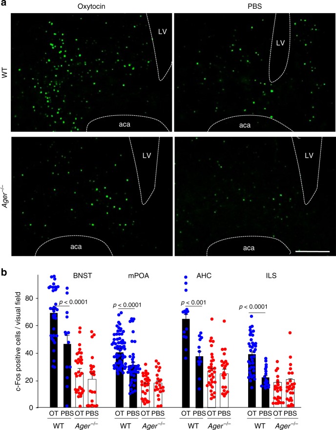
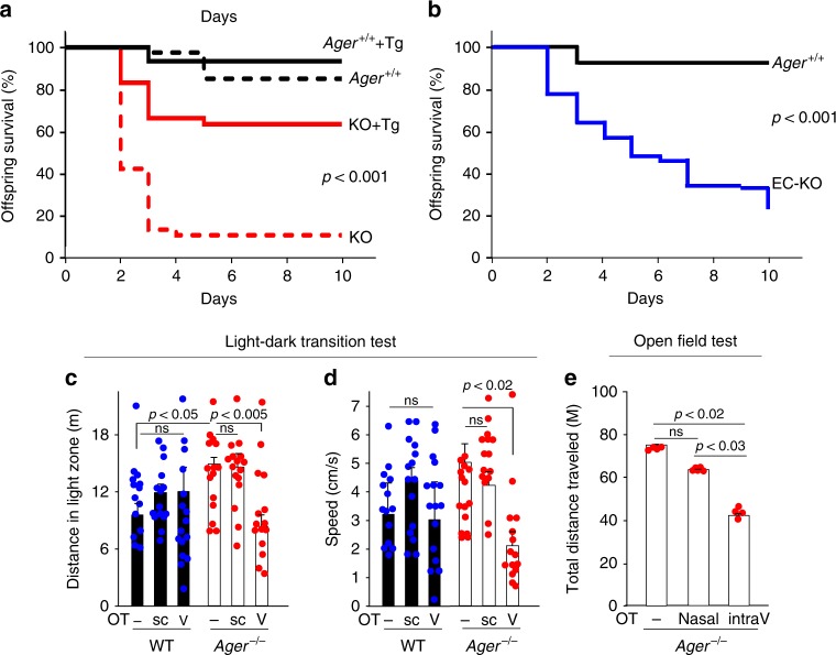
- Ager−/− mice recapitulated oxytocin-related behavioural deficits: maternal neglect reduced offspring survival, and endothelial RAGE re-expression rescued survival (63.4% KO+Tg vs 10.5% non-transgenic KO, P < 0.001). c-Fos increased after oxytocin in BNST, mPOA, AHC, and ILS of WT but not Ager−/− mice, and Ager−/− hyperactivity was normalised by intraventricular but not subcutaneous oxytocin.
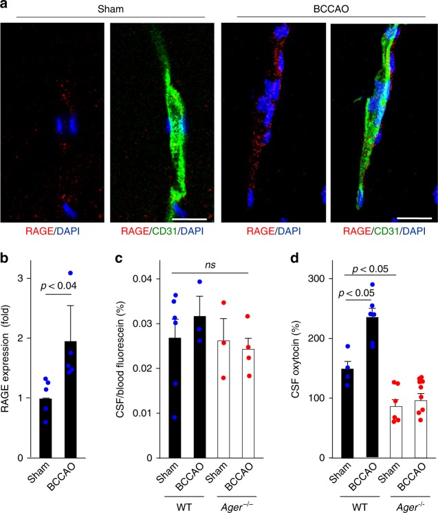
- After bilateral common carotid artery occlusion, capillary RAGE increased ~2-fold and exogenous oxytocin increased in CSF of WT but not Ager−/− male mice. The estimated availability of oxytocin in CSF was 0.2% after subcutaneous administration of 30 ng oxytocin in mice.
Methods
- Mouse models included WT, Ager−/−, endothelial RAGE-transgenic Ager−/− (KO+Tg), and endothelial RAGE-deficient (EC-KO) mice; both male and female mice were used, with behavioural and maternal survival assays, BCCAO, and oxytocin administration by subcutaneous, intravenous, or intranasal routes.
- Binding and transport were assessed by surface plasmon resonance, plate binding/competition assays, gel permeation chromatography, anti-RAGE affinity chromatography, LC-MS/MS, and an in vitro BBB model using monkey brain capillary endothelial cells with rat pericytes and astrocytes, including RAGE shRNA knockdown, TEER, and Papp calculations.
- In vivo oxytocin was measured by enzyme immunoassay in serum, cisterna magna CSF, third-ventricle CSF, and microperfusates from amygdala and PVN; c-Fos immunohistochemistry and behavioural tests (light-dark transition, open field) were used, with statistics by t-test, ANOVA, and Kaplan–Meier survival analysis.
Limitations
- Stated: the exact bioavailability of oxytocin reaching the CNS remains unclear, and findings across studies are inconsistent due to differences in treatment protocols, species, and detection methods. The mechanism by which RAGE transports oxytocin is also unclear, though vesicular endocytosis/transcytosis is proposed.
- Stated: current experiments provide no evidence for direct intranasal transport via olfactory or trigeminal routes; instead, nasally applied oxytocin may enter the bloodstream and then be transported by RAGE. Oxytocin measurement is also method-dependent, and plasma extraction may lower measured levels.
- Evident: the maternal behaviour and hyperactivity experiments were conducted largely in male mice, while female mice were used mainly to show RAGE-mediated transport; human data are limited to serum binding/co-elution and do not test behaviour or causality.
Model-written summary. Check it against the paper before relying on it.
Integrity signals
Registry
Cites
Statistics
Figures
Verification
Claims the paper makes
25 Own result · 18 Not checked yet · 8 Own method · 5 Own interpretation · 1 Partly supported · 1 No basis given · accuracy not yet measured
A result the paper reports from its own data — it cites no other source for it, so there is none to check. The quote shows it is in the paper.
In the paper
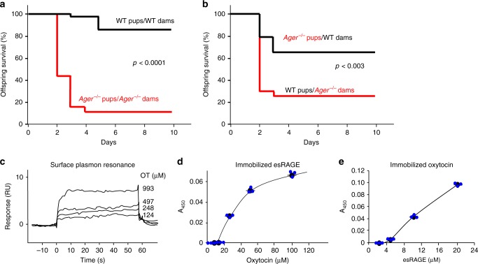
“Surface plasmon resonance methods showed concentration-dependent oxytocin binding to immobilised recombinant esRAGE, with an apparent dissociation constant (KD) of 179 nM (Fig. 1c).”
- Also stated in: § Oxytocin-RAGE interactions · plain
“Oxytocin-RAGE binding was further confirmed using plate assays where either oxytocin or esRAGE was immobilised, and esRAGE or oxytocin was in solution, respectively (Fig. 1d, e).”
§ Vascular RAGE transports oxytocin into the brain to elicit its maternal bonding behaviour in mice > Results > Oxytocin-RAGE interactions
plaincorrelational- Asserted: passes — The authors put it forward as true.
- Checkable: passes — It can be checked against evidence.
- Load-bearing: passes — The paper’s conclusions depend on it.
- Based: passes — The paper gives a reason why it holds.
- Also stated in: § Oxytocin-RAGE interactions · plain
A result the paper reports from its own data — it cites no other source for it, so there is none to check. The quote shows it is in the paper.
In the paper
“Endogenous oxytocin and soluble RAGE also associate in human circulation.”
- Also stated in: § Oxytocin-RAGE interactions · plain
“Using gel permeation chromatography to separate serum proteins based on molecular size, oxytocin immunoreactivity co-eluted with soluble RAGE (Supplementary Figure 1a).”
- Also stated in: § Oxytocin-RAGE interactions · plain
“Endogenous oxytocin in human serum (30 pmol/50 ml) also co-eluted with soluble RAGE following isolation by anti-RAGE antibody affinity chromatography (Supplementary Figure 1b).”
§ Vascular RAGE transports oxytocin into the brain to elicit its maternal bonding behaviour in mice > Results > Oxytocin-RAGE interactions
plaincorrelational- Asserted: passes — The authors put it forward as true.
- Checkable: passes — It can be checked against evidence.
- Load-bearing: passes — The paper’s conclusions depend on it.
- Based: passes — The paper gives a reason why it holds.
- Also stated in: § Oxytocin-RAGE interactions · plain
A result the paper reports from its own data — it cites no other source for it, so there is none to check. The quote shows it is in the paper.
In the paper
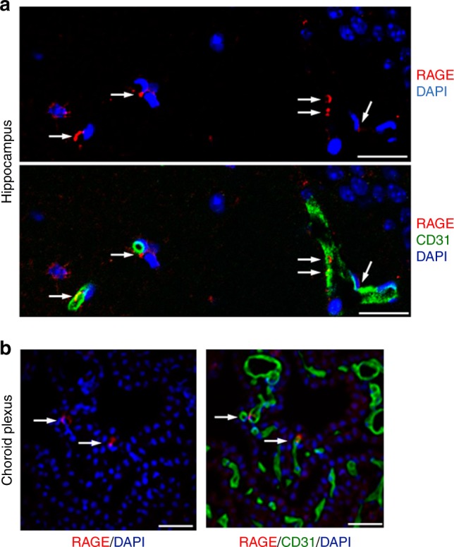
“Immunohistochemical analyses showed RAGE in vascular patterns limited to CD31 (platelet endothelial cell adhesion molecule)-positive endothelial cells23, which are the components of the BBB, of the hippocampus and choroid plexus of WT (Fig. 3a, b) but not of Ager−/− mice (Supplementary Figure 4a).”
- Also stated in: § Oxytocin transport across the BBB · plain
“We also performed confocal microscopic analyses to observe RAGE expression in the circumventricular organs (CVOs) of the brain, referred to as ‘windows of the brain’, and found that RAGE expression was clearly observable in the endothelial cells of the neurohypophysis (Supplementary Figure 4b), but barely in those of the sensory CVOs, including the organum vasculosum of the lamina terminalis, subf”
§ Vascular RAGE transports oxytocin into the brain to elicit its maternal bonding behaviour in mice > Results > Oxytocin transport across the BBB
plain- Asserted: passes — The authors put it forward as true.
- Checkable: passes — It can be checked against evidence.
- Load-bearing: passes — The paper’s conclusions depend on it.
- Based: passes — The paper gives a reason why it holds.
- Also stated in: § Oxytocin transport across the BBB · plain
A result the paper reports from its own data — it cites no other source for it, so there is none to check. The quote shows it is in the paper.
In the paper
“Analyses of the eluted fractions by liquid chromatography-tandem mass spectrometry (LC-MS/MS) showed that endogenous oxytocin in human serum is structurally indistinguishable from the synthetic oxytocin standard, as they eluted at identical times (Supplementary Figure 1d) and have identical parent ions (M+H+ 1008.2).”
§ Vascular RAGE transports oxytocin into the brain to elicit its maternal bonding behaviour in mice > Results > Oxytocin-RAGE interactions
plain- Asserted: passes — The authors put it forward as true.
- Checkable: passes — It can be checked against evidence.
- Load-bearing: unclear — It is unclear whether the paper’s conclusions depend on it.
- Based: passes — The paper gives a reason why it holds.
A result the paper reports from its own data — it cites no other source for it, so there is none to check. The quote shows it is in the paper.
In the paper
“Further analysis revealed co-localisation of RAGE with cavelolin-1, a marker of plasma membrane invaginations (caveolae), in endothelial cells and the choroid plexus in WT mice (Supplementary Figure 4c), suggesting a role of RAGE in the oxytocin transfer (trafficking) through caveolae-associated endocytosis and transcytosis (Supplementary Figure 4d).”
§ Vascular RAGE transports oxytocin into the brain to elicit its maternal bonding behaviour in mice > Results > Oxytocin transport across the BBB
plaincorrelational- Asserted: passes — The authors put it forward as true.
- Checkable: passes — It can be checked against evidence.
- Load-bearing: passes — The paper’s conclusions depend on it.
- Based: passes — The paper gives a reason why it holds.
A result the paper reports from its own data — it cites no other source for it, so there is none to check. The quote shows it is in the paper.
In the paper
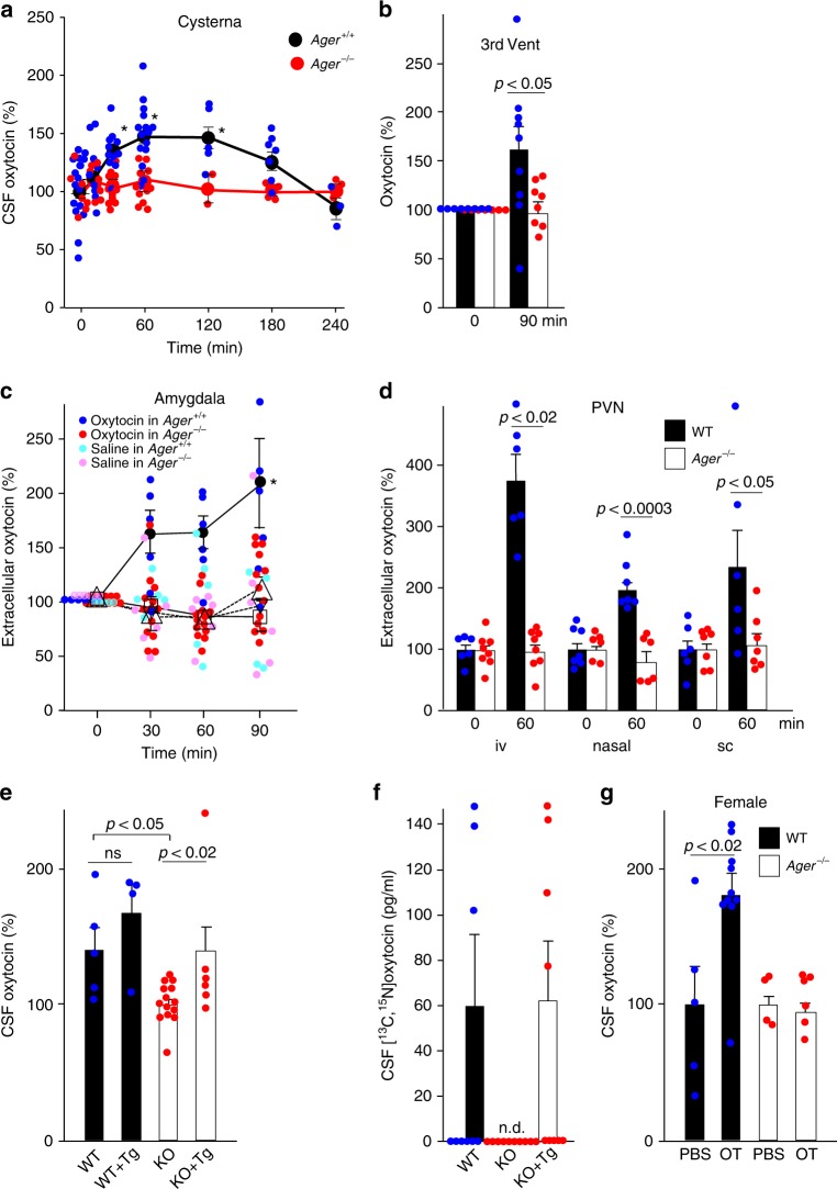
“Microperfusion methods showed that oxytocin also increased in relevant regions of the brain, including the amygdala and PVN of the hypothalamus, of WT but not Ager−/− male mice following subcutaneous, intravenous or intranasal administration of oxytocin but not saline carrier (Fig. 4c, d and Supplementary Figure 5c).”
§ Vascular RAGE transports oxytocin into the brain to elicit its maternal bonding behaviour in mice > Results > RAGE-dependent transport of oxytocin across the BBB
plaindirect- Asserted: passes — The authors put it forward as true.
- Checkable: passes — It can be checked against evidence.
- Load-bearing: passes — The paper’s conclusions depend on it.
- Based: passes — The paper gives a reason why it holds.
A result the paper reports from its own data — it cites no other source for it, so there is none to check. The quote shows it is in the paper.
In the paper
“By contrast, oxytocin receptors, which could theoretically act as oxytocin transporters, were not co-localised with endothelial cells in numerous sections of 5 brain regions of Venus mice expressing a fluorescent oxytocin receptor reporter47 (Supplementary Figures 4e and f).”
§ Vascular RAGE transports oxytocin into the brain to elicit its maternal bonding behaviour in mice > Results > Oxytocin transport across the BBB
plainn = 5- Asserted: passes — The authors put it forward as true.
- Checkable: passes — It can be checked against evidence.
- Load-bearing: passes — The paper’s conclusions depend on it.
- Based: passes — The paper gives a reason why it holds.
A result the paper reports from its own data — it cites no other source for it, so there is none to check. The quote shows it is in the paper.
In the paper
“Steady-state levels in blood (~20 pg/ml) increased rapidly, peaking within 10 min after injection and returning to baseline by 1–2 h in both WT and Ager−/− male mice (Supplementary Figure 5a).”
§ Vascular RAGE transports oxytocin into the brain to elicit its maternal bonding behaviour in mice > Results > RAGE-dependent transport of oxytocin across the BBB
plaindirect- Asserted: passes — The authors put it forward as true.
- Checkable: passes — It can be checked against evidence.
- Load-bearing: passes — The paper’s conclusions depend on it.
- Based: passes — The paper gives a reason why it holds.
A result the paper reports from its own data — it cites no other source for it, so there is none to check. The quote shows it is in the paper.
In the paper
“Although oxytocin has been found to produce sex-specific effects48–50, RAGE-mediated oxytocin transport was also observed in female WT-mice (Fig. 4g).”
§ Vascular RAGE transports oxytocin into the brain to elicit its maternal bonding behaviour in mice > Results > RAGE-dependent transport of oxytocin across the BBB
plaindirect- Asserted: passes — The authors put it forward as true.
- Checkable: passes — It can be checked against evidence.
- Load-bearing: passes — The paper’s conclusions depend on it.
- Based: passes — The paper gives a reason why it holds.
A result the paper reports from its own data — it cites no other source for it, so there is none to check. The quote shows it is in the paper.
In the paper
“Nevertheless, exogenous oxytocin increased in the CSF of WT but not Ager−/− male mice after bilateral carotid occlusion (Fig. 5d and Supplementary Figure 5f).”
§ Vascular RAGE transports oxytocin into the brain to elicit its maternal bonding behaviour in mice > Results > RAGE-dependent transport of oxytocin across the BBB
plaindirectn = 4- Asserted: passes — The authors put it forward as true.
- Checkable: passes — It can be checked against evidence.
- Load-bearing: passes — The paper’s conclusions depend on it.
- Based: passes — The paper gives a reason why it holds.
- Probably not a substantial claim. 19
- It may state two separate claims as one. 18
The cited sources support only part of this claim — read each source’s verdict below.
In the paper
“The three RAGE forms bind various ligands with similar affinities, including multiple advanced glycation end-products (AGEs) and amyloid β peptide31–37.”
§ Vascular RAGE transports oxytocin into the brain to elicit its maternal bonding behaviour in mice > Introduction
What the sources say
- Partly supportedcites [31] Mackic, J. B. et al. Human blood-brain barrier receptors for Alzheimer’s amyloid-beta 1- 40. Asymmetrical binding, endocytosis, and transcytosis at the apical side of brain microvascular endothelial cell monolayer. J. Clin. Invest. 102, 734–743 (1998). sourceExperimental — accuracy not yet measured
The Results report RAGE-dependent binding of amyloid-β 1–40, and the Discussion identifies RAGE as a receptor involved in its binding at the blood–brain barrier. The paper does not compare three RAGE forms or show that they bind AGEs and amyloid-β with similar affinities; AGEs appear only in a cited reference. (LLM, conf=0.86)
- Not checked yetcites [32] Yamamoto, Y. et al. Short-chain aldehyde-derived ligands for RAGE and their actions on endothelial cells. Diabetes Res. Clin. Pract. 77, S30–S40 (2007). source
- Not checked yetcites [33] Matsumoto, K. et al. Immunohistochemical analysis of transporters related to clearance of amyloid-β peptides through blood-cerebrospinal fluid barrier in human brain. Histochem. Cell. Biol. 144, 597–611 (2015). source
- Not checked yetcites [34] Deane, R. et al. RAGE mediates amyloid-beta peptide transport across the blood-brain barrier and accumulation in brain. Nat. Med. 9, 907–913 (2003). source
- Partly supportedcites [35] Deane, R. et al. A multimodal RAGE-specific inhibitor reduces amyloid β-mediated brain disorder in a mouse model of Alzheimer disease. J. Clin. Invest. 122, 1377–1392 (2012). sourceExperimental — accuracy not yet measured
The Introduction states that RAGE’s V domain binds multiple ligands, including AGE proteins and amyloid β. It does not say that three RAGE forms bind those ligands or that their binding affinities are similar; the Results report an affinity for amyloid β binding to RAGE but do not compare three forms. (LLM, conf=0.86)
- Not checked yetcites [36] Saito, R., Araki, S., Yamamoto, Y. & Kusuhara, K. Elevated endogenous secretory receptor for advanced glycation end products (esRAGE) levels are associated with circulating soluble RAGE levels in diabetic children. J. Pediatr. Endocrinol. Metab. 30, 63–69 (2017). source
- Partly supportedcites [37] Gasparotto, J. et al. Receptor for advanced glycation end products mediates sepsis-triggered amyloid-β accumulation, Tau phosphorylation, and cognitive impairment. J. Biol. Chem. 293, 226–244 (2018). sourceExperimental — accuracy not yet measured
The introductory passage labeled Abstract (page unavailable) describes RAGE as a multiligand receptor that interacts with AGEs and says amyloid-β binds and activates it. It does not say that three RAGE forms bind these ligands or that their binding affinities are similar. (LLM, conf=0.88)
plaincorrelational- Asserted: passes — The authors put it forward as true.
- Checkable: passes — It can be checked against evidence.
- Load-bearing: fails — The paper’s conclusions may not depend on it.
- Based: passes — The paper gives a reason why it holds.
Probably not a substantial claim. The paper’s conclusions may not depend on it.
- Partly supported
A result the paper reports from its own data — it cites no other source for it, so there is none to check. The quote shows it is in the paper.
In the paper
“oxytocin neither stimulates a RAGE-dependent NF-κB reporter nor inhibits reporter induction by AGEs, a ligand that activates the NF-κB reporter22,27 (Supplementary Figure 2a).”
- Also stated in: § Oxytocin-RAGE interactions · plain
“Furthermore, oxytocin did not activate NF-κB-independent Rac1 or Cdc42 signalling, which are also suggested to function downstream of RAGE24,41,42 (Supplementary Figures 2b and 2c).”
§ Vascular RAGE transports oxytocin into the brain to elicit its maternal bonding behaviour in mice > Results > Oxytocin-RAGE interactions
plain- Asserted: passes — The authors put it forward as true.
- Checkable: passes — It can be checked against evidence.
- Load-bearing: unclear — It is unclear whether the paper’s conclusions depend on it.
- Based: passes — The paper gives a reason why it holds.
It may state two separate claims as one.
- Also stated in: § Oxytocin-RAGE interactions · plain
A result the paper reports from its own data — it cites no other source for it, so there is none to check. The quote shows it is in the paper.
In the paper
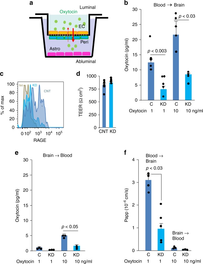
“Endothelial transport of oxytocin was dose-dependent (Fig. 2b) and of a similar rate to transport of centrally acting drugs46.”
- Also stated in: § Oxytocin transport across the BBB · plain
“Notably, oxytocin transport was reduced selectively in the blood-to-brain direction following RAGE knockdown (Fig. 2b).”
- Also stated in: § Oxytocin transport across the BBB · plain
“Reverse transport was much less efficient (Fig. 2e), and according to calculated apparent permeability (Papp) measures, inconsequential (Fig. 2f).”
§ Vascular RAGE transports oxytocin into the brain to elicit its maternal bonding behaviour in mice > Results > Oxytocin transport across the BBB
plaindirect- Asserted: passes — The authors put it forward as true.
- Checkable: passes — It can be checked against evidence.
- Load-bearing: passes — The paper’s conclusions depend on it.
- Based: passes — The paper gives a reason why it holds.
It may state two separate claims as one.
- Also stated in: § Oxytocin transport across the BBB · plain
A result the paper reports from its own data — it cites no other source for it, so there is none to check. The quote shows it is in the paper.
In the paper
“By contrast, levels of oxytocin in CSF from the cisterna magna of WT mice increased more gradually to maxima at 1–2 h before returning to baseline at 4 h (Fig. 4a).”
- Also stated in: § RAGE-dependent transport of oxytocin across the BBB · plain
“Despite equivalently high serum levels following injection of identical amounts of oxytocin (Supplementary Figure 5a), concentrations in the CSF of Ager−/− mice remained at baseline levels (Fig. 4a).”
- Also stated in: § RAGE-dependent transport of oxytocin across the BBB · plain
“Following subcutaneous injection, oxytocin increased in third ventricle CSF of WT but not Ager−/− male mice (Fig. 4b and Supplementary Figure 5b).”
§ Vascular RAGE transports oxytocin into the brain to elicit its maternal bonding behaviour in mice > Results > RAGE-dependent transport of oxytocin across the BBB
plaindirect- Asserted: passes — The authors put it forward as true.
- Checkable: passes — It can be checked against evidence.
- Load-bearing: passes — The paper’s conclusions depend on it.
- Based: passes — The paper gives a reason why it holds.
It may state two separate claims as one.
- Also stated in: § RAGE-dependent transport of oxytocin across the BBB · plain
A result the paper reports from its own data — it cites no other source for it, so there is none to check. The quote shows it is in the paper.
In the paper
“Following subcutaneous injection, [13C,15N]oxytocin was undetectable in CSF of Ager−/− male mice, whereas amounts in the CSF of WT or KO + Tg mice were equivalent (Fig. 4f and Supplementary Figure 5e).”
§ Vascular RAGE transports oxytocin into the brain to elicit its maternal bonding behaviour in mice > Results > RAGE-dependent transport of oxytocin across the BBB
plain- Asserted: passes — The authors put it forward as true.
- Checkable: passes — It can be checked against evidence.
- Load-bearing: passes — The paper’s conclusions depend on it.
- Based: passes — The paper gives a reason why it holds.
It may state two separate claims as one.
A result the paper reports from its own data — it cites no other source for it, so there is none to check. The quote shows it is in the paper.
In the paper
“Tg expression in Ager−/− mice increased oxytocin in CSF to the WT concentration, whereas Tg expression in WT mice minimally affected CSF oxytocin concentrations (Fig. 4e and Supplementary Figure 5d).”
§ Vascular RAGE transports oxytocin into the brain to elicit its maternal bonding behaviour in mice > Results > RAGE-dependent transport of oxytocin across the BBB
plaindirect- Asserted: passes — The authors put it forward as true.
- Checkable: passes — It can be checked against evidence.
- Load-bearing: passes — The paper’s conclusions depend on it.
- Based: passes — The paper gives a reason why it holds.
It may state two separate claims as one.
A result the paper reports from its own data — it cites no other source for it, so there is none to check. The quote shows it is in the paper.
In the paper
“During light-dark transition tests, the greater distances travelled (c) and greater average speed (d) in the light zone exhibited by Ager−/− male mice were both reduced by intraventricular (V) but not subcutaneous (sc) injection of oxytocin (OT) (n = 14–16).”
- Also stated in: § Social behaviour in RAGE knockout mice · plain
“Concordantly, intraventricular but not nasal administration of oxytocin normalised distances travelled by Ager−/− mice in open field tests (Fig. 7e).”
§ Vascular RAGE transports oxytocin into the brain to elicit its maternal bonding behaviour in mice > Results > Maternal behaviour
plaindirectn = 14- Asserted: passes — The authors put it forward as true.
- Checkable: passes — It can be checked against evidence.
- Load-bearing: passes — The paper’s conclusions depend on it.
- Based: passes — The paper gives a reason why it holds.
It may state two separate claims as one.
- Also stated in: § Social behaviour in RAGE knockout mice · plain
A result the paper reports from its own data — it cites no other source for it, so there is none to check. The quote shows it is in the paper.
In the paper
“Numbers of c-Fos positive nuclei increased after subcutaneous oxytocin administration in four distinct regions of the brain, the bed nucleus of the stria terminalis (BNST), medial preoptic area (mPOA), centre of the anterior hypothalamic area (AHC) and ventral portion of the intermediate lateral septal nucleus (ILS) (Fig. 6a, b).”
- Also stated in: § c-Fos as CNS actions of oxytocin · plain
“This is dependent on both oxytocin and RAGE, as there are no effects in Ager−/− male mice or when vehicle control was administered (Fig. 6b).”
§ Vascular RAGE transports oxytocin into the brain to elicit its maternal bonding behaviour in mice > Results > c-Fos as CNS actions of oxytocin
plaindirectn = 12- Asserted: passes — The authors put it forward as true.
- Checkable: passes — It can be checked against evidence.
- Load-bearing: passes — The paper’s conclusions depend on it.
- Based: passes — The paper gives a reason why it holds.
It may state two separate claims as one.
- Also stated in: § c-Fos as CNS actions of oxytocin · plain
A result the paper reports from its own data — it cites no other source for it, so there is none to check. The quote shows it is in the paper.
In the paper
“Survival of Ager gene null (Ager−/−) pups is often low due to maternal neglect, with even mild maternal stress leading to dramatic reductions in offspring survival (Fig. 1a).”
- Also stated in: § Oxytocin-RAGE interactions · boosted
“This is clearly a maternal deficit, as newborn Ager−/− pups transferred to wild-type (WT) post-partum mothers are nurtured and survive at near normal frequencies. By contrast, normal offspring of WT mothers do poorly when transferred to post-partum Ager−/− mothers (Fig. 1b).”
§ Vascular RAGE transports oxytocin into the brain to elicit its maternal bonding behaviour in mice > Results > Oxytocin-RAGE interactions
boosteddirect- Asserted: passes — The authors put it forward as true.
- Checkable: passes — It can be checked against evidence.
- Load-bearing: passes — The paper’s conclusions depend on it.
- Based: passes — The paper gives a reason why it holds.
It may state two separate claims as one.
- Also stated in: § Oxytocin-RAGE interactions · boosted
A result the paper reports from its own data — it cites no other source for it, so there is none to check. The quote shows it is in the paper.
In the paper
“Mass spectrometry of the eluted material from the immobilised esRAGE in human serum did not identify arginine-vasopressin; additionally, RAGE did not transport it in an in vitro blood–brain barrier (BBB) assay system (Supplementary Figure 7).”
§ Vascular RAGE transports oxytocin into the brain to elicit its maternal bonding behaviour in mice > Discussion
plain- Asserted: passes — The authors put it forward as true.
- Checkable: passes — It can be checked against evidence.
- Load-bearing: unclear — It is unclear whether the paper’s conclusions depend on it.
- Based: passes — The paper gives a reason why it holds.
It may state two separate claims as one.
None of the sources cited for this claim have been checked yet.
In the paper
“Oxytocin mediates both physiological and psychosocial events surrounding mammalian birth, including uterine contractions, initiation of lactation, and maternal bonding1,2.”
§ Vascular RAGE transports oxytocin into the brain to elicit its maternal bonding behaviour in mice > Introduction
What the sources say
plaindirect- Asserted: passes — The authors put it forward as true.
- Checkable: passes — It can be checked against evidence.
- Load-bearing: passes — The paper’s conclusions depend on it.
- Based: passes — The paper gives a reason why it holds.
Probably not a substantial claim.
None of the sources cited for this claim have been checked yet.
In the paper
“Exogenous oxytocin in humans also appears to have behavioural effects, particularly in such social deficit-related psychiatric disorders as autism and schizophrenia9–13.”
§ Vascular RAGE transports oxytocin into the brain to elicit its maternal bonding behaviour in mice > Introduction
What the sources say
- Not checked yetcites [9] Higashida, H., Yokoyama, S., Kikuchi, M. & Munesue, T. CD38 and its role in oxytocin secretion and social behavior. Horm. Behav. 61, 351–358 (2012). source
- Not checked yetcites [10] Insel, T. R. Translating oxytocin neuroscience to the clinic: A National Institute of Mental Health Perspective. Biol. Psychiatry 79, 153–154 (2016). source
- Not checked yetcites [11] Guastella, A. J. et al. Intranasal oxytocin improves emotion recognition for youth with autism spectrum disorders. Biol. Psychiatry 67, 692–694 (2010). source
- Not checked yetcites [12] Gordon, I. et al. Intranasal oxytocin enhances connectivity in the neural circuitry supporting social motivation and social perception in children with autism. Sci. Rep. 6, 35054 (2016). source
- Not checked yetcites [13] Feifel, D., Shilling, P. D. & MacDonald, K. A review of oxytocin’s effects on the positive, negative, and cognitive domains of schizophrenia. Biol. Psychiatry 79, 222–233 (2016). source
hedgedconditional- Asserted: passes — The authors put it forward as true.
- Checkable: passes — It can be checked against evidence.
- Load-bearing: fails — The paper’s conclusions may not depend on it.
- Based: passes — The paper gives a reason why it holds.
Probably not a substantial claim. The paper’s conclusions may not depend on it.
None of the sources cited for this claim have been checked yet.
In the paper
“Normal maternal behaviour is restored by peripheral oxcytocin replacement in Oxt−/− and Cd38−/−, but not Oxtr−/− mice, implying that circulating oxcytocin crosses the blood-brain barrier.”
- Also stated in: § Introduction · hedged
“This is restored by administration of exogenous oxytocin in Oxt−/− but not Oxtr−/− mice, suggesting that peripheral oxytocin has central effects.”
- Also stated in: § Introduction · plain
“Mice lacking CD38, a cyclic ADP ribose synthetase and hydrolase necessary for secretion of oxytocin, also display this characteristic behavioural phenotype, which is also reversed following administration of exogenous oxytocin5,9.”
§ Vascular RAGE transports oxytocin into the brain to elicit its maternal bonding behaviour in mice > Abstract
What the sources say
- Not checked yetcites [5] Jin, D. et al. CD38 is critical for social behaviour by regulating oxytocin secretion. Nature 446, 41–45 (2007). source
- Not checked yetcites [9] Higashida, H., Yokoyama, S., Kikuchi, M. & Munesue, T. CD38 and its role in oxytocin secretion and social behavior. Horm. Behav. 61, 351–358 (2012). source
plaindirect- Asserted: passes — The authors put it forward as true.
- Checkable: passes — It can be checked against evidence.
- Load-bearing: passes — The paper’s conclusions depend on it.
- Based: passes — The paper gives a reason why it holds.
It may state two separate claims as one.
- Also stated in: § Introduction · hedged
None of the sources cited for this claim have been checked yet.
In the paper
“RAGE, a member of the immunoglobulin superfamily of pattern recognition receptors, is not known to be related to oxytocin or oxytocin signalling22–33.”
§ Vascular RAGE transports oxytocin into the brain to elicit its maternal bonding behaviour in mice > Introduction
What the sources say
- Not checked yetcites [22] Senatus, L. M. & Schmidt, A. M. The AGE-RAGE axis: implications for age-associated arterial diseases. Front. Genet. 8, 187 (2017). source
- Not checked yetcites [23] Kamide, T. et al. RAGE mediates vascular injury and inflammation after global cerebral ischemia. Neurochem. Int. 60, 220–228 (2012). source
- Not checked yetcites [24] Manigrasso, M. B., Juranek, J., Ramasamy, R. & Schmidt, A. M. Unlocking the biology of RAGE in diabetic microvascular complications. Trends Endocrinol. Metab. 25, 15–22 (2014). source
- Not checked yetcites [25] Takuma, K. et al. RAGE-mediated signaling contributes to intraneuronal transport of amyloid-beta and neuronal dysfunction. Proc. Natl Acad. Sci. USA 106, 20021–20026 (2009). source
- Not checked yetcites [26] Yamamoto, Y. et al. Development and prevention of advanced diabetic nephropathy in RAGE-overexpressing mice. J. Clin. Invest. 108, 261–268 (2001). source
- Not checked yetcites [27] Yamamoto, Y. et al. Septic shock is associated with receptor for advanced glycation end products ligation of LPS. J. Immunol. 186, 3248–3257 (2011). source
- Not checked yetcites [28] Yamamoto, Y. & Yamamoto, H. RAGE-mediated inflammation, type 2 diabetes, and diabetic vascular complication. Front. Endocrinol. 4, 105 (2013). source
- Not checked yetcites [29] Yonekura, H. et al. Novel splice variants of the receptor for advanced glycation end-products expressed in human vascular endothelial cells and pericytes, and their putative roles in diabetes-induced vascular injury. Biochem. J. 370, 1097–1109 (2003). source
- Not checked yetcites [30] Motoyoshi, S. et al. cAMP ameliorates inflammation by modulation of macrophage receptor for advanced glycation end-products. Biochem. J. 463, 75–82 (2014). source
- Not checked yetcites [31] Mackic, J. B. et al. Human blood-brain barrier receptors for Alzheimer’s amyloid-beta 1- 40. Asymmetrical binding, endocytosis, and transcytosis at the apical side of brain microvascular endothelial cell monolayer. J. Clin. Invest. 102, 734–743 (1998). source
plain- Asserted: passes — The authors put it forward as true.
- Checkable: passes — It can be checked against evidence.
- Load-bearing: passes — The paper’s conclusions depend on it.
- Based: passes — The paper gives a reason why it holds.
Probably not a substantial claim.
What the paper did — it cites no source for this, so there is none to check. The quote shows it is in the paper.
In the paper
“As a control, we showed this procedure does not cause fluorescein to leak into the CSF (Fig. 5c), suggesting there is not a generalised increase in vascular permeability.”
§ Vascular RAGE transports oxytocin into the brain to elicit its maternal bonding behaviour in mice > Results > RAGE-dependent transport of oxytocin across the BBB
hedgedn = 3- Asserted: passes — The authors put it forward as true.
- Checkable: passes — It can be checked against evidence.
- Load-bearing: passes — The paper’s conclusions depend on it.
- Based: passes — The paper gives a reason why it holds.
It may state two separate claims as one.
In the paper
“The classical studies of Numan showed c-Fos positive neurons are activated during the acquisition of postpartum maternal behaviours51.”
§ Vascular RAGE transports oxytocin into the brain to elicit its maternal bonding behaviour in mice > Results > c-Fos as CNS actions of oxytocin
What the sources say
- Not checked yetcites [51] Numan, M. & Numan, M. J. Projection sites of medial preoptic area and ventral bed nucleus of the stria terminalis neurons that express Fos during maternal behavior in female rats. J. Neuroendocrinol. 9, 369–384 (1997). source
plaincorrelational- Asserted: passes — The authors put it forward as true.
- Checkable: passes — It can be checked against evidence.
- Load-bearing: fails — The paper’s conclusions may not depend on it.
- Based: passes — The paper gives a reason why it holds.
Probably not a substantial claim. The paper’s conclusions may not depend on it.
A conclusion the paper draws from its own results — it cites no other source for it, so there is none to check. The quote shows it is in the paper.
In the paper
“These results demonstrated that RAGE is both necessary and sufficient for oxytocin transport into the brain in both male and female mice.”
§ Vascular RAGE transports oxytocin into the brain to elicit its maternal bonding behaviour in mice > Results > RAGE-dependent transport of oxytocin across the BBB
boosteddirect- Asserted: passes — The authors put it forward as true.
- Checkable: passes — It can be checked against evidence.
- Load-bearing: passes — The paper’s conclusions depend on it.
- Based: passes — The paper gives a reason why it holds.
It may state two separate claims as one.
In the paper
“Since both of these are deleterious, pathological effects, RAGE is being targeted for inhibition in drug discovery efforts35.”
§ Vascular RAGE transports oxytocin into the brain to elicit its maternal bonding behaviour in mice > Discussion
What the sources say
- Not checked yetcites [35] Deane, R. et al. A multimodal RAGE-specific inhibitor reduces amyloid β-mediated brain disorder in a mouse model of Alzheimer disease. J. Clin. Invest. 122, 1377–1392 (2012). source
plain- Asserted: passes — The authors put it forward as true.
- Checkable: passes — It can be checked against evidence.
- Load-bearing: fails — The paper’s conclusions may not depend on it.
- Based: passes — The paper gives a reason why it holds.
Probably not a substantial claim. The paper’s conclusions may not depend on it.
None of the sources cited for this claim have been checked yet.
In the paper
“For example, RAGE has been shown to transport amyloid β peptide across the BBB, which promotes Alzheimer disease-like symptoms in susceptible mice.”
§ Vascular RAGE transports oxytocin into the brain to elicit its maternal bonding behaviour in mice > Discussion
boosteddirect- Asserted: passes — The authors put it forward as true.
- Checkable: passes — It can be checked against evidence.
- Load-bearing: fails — The paper’s conclusions may not depend on it.
- Based: passes — The paper gives a reason why it holds.
Probably not a substantial claim. The paper’s conclusions may not depend on it. It may state two separate claims as one.
In the paper
“The affinity of oxytocin binding to the oxytocin receptor has often been estimated to be KD ~100 nM, although a high affinity form with KD ~1 nM has also been reported60.”
§ Vascular RAGE transports oxytocin into the brain to elicit its maternal bonding behaviour in mice > Discussion
What the sources say
- Not checked yetcites [60] Gimpl, G. & Fahrenholz, F. The oxytocin receptor system: structure, function, and regulation. Physiol. Rev. 81, 629–683 (2001). source
plain- Asserted: passes — The authors put it forward as true.
- Checkable: passes — It can be checked against evidence.
- Load-bearing: fails — The paper’s conclusions may not depend on it.
- Based: passes — The paper gives a reason why it holds.
Probably not a substantial claim. The paper’s conclusions may not depend on it. It may state two separate claims as one.
In the paper
“Loss of maternal nurturing and social interactive behaviours in Cd38−/− mice have been restored following peripheral administration of oxytocin, consistent with transport of peripherally administered exogenous oxytocin into brain5.”
§ Vascular RAGE transports oxytocin into the brain to elicit its maternal bonding behaviour in mice > Results > Maternal behaviour
What the sources say
- Not checked yetcites [5] Jin, D. et al. CD38 is critical for social behaviour by regulating oxytocin secretion. Nature 446, 41–45 (2007). source
plaindirect- Asserted: passes — The authors put it forward as true.
- Checkable: passes — It can be checked against evidence.
- Load-bearing: passes — The paper’s conclusions depend on it.
- Based: passes — The paper gives a reason why it holds.
It may state two separate claims as one.
None of the sources cited for this claim have been checked yet.
In the paper
“Levels of oxytocin in the periphery are lower than those in the CSF, in humans16,61,62, providing a concentration gradient for what appears to be a transport mechanism: this point of view depends on the methods of oxytocin measurements in blood, because a recent report has shown much higher blood OT levels than previously thought using a robust nanoLC-MS platform63.”
§ Vascular RAGE transports oxytocin into the brain to elicit its maternal bonding behaviour in mice > Discussion
What the sources say
- Not checked yetcites [16] Striepens, N. et al. Elevated cerebrospinal fluid and blood concentrations of oxytocin following its intranasal administration in humans. Sci. Rep. 3, 3440 (2013). source
- Not checked yetcites [61] Kagerbauer, S. M. et al. Plasma oxytocin and vasopressin do not predict neuropeptide concentrations in human cerebrospinal fluid. J. Neuroendocrinol. 25, 668–673 (2013). source
- Not checked yetcites [62] Martin, J. et al. Vasopressin and oxytocin in CSF and plasma of patients with aneurysmal subarachnoid haemorrhage. Neuropeptides 48, 91–96 (2014). source
plaindirect- Asserted: passes — The authors put it forward as true.
- Checkable: passes — It can be checked against evidence.
- Load-bearing: unclear — It is unclear whether the paper’s conclusions depend on it.
- Based: passes — The paper gives a reason why it holds.
It may state two separate claims as one.
None of the sources cited for this claim have been checked yet.
In the paper
“Clinical studies have linked autism and Asperger’s disorders and attention-deficit/hyperactivity disorders to oxytocin deficiency7,8,54, and the potential use of exogenous oxytocin to treat these conditions is being assessed in clinical trials10,11,54–57.”
§ Vascular RAGE transports oxytocin into the brain to elicit its maternal bonding behaviour in mice > Results > Social behaviour in RAGE knockout mice
What the sources say
- Not checked yetcites [7] Insel, T. R. The challenge of translation in social neuroscience: a review of oxytocin, vasopressin, and affiliative behavior. Neuron 65, 768–779 (2010). source
- Not checked yetcites [8] Rilling, J. K. & Young, L. J. The biology of mammalian parenting and its effect on offspring social development. Science 345, 771–776 (2014). source
- Not checked yetcites [54] Feldman, R. et al. Sensitive parenting is associated with plasma oxytocin and polymorphisms in the OXTR and CD38 genes. Biol. Psychiatry 72, 175–181 (2012). source
plaincorrelational- Asserted: passes — The authors put it forward as true.
- Checkable: passes — It can be checked against evidence.
- Load-bearing: fails — The paper’s conclusions may not depend on it.
- Based: passes — The paper gives a reason why it holds.
Probably not a substantial claim. The paper’s conclusions may not depend on it.
The paper states this without a source or data behind it here, so there is nothing to check it against. The quote shows it is in the paper.
In the paper
“RAGE has also been implicated in the pathogenesis of diabetes complications.”
§ Vascular RAGE transports oxytocin into the brain to elicit its maternal bonding behaviour in mice > Discussion
hedgedcorrelational- Asserted: passes — The authors put it forward as true.
- Checkable: passes — It can be checked against evidence.
- Load-bearing: fails — The paper’s conclusions may not depend on it.
- Based: passes — The paper gives a reason why it holds.
Probably not a substantial claim. The paper’s conclusions may not depend on it.
What the paper did — it cites no source for this, so there is none to check. The quote shows it is in the paper.
In the paper
“The oxytocin assay had two linear ranges, which covers a lower concentration range from a few to 30 pg/ml, and a higher concentration range between 50 and 1000 pg/ml.”
§ Vascular RAGE transports oxytocin into the brain to elicit its maternal bonding behaviour in mice > Methods > Enzyme immunoassay for oxytocin
plain- Asserted: passes — The authors put it forward as true.
- Checkable: passes — It can be checked against evidence.
- Load-bearing: fails — The paper’s conclusions may not depend on it.
- Based: passes — The paper gives a reason why it holds.
Probably not a substantial claim. The paper’s conclusions may not depend on it.
What the paper did — it cites no source for this, so there is none to check. The quote shows it is in the paper.
In the paper
“All animal experiments were approved by the Committee on Animal Experimentation of Kanazawa University and performed in accordance with the Fundamental Guidelines for Proper Conduct of Animal Experiment and Related Activities in Academic Research Institutions under the jurisdiction of the Ministry of Education, Culture, Sports, Science and Technology of Japan.”
§ Vascular RAGE transports oxytocin into the brain to elicit its maternal bonding behaviour in mice > Methods > Animals
plain- Asserted: passes — The authors put it forward as true.
- Checkable: passes — It can be checked against evidence.
- Load-bearing: fails — The paper’s conclusions may not depend on it.
- Based: passes — The paper gives a reason why it holds.
Probably not a substantial claim. The paper’s conclusions may not depend on it. It may state two separate claims as one.
None of the sources cited for this claim have been checked yet.
In the paper
“CSF was collected according to the protocol for sampling CSF from mice without detectable plasma contamination77,78.”
§ Vascular RAGE transports oxytocin into the brain to elicit its maternal bonding behaviour in mice > Methods > Sampling of the cerebrospinal fluid (CSF) and blood
What the sources say
- Not checked yetcites [77] Lopatina, O. et al. H. Communication impairment in ultrasonic vocal repertoire during the suckling period of Cd157 knockout mice: transient improvement by oxytocin. Front. Neurosci. 11, 266 (2017). source
- Not checked yetcites [78] Liu, L. & Duff, K. A technique for serial collection of cerebrospinal fluid from the cisterna magna in mouse. J. Vis. Exp. 21, pii: 960 (2008).
plain- Asserted: passes — The authors put it forward as true.
- Checkable: passes — It can be checked against evidence.
- Load-bearing: passes — The paper’s conclusions depend on it.
- Based: passes — The paper gives a reason why it holds.
Probably not a substantial claim.
What the paper did — it cites no source for this, so there is none to check. The quote shows it is in the paper.
In the paper
“whereas an additional extraction step may have resulted in lower than actual oxytocin levels.”
§ Vascular RAGE transports oxytocin into the brain to elicit its maternal bonding behaviour in mice > Methods > Enzyme immunoassay for oxytocin
hedgedconditional- Asserted: passes — The authors put it forward as true.
- Checkable: passes — It can be checked against evidence.
- Load-bearing: fails — The paper’s conclusions may not depend on it.
- Based: passes — The paper gives a reason why it holds.
Probably not a substantial claim. The paper’s conclusions may not depend on it.
What the paper did — it cites no source for this, so there is none to check. The quote shows it is in the paper.
In the paper
“The inter-assay and intra-assay coefficients of variation were less than 15%.”
§ Vascular RAGE transports oxytocin into the brain to elicit its maternal bonding behaviour in mice > Methods > Enzyme immunoassay for oxytocin
plain- Asserted: passes — The authors put it forward as true.
- Checkable: passes — It can be checked against evidence.
- Load-bearing: fails — The paper’s conclusions may not depend on it.
- Based: passes — The paper gives a reason why it holds.
Probably not a substantial claim. The paper’s conclusions may not depend on it. It may state two separate claims as one.
What the paper did — it cites no source for this, so there is none to check. The quote shows it is in the paper.
In the paper
“In our experimental model, the mice that exhibited <15% of the baseline control microperfusion during the first minute of occlusion were used in subsequent experiments.”
§ Vascular RAGE transports oxytocin into the brain to elicit its maternal bonding behaviour in mice > Methods > Cerebral ischaemia model
plain- Asserted: passes — The authors put it forward as true.
- Checkable: passes — It can be checked against evidence.
- Load-bearing: fails — The paper’s conclusions may not depend on it.
- Based: passes — The paper gives a reason why it holds.
Probably not a substantial claim. The paper’s conclusions may not depend on it.
In the paper
“The in vitro BBB models were established within 4 days of seeding the cells according to the manufacturer’s instructions46.”
§ Vascular RAGE transports oxytocin into the brain to elicit its maternal bonding behaviour in mice > Methods > Blood-brain barrier (BBB) model
What the sources say
- Not checked yetcites [46] Nakagawa, S. et al. Pericytes from brain microvessels strengthen the barrier integrity in primary cultures of rat brain endothelial cells. Cell. Mol. Neurobiol. 27, 687–694 (2007). source
plain- Asserted: passes — The authors put it forward as true.
- Checkable: passes — It can be checked against evidence.
- Load-bearing: fails — The paper’s conclusions may not depend on it.
- Based: passes — The paper gives a reason why it holds.
Probably not a substantial claim. The paper’s conclusions may not depend on it.
What the paper did — it cites no source for this, so there is none to check. The quote shows it is in the paper.
In the paper
“though unextracted samples result in binding of complexes of oxytocin stuck to interacting proteins.”
§ Vascular RAGE transports oxytocin into the brain to elicit its maternal bonding behaviour in mice > Methods > Enzyme immunoassay for oxytocin
plaindirect- Asserted: passes — The authors put it forward as true.
- Checkable: passes — It can be checked against evidence.
- Load-bearing: fails — The paper’s conclusions may not depend on it.
- Based: passes — The paper gives a reason why it holds.
Probably not a substantial claim. The paper’s conclusions may not depend on it.
In the paper
“The open field test is meant to assess anxious behaviour80.”
§ Vascular RAGE transports oxytocin into the brain to elicit its maternal bonding behaviour in mice > Methods > Open field test
What the sources say
- Not checked yetcites [80] Zhong., J. et al. Cyclic ADP-ribose and heat regulate oxytocin release via CD38 and TRPM2 in the hypothalamus during social or psychological stress in mice. Front. Neurosci. 10, 304 (2016). source
plain- Asserted: passes — The authors put it forward as true.
- Checkable: passes — It can be checked against evidence.
- Load-bearing: fails — The paper’s conclusions may not depend on it.
- Based: passes — The paper gives a reason why it holds.
Probably not a substantial claim. The paper’s conclusions may not depend on it.
- Rests on an assumption: Mice naturally prefer to be near protective walls rather than being exposed to potential dangers in open spaces.
The paper relies on this without support.
Mice naturally prefer to be near protective walls rather than being exposed to potential dangers in open spaces.
“This test is based on the idea that mice naturally prefer to be near protective walls rather than being exposed to potential dangers in open spaces.”
The paper relies on this without support.
Sodium fluorescein (MW 376.3) does not cross an intact blood-brain barrier.
“Sodium fluorescein (MW 376.3) is a fluorescent tracer that does not cross an intact BBB.”
The paper relies on this without support.
Trans-endothelial electrical resistance (TEER) primarily reflects the flux of sodium ions through the cell layers in the culture conditions.
“Trans-endothelial electrical resistance (TEER), which primarily reflects the flux of sodium ions through the cell layers in the culture conditions, was measured with an epithelial-volt-ohm metre and an Endohm-6 chamber electrodes (World Precision Instruments, Sarasota, FL, USA).”
Claims other papers make about this one
Each is checked against this paper’s own text: does it say what the citing paper says it does?
In the citing paper
“However, bypassing the blood-brain barrier remains a perennial problem for pharmacological interventions including IN-OT”
From Translational opportunities for circuit-based social neuroscience: advancing 21st century psychiatry (2021) · cites this paper as [70]
In the citing paper
“and there are questions surrounding its ability to cross the blood-brain barrier and diffuse to subcortical structures [70,71]”
From Translational opportunities for circuit-based social neuroscience: advancing 21st century psychiatry (2021) · cites this paper as [70]
Self-check
Percentages, sums and reported differences re-computed from the paper’s own numbers.
Each contradiction between two passages listed here was confirmed by an independent check.
Rated major: it would touch a result — a value a result rests on, reported and recomputed p-values on opposite sides of the significance level, two passages that conflict with no sign which is right, or a cited work retracted before this paper was published.
In the paper
§ Vascular RAGE transports oxytocin into the brain to elicit its maternal bonding behaviour in mice > Results > Maternal behaviour
“nasal (100 ng/ml × 20 μl) administration of oxytocin (OT)”
§ Vascular RAGE transports oxytocin into the brain to elicit its maternal bonding behaviour in mice > Methods > Administration of oxytocin
“Synthetic oxytocin (1000 ng/ml × 20 μl) was administered nasally;”
Citations
Cites
The Mouse Brain in Stereotaxic Coordinates
Paxinos, G. (2001) · Cited by 12,859
Structure and function of the blood–brain barrier
Abbott, N. J., Patabendige, A. A. K., Dolman, D. E. M., Yusof, S. R., & Begley, D. J. (2010) · Neurobiology of Disease · doi:10.1016/j.nbd.2009.07.030 · Cited by 5,185
The oxytocin receptor system: structure, function, and regulation
Gimpl, G., & Fahrenholz, F. (2001) · Physiological Reviews · doi:10.1152/physrev.2001.81.2.629 · Cited by 3,089
Drug Transport across the Blood–Brain Barrier
Pardridge, W. M. (2012) · Journal of Cerebral Blood Flow & Metabolism · doi:10.1038/jcbfm.2012.126 · Cited by 1,994
RAGE mediates amyloid-β peptide transport across the blood-brain barrier and accumulation in brain
Deane, R., Du Yan, S., Submamaryan, R. K., LaRue, B., Jovanovic, S., Hogg, E., Welch, D., Manness, L., Lin, C., Yu, J., et al. (2003) · Nature Medicine · doi:10.1038/nm890 · Cited by 1,426
Social amnesia in mice lacking the oxytocin gene
Ferguson, J. N., Young, L. J., Hearn, E. F., Matzuk, M. M., Insel, T. R., & Winslow, J. T. (2000) · Nature Genetics · doi:10.1038/77040 · Cited by 1,130
Insel, T. R. (2010) · Neuron · doi:10.1016/j.neuron.2010.03.005 · Cited by 1,044
Intranasal oxytocin improves emotion recognition for youth with autism spectrum disorders.
Guastella, A. J., Einfeld, S. L., Gray, K. M., Rinehart, N. J., Tonge, B. J., Lambert, T. J., & Hickie, I. B. (2010) · Biological Psychiatry · doi:10.1016/j.biopsych.2009.09.020 · Cited by 962
Neumann, I. D., & Landgraf, R. (2012) · Trends in Neurosciences · doi:10.1016/j.tins.2012.08.004 · Cited by 954
Pervasive social deficits, but normal parturition, in oxytocin receptor-deficient mice
Takayanagi, Y., Yoshida, M., Bielsky, I. F., Ross, H. E., Kawamata, M., Onaka, T., Yanagisawa, T., Kimura, T., Matzuk, M. M., Young, L. J., et al. (2005) · Proceedings of the National Academy of Sciences · doi:10.1073/pnas.0505312102 · Cited by 806
Self-citation
Cited by
Sorted by citations
Items 101–171 of 171
Михайлович, Ч. С., Gerasimenko, M., Yuhi, T., Shabalova, A. A., Zhu, H., Yokoyama, S., Salmina, A. B., Munesue, S.-I., Harashima, A., Yamamoto, Y., et al. (2021) · Peptides · doi:10.1016/j.peptides.2021.170649 · Cited by 7
Self-citationKunimi, Y., Minami, M., Muchanga, S. M. J., Eitoku, M., Hayashi, K., Fujieda, M., Suganuma, N., & Maeda, N. (2021) · Journal of Affective Disorders · doi:10.1016/j.jad.2021.11.058 · Cited by 8
Gnanadesikan, G. E., Bray, E. E., Cook, E. N., Levy, K., Douglas, L. E. L. C., Kennedy, B. S., Tecot, S. R., & MacLean, E. L. (2024) · Hormones and Behavior · doi:10.1016/j.yhbeh.2024.105612 · Cited by 6
Chronic intranasal oxytocin increases acoustic eavesdropping and adult neurogenesis
Monari, P. K., Herro, Z. J., Bymers, J., & Marler, C. A. (2023) · Hormones and Behavior · doi:10.1016/j.yhbeh.2023.105443 · Cited by 6
Oral Oxytocin Blurs Sex Differences in Amygdala Responses to Emotional Scenes
Lan, C., Kou, J., Liu, Q., Qing, P., Zhang, X., Song, X., Xu, D., Zhang, Y., Chen, Y., Zhou, X., et al. (2024) · Biological Psychiatry Cognitive Neuroscience and Neuroimaging · doi:10.1016/j.bpsc.2024.05.010 · Cited by 7
Михайлович, Ч. С., Miura, R., Shabalova, A. A., Ichinose, W., Yokoyama, S., Fukuda, H., Watanabe, M., Higashida, H., & Shuto, S. (2019) · Bioorganic & Medicinal Chemistry · doi:10.1016/j.bmc.2019.06.018 · Cited by 6
Self-citationOxytocin-Trust Link in Oxytocin-Sensitive Participants and Those Without Autistic Traits
Kurokawa, H., Kinari, Y., Okudaira, H., Tsubouchi, K., Sai, Y., Kikuchi, M., Higashida, H., & Ohtake, F. (2021) · Frontiers in Neuroscience · doi:10.3389/fnins.2021.659737 · Cited by 6
Self-citationYonekura, H., Kato, I., Yamamoto, Y., Ikeda, T., Higashida, H., & Okamoto, H. (2023) · Endocrinology · doi:10.1210/endocr/bqad121 · Cited by 6
Self-citationMaternal oxytocin administration modulates gene expression in the brains of perinatal mice
Hsieh, F. F., Korsunsky, I., Shih, A., Moss, M. A., Chatterjee, P. K., Deshpande, J., Xue, X., Madankumar, S., Kumar, G., Rochelson, B. L., et al. (2021) · Journal of Perinatal Medicine · doi:10.1515/jpm-2020-0525 · Cited by 6
Plasma pharmacokinetics of intravenous and intranasal oxytocin in nonpregnant adults
Shafer, S. L., Ririe, D. G., Miller, S., Curry, R. S., Hsu, D. T., Sullivan, G. M., & Eisenach, J. C. (2025) · British Journal of Anaesthesia · doi:10.1016/j.bja.2024.12.046 · Cited by 5
Lu, Y., Qi, X., Cheng, Q., & Sun, J. (2025) · Journal of Psychosomatic Obstetrics & Gynecology · doi:10.1080/0167482x.2025.2519381 · Cited by 5
Effects of mesotocin manipulation on the behavior of male and female common waxbills
Trigo, S., Silva, P. A., Cardoso, G. C., & Soares, M. C. (2023) · Physiology & Behavior · doi:10.1016/j.physbeh.2023.114226 · Cited by 5
The central oxytocinergic system of the prairie vole
Ramos, E., Jiron, G. M., Danoff, J. S., Anderson, Z., Carter, C. S., Perkeybile, A. M., Connelly, J. J., & Erişir, A. (2024) · Brain Structure and Function · doi:10.1007/s00429-024-02832-1 · Cited by 4
Zablocki‐Thomas, P., Lau, A. R., Witczak, L. R., Dufek, M. E., Wright, A., Savidge, L. E., Paulus, J. P., BAXTER, A. B., Karaskiewicz, C. L., Seelke, A. M. H., et al. (2023) · Journal of Neuroendocrinology · doi:10.1111/jne.13339 · Cited by 3
Budniok, S., Bakermans‐Kranenburg, M. J., & Bosmans, G. (2024) · The Journal of Early Adolescence · doi:10.1177/02724316241296180 · Cited by 5
Henningsson, S., Leknes, S., Asperholm, M., Eikemo, M., & Westberg, L. (2021) · Biological Psychology · doi:10.1016/j.biopsycho.2021.108164 · Cited by 4
Oxytocin and opioid antagonists: A dual approach to improving social behavior
Monte, O. D., Gangopadhyay, P., Putnam, P. T., & Chang, S. W. C. (2025) · Annals of the New York Academy of Sciences · doi:10.1111/nyas.15376 · Cited by 3
Higashida, H., Gerasimenko, M., & Yamamoto, Y. (2022) · Comprehensive Psychoneuroendocrinology · doi:10.1016/j.cpnec.2022.100146 · Cited by 3
Self-citationNasal dominance potentiates intranasal oxytocin’s anxiolytic effects
Julius, N., Nicholls, D., Nowell, J., & Burmester, V. (2025) · Scientific Reports · doi:10.1038/s41598-025-95148-x · Cited by 3
ZFP57 is a regulator of postnatal growth and life-long health
Hanin, G., AlSulaiti, B., Costello, K. R., Tavares, H., Takahashi, N., Mikheeva, L. A., Freeman, A. K., Patel, S. J., Jenkins, B. H., Koulman, A., et al. (2026) · Nature Communications · doi:10.1038/s41467-026-68608-9 · Cited by 2
Kurokawa, H., Kinari, Y., Okudaira, H., Tsubouchi, K., Sai, Y., Kikuchi, M., Higashida, H., & Ohtake, F. (2020) · Scientific Reports · doi:10.1038/s41598-020-68445-w · Cited by 2
Self-citationVascular Component of Neuroinflammation in Experimental Alzheimer’s Disease in Mice
Morgun, A. V., Osipova, E. D., Boitsova, E. B., Lopatina, O. L., Gorina, Y. V., Пожиленкова, Е. А., & Salmina, A. B. (2020) · Cell and Tissue Biology · doi:10.1134/s1990519x20040057 · Cited by 2
Self-citationOxytocin-Cholinergic Central Interaction: Implications for Non-Social Memory Formation
Medina, C., Krawczyk, M. C., Millan, J., Blake, M. G., & Boccia, M. M. (2022) · Neuroscience · doi:10.1016/j.neuroscience.2022.06.028 · Cited by 2
Numan, M. (2020) · Oxford University Press eBooks · doi:10.1093/oso/9780190848675.003.0008 · Cited by 2
Hormonal Control of Maternal Behavior in Nonhuman Mammals
Numan, M. (2020) · Oxford University Press eBooks · doi:10.1093/oso/9780190848675.003.0003 · Cited by 2
Assessment of the Level of Rage in Cells Blood-Brain Barrier in Experimental Alzheimer’s Disease
Gorina, Y. V., Osipova, E. D., Morgun, A. V., Boytsova, E. B., Lopatina, O. L., & Salmina, A. B. (2021) · Cell and Tissue Biology · doi:10.1134/s1990519x21030032 · Cited by 2
Self-citationNuman, M. (2020) · Oxford University Press eBooks · doi:10.1093/oso/9780190848675.003.0002 · Cited by 1
Paul, S., Verma, J., & Mehan, S. (2026) · Neuropeptides · doi:10.1016/j.npep.2026.102597 · Cited by 1
Fujima, S., & Sato, M. (2026) · Frontiers in Neural Circuits · doi:10.3389/fncir.2026.1791625 · Cited by 1
Ishii, H., Demura, M., Hattori, T., Aida, Y., Takarada‐Iemata, M., Fan, Q., Okuma, K., Munesue, S., Yamamoto, H., Schmidt, A. M., et al. (2026) · Journal of Cerebral Blood Flow & Metabolism · doi:10.1177/0271678x261454113 · Cited by 2
Self-citationBirgitsdotter, L. M., Borg, H. F., & Kendall, A. T. (2026) · Journal of Veterinary Internal Medicine · doi:10.1093/jvimsj/aalag131 · Cited by 1
Salivary and blood plasma oxytocin after oxytocin injection and during machine milking in dairy cows
Wellnitz, O., Jenni, B., Stoffel, N., Weber, S., Aybek, S., & Bruckmaier, R. M. (2025) · JDS Communications · doi:10.3168/jdsc.2025-0761 · Cited by 1
Numan, M. (2020) · Oxford University Press eBooks · doi:10.1093/oso/9780190848675.003.0001 · Cited by 1
Central Neural Circuits Regulating Maternal Behavior in Nonhuman Mammals
Numan, M. (2020) · Oxford University Press eBooks · doi:10.1093/oso/9780190848675.003.0005 · Cited by 1
Alloparental Behavior and Paternal Behavior in Nonhuman Mammals
Numan, M. (2020) · Oxford University Press eBooks · doi:10.1093/oso/9780190848675.003.0007 · Cited by 1
Facteurs psychosomatiques intervenant dans le développement de l’enfant
Hascoët, J. (2021) · Bulletin de l Académie Nationale de Médecine · doi:10.1016/j.banm.2021.03.008 · Cited by 1
Use of Cocultures to Measure the Blood–Brain Barrier Permeability of Oxytocin
Yamamoto, Y., Harashima, A., Munesue, S., & Higashida, H. (2021) · Methods in molecular biology · doi:10.1007/978-1-0716-1759-5_15 · Cited by 1
Self-citationEvolutionary Perspectives on the Parental Brain
Numan, M. (2020) · Oxford University Press eBooks · doi:10.1093/oso/9780190848675.003.0011
Anxiety Reduction and Maternal Aggression in Postpartum Nonhuman Mammals
Numan, M. (2020) · Oxford University Press eBooks · doi:10.1093/oso/9780190848675.003.0006
Development of the Parental Brain in Nonhuman Mammals
Numan, M. (2020) · Oxford University Press eBooks · doi:10.1093/oso/9780190848675.003.0009
Development of the Parental Brain in Humans
Numan, M. (2020) · Oxford University Press eBooks · doi:10.1093/oso/9780190848675.003.0010
Brain Mechanisms Regulating Maternal Behavior in Nonhuman Mammals
Numan, M. (2020) · Oxford University Press eBooks · doi:10.1093/oso/9780190848675.003.0004
Pharmacological Characterisation of Novel Oxytocin and Vasopressin Receptor Ligands
Gulliver, D. W. (2021) · The Sydney eScholarship Repository (The University of Sydney)
Evolution and diabetic vasculopathy
Yamamoto, H., & Yamamoto, Y. (2022) · Journal of Diabetes Investigation · doi:10.1111/jdi.13843
Self-citationPVN-mPFC OT projections modulates pup-directed pup care or attacking in virgin mandarin volesPreprint
Li, L., He, Z., Yin, L., Huang, C., Hou, W., Lv, Z., Zhang, L., Qu, Y., Sun, Y., Huang, K., et al. (2024) · eLife · doi:10.7554/elife.96543.1
Numan, M. (2020) · Oxford University Press eBooks · doi:10.1093/oso/9780190848675.002.0006
Numan, M. (2020) · Oxford University Press eBooks · doi:10.1093/oso/9780190848675.002.0003
Abbreviations Used for Major Brain Regions
Numan, M. (2020) · Oxford University Press eBooks · doi:10.1093/oso/9780190848675.002.0007
Numan, M. (2020) · Oxford University Press eBooks · doi:10.1093/oso/9780190848675.002.0004
PVN-mPFC OT projections modulates pup-directed pup care or attacking in virgin mandarin volesPreprint
Li, L., Li, Y., Huang, C., Hou, W., Lv, Z., Zhang, L., Qu, Y., Sun, Y., Huang, K., Han, X., et al. (2024) · eLife · doi:10.7554/elife.96543.2
Repetitive daily oxytocin treatment reduces weight gain but not acute neonatal procedural pain
Baudat, M., Joosten, E. A. J., & Simons, S. H. P. (2024) · Pediatric Research · doi:10.1038/s41390-024-03680-9
NUNOBIKI, S., YOSHIMOTO, A., Ito, M., YONOICHI, S., Yukako, H. A. R. A., ISHIDA, Y., Morishita, R., MANTANI, Y., YOKOYAMA, T., Hirano, T., et al. (2025) · Journal of Veterinary Medical Science · doi:10.1292/jvms.24-0372
Михайлович, Ч. С., Gerasimenko, M., Yuhi, T., Furuhara, K., Tsuji, C., Yokoyama, S., Nakayama, K. I., Nishiyama, M., & Higashida, H. (2021) · Figshare · doi:10.6084/m9.figshare.14525969
Self-citationCherepanov, S. M., Gerasimenko, M., Yuhi, T., Furuhara, K., Tsuji, C., Yokoyama, S., Nakayama, K. I., Nishiyama, M., & Higashida, H. (2021) · Figshare · doi:10.6084/m9.figshare.14525966
Self-citationМихайлович, Ч. С., Gerasimenko, M., Yuhi, T., Furuhara, K., Tsuji, C., Yokoyama, S., Nakayama, K. I., Nishiyama, M., & Higashida, H. (2021) · Figshare · doi:10.6084/m9.figshare.14525963
Self-citationHu, S., Wang, Y., Han, X., Dai, M., Zhang, Y., Ma, Y., Weng, S., & Xiao, L. (2022) · Figshare · doi:10.6084/m9.figshare.21175657
Hu, S., Wang, Y., Han, X., Dai, M., Zhang, Y., Ma, Y., Weng, S., & Xiao, L. (2022) · Figshare · doi:10.6084/m9.figshare.21175651
Yamamoto, Y., Zhang, A., Shabalova, A. A., Harashima, A., Fujita, K., Yuhi, T., Oshima, Y., Fu, P., Munesue, S., Minami, K., et al. (2026) · Brain Sciences · doi:10.3390/brainsci16010081
Self-citationIdentification of intranasal oxytocin plasma proteome signatures
Breithaupt, L. E., Harshman, S. G., Botros, P., Zhang, Z., Holman, K., Boutin, R., Galbiati, F., Aulinas, A., Lo, J., Soukas, A. A., et al. (2026) · Endocrine · doi:10.1007/s12020-025-04529-9
Royer, J. (2021) · theses.fr (ABES) · doi:10.70675/9561320dz3197z4755zbb24zf51613d38d02
New regulators in oxytocin and vasopressin release : role of the endolysosomal two-pore channels
Martucci, L. L. (2021) · theses.fr (ABES) · doi:10.70675/027d1ae2zd2ebz4677za019ze6ccd94cafe9
Jung, D., Han, H.-B., Kim, J., Kim, J. H., Froemke, R. C., & Choi, J. H. (2026) · Proceedings of the National Academy of Sciences · doi:10.1073/pnas.2603966123
Nipper, M. A., Zweig, J. A., Johnson, M. C., & Ryabinin, A. E. (2026) · Alcoholism Clinical and Experimental Research · doi:10.1111/acer.70374
Vasotocin influences mother–offspring associations in a facultatively family-living lizard
Lake, J., While, G. M., Ophir, A. G., Rubenstein, D. R., Witczak, L. R., & Whiting, M. J. (2026) · Behavioral Ecology · doi:10.1093/beheco/arag082
Srithongchai, Y., Harashima, A., Munesue, S., Kimura, K., Oshima, Y., Tanaka, M., Takahashi, H., Sakulsak, P., Uramoto, C., Leerach, N., et al. (2026) · Frontiers in Clinical Diabetes and Healthcare · doi:10.3389/fcdhc.2026.1854214 · Cited by 1
Self-citationMancini, C., Segni, M. D., Pizzetti, S., Mancini, G., Marino, G., Denitti, M., & Ventura, R. (2026) · Neuroscience & Biobehavioral Reviews · doi:10.1016/j.neubiorev.2026.106989
Kiyokawa, Y., Kan, R., & Takeuchi, Y. (2026) · Social Neuroscience · doi:10.1080/17470919.2026.2742155
A silent RAGE: The inflammatory pathway to dementia in Parkinson’s disease
Peixoto, D. O., Gasparotto, J., Ojo, O. R., Aro, O. P., Godswill, U.-A. S., de Abreu, L. F., Lopes, L. C., Moreira, J. C. F., & Gelain, D. P. (2026) · Ageing Research Reviews · doi:10.1016/j.arr.2026.103392
Metadata
- Authors
- Yamamoto, Y., Liang, M., Munesue, S., Deguchi, K., Harashima, A., Furuhara, K., Yuhi, T., Zhong, J., Akther, S., Goto, H., Eguchi, Y., Kitao, Y., Hori, O., Shiraishi, Y., Ozaki, N., Shimizu, Y., Kamide, T., Yoshikawa, A., Hayashi, Y., Nakada, M., Lopatina, O., Gerasimenko, M., Komleva, Y., Malinovskaya, N., Salmina, A. B., Asano, M., Nishimori, K., Shoelson, S. E., Yamamoto, H., & Higashida, H.
- Affiliations
- Kanazawa University (JP)
- Kanazawa Medical University (JP)
- Krasnoyarsk State Medical University (RU)
- Tohoku University (JP)
- Joslin Diabetes Center (US)
- Corresponding author
- Yamamoto, Yasuhiko
- Higashida, Haruhiro
- ORCID
- Yamamoto, Yasuhiko
- Liang, Mingkun
- Yuhi, Teruko
- Zhong, Jing
- Goto, Hisanori
- Hori, Osamu
- Shimizu, Yu
- Kamide, Tomoya
- Yoshikawa, Akifumi
- Hayashi, Yasuhiko
- Nakada, Mitsutoshi
- Lopatina, Olga
- Gerasimenko, Maria
- Komleva, Yulia
- Malinovskaya, Natalia
- Salmina, Alla B.
- Asano, Masahide
- Nishimori, Katsuhiko
- Shoelson, Steven E.
- Higashida, Haruhiro
- Venue
- Communications Biology
- Published
- 2019-02-25
- Volume
- 2
- Issue
- 1
- Pages
- 76-76
- ISSN
- 2399-3642 (ISSN-L)
- PMC
- PMC6389896
- OpenAlex
- W2915217457
- Semantic Scholar
- 15105180d45078885146968dd9ad52c15161b461
- Cited by
- 171
- References
- 84
- Citations by year
- 2026: 14 · 2025: 9 · 2024: 13 · 2023: 17 · 2022: 23 · 2021: 40 · 2020: 44 · 2019: 11
- FWCI
- 37.36
- Type
- journal article
- Language
- en
- Open access
- gold
- doi.org
- Locations
- Communications Biology (publishedVersion, cc-by)
- Europe PMC (PubMed Central) (publishedVersion, other-oa)
- PubMed Central (publishedVersion, cc-by)
- PubMed (publishedVersion)
- DOAJ (DOAJ: Directory of Open Access Journals) (submittedVersion)
- Funders
- Kanazawa University
- Ministry of Education, Culture, Sports, Science and Technology
- Japan Society for the Promotion of Science (17K09031, 18K06889, 25461335, 18KK0255, 24590375, 18K06500, 18K07551)
- National Institute of Diabetes and Digestive and Kidney Diseases (P30DK036836)
- Topics
- Life Sciences › Neuroscience › Endocrine and Autonomic Systems › Neuroendocrine regulation and behavior
- Life Sciences › Neuroscience › Neurology › Barrier Structure and Function Studies
- Life Sciences › Neuroscience › Cellular and Molecular Neuroscience › Neuropeptides and Animal Physiology
- Keywords
- oxytocin, receptor for advanced glycation end products, RAGE, blood-brain barrier, brain microvascular endothelial cells, maternal behavior, social bonding, hyperactivity, oxytocin receptor
- MeSH terms
- Receptor for Advanced Glycation End Products, Animals, Brain, Female, Humans, Male, Maternal Behavior, Mice, Inbred C57BL, Mice, Transgenic, Object Attachment, Oxytocin, Receptors, Oxytocin, Mice, Knockout, Endothelial Cells
About these checks
Registry, retraction, Cites and statistics results are computed, not judged, with one exception: a printed reference that no index lists is read once by a model before it counts as a citation error. Claims are extracted by a model and kept only when their quote appears verbatim in the text; each cited source is then checked, with a model’s help, for whether it says what the claim relies on. Those verdicts can be wrong — read the claim’s quote and the cited source before relying on one.
A language model drew the claims from the paper on 2026-10-09, under version 3 of the claim definition (model deepseek-v4.1-flash, prompt extract-v3).
A second model labelled each claim on 2026-10-09 (model jev-1.13-20260917, definition revision gold-guideline-v0.2).
Cited sources are checked with a language model’s help (gpt-6-sol).
Statistics and arithmetic are recomputed, not judged.
Confidence bands are shown only for fields calibrated on our annotated test set. None is calibrated yet, so no band is shown.
What counts as a claim ›How we measure confidence ›How we check this ›
Are you an author, or do you see a problem with this page? Request a review
