

The DOI matches its Crossref record; 18 of the 18 references it cites were found in a registry or an index.
Mitochondrial uncoupling, energy substrate utilization, and brown adipose tissue as therapeutic targets in cancer
Abstract
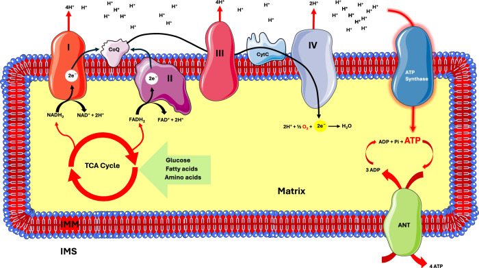
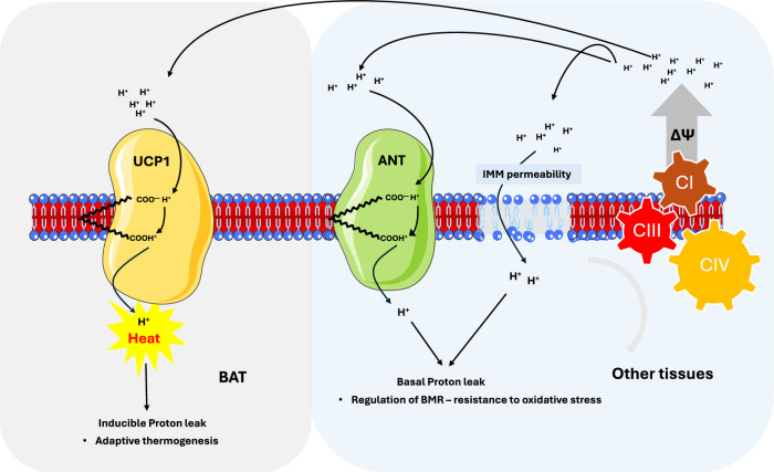
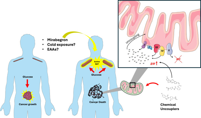
Mitochondria play a central role in regulating cellular energy metabolism, redox homeostasis, and biosynthesis. Mitochondrial uncoupling, through the alteration in the permeability of the inner mitochondrial membrane (IMM) to the leak of protons without adenosine triphosphate (ATP) synthesis, regulates thermogenesis, glucose and lipid metabolism, and reactive oxygen species (ROS) generation. In brown adipose tissue (BAT), proton leak via uncoupling protein 1 (UCP1) is essential for thermogenesis and has been shown to improve systemic glucose homeostasis, and recent studies indicate that BAT activation can also suppress tumor growth by competing with cancer cells for glucose. Several small-molecule mitochondrial uncouplers have demonstrated anticancer effects in preclinical models, although endogenous UCPs—particularly UCP2—are often upregulated in tumors, where they may support tumor growth by buffering ROS and increasing metabolic flexibility. These seemingly contradictory observations highlight the context-dependent effects of mitochondrial uncoupling in cancer. Here, we review current understanding of mitochondrial uncoupling mechanisms, the roles of UCP isoforms, and the metabolic interplay between BAT, cancer cells, and the tumor microenvironment, with a focus on therapeutic implications.
Overview
The claims and statistics of this paper haven’t been checked yet.
Run the check — free, no account needed. It reads the open-access text and checks the sources it cites.
Integrity signals
Registry
Cites
Statistics
Figures
Verification
Claims the paper makes
The claims and statistics of this paper haven’t been checked yet.
Run the check — free, no account needed. It reads the open-access text and checks the sources it cites.
Claims other papers make about this one
Each is checked against this paper’s own text: does it say what the citing paper says it does?
In the citing paper
“Regarding the relationship between mitochondrial uncoupling and its role in cancer, it has been suggested that it is a complex one, resulting in the promotion of cancer cell survival in some cases and limiting tumour progression in others [56].”
From Maceration, Soxhlet Extraction, and Steam Hydrodistillation of Anethum graveolens Seeds: Phytochemical Profiles and Cytotoxic Effects (2026) · cites this paper as [56]
Self-check
Percentages, sums and reported differences re-computed from the paper’s own numbers.
The self-check needs the paper’s full text.
Citations
Cites
A role for brown adipose tissue in diet-induced thermogenesis
Rothwell, N. J., & Stock, M. J. (1979) · Nature · doi:10.1038/281031a0 · Cited by 1,606
Superoxide activates mitochondrial uncoupling proteins
Echtay, K. S., Roussel, D., St-Pierre, J., Jekabsons, M. B., Cadenas, S., Stuart, J. A., Harper, J. A., Roebuck, S. J., Morrison, A., Pickering, S., et al. (2002) · Nature · doi:10.1038/415096a · Cited by 1,382
Roberts, L. D., Boström, P., O’Sullivan, J. F., Schinzel, R. T., Lewis, G. D., Dejam, A., Lee, Y.-K., Palma, M. J., Calhoun, S., Georgiadi, A., et al. (2014) · Cell Metabolism · doi:10.1016/j.cmet.2013.12.003 · Cited by 643
Nedergaard, J., Golozoubova, V., Matthias, A., Asadi, A., Jacobsson, A., & Cannon, B. (2001) · Biochimica et Biophysica Acta (BBA) - Bioenergetics · doi:10.1016/s0005-2728(00)00247-4 · Cited by 615
Mapping of human brown adipose tissue in lean and obese young men
Leitner, B. P., Huang, S., Brychta, R. J., Duckworth, C. J., Baskin, A. S., McGehee, S., Tal, I., Dieckmann, W., Gupta, G., Kolodny, G. M., et al. (2017) · Proceedings of the National Academy of Sciences · doi:10.1073/pnas.1705287114 · Cited by 530
The basal proton conductance of mitochondria depends on adenine nucleotide translocase content
Brand, M. D., Pakay, J. L., Ocloo, A., Kokoszka, J., Wallace, D. C., Brookes, P. S., & Cornwall, E. J. (2005) · Biochemical Journal · doi:10.1042/bj20050890 · Cited by 410
Mitochondria as biosynthetic factories for cancer proliferation
Ahn, C. S., & Metallo, C. M. (2015) · Cancer & Metabolism · doi:10.1186/s40170-015-0128-2 · Cited by 399
Gluttony and thermogenesis revisited
Stock, M. J. (1999) · International Journal of Obesity · doi:10.1038/sj.ijo.0801108 · Cited by 242 · cited 1×
“Instead, these examples suggest a specialized role for BAT in DIT under conditions of nutrient imbalance or protein scarcity 73,74 .”
Iron and Reactive Oxygen Species: Friends or Foes of Cancer Cells?
Bystrom, L. M., Guzman, M. L., & Rivella, S. (2014) · Antioxidants & Redox Signaling · doi:10.1089/ars.2012.5014 · Cited by 212
Postprandial Oxidative Metabolism of Human Brown Fat Indicates Thermogenesis
U Din, M., Saari, T., Raiko, J., Kudomi, N., Maurer, S. F., Lahesmaa, M., Fromme, T., Amri, E.-Z., Klingenspor, M., Solin, O., et al. (2018) · Cell Metabolism · doi:10.1016/j.cmet.2018.05.020 · Cited by 196
Samudio, I., Fiegl, M., McQueen, T., Clise-Dwyer, K., & Andreeff, M. (2008) · Cancer Research · doi:10.1158/0008-5472.can-08-0555 · Cited by 168
USE OF DINITROPHENOL IN OBESITY AND RELATED CONDITIONS: A PROGRESS REPORT
TAINTER, M. L., STOCKTON, A. B., & CUTTING, W. C. (1933) · Journal of the American Medical Association · doi:10.1001/jama.1933.02740440032009 · Cited by 112
Mitochondrial (‘mild’) uncoupling and ROS production: Physiologically relevant or not?
Shabalina, I. G., & Nedergaard, J. (2011) · Biochemical Society Transactions · doi:10.1042/bst0391305 · Cited by 110
Son, J. S., Zhao, L., Chen, Y., Chen, K., Chae, S. A., de Avila, J. M., Wang, H., Zhu, M.-J., Jiang, Z., & Du, M. (2020) · Science Advances · doi:10.1126/sciadv.aaz0359 · Cited by 101
HARPER, J. A., STUART, J. A., JEKABSONS, M. B., ROUSSEL, D., BRINDLE, K. M., DICKINSON, K., JONES, R. B., & BRAND, M. D. (2002) · Biochemical Journal · doi:10.1042/bj3610049 · Cited by 87
Moreno, M., Lombardi, A., De Lange, P., Silvestri, E., Ragni, M., Lanni, A., & Goglia, F. (2003) · The FASEB Journal · doi:10.1096/fj.02-0839fje · Cited by 42
Self-citationKlumpe, I., Savvatis, K., Westermann, D., Tschöpe, C., Rauch, U., Landmesser, U., Schultheiss, H.-P., & Dörner, A. (2016) · Journal of Molecular Medicine · doi:10.1007/s00109-016-1413-4 · Cited by 35
Singhal, S. S., Figarola, J., Singhal, J., Leake, K., Nagaprashantha, L., Lincoln, C., Gabriel Gugiu, B., Horne, D., Jove, R., Awasthi, S., et al. (2012) · Biochemical Pharmacology · doi:10.1016/j.bcp.2012.08.020 · Cited by 20
Research works printed but not linked: 191
- Hanahan, D. Hallmarks of cancer: new dimensions. Cancer Discov. 12, 31–46 (2022). · DOI · Paper page
- Di Gregorio, E. et al. The distinguishing electrical properties of cancer cells. Phys. Life Rev. 43, 139–188 (2022). · DOI
- Fantin, V. R., St-Pierre, J. & Leder, P. Attenuation of LDH-A expression uncovers a link between glycolysis, mitochondrial physiology, and tumor maintenance. Cancer Cell 9, 425–434 (2006). · DOI · Paper page
- Mitchell, P. Coupling of phosphorylation to electron and hydrogen transfer by a chemi-osmotic type of mechanism. Nature 191, 144–148 (1961). · DOI · Paper page
- Seki, T. et al. Brown-fat-mediated tumour suppression by cold-altered global metabolism. Nature 608, 421–428 (2022). · DOI
- Sun, X. et al. Mirabegron displays anticancer effects by globally browning adipose tissues. Nat. Commun. 14 (2023). · DOI
- Shrestha, R., Johnson, E. & Byrne, F. L. Exploring the therapeutic potential of mitochondrial uncouplers in cancer. Mol. Metab. 51 https://doi.org/10.1016/j.molmet.2021.101222 (2021). · DOI · Paper page
- Rolfe, D. F. S. & Brand, M. D. Contribution of mitochondrial proton leak to skeletal muscle respiration and to standard metabolic rate. Am. J. Physiol.—Cell Physiol. 271 (1996). · DOI
- Divakaruni, A. S. & Brand, M. D. The regulation and physiology of mitochondrial proton leak. Physiol. (Bethesda, Md.) 26, 192–205 (2011).
- Nobes, C. D., Brown, G. C., Olive, P. N. & Brand, M. D. Non-ohmic proton conductance of the mitochondrial inner membrane in hepatocytes. J. Biol. Chem. 265, 12903–12909 (1990). · DOI
- Buttgereit, F., Brand, M. D. & Müller, M. ConA induced changes in energy metabolism of rat thymocytes. Biosci. Rep. 12, 381–386 (1992). · DOI
- Jekabsons, M. B. & Nicholls, D. G. In Situ respiration and bioenergetic status of mitochondria in primary cerebellar granule neuronal cultures exposed continuously to glutamate. J. Biol. Chem. 279, 32989–33000 (2004). · DOI
- Hafner, R. P. & Brand, M. D. Hypothyroidism in rats does not lower mitochondrial ADP/O and H+/O ratios. Biochem. J. 250, 477–484 (1988). · DOI
- Silvestri, E. et al. Age-related changes in renal and hepatic cellular mechanisms associated with variations in rat serum thyroid hormone levels. Am. J. Physiol.—Endocrinol. Metab. 294 (2008). · DOI
- Lombardi, A. et al. 3,5-Diiodo-L-thyronine rapidly enhances mitochondrial fatty acid oxidation rate and thermogenesis in rat skeletal muscle: AMP-activated protein kinase involvement. Am. J. Physiol— Endocrinol. Metab. 296 (2009). · DOI
- Brand, M. D., Couture, P., Else, P. L., Withers, K. W. & Hulbert, A. J. Evolution of energy metabolism. Proton permeability of the inner membrane of liver mitochondria is greater in a mammal than in a reptile. Biochem. J. 275, 81–86 (1991). · DOI
- Brookes, P. S., Buckingham, J. A., Tenreiro, A. M., Hulbert, A. J. & Brand, M. D. The proton permeability of the inner membrane of liver mitochondria from ectothermic and endothermic vertebrates and from obese rats: correlations with standard metabolic rate and phospholipid fatty acid composition. Comp. Biochem. Physiol. B: Biochem. Mol. Biol. 119, 325–334 (1998). · DOI
- Stuart, J. A., Brindle, K. M., Harper, J. A. & Brand, M. D. Mitochondrial proton leak and the uncoupling proteins. J. Bioenerg. Biomembr. 31, 517–524, https://doi.org/10.1023/A:1005456725549 (1999). · DOI
- Pehowich, D. J. Thyroid hormone status and membrane n-3 fatty acid content influence mitochondrial proton leak. Biochim. Biophys. Acta—Bioenerg. 1411, 192–200 (1999). · DOI
- Brookes, P. S., Hulbert, A. J. & Brand, M. D. The proton permeability of liposomes made from mitochondrial inner membrane phospholipids: No effect of fatty acid composition. Biochim. Biophys. Acta—Biomembr. 1330, 157–164 (1997). · DOI
- Onukwufor, J. O., Berry, B. J. & Wojtovich, A. P. Physiologic implications of reactive oxygen species production by mitochondrial complex I reverse electron transport. Antioxidants 8 https://doi.org/10.3390/antiox8080285 (2019). · DOI
- Nicholls, D. G. Mitochondrial proton leaks and uncoupling proteins. Biochim. Biophys. Acta—Bioenergetics 1862 https://doi.org/10.1016/j.bbabio.2021.148428 (2021). · DOI
- Speakman, J. R. et al. Uncoupled and surviving: Individual mice with high metabolism have greater mitochondrial uncoupling and live longer. Aging Cell 3, 87–95 (2004). · DOI · Paper page
- Caldeira Da Silva, C. C., Cerqueira, F. M., Barbosa, L. F., Medeiros, M. H. G. & Kowaltowski, A. J. Mild mitochondrial uncoupling in mice affects energy metabolism, redox balance and longevity. Aging Cell 7, 552–560 (2008). · DOI · Paper page
- Brys, K., Vanfleteren, J. R. & Braeckman, B. P. Testing the rate-of-living/oxidative damage theory of aging in the nematode model Caenorhabditis elegans. Exp. Gerontol. 42, 845–851 (2007). · DOI
- Stöckl, P. et al. Partial uncoupling of oxidative phosphorylation induces premature senescence in human fibroblasts and yeast mother cells. Free Radic. Biol. Med. 43, 947–958 (2007). · DOI
- Samartsev, V. N., Mokhova, E. N. & Skulachev, V. P. The pH-dependent reciprocal changes in contributions of ADP/ATP antiporter and aspartate/glutamate antiporter to the fatty acid-induced uncoupling. FEBS Lett. 412, 179–182 (1997). · DOI
- Simonyan, R. A. & Skulachev, V. P. Thermoregulatory uncoupling in heart muscle mitochondria: Involvement of the ATP/ADP antiporter and uncoupling protein. FEBS Lett. 436, 81–84 (1998). · DOI
- Andreyev, A. Y. et al. The ATP/ADP-antiporter is involved in the uncoupling effect of fatty acids on mitochondria. Eur. J. Biochem. 182, 585–592 (1989). · DOI
- Skulachev, V. P. Fatty acid circuit as a physiological mechanism of uncoupling of oxidative phosphorylation. FEBS Lett. 294, 158–162 (1991). · DOI · Paper page
- Hermesh, O., Kalderon, B. & Bar-Tana, J. Mitochondria uncoupling by a long chain fatty acyl analogue. J. Biol. Chem. 273, 3937–3742 (1998). · DOI
- Parker, N., Affourtit, C., Vidal-Puig, A. & Brand, M. D. Energization-dependent endogenous activation of proton conductance in skeletal muscle mitochondria. Biochem. J. 412, 131–139 (2008). · DOI
- Dümmler, K., Müller, S. & Seitz, H. J. Regulation of adenine nucleotide translocase and glycerol 3-phosphate dehydrogenase expression by thyroid hormones in different rat tissues. Biochem. J. 317, 913–918 (1996). · DOI
- Seitz, H. J., Muller, M. J. & Soboll, S. Rapid thyroid-hormone effect on mitochondrial and cytosolic ATP/ADP ratios in the intact liver cell. Biochem. J. 227, 149–153 (1985). · DOI
- Bertholet, A. M. et al. H+ transport is an integral function of the mitochondrial ADP/ATP carrier. Nature 571, 515–520 (2019). · DOI
- Bertholet, A. M. et al. Mitochondrial uncouplers induce proton leak by activating AAC and UCP1. Nature 606, 180–187 (2022). · DOI
- Shabalina, I. G., Jiménez, B., Sousa-Filho, C. P. B., Cannon, B. & Nedergaard, J. In isolated brown adipose tissue mitochondria, UCP1 is not essential for - nor involved in - the uncoupling effects of the classical uncouplers FCCP and DNP. Biochim. Biophys. Acta—Bioenerg. 1866, 149516 (2025). · DOI
- Morrow, R. M. et al. Mitochondrial energy deficiency leads to hyperproliferation of skeletal muscle mitochondria and enhanced insulin sensitivity. Proc. Natl. Acad. Sci. USA 114, 2705–2710 (2017). · DOI
- Cho, J. et al. Mitochondrial ATP transporter depletion protects mice against liver steatosis and insulin resistance. Nat. Commun. 8 (2017). · DOI
- Moon, J. S. et al. ANT2 drives proinflammatory macrophage activation in obesity. JCI Insight 6 (2021). · DOI
- Walther, T. et al. Accelerated mitochondrial adenosine diphosphate/adenosine triphosphate transport improves hypertension-induced heart disease. Circulation 115, 333–344 (2007). · DOI
- Wang, Y. et al. Myocardial overexpression of adenine nucleotide translocase 1 ameliorates diabetic cardiomyopathy in mice. Exp. Physiol. 94, 220–227 (2009). · DOI
- Bal, N. C. et al. Sarcolipin is a newly identified regulator of muscle-based thermogenesis in mammals. Nat. Med. 18, 1575–1579 (2012). · DOI
- Seale, P. et al. PRDM16 controls a brown fat/skeletal muscle switch. Nature 454, 961–967 (2008). · DOI · Paper page
- Esser, V., Brown, N. F., Cowan, A. T., Foster, D. W. & McGarry, J. D. Expression of a cDNA isolated from rat brown adipose tissue and heart identifies the product as the muscle isoform of carnitine palmitoyltransferase I (M-CPT I): M-CPT I is the predominant CPT I isoform expressed in both white (epididymal) and brown adipo. J. Biol. Chem. 271, 6972–6977 (1996). · DOI
- Himms-Hagen, J. & Harper, M. E. Physiological role of UCP3 may be export of fatty acids from mitochondria when fatty acid oxidation predominates: an hypothesis. Proc. Soc. Exp. Biol. Med. 226, 78–84 (2001). · DOI
- Daikoku, T., Shinohara, Y., Shima, A., Yamazaki, N. & Terada, H. Specific elevation of transcript levels of particular protein subtypes induced in brown adipose tissue by cold exposure. Biochim. Biophys. Acta—Bioenerg. 1457, 263–272 (2000). · DOI
- Boström, P. et al. A PGC1-α-dependent myokine that drives brown-fat-like development of white fat and thermogenesis. Nature 481, 463–468 (2012). · DOI · Paper page
- Lund, J. et al. The anorectic and thermogenic effects of pharmacological lactate in male mice are confounded by treatment osmolarity and co-administered counterions. Nat. Metab. 5, 677–698 (2023). · DOI
- Stanford, K. I. et al. 12,13-diHOME: An Exercise-Induced Lipokine that Increases Skeletal Muscle Fatty Acid Uptake. Cell Metab. 27, 1111–1120.e3 (2018). · DOI
- Ruocco, C. et al. Manipulation of dietary amino acids prevents and reverses obesity in mice through multiple mechanisms that modulate energy homeostasis. Diabetes 69, 2324–2339 (2020). · DOI
- Ragni, M. et al. Dietary essential amino acids for the treatment of heart failure with reduced ejection fraction. Cardiovasc. Res. 119, 982–997 (2023). · DOI
- Ragni, M. et al. An amino acid-defined diet impairs tumour growth in mice by promoting endoplasmic reticulum stress and mTOR inhibition. Mol. Metab. 60, 101478 (2022). · DOI
- Chang, J. C. et al. Adaptive adipose tissue stromal plasticity in response to cold stress and antibody-based metabolic therapy. Sci. Rep. 9 (2019). · DOI
- Bing, C. et al. Hyperphagia in cold-exposed rats is accompanied by decreased plasma leptin but unchanged hypothalamic NPY. Am. J. Physiol.—Regul. Integr. Comp. Physiol. 274 (1998). · DOI
- Holloszy, J. O. & Smith, E. K. Longevity of cold-exposed rats: a reevaluation of the ‘rate-of-living theory’. J. Appl. Physiol. 61, 1656–1660 (1986). · DOI
- Barnett, S. A. Adaptation of Mice To Cold. Biol. Rev. Camb. Philos. Soc. 40, 5–51 (1965). · DOI
- Zhu, P. et al. Cold exposure promotes obesity and impairs glucose homeostasis in mice subjected to a high-fat diet. Mol. Med. Rep. 18, 3923–3931 (2018).
- von Essen, G., Lindsund, E., Cannon, B. & Nedergaard, J. Adaptive facultative diet-induced thermogenesis in wild-type but not in UCP1-ablated mice. Am. J. Physiol.—Endocrinol. Metab. 313, E515–E527 (2017). · DOI
- Kozak, L. P. Brown fat and the myth of diet-induced thermogenesis. Cell Metab. 11, 263–267 (2010). · DOI
- Kleitman, N. Basal metabolism in prolonged fasting in man. Am. J. Physiol. Content 77, 233–244 (1926). · DOI
- Leibel, R. L., Rosenbaum, M. & Hirsch, J. Changes in energy expenditure resulting from altered body weight. N. Engl. J. Med. 332, 621–628 (1995). · DOI · Paper page
- Hamilton, T. S. The heat increments of diets balanced and unbalanced with respect to protein. J. Nutr. 17, 583–599 (1939). · DOI
- Miller, D. S. & Payne, P. R. Weight maintenance and food intake. J. Nutr. 78, 255–262 (1962). · DOI
- Rothwell, N. J., Stock, M. J. & Tyzbir, R. S. Energy balance and mitochondrial function in liver and brown fat of rats fed ‘cafeteria’ diets of varying protein content. J. Nutr. 112, 1663–1672 (1982). · DOI
- Rothwell, N. J. & Stock, M. J. Thermogenic capacity and brown adipose tissue activity in the common marmoset. Compar. Biochem. Physiol. A: Physiol 81, 683–686 (1985). · DOI
- Cramer, W. On glandular adipose tissue, and its relation to other endocrine organs and to the vitamin problem. Br. J. Exp. Pathol. 1, 184 (1920).
- Heaton, J. M. The distribution of brown adipose tissue in the human. J. Anat. 112, 35–39 (1972).
- Huttunen, P., Hirvonen, J. & Kinnula, V. The occurrence of brown adipose tissue in outdoor workers. Eur. J. Appl. Physiol. Occup. Physiol. 46, 339–345 (1981). · DOI
- Cypess, A. M. et al. Identification and importance of brown adipose tissue in adult humans. N. Engl. J. Med. 360, 1509–1517 (2009). · DOI · Paper page
- van Marken Lichtenbelt, W. D. et al. Cold-activated brown adipose tissue in healthy men. N. Engl. J. Med. 360, 1500–1508 (2009). · DOI · Paper page
- Ouellet, V. et al. Outdoor temperature, age, sex, body mass index, and diabetic status determine the prevalence, mass, and glucose-uptake activity of 18F-FDG-detected BAT in humans. J. Clin. Endocrinol. Metab. 96, 192–199 (2011). · DOI
- Brychta, R. J. & Chen, K. Y. Cold-induced thermogenesis in humans. Eur. J. Clin. Nutr. 71, 345–352 (2017). · DOI
- Ouellet, V. et al. Brown adipose tissue oxidative metabolism contributes to energy expenditure during acute cold exposure in humans. J. Clin. Invest. 122, 545–552 (2012). · DOI
- Yoneshiro, T. et al. Brown adipose tissue, whole-body energy expenditure, and thermogenesis in healthy adult men. Obesity 19, 13–16 (2011). · DOI
- Van Der Lans, A. A. J. J. et al. Cold acclimation recruits human brown fat and increases nonshivering thermogenesis. J. Clin. Invest. 123, 3395–3403 (2013). · DOI
- Blondin, D. P. et al. Selective impairment of glucose but not fatty acid or oxidative metabolism in brown adipose tissue of subjects with type 2 diabetes. Diabetes 64, 2388–2397 (2015). · DOI
- Muzik, O. et al. 15O PET measurement of blood flow and oxygen consumption in cold-activated human brown fat. J. Nucl. Med. 54, 523–531 (2013). · DOI
- Chondronikola, M. et al. Brown adipose tissue improves whole-body glucose homeostasis and insulin sensitivity in humans. Diabetes 63, 4089–4099 (2014). · DOI
- Lee, P. et al. Temperature-acclimated brown adipose tissue modulates insulin sensitivity in humans. Diabetes 63, 3686–3698 (2014). · DOI
- Blondin, D. P. et al. Increased brown adipose tissue oxidative capacity in cold-acclimated humans. J. Clin. Endocrinol. Metab. 99 (2014). · DOI
- Hanssen, M. J. W. et al. Short-term cold acclimation improves insulin sensitivity in patients with type 2 diabetes mellitus. Nat. Med. 21, 863–865 (2015). · DOI
- Yoneshiro, T. et al. Recruited brown adipose tissue as an antiobesity agent in humans. J. Clin. Invest. 123, 3404–3408 (2013). · DOI
- Matsushita, M. et al. Impact of brown adipose tissue on body fatness and glucose metabolism in healthy humans. Int. J. Obes. 38, 812–817 (2014). · DOI
- Herz, C. T. et al. Active brown adipose tissue is associated with a healthier metabolic phenotype in obesity. Diabetes 71, 93–103 (2022). · DOI
- Vosselman, M. J. et al. Brown adipose tissue activity after a high-calorie meal in humans. Am. J. Clin. Nutr. 98, 57–64 (2013). · DOI
- Vrieze, A. et al. Fasting and postprandial activity of brown adipose tissue in healthy men. J. Nucl. Med. 53, 1407–1410 (2012). · DOI
- Carpentier, A. C., Blondin, D. P., Haman, F. & Richard, D. Brown adipose tissue—a translational perspective. Endocr. Rev. 44, 143–192 (2023). · DOI
- Hibi, M. et al. Brown adipose tissue is involved in diet-induced thermogenesis and whole-body fat utilization in healthy humans. Int. J. Obes. 40, 1655–1661 (2016). · DOI
- Loeliger, R. C. et al. Relation of diet-induced thermogenesis to brown adipose tissue activity in healthy men. Am. J. Physiol.—Endocrinol. Metab. 320, E93–E101 (2021). · DOI
- Alexander Iwen, K. et al. Cold-induced brown adipose tissue activity alters plasma fatty acids and improves glucose metabolism in men. J. Clin. Endocrinol. Metab. 102, 4226–4234 (2017). · DOI
- O’Mara, A. E. et al. Chronic mirabegron treatment increases human brown fat, HDL cholesterol, and insulin sensitivity. J. Clin. Invest. 130, 2209–2219 (2020). · DOI
- Finlin, B. S. et al. The β3-adrenergic receptor agonist mirabegron improves glucose homeostasis in obese humans. J. Clin. Invest. 130, 2319–2331 (2020). · DOI
- Becher, T. et al. Brown adipose tissue is associated with cardiometabolic health. Nat. Med. 27, 58–65 (2021). · DOI · Paper page
- Wang, Z. et al. Chronic cold exposure enhances glucose oxidation in brown adipose tissue. EMBO Rep. 21 (2020). · DOI
- Albert, V. et al. mTORC 2 sustains thermogenesis via Akt-induced glucose uptake and glycolysis in brown adipose tissue. EMBO Mol. Med. 8, 232–246 (2016). · DOI
- Jeong, J. H., Chang, J. S. & Jo, Y. H. Intracellular glycolysis in brown adipose tissue is essential for optogenetically induced nonshivering thermogenesis in mice. Sci. Rep. 8 (2018). · DOI
- Bornstein, M. R. et al. Comprehensive quantification of metabolic flux during acute cold stress in mice. Cell Metab. 35, 2077–2092.e6 (2023). · DOI
- Cori, C. F. & Cori, G. T. The carbohydrate metabolism of tumors. II. Changes in the sugar, lactic acid, and co-combining power of blood passing through a tumor. J. Biol. Chem. 65, 397–405 (1925). · DOI
- Chen, R. et al. Effective prevention and treatment of acute leukemias in mice by activation of thermogenic adipose tissues. Adv. Sci. https://doi.org/10.1002/advs.202402332 (2024) · DOI
- Pfannenberg, C. et al. Impact of age on the relationships of brown adipose tissue with sex and adiposity in humans. Diabetes 59, 1789–1793 (2010). · DOI
- Olsen, J. M. et al. 3-Adrenergically induced glucose uptake in brown adipose tissue is independent of UCP1 presence or activity: Mediation through the mTOR pathway. Mol. Metab. 6, 611–619 (2017). · DOI
- Inokuma, K. I. et al. Uncoupling protein 1 is necessary for norepinephrine-induced glucose utilization in brown adipose tissue. Diabetes 54, 1385–1391 (2005). · DOI
- Heiden, M. G. V., Cantley, L. C. & Thompson, C. B. Understanding the warburg effect: The metabolic requirements of cell proliferation. Science 324, 1029–1033 (2009). · DOI · Paper page
- Ragni, M., Fornelli, C., Nisoli, E. & Penna, F. Amino acids in cancer and cachexia: an integrated view. Cancers 14, 5691 (2022). · DOI
- Harper, M. et al. Characterization of a novel metabolic strategy used by drug-resistant tumor cells. FASEB J. 16, 1550–1557 (2002). · DOI
- Zhau, H. E. et al. Human prostate cancer harbors the stem cell properties of bone marrow mesenchymal stem cells. Clin. Cancer Res. 17, 2159–2169 (2011). · DOI
- Giatromanolaki, A. et al. Thermogenic protein UCP1 and UCP3 expression in nonsmall cell lung cancer: relation with glycolysis and anaerobic metabolism. Cancer Biol. Med. 14, 396–404 (2017). · DOI
- Xia, J. et al. Mitochondrial protein UCP1 inhibits the malignant behaviors of triple-negative breast cancer through activation of mitophagy and pyroptosis. Int. J. Biol. Sci. 18, 2949–2961 (2022). · DOI
- Kawashima, M. et al. Disruption of hypoxia-inducible fatty acid binding protein 7 induces beige fat-like differentiation and thermogenesis in breast cancer cells. Cancer Metab. 8 (2020). · DOI
- Wu, Y., Li, X., Li, Q., Cheng, C. & Zheng, L. Adipose tissue-to-breast cancer crosstalk: comprehensive insights. Biochim. Biophys. Acta—Rev. Cancer 1877 https://doi.org/10.1016/j.bbcan.2022.188800 (2022). · DOI
- Morroni, M. et al. Reversible transdifferentiation of secretory epithelial cells into adipocytes in the mammary gland. Proc. Natl. Acad. Sci. USA 101, 16801–16806 (2004). · DOI
- Giordano, A., Smorlesi, A., Frontini, A., Barbatelli, G. & Cint, S. White, brown and pink adipocytes: the extraordinary plasticity of the adipose organ. Eur. J. Endocrinol. 170 https://doi.org/10.1530/EJE-13-0945 (2014). · DOI
- Ricquier, D. Uncoupling protein 1 of brown adipocytes, the only uncoupler: a historical perspective. Front. Endocrinol. 2 https://doi.org/10.3389/fendo.2011.00085 (2011). · DOI
- Sreedhar, A., Petruska, P., Miriyala, S., Panchatcharam, M. & Zhao, Y. UCP2 overexpression enhanced glycolysis via activation of PFKFB2 during skin cell transformation. Oncotarget 8, 95504–95515 (2017). · DOI
- Nègre-Salvayre, A. et al. A role for uncoupling protein-2 as a regulator of mitochondrial hydrogen peroxide generation. FASEB J. 11, 809–815 (1997). · DOI
- Arsenijevic, D. et al. Disruption of the uncoupling protein-2 gene in mice reveals a role in immunity and reactive oxygen species production. Nat. Genet. 26, 435–439 (2000). · DOI
- Derdak, Z. et al. The mitochondrial uncoupling protein-2 promotes chemoresistance in cancer cells. Cancer Res. 68, 2813–2819 (2008). · DOI · Paper page
- Sreedhar, A. et al. UCP2 overexpression redirects glucose into anabolic metabolic pathways. Proteomics 19 (2019). · DOI
- Vozza, A. et al. UCP2 transports C4 metabolites out of mitochondria, regulating glucose and glutamine oxidation. Proc. Natl. Acad. Sci. USA111, 960–965 (2014). · DOI · Paper page
- Raho, S. et al. KRAS-regulated glutamine metabolism requires UCP2-mediated aspartate transport to support pancreatic cancer growth. Nat. Metab. 2, 1373–1381 (2020). · DOI
- Cheng, W. C. et al. Uncoupling protein 2 reprograms the tumor microenvironment to support the anti-tumor immune cycle. Nat. Immunol. 20, 206–217 (2019). · DOI
- Esteves, P. et al. Mitochondrial retrograde signaling mediated by UCP2 inhibits cancer cell proliferation and tumorigenesis. Cancer Res. 74, 3971–3982 (2014). · DOI
- Derdák, Z. et al. Enhanced colon tumor induction in uncoupling protein-2 deficient mice is associated with NF-κB activation and oxidative stress. Carcinogenesis https://doi.org/10.1093/carcin/bgi335 (2006) · DOI
- Pecqueur, C. et al. Uncoupling protein 2, in vivo distribution, induction upon oxidative stress, and evidence for translational regulation. J. Biol. Chem. 276, 8705–8712 (2001). · DOI
- Stuart, J. A., Harper, J. A., Brindle, K. M., Jekabsons, M. B. & Brand, M. D. A mitochondrial uncoupling artifact can be caused by expression of uncoupling protein 1 in yeast. Biochem. J. 356, 779–789 (2001). · DOI
- Nedergaard, J. & Cannon, B. The ‘novel’ ‘uncoupling’ UCP2 and UCP3: What do they really do? Pros and cons for suggested functions. Exp. Physiol. 88, 65–84 (2003). · DOI
- Han, Z. et al. Niclosamide induces cell cycle arrest in G1 phase in head and neck squamous cell carcinoma through let-7d/Cdc34 axis. Front. Pharmacol. 9 (2019). · DOI
- Satoh, K. et al. Identification of niclosamide as a novel anticancer agent for adrenocortical carcinoma. Clin. Cancer Res. 22, 3458–3466 (2016). · DOI
- Wei, W., Liu, H., Yuan, J. & Yao, Y. Targeting Wnt/β-catenin by anthelmintic drug niclosamide overcomes paclitaxel resistance in esophageal cancer. Fundam. Clin. Pharmacol. 35, 165–173 (2021). · DOI
- Senkowski, W. et al. Three-dimensional cell culture-based screening identifies the anthelmintic drug nitazoxanide as a candidate for treatment of colorectal cancer. Mol. Cancer Ther. 14, 1504–1516 (2015). · DOI
- Wang, X. et al. Nitazoxanide, an antiprotozoal drug, inhibits late-stage autophagy and promotes ING1-induced cell cycle arrest in glioblastoma. Cell Death Dis. 9 (2018). · DOI
- Fan-Minogue, H. et al. A c-Myc activation sensor-based high-throughput drug screening identifies an antineoplastic effect of nitazoxanide. Mol. Cancer Ther. 12, 1896–1905 (2013). · DOI
- Zhang, W. et al. An Underlying Mechanism of Dual Wnt Inhibition and AMPK Activation: Mitochondrial Uncouplers Masquerading as Wnt Inhibitors. J. Med. Chem. 62, 11348–11358 (2019). · DOI
- Turcios, L. et al. Autophagic flux modulation by Wnt/β-catenin pathway inhibition in hepatocellular carcinoma. PLoS ONE 14 (2019). · DOI
- Chen, Y. et al. FH535 inhibits proliferation and motility of colon cancer cells by targeting Wnt/ß-catenin signaling pathway. J. Cancer 8, 3142–3153 (2017). · DOI
- Singhal, S. S. et al. Novel compound 1,3-bis (3,5-dichlorophenyl) urea inhibits lung cancer progression. Biochem. Pharmacol. 86, 1664–1672 (2013). · DOI · Paper page
- Forrest, M. D. Why cancer cells have a more hyperpolarised mitochondrial membrane potential and emergent prospects for therapy. bioRxiv 1–42. https://doi.org/10.1101/025197 (2015). · DOI
- Summerhayes, I. C. et al. Unusual retention of rhodamine 123 by mitochondria in muscle and carcinoma cells. Proc. Natl. Acad. Sci. USA 79, 5292–5296 (1982). · DOI
- Bernal, S. D., Lampidis, T. J., Summerhayes, I. C. & Chen, L. B. Rhodamine-123 selectively reduces clonal growth of carcinoma cells in vitro. Science 218, 1117–1119 (1982). · DOI
- Argilés, J. M., Busquets, S., Stemmler, B. & López-Soriano, F. J. Cancer cachexia: understanding the molecular basis. Nat. Rev. Cancer 14, 754–762 (2014). · DOI
- Brooks, S. L., Neville, A. M., Rothwell, N. J., Stock, M. J. & Wilson, S. Sympathetic activation of brown-adipose-tissue thermogenesis in cachexia. Biosci. Rep. 1, 509–517 (1981). · DOI
- Tsoli, M. et al. Activation of thermogenesis in brown adipose tissue and dysregulated lipid metabolism associated with cancer cachexia in mice. Cancer Res. 72, 4372–4382 (2012). · DOI
- Petruzzelli, M. et al. A switch from white to brown fat increases energy expenditure in cancer-associated cachexia. Cell Metab. 20, 433–447 (2014). · DOI
- Bianchi, A. et al. Increased brown adipose tissue activity in children with malignant disease. Horm. Metab. Res. 21, 640–641 (1989). · DOI
- Shellock, F. G., Riedinger, M. S. & Fishbein, M. C. Brown adipose tissue in cancer patients: Possible cause of cancer-induced cachexia. J. Cancer Res. Clin. Oncol. 111, 82–85 (1986). · DOI
- Rousseau, C. et al. Brown fat in breast cancer patients: Analysis of serial 18F-FDG PET/CT scans. Eur. J. Nucl. Med. Mol. Imaging 33, 785–791 (2006). · DOI
- Gilsanz, V. et al. The depiction of brown adipose tissue is related to disease status in pediatric patients with lymphoma. Am. J. Roentgenol. 198, 909–913 (2012). · DOI
- Lee, P., Greenfield, J. R., Ho, K. K. Y. & Fulham, M. J. A critical appraisal of the prevalence and metabolic significance of brown adipose tissue in adult humans. Am. J. Physiol.—Endocrinol. Metab. 299 (2010). · DOI
- Bos, S. A., Gill, C. M., Martinez-Salazar, E. L., Torriani, M. & Bredella, M. A. Preliminary investigation of brown adipose tissue assessed by PET/CT and cancer activity. Skelet. Radiol. 48, 413–419 (2019). · DOI
- Eljalby, M. et al. Brown adipose tissue is not associated with cachexia or increased mortality in a retrospective study of patients with cancer. Am. J. Physiol. Endocrinol. Metab. 324, E144–E153 (2023). · DOI
- Becker, A. S. et al. Brown fat does not cause cachexia in cancer patients: a large retrospective longitudinal FDG-PET/CT cohort study. PLoS ONE 15 (2020). · DOI
- Panagiotou, G. et al. Brown adipose tissue is associated with reduced weight loss and risk of cancer cachexia: A retrospective cohort study. Clin. Nutr. 45, 262–269 (2025). · DOI
- Beijer, E. et al. A role of active brown adipose tissue in cancer cachexia? Oncol. Rev. 6, 88–94 (2012). · DOI
- Sadeghi, M. et al. Cancer cachexia: diagnosis, assessment, and treatment. Crit. Rev. Oncol. Hematol. 127, 91–104 (2018). · DOI
- John, D. et al. Ponsegromab for the Treatment of Cancer Cachexia. N. Engl. J. Med. 391, 2291–2303 (2024). · DOI
- Watanabe, H. & Oshima, T. The latest treatments for cancer cachexia: an overview. Anticancer Res. 43, 511–521 (2023). · DOI
- Zhang, F. et al. UCP1 regulates ALDH-positive breast cancer stem cells through releasing the suppression of Snail on FBP1. Cell Biol. Toxicol. 37, 277–291 (2021). · DOI
- Alnabulsi, A. et al. The expression of brown fat-associated proteins in colorectal cancer and the relationship of uncoupling protein 1 with prognosis. Int. J. Cancer 145, 1138–1147 (2019). · DOI
- Zhao, J. et al. Myeloid-derived suppressor cells in the tumor microenvironment reduce uncoupling protein 1 expression to boost immunosuppressive activity. Biochem. Biophys. Res. Commun. 732, 150408 (2024). · DOI
- Xiong, Z. et al. Tumor cell “slimming” regulates tumor progression through PLCL1/UCP1-mediated lipid browning. Adv. Sci. 6 (2019). · DOI
- Yu, X. et al. Identification of UCP1 and UCP2 as potential prognostic markers in breast cancer: a study based on immunohistochemical analysis and bioinformatics. Front. Cell Dev. Biol. 10 (2022). · DOI
- Horimoto, M. et al. Expression of uncoupling protein-2 in human colon cancer. Clin. Cancer Res. 10, 6203–6207 (2004). · DOI
- Kuai, X. Y., Ji, Z. Y. & Zhang, H. J. Mitochondrial uncoupling protein 2 expression in colon cancer and its clinical significance. World J. Gastroenterol. 16, 5773–5778 (2010). · DOI
- Sayeed, A. et al. Negative regulation of UCP2 by TGFβ signaling characterizes low and intermediate-grade primary breast cancer. Cell Death Dis. 1, 53 (2010). · DOI
- Ayyasamy, V. et al. Cellular model of Warburg Effect identifies tumor promoting function of UCP2 in breast cancer and its suppression by genipin. PLoS ONE 6 (2011). · DOI
- Li, W., Nichols, K., Nathan, C. A. & Zhao, Y. Mitochondrial uncoupling protein 2 is up-regulated in human head and neck, skin, pancreatic, and prostate tumors. Cancer Biomark. 13, 377–383 (2013). · DOI
- Alvero, A. B. et al. Multiple blocks in the engagement of oxidative phosphorylation in putative ovarian cancer stem cells: Implication for maintenance therapy with glycolysis inhibitors. Oncotarget 5, 8703–8715 (2014). · DOI
- Lim, H. Y. et al. Metabolic signatures of renal cell carcinoma. Biochem. Biophys. Res. Commun. 460, 938–943 (2015). · DOI
- Imai, K. et al. UCP2 expression may represent a predictive marker of neoadjuvant chemotherapy effectiveness for locally advanced uterine cervical cancer. Oncol. Lett. 14, 951–957 (2017). · DOI
- Kawanishi, M. et al. Expression of UCP2 is associated with sensitivity to platinum-based chemotherapy for ovarian serous carcinoma. Oncol. Lett. 15, 9923–9928 (2018).
- Aguilar, E. et al. UCP2 deficiency increases colon tumorigenesis by promoting lipid synthesis and depleting NADPH for antioxidant defenses. Cell Rep. 28, 2306–2316.e5 (2019). · DOI
- Yu, J., Shi, L., Lin, W., Lu, B. & Zhao, Y. UCP2 promotes proliferation and chemoresistance through regulating the NF-κB/β-catenin axis and mitochondrial ROS in gallbladder cancer. Biochem. Pharmacol. 172 (2020). · DOI
- Wang, A. et al. Role and mechanism of FLNa and UCP2 in the development of cervical cancer. Oncol. Rep. 44, 2656–2668 (2020). · DOI
- Braun, N. et al. UCP-3 uncoupling protein confers hypoxia resistance to renal epithelial cells and is upregulated in renal cell carcinoma. Sci. Rep. 5 (2015). · DOI
- Kazak, L. et al. A creatine-driven substrate cycle enhances energy expenditure and thermogenesis in beige fat. Cell 163, 643–655 (2015). · DOI · Paper page
- Rahbani, J. F. et al. Creatine kinase B controls futile creatine cycling in thermogenic fat. Nature 590, 480–485 (2021). · DOI
- Sun, Y. et al. Mitochondrial TNAP controls thermogenesis by hydrolysis of phosphocreatine. Nature 593, 580–585 (2021). · DOI
- Kazak, L. et al. Genetic depletion of adipocyte creatine metabolism inhibits diet-induced thermogenesis and drives obesity. Cell Metab. 26, 660–671.e3 (2017). · DOI
- Nicholls, D. G. & Brand, M. D. A critical assessment of the role of creatine in brown adipose tissue thermogenesis. Nat. Metab. 5, 21–28 (2023). · DOI
- Bunk, J. et al. The Futile Creatine Cycle powers UCP1-independent thermogenesis in classical BAT. Nat. Commun. 16 (2025). · DOI
- Ikeda, K. et al. UCP1-independent signaling involving SERCA2bmediated calcium cycling regulates beige fat thermogenesis and systemic glucose homeostasis. Nat. Med. 23, 1454–1465 (2017). · DOI
- Vaughan, M. The production and release of glycerol by adipose tissue incubated in vitro. J. Biol. Chem. 237, 3354–3358 (1962). · DOI
- Jensen, M. D., Ekberg, K. & Landau, B. R. Lipid metabolism during fasting. Am. J. Physiol.—Endocrinol. Metab. 281 (2001). · DOI
- Newsholme, E. A. & Crabtree, B. Substrate cycles in metabolic regulation and in heat generation. Biochem. Soc. Symp.41, 61–109 (1976).
- Brownstein, A. J., Veliova, M., Acin-Perez, R., Liesa, M. & Shirihai, O. S. ATP-consuming futile cycles as energy dissipating mechanisms to counteract obesity. Rev. Endocr. Metab. Disord. 23, 121–131 (2022). · DOI
- Yu, X. X., Lewin, D. A., Forrest, W. & Adams, S. H. Cold elicits the simultaneous induction of fatty acid synthesis and β-oxidation in murine brown adipose tissue: prediction from differential gene expression and confirmation in vivo. FASEB J. 16, 155–168 (2002). · DOI
- Trayhurn, P. Fatty acid synthesis in mouse brown adipose tissue the influence of environmental temperature on the proportion of whole-body fatty acid synthesis in brown adipose tissue and the liver. Biochim. Biophys. Acta (BBA)/Lipids Lipid Metab. 664, 549–560 (1981). · DOI
- Brooks, B. J., Arch, J. R. S. & Newsholme, E. A. Effect of some hormones on the rate of the triacylglycerol/fatty-acid substrate cycle in adipose tissue of the mouse in vivo. Biosci. Rep. 3, 263–267 (1983). · DOI
- Flachs, P. et al. Induction of lipogenesis in white fat during cold exposure in mice: Link to lean phenotype. Int. J. Obes. 41, 372–380 (2017). · DOI
- Guan, H. P. et al. A futile metabolic cycle activated in adipocytes by antidiabetic agents. Nat. Med. 8, 1122–1128 (2002). · DOI
Cited by
Goldiș, C., Racoviceanu, R., Negrea-Ghiulai, R., Mioc, A., Prodea, A., Atyim, E., Maksimovic, T., Lukinich-Gruia, A. T., Pricop, M.-A., & Șoica, C. (2026) · Molecules · doi:10.3390/molecules31183337
Metadata
- Authors
- Ragni, M., Ruocco, C., & Nisoli, E.
- Venue
- npj Metabolic Health and Disease
- Published
- 2025-09-22
- Volume
- 3
- Issue
- 1
- ISSN
- 2948-2828
- PMC
- PMC12454651
- Cited by
- 16
- References
- 18
- Language
- en
- Funders
- National Recovery and Resilience Plan (SOE_0000181)
- PRIN (20205X4C9E)
About these checks
Registry, retraction, Cites and statistics results are computed, not judged, with one exception: a printed reference that no index lists is read once by a model before it counts as a citation error. Claims are extracted by a model and kept only when their quote appears verbatim in the text; each cited source is then checked, with a model’s help, for whether it says what the claim relies on. Those verdicts can be wrong — read the claim’s quote and the cited source before relying on one.
Statistics and arithmetic are recomputed, not judged.
Confidence bands are shown only for fields calibrated on our annotated test set. None is calibrated yet, so no band is shown.
What counts as a claim ›How we measure confidence ›How we check this ›
Are you an author, or do you see a problem with this page? Request a review
Data sources
Bibliographic data: Semantic Scholar, OpenAlex, Crossref.
Contains information from the Semantic Scholar Academic Graph Datasets, made available under the ODC Attribution License (ODC-BY 1.0).
Semantic Scholar’s data is described in: Kinney, R., et al. (2023). The Semantic Scholar Open Data Platform. arXiv:2301.10140.
