

The DOI matches its Crossref record; 12 of the 12 references it cites were found in a registry or an index.
The miRNA–target interactions: An underestimated intricacy
Abstract
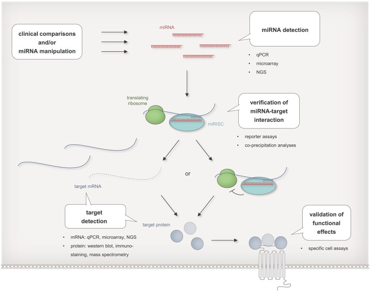
MicroRNAs (miRNAs) play indispensable roles in posttranscriptional gene regulation. Their cellular regulatory impact is determined not solely by their sheer number, which likely amounts to >2000 individual miRNAs in human, than by the regulatory effectiveness of single miRNAs. Although, one begins to develop an understanding of the complex mechanisms underlying miRNA–target interactions (MTIs), the overall knowledge of MTI functionality is still rather patchy. In this critical review, we summarize key features of mammalian MTIs. We especially highlight latest insights on (i) the dynamic make-up of miRNA binding sites including non-canonical binding sites, (ii) the cooperativity between miRNA binding sites, (iii) the adaptivity of MTIs through sequence modifications, (iv) the bearing of intra-cellular miRNA localization changes and (v) the role of cell type and cell status specific miRNA interaction partners. The MTI biology is discussed against the background of state-of-the-art approaches with particular emphasis on experimental strategies for evaluating miRNA functionality.
Overview
The claims and statistics of this paper haven’t been checked yet.
Run the check — free, no account needed. It reads the open-access text and checks the sources it cites.
Integrity signals
Registry
Cites
Statistics
Figures
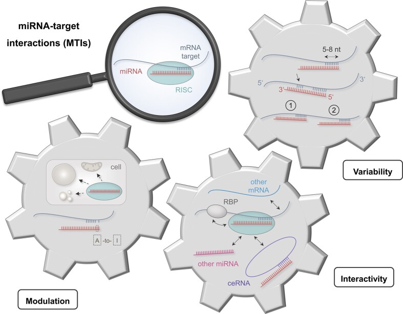
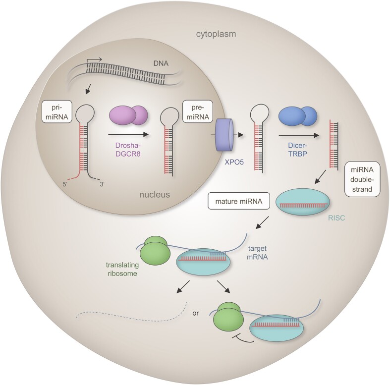
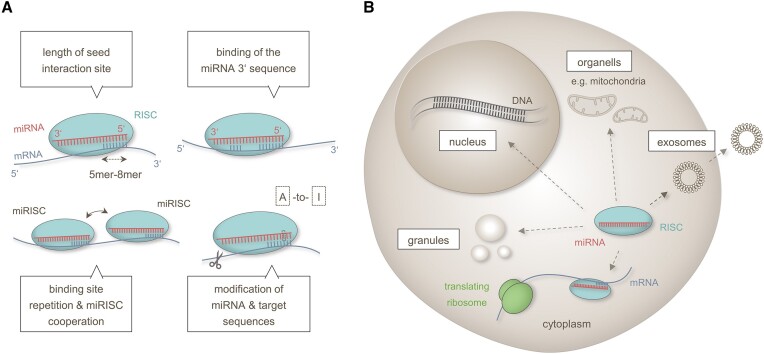
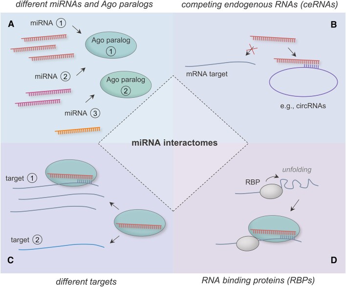
Verification
Claims the paper makes
The claims and statistics of this paper haven’t been checked yet.
Run the check — free, no account needed. It reads the open-access text and checks the sources it cites.
Claims other papers make about this one
Each is checked against this paper’s own text: does it say what the citing paper says it does?
The cited source says what the claim attributes to it.
In the citing paper
“Another fundamental principle of miRNA biology is that miRNAs may not uniformly or simultaneously repress all their targets. Instead, most miRNAs prioritize their targets depending on the cellular context, the presence or absence of additional triggers, and the abundance of both other miRNAs and mRNA transcripts [12, 113, 114].”
From microRNA-132 attenuates inflammation in induced pluripotent stem cell-derived microglia from Alzheimer’s disease patients (2026) · cites this paper as [114]
Self-check
Percentages, sums and reported differences re-computed from the paper’s own numbers.
The self-check needs the paper’s full text.
Citations
Cites
The C. elegans heterochronic gene lin-4 encodes small RNAs with antisense complementarity to lin-14
Lee, R. C., Feinbaum, R. L., & Ambros, V. (1993) · Cell · doi:10.1016/0092-8674(93)90529-y · Cited by 12,948
Most mammalian mRNAs are conserved targets of microRNAs
Friedman, R. C., Farh, K. K.-H., Burge, C. B., & Bartel, D. P. (2009) · Genome Research · doi:10.1101/gr.082701.108 · Cited by 8,462
Natural RNA circles function as efficient microRNA sponges
Hansen, T. B., Jensen, T. I., Clausen, B. H., Bramsen, J. B., Finsen, B., Damgaard, C. K., & Kjems, J. (2013) · Nature · doi:10.1038/nature11993 · Cited by 7,985
Global quantification of mammalian gene expression control
Schwanhäusser, B., Busse, D., Li, N., Dittmar, G., Schuchhardt, J., Wolf, J., Chen, W., & Selbach, M. (2011) · Nature · doi:10.1038/nature10098 · Cited by 6,566 · cited 1×
“The same applies to miRNA targets, which can be measured as mRNAs or proteins, each with their specific cellular half-lives ( 140 ,141 ).”
BackgroundThe multilayered complexity of ceRNA crosstalk and competition
Tay, Y., Rinn, J., & Pandolfi, P. P. (2014) · Nature · doi:10.1038/nature12986 · Cited by 3,910
Widespread changes in protein synthesis induced by microRNAs
Selbach, M., Schwanhäusser, B., Thierfelder, N., Fang, Z., Khanin, R., & Rajewsky, N. (2008) · Nature · doi:10.1038/nature07228 · Cited by 3,429 · cited 1×
“MiRNA regulations usually result in rather moderate effects on the expression levels of single targets ( 121 ).”
BackgroundAn integrated expression atlas of miRNAs and their promoters in human and mouse
The FANTOM Consortium, de Rie, D., Abugessaisa, I., Alam, T., Arner, E., Arner, P., Ashoor, H., Åström, G., Babina, M., Bertin, N., et al. (2017) · Nature Biotechnology · doi:10.1038/nbt.3947 · Cited by 561
Pairing beyond the Seed Supports MicroRNA Targeting Specificity
Broughton, J. P., Lovci, M. T., Huang, J. L., Yeo, G. W., & Pasquinelli, A. E. (2016) · Molecular Cell · doi:10.1016/j.molcel.2016.09.004 · Cited by 487
Denzler, R., McGeary, S. E., Title, A. C., Agarwal, V., Bartel, D. P., & Stoffel, M. (2016) · Molecular Cell · doi:10.1016/j.molcel.2016.09.027 · Cited by 340
Moore, M. J., Scheel, T. K. H., Luna, J. M., Park, C. Y., Fak, J. J., Nishiuchi, E., Rice, C. M., & Darnell, R. B. (2015) · Nature Communications · doi:10.1038/ncomms9864 · Cited by 326
Cross-linking, ligation, and sequencing of hybrids reveals RNA–RNA interactions in yeast
Kudla, G., Granneman, S., Hahn, D., Beggs, J. D., & Tollervey, D. (2011) · Proceedings of the National Academy of Sciences · doi:10.1073/pnas.1017386108 · Cited by 308
Hoffman, Y., Bublik, D. R., P. Ugalde, A., Elkon, R., Biniashvili, T., Agami, R., Oren, M., & Pilpel, Y. (2016) · PLOS Genetics · doi:10.1371/journal.pgen.1005879 · Cited by 90
Research works printed but not linked: 168
- Diener, 2022, Emerging concepts of miRNA therapeutics: from cells to clinic, Trends Genet., 38, 613, 10.1016/j.tig.2022.02.006 · DOI
- Fehlmann, 2020, Common diseases alter the physiological age-related blood microRNA profile, Nat. Commun., 11, 5958, 10.1038/s41467-020-19665-1 · DOI
- Brancati, 2018, An interplay of miRNA abundance and target site architecture determines miRNA activity and specificity, Nucleic Acids Res., 46, 3259, 10.1093/nar/gky201 · DOI
- Turunen, 2019, Changes in nuclear and cytoplasmic microRNA distribution in response to hypoxic stress, Sci. Rep., 9, 10332, 10.1038/s41598-019-46841-1 · DOI
- Bartel, 2004, MicroRNAs: genomics, biogenesis, mechanism, and function, Cell, 116, 281, 10.1016/S0092-8674(04)00045-5 · DOI
- Leitao, 2022, A structural view of miRNA biogenesis and function, Noncoding RNA, 8, 10
- Gregory, 2005, Human RISC couples microRNA biogenesis and posttranscriptional gene silencing, Cell, 123, 631, 10.1016/j.cell.2005.10.022 · DOI · Paper page
- Lewis, 2005, Conserved seed pairing, often flanked by adenosines, indicates that thousands of human genes are microRNA targets, Cell, 120, 15, 10.1016/j.cell.2004.12.035 · DOI · Paper page
- Gu, 2009, Biological basis for restriction of microRNA targets to the 3′ untranslated region in mammalian mRNAs, Nat. Struct. Mol. Biol., 16, 144, 10.1038/nsmb.1552 · DOI
- Lytle, 2007, Target mRNAs are repressed as efficiently by microRNA-binding sites in the 5′ UTR as in the 3′ UTR, Proc. Natl. Acad. Sci. U.S.A., 104, 9667, 10.1073/pnas.0703820104 · DOI
- Wu, 2006, MicroRNAs direct rapid deadenylation of mRNA, Proc. Natl. Acad. Sci. U.S.A., 103, 4034, 10.1073/pnas.0510928103 · DOI
- Behm-Ansmant, 2006, mRNA degradation by miRNAs and GW182 requires both CCR4:NOT deadenylase and DCP1:DCP2 decapping complexes, Genes Dev., 20, 1885, 10.1101/gad.1424106 · DOI
- Eichhorn, 2014, mRNA destabilization is the dominant effect of mammalian microRNAs by the time substantial repression ensues, Mol. Cell, 56, 104, 10.1016/j.molcel.2014.08.028 · DOI · Paper page
- Oliveto, 2017, Role of microRNAs in translation regulation and cancer, World J. Biol. Chem., 8, 45, 10.4331/wjbc.v8.i1.45 · DOI
- Vasudevan, 2007, Switching from repression to activation: microRNAs can up-regulate translation, Science, 318, 1931, 10.1126/science.1149460 · DOI
- Ni, 2016, miRNA-dependent activation of mRNA translation, Microrna, 5, 83, 10.2174/2211536605666160825151201 · DOI
- Ludwig, 2017, Bias in recent miRBase annotations potentially associated with RNA quality issues, Sci. Rep., 7, 5162, 10.1038/s41598-017-05070-0 · DOI · Paper page
- Kozomara, 2019, miRBase: from microRNA sequences to function, Nucleic Acids Res., 47, D155, 10.1093/nar/gky1141 · DOI · Paper page
- Alles, 2019, An estimate of the total number of true human miRNAs, Nucleic Acids Res., 47, 3353, 10.1093/nar/gkz097 · DOI · Paper page
- Fromm, 2015, A uniform system for the annotation of vertebrate microRNA genes and the evolution of the human microRNAome, Annu. Rev. Genet., 49, 213, 10.1146/annurev-genet-120213-092023 · DOI
- Johnson, 2022, Prioritizing annotated miRNAs: only a small percentage are candidates for biological regulation, 10.1101/2022.10.18.512653 · DOI
- Fromm, 2020, Quo vadis microRNAs?, Trends Genet., 36, 461, 10.1016/j.tig.2020.03.007 · DOI · Paper page
- Backes, 2018, miRCarta: a central repository for collecting miRNA candidates, Nucleic Acids Res., 46, D160, 10.1093/nar/gkx851 · DOI
- Fromm, 2022, MirGeneDB 2.1: toward a complete sampling of all major animal phyla, Nucleic Acids Res., 50, D204, 10.1093/nar/gkab1101 · DOI · Paper page
- Kim, 2021, A quantitative map of human primary microRNA processing sites, Mol. Cell, 81, 3422, 10.1016/j.molcel.2021.07.002 · DOI
- Rice, 2020, Functional atlas of primary miRNA maturation by the microprocessor, Mol. Cell, 80, 892, 10.1016/j.molcel.2020.10.028 · DOI
- Vlachos, 2015, DIANA-TarBase v7.0: indexing more than half a million experimentally supported miRNA:mRNA interactions, Nucleic Acids Res., 43, D153, 10.1093/nar/gku1215 · DOI
- Huang, 2022, miRTarBase update 2022: an informative resource for experimentally validated miRNA–target interactions, Nucleic Acids Res., 50, D222, 10.1093/nar/gkab1079 · DOI
- McGeary, 2019, The biochemical basis of microRNA targeting efficacy, Science, 366, eaav1741, 10.1126/science.aav1741 · DOI
- Bartel, 2009, MicroRNAs: target recognition and regulatory functions, Cell, 136, 215, 10.1016/j.cell.2009.01.002 · DOI · Paper page
- Hart, 2018, The deterministic role of 5-mers in microRNA-gene targeting, RNA Biol., 15, 819
- Grimson, 2007, MicroRNA targeting specificity in mammals: determinants beyond seed pairing, Mol. Cell, 27, 91, 10.1016/j.molcel.2007.06.017 · DOI · Paper page
- McGeary, 2022, MicroRNA 3′-compensatory pairing occurs through two binding modes, with affinity shaped by nucleotide identity and position, eLife, 11, e69803, 10.7554/eLife.69803 · DOI
- Lal, 2009, miR-24 Inhibits cell proliferation by targeting E2F2, MYC, and other cell-cycle genes via binding to “seedless” 3′UTR microRNA recognition elements, Mol. Cell, 35, 610, 10.1016/j.molcel.2009.08.020 · DOI
- Flamand, 2017, A non-canonical site reveals the cooperative mechanisms of microRNA-mediated silencing, Nucleic Acids Res., 45, 7212, 10.1093/nar/gkx340 · DOI
- Farberov, 2023, Multiple copies of microRNA binding sites in long 3′UTR variants regulate axonal translation, Cells, 12, 233, 10.3390/cells12020233 · DOI
- Gebert, 2019, Regulation of microRNA function in animals, Nat. Rev. Mol. Cell Biol., 20, 21, 10.1038/s41580-018-0045-7 · DOI · Paper page
- Broderick, 2011, Argonaute protein identity and pairing geometry determine cooperativity in mammalian RNA silencing, RNA, 17, 1858, 10.1261/rna.2778911 · DOI
- Shu, 2017, Dynamic and modularized MicroRNA regulation and its implication in human cancers, Sci. Rep., 7, 13356, 10.1038/s41598-017-13470-5 · DOI
- Saetrom, 2007, Distance constraints between microRNA target sites dictate efficacy and cooperativity, Nucleic Acids Res., 35, 2333, 10.1093/nar/gkm133 · DOI
- Diener, 2023, Outside the limit: questioning the distance restrictions for cooperative miRNA binding sites, Cell. Mol. Biol. Lett., 28, 8, 10.1186/s11658-023-00421-4 · DOI
- Ishiguro, 2018, Base-pairing probability in the microRNA stem region affects the binding and editing specificity of human A-to-I editing enzymes ADAR1-p110 and ADAR2, RNA Biol., 15, 976, 10.1080/15476286.2018.1486658 · DOI
- Correia de Sousa, 2019, Deciphering miRNAs’ action through miRNA editing, Int. J. Mol. Sci., 20, 6249, 10.3390/ijms20246249 · DOI
- Li, 2018, The landscape of miRNA editing in animals and its impact on miRNA biogenesis and targeting, Genome Res., 28, 132, 10.1101/gr.224386.117 · DOI
- Marceca, 2021, Detecting and characterizing A-To-I microRNA editing in cancer, Cancers (Basel), 13, 1699, 10.3390/cancers13071699 · DOI
- Sharma, 2017, Transient overexpression of exogenous APOBEC3A causes C-to-U RNA editing of thousands of genes, RNA Biol., 14, 603, 10.1080/15476286.2016.1184387 · DOI
- Aparicio-Puerta, 2023, isomiRdb: microRNA expression at isoform resolution, Nucleic Acids Res., 51, D179, 10.1093/nar/gkac884 · DOI
- Wang, 2021, MiR-CLIP reveals iso-miR selective regulation in the miR-124 targetome, Nucleic Acids Res., 49, 25, 10.1093/nar/gkaa1117 · DOI
- Kim, 2019, Bias-minimized quantification of microRNA reveals widespread alternative processing and 3′ end modification, Nucleic Acids Res., 47, 2630, 10.1093/nar/gky1293 · DOI
- Yu, 2017, Naturally existing isoforms of miR-222 have distinct functions, Nucleic Acids Res., 45, 11371, 10.1093/nar/gkx788 · DOI
- Brummer, 2017, Structure-mediated modulation of mRNA abundance by A-to-I editing, Nat. Commun., 8, 1255, 10.1038/s41467-017-01459-7 · DOI
- Navarro, 2021, Dynamic variations of 3′UTR length reprogram the mRNA regulatory landscape, Biomedicines, 9, 1560, 10.3390/biomedicines9111560 · DOI
- Gan, 2019, The role of tertiary structure in MicroRNA target recognition, Methods Mol. Biol., 1970, 43, 10.1007/978-1-4939-9207-2_4 · DOI
- Rolle, 2016, The sequence and structure determine the function of mature human miRNAs, PLoS One, 11, e0151246, 10.1371/journal.pone.0151246 · DOI
- Akgul, 2018, Intracytoplasmic re-localization of miRISC Complexes, Front. Genet., 9, 403, 10.3389/fgene.2018.00403 · DOI
- Younger, 2011, Transcriptional gene silencing in mammalian cells by miRNA mimics that target gene promoters, Nucleic Acids Res., 39, 5682, 10.1093/nar/gkr155 · DOI
- Johnson, 2023, RNAi in cell nuclei: potential for a new layer of biological regulation and a new strategy for therapeutic discovery, RNA, 29, 415, 10.1261/rna.079500.122 · DOI
- Kim, 2008, MicroRNA-directed transcriptional gene silencing in mammalian cells, Proc. Natl. Acad. Sci. U.S.A., 105, 16230, 10.1073/pnas.0808830105 · DOI
- Huang, 2013, Ago1 Interacts with RNA polymerase II and binds to the promoters of actively transcribed genes in human cancer cells, PLoS Genet., 9, e1003821, 10.1371/journal.pgen.1003821 · DOI
- Wei, 2014, Importin 8 regulates the transport of mature microRNAs into the cell nucleus, J. Biol. Chem., 289, 10270, 10.1074/jbc.C113.541417 · DOI
- Chaston, 2017, Structural characterisation of TNRC6A nuclear localisation signal in complex with importin-alpha, PLoS One, 12, e0183587, 10.1371/journal.pone.0183587 · DOI
- Castanotto, 2009, CRM1 mediates nuclear-cytoplasmic shuttling of mature microRNAs, Proc. Natl. Acad. Sci. U.S.A., 106, 21655, 10.1073/pnas.0912384106 · DOI
- Hwang, 2007, A hexanucleotide element directs microRNA nuclear import, Science, 315, 97, 10.1126/science.1136235 · DOI
- Jeffries, 2011, Nuclear and cytoplasmic localization of neural stem cell microRNAs, RNA, 17, 675, 10.1261/rna.2006511 · DOI
- Sadakierska-Chudy, 2020, MicroRNAs: diverse mechanisms of action and heir potential applications as cancer epi-therapeutics, Biomolecules, 10, 1285, 10.3390/biom10091285 · DOI
- Liu, 2018, Nuclear functions of mammalian MicroRNAs in gene regulation, immunity and cancer, Mol. Cancer, 17, 64, 10.1186/s12943-018-0765-5 · DOI
- Chaluvally-Raghavan, 2016, Direct upregulation of STAT3 by MicroRNA-551b-3p deregulates growth and metastasis of ovarian cancer, Cell Rep., 15, 1493, 10.1016/j.celrep.2016.04.034 · DOI
- Guo, 2021, Nuclear miR-30b-5p suppresses TFEB-mediated lysosomal biogenesis and autophagy, Cell Death Differ., 28, 320, 10.1038/s41418-020-0602-4 · DOI
- Yan, 2019, Mitochondrial miR-762 regulates apoptosis and myocardial infarction by impairing ND2, Cell Death. Dis., 10, 500, 10.1038/s41419-019-1734-7 · DOI
- Diener, 2023, Time-resolved RNA signatures of CD4+ T cells in Parkinson's disease, Cell Death Discov., 9, 18, 10.1038/s41420-023-01333-0 · DOI
- Shepherd, 2017, Exploring the mitochondrial microRNA import pathway through polynucleotide phosphorylase (PNPase), J. Mol. Cell Cardiol., 110, 15, 10.1016/j.yjmcc.2017.06.012 · DOI
- Vendramin, 2017, Non-coding RNAs: the dark side of nuclear-mitochondrial communication, EMBO J., 36, 1123, 10.15252/embj.201695546 · DOI
- Luo, 2018, P-Bodies: composition, properties, and functions, Biochemistry, 57, 2424, 10.1021/acs.biochem.7b01162 · DOI
- Pitchiaya, 2019, Dynamic recruitment of single RNAs to processing bodies depends on RNA functionality, Mol. Cell, 74, 521, 10.1016/j.molcel.2019.03.001 · DOI
- Valadi, 2007, Exosome-mediated transfer of mRNAs and microRNAs is a novel mechanism of genetic exchange between cells, Nat. Cell Biol., 9, 654, 10.1038/ncb1596 · DOI · Paper page
- Garcia-Martin, 2022, MicroRNA sequence codes for small extracellular vesicle release and cellular retention, Nature, 601, 446, 10.1038/s41586-021-04234-3 · DOI
- Narayan, 2017, Functionally distinct roles for different miR-155 expression levels through contrasting effects on gene expression, in acute myeloid leukaemia, Leukemia, 31, 808, 10.1038/leu.2016.279 · DOI
- Brown, 2007, Endogenous microRNA can be broadly exploited to regulate transgene expression according to tissue, lineage and differentiation state, Nat. Biotechnol., 25, 1457, 10.1038/nbt1372 · DOI
- Sabi, 2019, Modelling and measuring intracellular competition for finite resources during gene expression, J. R. Soc. Interface, 16, 20180887, 10.1098/rsif.2018.0887 · DOI
- Grimm, 2011, The dose can make the poison: lessons learned from adverse in vivo toxicities caused by RNAi overexpression, Silence, 2, 8, 10.1186/1758-907X-2-8 · DOI
- Diener, 2020, Quantitative and time-resolved miRNA pattern of early human T cell activation, Nucleic Acids Res., 48, 10164, 10.1093/nar/gkaa788 · DOI
- Bosson, 2014, Endogenous miRNA and target concentrations determine susceptibility to potential ceRNA competition, Mol. Cell, 56, 347, 10.1016/j.molcel.2014.09.018 · DOI
- Lai, 2019, Systems biology-based investigation of cooperating microRNAs as monotherapy or adjuvant therapy in cancer, Nucleic Acids Res., 47, 7753, 10.1093/nar/gkz638 · DOI
- Baccarini, 2011, Kinetic analysis reveals the fate of a microRNA following target regulation in mammalian cells, Curr. Biol., 21, 369, 10.1016/j.cub.2011.01.067 · DOI
- Hutvagner, 2002, A microRNA in a multiple-turnover RNAi enzyme complex, Science, 297, 2056, 10.1126/science.1073827 · DOI · Paper page
- Haley, 2004, Kinetic analysis of the RNAi enzyme complex, Nat. Struct. Mol. Biol., 11, 599, 10.1038/nsmb780 · DOI · Paper page
- Su, 2009, Essential and overlapping functions for mammalian Argonautes in microRNA silencing, Genes Dev., 23, 304, 10.1101/gad.1749809 · DOI
- Nakanishi, 2022, Anatomy of four human Argonaute proteins, Nucleic Acids Res., 50, 6618, 10.1093/nar/gkac519 · DOI
- Voller, 2016, Argonaute family protein expression in normal tissue and cancer entities, PLoS One, 11, e0161165, 10.1371/journal.pone.0161165 · DOI
- Dueck, 2012, microRNAs associated with the different human Argonaute proteins, Nucleic Acids Res., 40, 9850, 10.1093/nar/gks705 · DOI · Paper page
- Turchinovich, 2012, Distinct AGO1 and AGO2 associated miRNA profiles in human cells and blood plasma, RNA Biol., 9, 1066, 10.4161/rna.21083 · DOI · Paper page
- Burroughs, 2011, Deep-sequencing of human Argonaute-associated small RNAs provides insight into miRNA sorting and reveals Argonaute association with RNA fragments of diverse origin, RNA Biol., 8, 158, 10.4161/rna.8.1.14300 · DOI
- Liu, 2021, Repressing Ago2 mRNA translation by Trim71 maintains pluripotency through inhibiting let-7 microRNAs, eLife, 10, e66288, 10.7554/eLife.66288 · DOI
- Pawlica, 2019, How complementary targets expose the microRNA 3′ end for tailing and trimming during target-directed microRNA degradation, Cold Spring Harb. Symp. Quant. Biol., 84, 179, 10.1101/sqb.2019.84.039321 · DOI
- Park, 2017, Non-canonical targets destabilize microRNAs in human Argonautes, Nucleic Acids Res., 45, 1569
- Ghini, 2018, Endogenous transcripts control miRNA levels and activity in mammalian cells by target-directed miRNA degradation, Nat. Commun., 9, 3119, 10.1038/s41467-018-05182-9 · DOI
- Haas, 2016, Identification of factors involved in target RNA-directed microRNA degradation, Nucleic Acids Res., 44, 2873, 10.1093/nar/gkw040 · DOI
- Sheu-Gruttadauria, 2019, Structural Basis for Target-Directed MicroRNA Degradation, Mol. Cell, 75, 1243, 10.1016/j.molcel.2019.06.019 · DOI · Paper page
- Mukherji, 2011, MicroRNAs can generate thresholds in target gene expression, Nat. Genet., 43, 854, 10.1038/ng.905 · DOI
- Thomson, 2016, Endogenous microRNA sponges: evidence and controversy, Nat. Rev. Genet., 17, 272, 10.1038/nrg.2016.20 · DOI · Paper page
- Cremer, 2019, Hematopoietic deficiency of the long noncoding RNA MALAT1 promotes atherosclerosis and plaque inflammation, Circulation, 139, 1320, 10.1161/CIRCULATIONAHA.117.029015 · DOI
- Karagkouni, 2021, Characterizing miRNA-lncRNA Interplay, Methods Mol. Biol., 2372, 243, 10.1007/978-1-0716-1697-0_21 · DOI
- Bosia, 2013, Modelling competing endogenous RNA networks, PLoS One, 8, e66609, 10.1371/journal.pone.0066609 · DOI
- Kakumani, 2022, AGO-RBP crosstalk on target mRNAs: implications in miRNA-guided gene silencing and cancer, Transl Oncol., 21, 101434, 10.1016/j.tranon.2022.101434 · DOI
- Kim, 2021, The regulatory impact of RNA-binding proteins on microRNA targeting, Nat. Commun., 12, 5057, 10.1038/s41467-021-25078-5 · DOI
- Li, 2018, Transcriptome-wide identification and validation of interactions between the miRNA Machinery and HuR on mRNA targets, J. Mol. Biol., 430, 285, 10.1016/j.jmb.2017.12.006 · DOI
- Cottrell, 2018, PTRE-seq reveals mechanism and interactions of RNA binding proteins and miRNAs, Nat. Commun., 9, 301, 10.1038/s41467-017-02745-0 · DOI
- Nussbacher, 2018, Systematic discovery of RNA binding proteins that regulate MicroRNA levels, Mol. Cell, 69, 1005, 10.1016/j.molcel.2018.02.012 · DOI
- White, 2017, AUF1 regulation of coding and noncoding RNA, Wiley Interdiscip. Rev. RNA, 8, 10.1002/wrna.1393 · DOI
- Lambert, 2018, The RNA helicase DDX17 controls the transcriptional activity of REST and the expression of proneural microRNAs in neuronal differentiation, Nucleic Acids Res., 46, 7686, 10.1093/nar/gky545 · DOI
- Treiber, 2017, A Compendium of RNA-Binding Proteins that Regulate MicroRNA Biogenesis, Mol. Cell, 66, 270, 10.1016/j.molcel.2017.03.014 · DOI
- Kern, 2021, Validation of human microRNA target pathways enables evaluation of target prediction tools, Nucleic Acids Res., 49, 127, 10.1093/nar/gkaa1161 · DOI
- Hart, 2019, miR-34a as hub of T cell regulation networks, J. Immunother. Cancer, 7, 187, 10.1186/s40425-019-0670-5 · DOI
- Kehl, 2020, miRPathDB 2.0: a novel release of the miRNA Pathway Dictionary Database, Nucleic Acids Res., 48, D142, 10.1093/nar/gkz1022 · DOI
- Kern, 2020, What's the target: understanding two decades of in silico microRNA–target prediction, Brief Bioinform., 21, 1999, 10.1093/bib/bbz111 · DOI
- Gerstner, 2020, GeneTrail 3: advanced high-throughput enrichment analysis, Nucleic Acids Res., 48, W515, 10.1093/nar/gkaa306 · DOI
- Hart, 2023, miR-34a-5p as molecular hub of pathomechanisms in Huntington's disease, Mol. Med., 29, 43, 10.1186/s10020-023-00640-7 · DOI
- Khan, 2009, Transfection of small RNAs globally perturbs gene regulation by endogenous microRNAs, Nat. Biotechnol., 27, 549, 10.1038/nbt.1543 · DOI · Paper page
- Patutina, 2022, Bulge-forming miRNases cleave oncogenic miRNAs at the central loop region in a sequence-specific manner, Int. J. Mol. Sci., 23, 6562, 10.3390/ijms23126562 · DOI
- Meng, 2017, Small RNA zippers lock miRNA molecules and block miRNA function in mammalian cells, Nat. Commun., 8, 13964, 10.1038/ncomms13964 · DOI
- Schaefer, 2022, Global and precise identification of functional miRNA targets in mESCs by integrative analysis, EMBO Rep., 23, e54762, 10.15252/embr.202254762 · DOI
- Chang, 2016, CRISPR/cas9, a novel genomic tool to knock down microRNA in vitro and in vivo, Sci. Rep., 6, 22312, 10.1038/srep22312 · DOI
- Jeong, 2017, Knockout of miR-221 and miR-222 reveals common and specific targets for paralogous miRNAs, RNA Biol., 14, 197, 10.1080/15476286.2016.1269994 · DOI
- Menegatti, 2022, Changes of protein expression after CRISPR/Cas9 knockout of miRNA-142 in cell lines derived from diffuse large B-cell lymphoma, Cancers (Basel), 14, 5031, 10.3390/cancers14205031 · DOI
- Murakami, 2014, Tiny masking locked nucleic acids effectively bind to mRNA and inhibit binding of microRNAs in relation to thermodynamic stability, Biomed. Rep., 2, 509, 10.3892/br.2014.260 · DOI
- Sultan, 2020, A peptide nucleic acid (PNA) masking the miR-145-5p binding site of the 3′UTR of the cystic fibrosis transmembrane conductance regulator (CFTR) mRNA enhances CFTR expression in Calu-3 cells, Molecules, 25, 1677, 10.3390/molecules25071677 · DOI
- Jin, 2015, Transfection of microRNA mimics should be used with caution, Front. Genet., 6, 340, 10.3389/fgene.2015.00340 · DOI
- Pitchiaya, 2017, Resolving subcellular miRNA trafficking and turnover at single-molecule resolution, Cell Rep., 19, 630, 10.1016/j.celrep.2017.03.075 · DOI · Paper page
- Kingston, 2019, Global analyses of the dynamics of mammalian microRNA metabolism, Genome Res., 29, 1777, 10.1101/gr.251421.119 · DOI · Paper page
- Huang, 2013, Proteomics for understanding miRNA biology, Proteomics, 13, 558, 10.1002/pmic.201200339 · DOI
- Wen, 2015, Transcriptome dynamics of the microRNA inhibition response, Nucleic Acids Res., 43, 6207, 10.1093/nar/gkv603 · DOI
- Sindhu, 2021, MicroRNA interactome multiomics characterization for cancer research and personalized medicine: an expert review, OMICS, 25, 545, 10.1089/omi.2021.0087 · DOI
- Seo, 2017, Integration of MicroRNA, mRNA, and protein expression data for the identification of cancer-related MicroRNAs, PLoS One, 12, e0168412, 10.1371/journal.pone.0168412 · DOI
- Git, 2010, Systematic comparison of microarray profiling, real-time PCR, and next-generation sequencing technologies for measuring differential microRNA expression, RNA, 16, 991, 10.1261/rna.1947110 · DOI · Paper page
- Guo, 2010, Mammalian microRNAs predominantly act to decrease target mRNA levels, Nature, 466, 835, 10.1038/nature09267 · DOI · Paper page
- Martinez-Sanchez, 2013, MicroRNA target identification-experimental approaches, Biology (Basel), 2, 189
- Thomson, 2011, Experimental strategies for microRNA target identification, Nucleic Acids Res., 39, 6845, 10.1093/nar/gkr330 · DOI · Paper page
- Filip, 2021, Profiling of MicroRNA targets using activity-based protein profiling: linking enzyme activity to microRNA-185 function, Cell Chem. Biol., 28, 202, 10.1016/j.chembiol.2020.12.009 · DOI
- O'Sullivan, 2017, Parallel mRNA, proteomics and miRNA expression analysis in cell line models of the intestine, World J. Gastroenterol., 23, 7369, 10.3748/wjg.v23.i41.7369 · DOI
- Sedaghat, 2018, Combining supervised and unsupervised learning for improved miRNA target prediction, IEEE/ACM Trans. Comput. Biol. Bioinform., 15, 1594, 10.1109/TCBB.2017.2727042 · DOI
- Rokavec, 2023, Meta-analysis of miR-34 target mRNAs using an integrative online application, Comput. Struct. Biotechnol. J., 21, 267, 10.1016/j.csbj.2022.12.003 · DOI
- Wang, 2009, Correlation of expression profiles between microRNAs and mRNA targets using NCI-60 data, Bmc Genomics [Electronic Resource], 10, 218
- Abel, 2021, Gene reporter assays to study miRNA function, Methods Mol. Biol., 2300, 119, 10.1007/978-1-0716-1386-3_12 · DOI
- Chou, 2018, miRTarBase update 2018: a resource for experimentally validated microRNA–target interactions, Nucleic Acids Res., 46, D296, 10.1093/nar/gkx1067 · DOI
- Kuhn, 2008, Experimental validation of miRNA targets, Methods, 44, 47, 10.1016/j.ymeth.2007.09.005 · DOI
- Riolo, 2020, miRNA targets: from prediction tools to experimental validation, Methods Protoc., 4, 1, 10.3390/mps4010001 · DOI
- Hu, 2022, Survey of the binding preferences of RNA-binding proteins to RNA editing events, Genome Biol., 23, 169, 10.1186/s13059-022-02741-8 · DOI
- Arnold, 2012, Cis-acting polymorphisms affect complex traits through modifications of microRNA regulation pathways, PLoS One, 7, e36694, 10.1371/journal.pone.0036694 · DOI
- Bhattacharya, 2017, Systematic prediction of the impacts of mutations in MicroRNA seed sequences, J. Integr. Bioinform., 14, 20170001, 10.1515/jib-2017-0001 · DOI
- Donnelly, 2022, The developmentally timed decay of an essential microRNA family is seed-sequence dependent, Cell Rep., 40, 111154, 10.1016/j.celrep.2022.111154 · DOI
- Yang, 2020, In vivo CRISPR screening for phenotypic targets of the mir-35-42 family in C. elegans, Genes Dev., 34, 1227, 10.1101/gad.339333.120 · DOI
- Patel, 2020, Robust partitioning of microRNA targets from downstream regulatory changes, Nucleic Acids Res., 48, 9724, 10.1093/nar/gkaa687 · DOI
- Kazmierczak, 2021, Construction of a full-length 3′UTR reporter system for identification of cell-cycle regulating MicroRNAs, Methods Mol. Biol., 2329, 81, 10.1007/978-1-0716-1538-6_7 · DOI
- Wolter, 2014, 3′LIFE: a functional assay to detect miRNA targets in high-throughput, Nucleic Acids Res., 42, e132, 10.1093/nar/gku626 · DOI
- Brustikova, 2018, Cell-based reporter system for high-throughput screening of MicroRNA pathway inhibitors and its limitations, Front. Genet., 9, 45, 10.3389/fgene.2018.00045 · DOI
- Beitzinger, 2007, Identification of human microRNA targets from isolated argonaute protein complexes, RNA Biol., 4, 76, 10.4161/rna.4.2.4640 · DOI · Paper page
- Chi, 2009, Argonaute HITS-CLIP decodes microRNA-mRNA interaction maps, Nature, 460, 479, 10.1038/nature08170 · DOI · Paper page
- Helwak, 2013, Mapping the human miRNA interactome by CLASH reveals frequent noncanonical binding, Cell, 153, 654, 10.1016/j.cell.2013.03.043 · DOI · Paper page
- Petri, 2018, Identifying miRNA targets using AGO-RIPseq, Methods Mol. Biol., 1720, 131, 10.1007/978-1-4939-7540-2_9 · DOI
- Tichy, 2018, Experimental design and data analysis of Ago-RIP-Seq experiments for the identification of microRNA targets, Brief. Bioinform., 19, 918, 10.1093/bib/bbx032 · DOI
- Xun, 2019, Purification and Identification of miRNA Target Sites in Genome Using DNA Affinity Precipitation, Front. Genet., 10, 778, 10.3389/fgene.2019.00778 · DOI
- Hafner, 2012, Genome-wide identification of miRNA targets by PAR-CLIP, Methods, 58, 94, 10.1016/j.ymeth.2012.08.006 · DOI
- Liu, 2013, CLIP-based prediction of mammalian microRNA binding sites, Nucleic Acids Res., 41, e138, 10.1093/nar/gkt435 · DOI
- Kaur, 2012, Cell lines: valuable tools or useless artifacts, Spermatogenesis, 2, 1, 10.4161/spmg.19885 · DOI
- Li, 2020, High-resolution in vivo identification of miRNA targets by halo-enhanced Ago2 pull-down, Mol. Cell, 79, 167, 10.1016/j.molcel.2020.05.009 · DOI
- Chu, 2020, Argonaute binding within 3′-untranslated regions poorly predicts gene repression, Nucleic Acids Res., 48, 7439
- Reinkens, 2022, Ago-RIP sequencing identifies new MicroRNA-449a-5p target genes increasing sorafenib efficacy in hepatocellular carcinoma, J. Cancer, 13, 62, 10.7150/jca.66016 · DOI
- Androsavich, 2017, Competitive Argonaute-based RNA immunoprecipitation for investigation of transcriptomic response to anti-miR, Methods Mol. Biol., 1517, 91, 10.1007/978-1-4939-6563-2_6 · DOI
Cited by
Sorted by citations
Xu, M., & Liu, X. (2026) · Biomolecules · doi:10.3390/biom16030406 · Cited by 6
Penning, A., Snoeck, S., Ormaechea, O. R., Ayyildiz, D., Polzer, O., Buitrago-Arango, M., Capobianco, R., de Winter, F., Balusu, S., Verhaagen, J., et al. (2026) · Acta Neuropathologica Communications · doi:10.1186/s40478-026-02228-8 · Cited by 6
Metadata
- Authors
- Diener, C., Keller, A., & Meese, E.
- Affiliations
- Saarland University (USAAR), Institute of Human Genetics , 66421 Homburg , Germany
- Saarland University (USAAR), Chair for Clinical Bioinformatics , 66123 Saarbrücken, Germany
- Venue
- Nucleic Acids Research
- Publisher
- Oxford University Press (OUP)
- Published
- 2023-11-30
- Volume
- 52
- Issue
- 4
- Pages
- 1544-1557
- ISSN
- 0305-1048
- 1362-4962
- PMC
- PMC10899768
- Cited by
- 248
- References
- 12
- Language
- en
- Funders
- Hans-und-Ruth-Giessen-Stiftung
- Hedwig-Stalter-Stiftung
About these checks
Registry, retraction, Cites and statistics results are computed, not judged, with one exception: a printed reference that no index lists is read once by a model before it counts as a citation error. Claims are extracted by a model and kept only when their quote appears verbatim in the text; each cited source is then checked, with a model’s help, for whether it says what the claim relies on. Those verdicts can be wrong — read the claim’s quote and the cited source before relying on one.
Statistics and arithmetic are recomputed, not judged.
Confidence bands are shown only for fields calibrated on our annotated test set. None is calibrated yet, so no band is shown.
What counts as a claim ›How we measure confidence ›How we check this ›
Are you an author, or do you see a problem with this page? Request a review
