

Dali server: conservation mapping in 3D
Open accessThe DOI matches its Crossref record; 19 of the 19 references it cites were found in a registry or an index.
Abstract
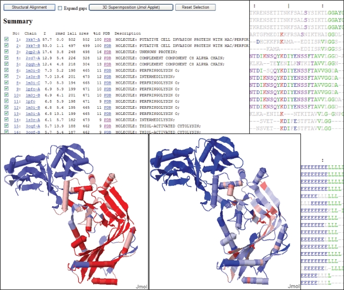
Our web site (http://ekhidna.biocenter.helsinki.fi/dali_server) runs the Dali program for protein structure comparison. The web site consists of three parts: (i) the Dali server compares newly solved structures against structures in the Protein Data Bank (PDB), (ii) the Dali database allows browsing precomputed structural neighbourhoods and (iii) the pairwise comparison generates suboptimal alignments for a pair of structures. Each part has its own query form and a common format for the results page. The inputs are either PDB identifiers or novel structures uploaded by the user. The results pages are hyperlinked to aid interactive analysis. The web interface is simple and easy to use. The key purpose of interactive analysis is to check whether conserved residues line up in multiple structural alignments and how conserved residues and ligands cluster together in multiple structure superimpositions. In favourable cases, protein structure comparison can lead to evolutionary discoveries not detected by sequence analysis.
Overview
The claims and statistics of this paper haven’t been checked yet.
Free: it reads the open-access text and checks the first 5 claims that cite a source we can check. Create a free account to check every claim.
Integrity signals
Registry
Cites
Statistics
Figures
Verification
Claims the paper makes
The claims and statistics of this paper haven’t been checked yet.
Free: it reads the open-access text and checks the first 5 claims that cite a source we can check. Create a free account to check every claim.
Claims other papers make about this one
Each is checked against this paper’s own text: does it say what the citing paper says it does?
The cited source says what the claim attributes to it.
In the citing paper
“Structural comparison with all protein structures deposited in the Protein Data Bank (PDB) using the DALI server [67] gave a single hit, the 55-residue fragment of the closely related pneumococcal PhtA protein (PDB code 2CS7 [43]).”
From New Insights into Histidine Triad Proteins: Solution Structure of a Streptococcus pneumoniae PhtD Domain and Zinc Transfer to AdcAII (2013) · cites this paper as [67]
The cited source says what the claim attributes to it.
In the citing paper
“A homology search using Dali34 shows that the overall structure of PSS1 does not share any similar fold with membrane proteins whose structures have been determined before.”
From Molecular insights into human phosphatidylserine synthase 1 reveal its inhibition promotes LDL uptake (2024) · cites this paper as [34]
The cited source says what the claim attributes to it.
In the citing paper
“Despite the lack of sequence homology to any protein in the PDB, a DALI16 search of YPL067C (Supplementary Table 1) found that it is structurally similar to members of the widely conserved superfamily of histidine triad (HIT) proteins.”
From Determining crystal structures through crowdsourcing and coursework (2016) · cites this paper as [16]
The cited source says what the claim attributes to it.
In the citing paper
“The top DALI16 match to the crystal structure of YPL067C was to a HIT protein of unknown function from Clostridium difficile (PDB: 4EGU), with a Z-score of 4.9.”
From Determining crystal structures through crowdsourcing and coursework (2016) · cites this paper as [16]
Self-check
Percentages, sums and reported differences re-computed from the paper’s own numbers.
The self-check needs the paper’s full text.
Citations
Cites
Krissinel, E., & Henrick, K. (2004) · Acta Crystallographica Section D Biological Crystallography · doi:10.1107/s0907444904026460 · Cited by 3,760
Holm, L., & Sander, C. (1996) · Science · doi:10.1126/science.273.5275.595 · Cited by 1,497
Self-citationDali: a network tool for protein structure comparison
Holm, L., & Sander, C. (1995) · Trends in Biochemical Sciences · doi:10.1016/s0968-0004(00)89105-7 · Cited by 1,469
Self-citationConSurf 2005: the projection of evolutionary conservation scores of residues on protein structures
Landau, M., Mayrose, I., Rosenberg, Y., Glaser, F., Martz, E., Pupko, T., & Ben-Tal, N. (2005) · Nucleic Acids Research · doi:10.1093/nar/gki370 · Cited by 1,413
Surprising similarities in structure comparison
Gibrat, J.-F., Madej, T., & Bryant, S. H. (1996) · Current Opinion in Structural Biology · doi:10.1016/s0959-440x(96)80058-3 · Cited by 1,049
Searching protein structure databases with DaliLite v.3
Holm, L., Kääriäinen, S., Rosenström, P., & Schenkel, A. (2008) · Bioinformatics · doi:10.1093/bioinformatics/btn507 · Cited by 1,018
Self-citationData growth and its impact on the SCOP database: new developments
Andreeva, A., Howorth, D., Chandonia, J.-M., Brenner, S. E., Hubbard, T. J. P., Chothia, C., & Murzin, A. G. (2007) · Nucleic Acids Research · doi:10.1093/nar/gkm993 · Cited by 944
ProFunc: a server for predicting protein function from 3D structure
Laskowski, R. A., Watson, J. D., & Thornton, J. M. (2005) · Nucleic Acids Research · doi:10.1093/nar/gki414 · Cited by 655
Advances and pitfalls of protein structural alignment
Hasegawa, H., & Holm, L. (2009) · Current Opinion in Structural Biology · doi:10.1016/j.sbi.2009.04.003 · Cited by 368
Self-citationA Common Fold Mediates Vertebrate Defense and Bacterial Attack
Rosado, C. J., Buckle, A. M., Law, R. H. P., Butcher, R. E., Kan, W.-T., Bird, C. H., Ung, K., Browne, K. A., Baran, K., Bashtannyk-Puhalovich, T. A., et al. (2007) · Science · doi:10.1126/science.1144706 · Cited by 278
Cited by
The HCV Life Cycle: In vitro Tissue Culture Systems and Therapeutic Targets
Gerold, G., & Pietschmann, T. (2014) · Digestive Diseases · doi:10.1159/000360830 · Cited by 4,854
Pore-forming activity and structural autoinhibition of the gasdermin family
Ding, J., Wang, K., Liu, W., She, Y., Sun, Q., Shi, J., Sun, H., Wang, D.-C., & Shao, F. (2016) · Nature · doi:10.1038/nature18590 · Cited by 2,771
Crystal structure of Cas9 in complex with guide RNA and target DNA
Nishimasu, H., Ran, F. A., Hsu, P. D., Konermann, S., Shehata, S. I., Dohmae, N., Ishitani, R., Zhang, F., & Nureki, O. (2014) · Cell · doi:10.1016/j.cell.2014.02.001 · Cited by 2,306
“A Dali search (Holm and Rosenstrom, 2010) revealed that the REC lobe does not share structural similarity with other known proteins, indicating that it is a Cas9-specific functional domain.”
BackgroundRogers, C., Fernandes-Alnemri, T., Mayes, L., Alnemri, D., Cingolani, G., & Alnemri, E. S. (2017) · Nature Communications · doi:10.1038/ncomms14128 · Cited by 1,510
Neuropilin-1 is a host factor for SARS-CoV-2 infection
Daly, J. L., Simonetti, B., Klein, K., Chen, K.-E., Williamson, M. K., Antón-Plágaro, C., Shoemark, D. K., Simón-Gracia, L., Bauer, M., Hollandi, R., et al. (2020) · Science · doi:10.1126/science.abd3072 · Cited by 1,251
Holm, L., & Laakso, L. M. (2016) · Nucleic Acids Research · doi:10.1093/nar/gkw357 · Cited by 868
Self-citationCrystal Structure of Cpf1 in Complex with Guide RNA and Target DNA
Yamano, T., Nishimasu, H., Zetsche, B., Hirano, H., Slaymaker, I. M., Li, Y., Fedorova, I., Nakane, T., Makarova, K. S., Koonin, E. V., et al. (2016) · Cell · doi:10.1016/j.cell.2016.04.003 · Cited by 764
Molecular basis of binding between novel human coronavirus MERS-CoV and its receptor CD26
Lu, G., Hu, Y., Wang, Q., Qi, J., Gao, F., Li, Y., Zhang, Y., Zhang, W., Yuan, Y., Bao, J., et al. (2013) · Nature · doi:10.1038/nature12328 · Cited by 739
Nsp3 of coronaviruses: Structures and functions of a large multi-domain protein
Lei, J., Kusov, Y., & Hilgenfeld, R. (2018) · Antiviral Research · doi:10.1016/j.antiviral.2017.11.001 · Cited by 727
The genome of Chenopodium quinoa
Jarvis, D. E., Ho, Y. S., Lightfoot, D. J., Schmöckel, S. M., Li, B., Borm, T. J. A., Ohyanagi, H., Mineta, K., Michell, C. T., Saber, N., et al. (2017) · Nature · doi:10.1038/nature21370 · Cited by 719
A Highly Conserved Program of Neuronal Microexons Is Misregulated in Autistic Brains
Irimia, M., Weatheritt, R. J., Ellis, J. D., Parikshak, N. N., Gonatopoulos-Pournatzis, T., Babor, M., Quesnel-Vallières, M., Tapial, J., Raj, B., O’Hanlon, D., et al. (2014) · Cell · doi:10.1016/j.cell.2014.11.035 · Cited by 693
“We thus used the Dali server (Holm and Rosenström, 2010) to identify structures that have been resolved in the presence of a protein ligand and that most closely resembles the APBB1-PTB1 domain.”
MethodDALI and the persistence of protein shape
Holm, L. (2020) · Protein Science · doi:10.1002/pro.3749 · Cited by 692
Self-citationTools for macromolecular model building and refinement into electron cryo-microscopy reconstructions
Brown, A., Long, F., Nicholls, R. A., Toots, J., Emsley, P., & Murshudov, G. (2015) · Acta Crystallographica Section D Biological Crystallography · doi:10.1107/s1399004714021683 · Cited by 618
Structural Basis for Sequence-Specific Recognition of DNA by TAL Effectors
Deng, D., Yan, C., Pan, X., Mahfouz, M., Wang, J., Zhu, J.-K., Shi, Y., & Yan, N. (2012) · Science · doi:10.1126/science.1215670 · Cited by 601
Structure of the Yeast Mitochondrial Large Ribosomal Subunit
Amunts, A., Brown, A., Bai, X.-C., Llácer, J. L., Hussain, T., Emsley, P., Long, F., Murshudov, G., Scheres, S. H. W., & Ramakrishnan, V. (2014) · Science · doi:10.1126/science.1249410 · Cited by 579
Cryo-electron microscopy structure of a coronavirus spike glycoprotein trimer
Walls, A. C., Tortorici, M. A., Bosch, B.-J., Frenz, B., Rottier, P. J. M., DiMaio, F., Rey, F. A., & Veesler, D. (2016) · Nature · doi:10.1038/nature16988 · Cited by 538
The structure of the human mitochondrial ribosome
Amunts, A., Brown, A., Toots, J., Scheres, S. H. W., & Ramakrishnan, V. (2015) · Science · doi:10.1126/science.aaa1193 · Cited by 533
Three-Dimensional Structures of Membrane Proteins from Genomic Sequencing
Hopf, T. A., Colwell, L. J., Sheridan, R., Rost, B., Sander, C., & Marks, D. S. (2012) · Cell · doi:10.1016/j.cell.2012.04.012 · Cited by 512
Defense Islands in Bacterial and Archaeal Genomes and Prediction of Novel Defense Systems
Makarova, K. S., Wolf, Y. I., Snir, S., & Koonin, E. V. (2011) · Journal of Bacteriology · doi:10.1128/jb.05535-11 · Cited by 489
Crystal structure of the human σ1 receptor
Schmidt, H. R., Zheng, S., Gurpinar, E., Koehl, A., Manglik, A., & Kruse, A. C. (2016) · Nature · doi:10.1038/nature17391 · Cited by 482
Structural basis and functional analysis of the SARS coronavirus nsp14–nsp10 complex
Ma, Y., Wu, L., Shaw, N., Gao, Y., Wang, J., Sun, Y., Lou, Z., Yan, L., Zhang, R., & Rao, Z. (2015) · Proceedings of the National Academy of Sciences · doi:10.1073/pnas.1508686112 · Cited by 482
Makarova, K. S., Aravind, L., Wolf, Y. I., & Koonin, E. V. (2011) · Biology Direct · doi:10.1186/1745-6150-6-38 · Cited by 470
Crystal Structure of TET2-DNA Complex: Insight into TET-Mediated 5mC Oxidation
Hu, L., Li, Z., Cheng, J., Rao, Q., Gong, W., Liu, M., Shi, Y. G., Zhu, J., Wang, P., & Xu, Y. (2013) · Cell · doi:10.1016/j.cell.2013.11.020 · Cited by 450
Structural Basis for DNA Binding Specificity by the Auxin-Dependent ARF Transcription Factors
Boer, D. R., Freire-Rios, A., van den Berg, W. A. M., Saaki, T., Manfield, I. W., Kepinski, S., López-Vidrieo, I., Franco-Zorrilla, J. M., de Vries, S. C., Solano, R., et al. (2014) · Cell · doi:10.1016/j.cell.2013.12.027 · Cited by 450
Structure of a mammalian ryanodine receptor
Zalk, R., Clarke, O. B., des Georges, A., Grassucci, R. A., Reiken, S., Mancia, F., Hendrickson, W. A., Frank, J., & Marks, A. R. (2015) · Nature · doi:10.1038/nature13950 · Cited by 437
Bacterial ferrous iron transport: the Feo system
Lau, C. K. Y., Krewulak, K. D., & Vogel, H. J. (2016) · FEMS Microbiology Reviews · doi:10.1093/femsre/fuv049 · Cited by 428
Walls, A. C., Tortorici, M. A., Frenz, B., Snijder, J., Li, W., Rey, F. A., DiMaio, F., Bosch, B.-J., & Veesler, D. (2016) · Nature Structural & Molecular Biology · doi:10.1038/nsmb.3293 · Cited by 412
Polymerase IV occupancy at RNA-directed DNA methylation sites requires SHH1
Law, J. A., Du, J., Hale, C. J., Feng, S., Krajewski, K., Palanca, A. M. S., Strahl, B. D., Patel, D. J., & Jacobsen, S. E. (2013) · Nature · doi:10.1038/nature12178 · Cited by 393
A prevalent peptide-binding domain guides ribosomal natural product biosynthesis
Burkhart, B. J., Hudson, G. A., Dunbar, K. L., & Mitchell, D. A. (2015) · Nature Chemical Biology · doi:10.1038/nchembio.1856 · Cited by 374
Structure of paused transcription complex Pol II–DSIF–NELF
Vos, S. M., Farnung, L., Urlaub, H., & Cramer, P. (2018) · Nature · doi:10.1038/s41586-018-0442-2 · Cited by 374
Structure and function of Parkin E3 ubiquitin ligase reveals aspects of RING and HECT ligases
Riley, B. E., Lougheed, J. C., Callaway, K., Velasquez, M., Brecht, E., Nguyen, L., Shaler, T., Walker, D., Yang, Y., Regnstrom, K., et al. (2013) · Nature Communications · doi:10.1038/ncomms2982 · Cited by 371
Structural Basis for Assembly and Function of a Heterodimeric Plant Immune Receptor
Williams, S. J., Sohn, K. H., Wan, L., Bernoux, M., Sarris, P. F., Segonzac, C., Ve, T., Ma, Y., Saucet, S. B., Ericsson, D. J., et al. (2014) · Science · doi:10.1126/science.1247357 · Cited by 370
Abdul Rehman, S. A., Kristariyanto, Y. A., Choi, S.-Y., Nkosi, P. J., Weidlich, S., Labib, K., Hofmann, K., & Kulathu, Y. (2016) · Molecular Cell · doi:10.1016/j.molcel.2016.05.009 · Cited by 363
Bernoux, M., Ve, T., Williams, S., Warren, C., Hatters, D., Valkov, E., Zhang, X., Ellis, J. G., Kobe, B., & Dodds, P. N. (2011) · Cell Host & Microbe · doi:10.1016/j.chom.2011.02.009 · Cited by 362
Cap-specific terminal N 6 -methylation of RNA by an RNA polymerase II–associated methyltransferase
Akichika, S., Hirano, S., Shichino, Y., Suzuki, T., Nishimasu, H., Ishitani, R., Sugita, A., Hirose, Y., Iwasaki, S., Nureki, O., et al. (2019) · Science · doi:10.1126/science.aav0080 · Cited by 361
Joshi, R. S., Jagdale, S. S., Bansode, S. B., Shankar, S. S., Tellis, M. B., Pandya, V. K., Chugh, A., Giri, A. P., & Kulkarni, M. J. (2020) · Journal of Biomolecular Structure and Dynamics · doi:10.1080/07391102.2020.1760137 · Cited by 354
Ouyang, S., Song, X., Wang, Y., Ru, H., Shaw, N., Jiang, Y., Niu, F., Zhu, Y., Qiu, W., Parvatiyar, K., et al. (2012) · Immunity · doi:10.1016/j.immuni.2012.03.019 · Cited by 350
The Dendritic Cell Receptor Clec9A Binds Damaged Cells via Exposed Actin Filaments
Zhang, J.-G., Czabotar, P. E., Policheni, A. N., Caminschi, I., San Wan, S., Kitsoulis, S., Tullett, K. M., Robin, A. Y., Brammananth, R., van Delft, M. F., et al. (2012) · Immunity · doi:10.1016/j.immuni.2012.03.009 · Cited by 342
MMDB and VAST+: tracking structural similarities between macromolecular complexes
Madej, T., Lanczycki, C. J., Zhang, D., Thiessen, P. A., Geer, R. C., Marchler-Bauer, A., & Bryant, S. H. (2014) · Nucleic Acids Research · doi:10.1093/nar/gkt1208 · Cited by 339
The structural biochemistry of Zucchini implicates it as a nuclease in piRNA biogenesis
Ipsaro, J. J., Haase, A. D., Knott, S. R., Joshua-Tor, L., & Hannon, G. J. (2012) · Nature · doi:10.1038/nature11502 · Cited by 334
Two Distant Catalytic Sites Are Responsible for C2c2 RNase Activities
Liu, L., Li, X., Wang, J., Wang, M., Chen, P., Yin, M., Li, J., Sheng, G., & Wang, Y. (2017) · Cell · doi:10.1016/j.cell.2016.12.031 · Cited by 328
Structure of the large ribosomal subunit from human mitochondria
Brown, A., Amunts, A., Bai, X.-C., Sugimoto, Y., Edwards, P. C., Murshudov, G., Scheres, S. H. W., & Ramakrishnan, V. (2014) · Science · doi:10.1126/science.1258026 · Cited by 324
Selective depletion of uropathogenic E. coli from the gut by a FimH antagonist
Spaulding, C. N., Klein, R. D., Ruer, S., Kau, A. L., Schreiber, H. L., Cusumano, Z. T., Dodson, K. W., Pinkner, J. S., Fremont, D. H., Janetka, J. W., et al. (2017) · Nature · doi:10.1038/nature22972 · Cited by 315
Korasick, D. A., Westfall, C. S., Lee, S. G., Nanao, M. H., Dumas, R., Hagen, G., Guilfoyle, T. J., Jez, J. M., & Strader, L. C. (2014) · Proceedings of the National Academy of Sciences · doi:10.1073/pnas.1400074111 · Cited by 306
The cytotoxic Staphylococcus aureus PSMα3 reveals a cross-α amyloid-like fibril
Tayeb-Fligelman, E., Tabachnikov, O., Moshe, A., Goldshmidt-Tran, O., Sawaya, M. R., Coquelle, N., Colletier, J.-P., & Landau, M. (2017) · Science · doi:10.1126/science.aaf4901 · Cited by 304
DNA-binding domains of plant-specific transcription factors: structure, function, and evolution
Yamasaki, K., Kigawa, T., Seki, M., Shinozaki, K., & Yokoyama, S. (2013) · Trends in Plant Science · doi:10.1016/j.tplants.2012.09.001 · Cited by 303
Polycomb-like proteins link the PRC2 complex to CpG islands
Li, H., Liefke, R., Jiang, J., Kurland, J. V., Tian, W., Deng, P., Zhang, W., He, Q., Patel, D. J., Bulyk, M. L., et al. (2017) · Nature · doi:10.1038/nature23881 · Cited by 289
Circadian Rhythms in Cyanobacteria
Cohen, S. E., & Golden, S. S. (2015) · Microbiology and Molecular Biology Reviews · doi:10.1128/mmbr.00036-15 · Cited by 288
Sabbadin, F., Hemsworth, G. R., Ciano, L., Henrissat, B., Dupree, P., Tryfona, T., Marques, R. D. S., Sweeney, S. T., Besser, K., Elias, L., et al. (2018) · Nature Communications · doi:10.1038/s41467-018-03142-x · Cited by 282
Multiple origins of viral capsid proteins from cellular ancestors
Krupovic, M., & Koonin, E. V. (2017) · Proceedings of the National Academy of Sciences · doi:10.1073/pnas.1621061114 · Cited by 282
Boosting the Full Potential of PyMOL with Structural Biology Plugins
Rosignoli, S., & Paiardini, A. (2022) · Biomolecules · doi:10.3390/biom12121764 · Cited by 276
Structure of the core ectodomain of the hepatitis C virus envelope glycoprotein 2
Khan, A. G., Whidby, J., Miller, M. T., Scarborough, H., Zatorski, A. V., Cygan, A., Price, A. A., Yost, S. A., Bohannon, C. D., Jacob, J., et al. (2014) · Nature · doi:10.1038/nature13117 · Cited by 274
Structure-function analysis identifies highly sensitive strigolactone receptors in Striga
Toh, S., Holbrook-Smith, D., Stogios, P. J., Onopriyenko, O., Lumba, S., Tsuchiya, Y., Savchenko, A., & McCourt, P. (2015) · Science · doi:10.1126/science.aac9476 · Cited by 273
Structure of the Human Lipid Exporter ABCA1
Qian, H., Zhao, X., Cao, P., Lei, J., Yan, N., & Gong, X. (2017) · Cell · doi:10.1016/j.cell.2017.05.020 · Cited by 272
Bacterial killing via a type IV secretion system
Souza, D. P., Oka, G. U., Alvarez-Martinez, C. E., Bisson-Filho, A. W., Dunger, G., Hobeika, L., Cavalcante, N. S., Alegria, M. C., Barbosa, L. R. S., Salinas, R. K., et al. (2015) · Nature Communications · doi:10.1038/ncomms7453 · Cited by 271
Structural basis for organohalide respiration
Bommer, M., Kunze, C., Fesseler, J., Schubert, T., Diekert, G., & Dobbek, H. (2014) · Science · doi:10.1126/science.1258118 · Cited by 268
Whitney, J. C., Quentin, D., Sawai, S., LeRoux, M., Harding, B. N., Ledvina, H. E., Tran, B. Q., Robinson, H., Goo, Y. A., Goodlett, D. R., et al. (2015) · Cell · doi:10.1016/j.cell.2015.09.027 · Cited by 263
CENP-T-W-S-X Forms a Unique Centromeric Chromatin Structure with a Histone-like Fold
Nishino, T., Takeuchi, K., Gascoigne, K. E., Suzuki, A., Hori, T., Oyama, T., Morikawa, K., Cheeseman, I. M., & Fukagawa, T. (2012) · Cell · doi:10.1016/j.cell.2011.11.061 · Cited by 263
Structural Insight into the Ion-Exchange Mechanism of the Sodium/Calcium Exchanger
Liao, J., Li, H., Zeng, W., Sauer, D. B., Belmares, R., & Jiang, Y. (2012) · Science · doi:10.1126/science.1215759 · Cited by 261
Roth, C., Wei, R., Oeser, T., Then, J., Föllner, C., Zimmermann, W., & Sträter, N. (2014) · Applied Microbiology and Biotechnology · doi:10.1007/s00253-014-5672-0 · Cited by 260
Structural and functional aspects of PR‐10 proteins
Fernandes, H., Michalska, K., Sikorski, M., & Jaskolski, M. (2013) · The FEBS Journal · doi:10.1111/febs.12114 · Cited by 258
Structural basis of indisulam-mediated RBM39 recruitment to DCAF15 E3 ligase complex
Bussiere, D. E., Xie, L., Srinivas, H., Shu, W., Burke, A., Be, C., Zhao, J., Godbole, A., King, D., Karki, R. G., et al. (2020) · Nature Chemical Biology · doi:10.1038/s41589-019-0411-6 · Cited by 257
Wagner, S., Stuttmann, J., Rietz, S., Guerois, R., Brunstein, E., Bautor, J., Niefind, K., & Parker, J. E. (2013) · Cell Host & Microbe · doi:10.1016/j.chom.2013.11.006 · Cited by 250
Kwon, S. C., Nguyen, T. A., Choi, Y.-G., Jo, M. H., Hohng, S., Kim, V. N., & Woo, J.-S. (2016) · Cell · doi:10.1016/j.cell.2015.12.019 · Cited by 241
Mechanism of Foreign DNA Selection in a Bacterial Adaptive Immune System
Sashital, D. G., Wiedenheft, B., & Doudna, J. A. (2012) · Molecular Cell · doi:10.1016/j.molcel.2012.03.020 · Cited by 237
Structures of β-klotho reveal a ‘zip code’-like mechanism for endocrine FGF signalling
Lee, S., Choi, J., Mohanty, J., Sousa, L. P., Tome, F., Pardon, E., Steyaert, J., Lemmon, M. A., Lax, I., & Schlessinger, J. (2018) · Nature · doi:10.1038/nature25010 · Cited by 236
A Salmonella Toxin Promotes Persister Formation through Acetylation of tRNA
Cheverton, A. M., Gollan, B., Przydacz, M., Wong, C. T., Mylona, A., Hare, S. A., & Helaine, S. (2016) · Molecular Cell · doi:10.1016/j.molcel.2016.05.002 · Cited by 235
de Guillen, K., Ortiz-Vallejo, D., Gracy, J., Fournier, E., Kroj, T., & Padilla, A. (2015) · PLOS Pathogens · doi:10.1371/journal.ppat.1005228 · Cited by 235
Crystal structures of STING protein reveal basis for recognition of cyclic di-GMP
Shang, G., Zhu, D., Li, N., Zhang, J., Zhu, C., Lu, D., Liu, C., Yu, Q., Zhao, Y., Xu, S., et al. (2012) · Nature Structural & Molecular Biology · doi:10.1038/nsmb.2332 · Cited by 233
The prospects of quantum computing in computational molecular biology
Outeiral, C., Strahm, M., Shi, J., Morris, G. M., Benjamin, S. C., & Deane, C. M. (2021) · WIREs Computational Molecular Science · doi:10.1002/wcms.1481 · Cited by 232
Architectures of Lipid Transport Systems for the Bacterial Outer Membrane
Ekiert, D. C., Bhabha, G., Isom, G. L., Greenan, G., Ovchinnikov, S., Henderson, I. R., Cox, J. S., & Vale, R. D. (2017) · Cell · doi:10.1016/j.cell.2017.03.019 · Cited by 230
Structural diversity of lytic polysaccharide monooxygenases
Vaaje-Kolstad, G., Forsberg, Z., Loose, J. S., Bissaro, B., & Eijsink, V. G. (2017) · Current Opinion in Structural Biology · doi:10.1016/j.sbi.2016.12.012 · Cited by 226
Structural basis for nutrient acquisition by dominant members of the human gut microbiota
Glenwright, A. J., Pothula, K. R., Bhamidimarri, S. P., Chorev, D. S., Baslé, A., Firbank, S. J., Zheng, H., Robinson, C. V., Winterhalter, M., Kleinekathöfer, U., et al. (2017) · Nature · doi:10.1038/nature20828 · Cited by 225
Dunne, M., Rupf, B., Tala, M., Qabrati, X., Ernst, P., Shen, Y., Sumrall, E., Heeb, L., Plückthun, A., Loessner, M. J., et al. (2019) · Cell Reports · doi:10.1016/j.celrep.2019.09.062 · Cited by 225
Wagner, T., Koch, J., Ermler, U., & Shima, S. (2017) · Science · doi:10.1126/science.aan0425 · Cited by 219
Structural basis of inhibition of Mycobacterium tuberculosis DprE1 by benzothiazinone inhibitors
Batt, S. M., Jabeen, T., Bhowruth, V., Quill, L., Lund, P. A., Eggeling, L., Alderwick, L. J., Fütterer, K., & Besra, G. S. (2012) · Proceedings of the National Academy of Sciences · doi:10.1073/pnas.1205735109 · Cited by 218
Li, Z., & Nair, S. K. (2012) · Protein Science · doi:10.1002/pro.2132 · Cited by 216
Crystal structures of the human adiponectin receptors
Tanabe, H., Fujii, Y., Okada-Iwabu, M., Iwabu, M., Nakamura, Y., Hosaka, T., Motoyama, K., Ikeda, M., Wakiyama, M., Terada, T., et al. (2015) · Nature · doi:10.1038/nature14301 · Cited by 214
The structural basis for the sensing and binding of cyclic di-GMP by STING
Huang, Y.-H., Liu, X.-Y., Du, X.-X., Jiang, Z.-F., & Su, X.-D. (2012) · Nature Structural & Molecular Biology · doi:10.1038/nsmb.2333 · Cited by 213
A protein fold switch joins the circadian oscillator to clock output in cyanobacteria
Chang, Y.-G., Cohen, S. E., Phong, C., Myers, W. K., Kim, Y.-I., Tseng, R., Lin, J., Zhang, L., Boyd, J. S., Lee, Y., et al. (2015) · Science · doi:10.1126/science.1260031 · Cited by 212
Structure of the mechanosensitive OSCA channels
Zhang, M., Wang, D., Kang, Y., Wu, J.-X., Yao, F., Pan, C., Yan, Z., Song, C., & Chen, L. (2018) · Nature Structural & Molecular Biology · doi:10.1038/s41594-018-0117-6 · Cited by 212
The structure of the yeast mitochondrial ribosome
Desai, N., Brown, A., Amunts, A., & Ramakrishnan, V. (2017) · Science · doi:10.1126/science.aal2415 · Cited by 211
Employing a biochemical protecting group for a sustainable indigo dyeing strategy
Hsu, T. M., Welner, D. H., Russ, Z. N., Cervantes, B., Prathuri, R. L., Adams, P. D., & Dueber, J. E. (2018) · Nature Chemical Biology · doi:10.1038/nchembio.2552 · Cited by 211
Yeast Alcohol Dehydrogenase Structure and Catalysis
Raj, S. B., Ramaswamy, S., & Plapp, B. V. (2014) · Biochemistry · doi:10.1021/bi5006442 · Cited by 211
Secreted pectin monooxygenases drive plant infection by pathogenic oomycetes
Sabbadin, F., Urresti, S., Henrissat, B., Avrova, A. O., Welsh, L. R. J., Lindley, P. J., Csukai, M., Squires, J. N., Walton, P. H., Davies, G. J., et al. (2021) · Science · doi:10.1126/science.abj1342 · Cited by 208
Hajighasemi, M., Nocek, B. P., Tchigvintsev, A., Brown, G., Flick, R., Xu, X., Cui, H., Hai, T., Joachimiak, A., Golyshin, P. N., et al. (2016) · Biomacromolecules · doi:10.1021/acs.biomac.6b00223 · Cited by 208
X-domain of peptide synthetases recruits oxygenases crucial for glycopeptide biosynthesis
Haslinger, K., Peschke, M., Brieke, C., Maximowitsch, E., & Cryle, M. J. (2015) · Nature · doi:10.1038/nature14141 · Cited by 208
Structure and mechanism of the uracil transporter UraA
Lu, F., Li, S., Jiang, Y., Jiang, J., Fan, H., Lu, G., Deng, D., Dang, S., Zhang, X., Wang, J., et al. (2011) · Nature · doi:10.1038/nature09885 · Cited by 207
Kim, K. H., An, D. R., Song, J., Yoon, J. Y., Kim, H. S., Yoon, H. J., Im, H. N., Kim, J., Kim, D. J., Lee, S. J., et al. (2012) · Proceedings of the National Academy of Sciences · doi:10.1073/pnas.1120251109 · Cited by 203
Carpenter, B., & Tate, C. G. (2016) · Protein Engineering Design and Selection · doi:10.1093/protein/gzw049 · Cited by 202
Borisova, A. S., Isaksen, T., Dimarogona, M., Kognole, A. A., Mathiesen, G., Várnai, A., Røhr, Å. K., Payne, C. M., Sørlie, M., Sandgren, M., et al. (2015) · Journal of Biological Chemistry · doi:10.1074/jbc.m115.660183 · Cited by 199
Widespread anti-CRISPR proteins in virulent bacteriophages inhibit a range of Cas9 proteins
Hynes, A. P., Rousseau, G. M., Agudelo, D., Goulet, A., Amigues, B., Loehr, J., Romero, D. A., Fremaux, C., Horvath, P., Doyon, Y., et al. (2018) · Nature Communications · doi:10.1038/s41467-018-05092-w · Cited by 198
Lau, R. K., Ye, Q., Birkholz, E. A., Berg, K. R., Patel, L., Mathews, I. T., Watrous, J. D., Ego, K., Whiteley, A. T., Lowey, B., et al. (2020) · Molecular Cell · doi:10.1016/j.molcel.2019.12.010 · Cited by 198
Twenty-Five Years of Structural Parvovirology
Mietzsch, M., Pénzes, J. J., & Agbandje-McKenna, M. (2019) · Viruses · doi:10.3390/v11040362 · Cited by 197
Structures of the calcium-activated, non-selective cation channel TRPM4
Guo, J., She, J., Zeng, W., Chen, Q., Bai, X.-C., & Jiang, Y. (2017) · Nature · doi:10.1038/nature24997 · Cited by 197
Protein AMPylation by an Evolutionarily Conserved Pseudokinase
Sreelatha, A., Yee, S. S., Lopez, V. A., Park, B. C., Kinch, L. N., Pilch, S., Servage, K. A., Zhang, J., Jiou, J., Karasiewicz-Urbańska, M., et al. (2018) · Cell · doi:10.1016/j.cell.2018.08.046 · Cited by 195
Kwasna, D., Abdul Rehman, S. A., Natarajan, J., Matthews, S., Madden, R., De Cesare, V., Weidlich, S., Virdee, S., Ahel, I., Gibbs-Seymour, I., et al. (2018) · Molecular Cell · doi:10.1016/j.molcel.2018.02.023 · Cited by 190
Structures of Phytophthora RXLR Effector Proteins
Boutemy, L. S., King, S. R. F., Win, J., Hughes, R. K., Clarke, T. A., Blumenschein, T. M. A., Kamoun, S., & Banfield, M. J. (2011) · Journal of Biological Chemistry · doi:10.1074/jbc.m111.262303 · Cited by 188
Brand, L. H., Fischer, N. M., Harter, K., Kohlbacher, O., & Wanke, D. (2013) · Nucleic Acids Research · doi:10.1093/nar/gkt732 · Cited by 187
Zhang, T., Liu, J., Fellner, M., Zhang, C., Sui, D., & Hu, J. (2017) · Science Advances · doi:10.1126/sciadv.1700344 · Cited by 187
Metadata
- Authors
- Holm, L., & Rosenstr�m, P.
- Affiliations
- University of Helsinki (FI)
- Corresponding author
- Holm, Liisa
- ORCID
- Holm, Liisa
- Venue
- Nucleic Acids Research
- Publisher
- Oxford University Press (OUP)
- Published
- 2010-05-10
- Volume
- 38
- Issue
- suppl_2
- Pages
- W545-W549
- ISSN
- 0305-1048 (ISSN-L)
- 1362-4962
- PMC
- PMC2896194
- OpenAlex
- W2030517878
- Cited by
- 3,640
- References
- 18
- Citations by year
- 2026: 42 · 2025: 67 · 2024: 89 · 2023: 139 · 2022: 128 · 2021: 120 · 2020: 163 · 2019: 225 · 2018: 304 · 2017: 358 · 2016: 379 · 2015: 363 · 2014: 385 · 2013: 380 · 2012: 281
- FWCI
- 113.03
- Type
- journal article
- Language
- en
- Open access
- gold
- doi.org
- Licence
- Locations
- Nucleic Acids Research (publishedVersion, cc-by-nc)
- PubMed Central (publishedVersion, cc-by-nc)
- PubMed (publishedVersion)
- Työväentutkimus Vuosikirja (submittedVersion)
- HighWire Press Open Archive (submittedVersion)
- Topics
- Life Sciences › Biochemistry, Genetics and Molecular Biology › Molecular Biology › Protein Structure and Dynamics
- Life Sciences › Biochemistry, Genetics and Molecular Biology › Structural Biology › Enzyme Structure and Function
- Life Sciences › Biochemistry, Genetics and Molecular Biology › Molecular Biology › Genomics and Phylogenetic Studies
- Keywords
- protein structure comparison, structural alignment, conserved residues, ligand clustering, Protein Data Bank, web server
- MeSH terms
- Bacterial Proteins, Software, User-Computer Interface, Internet, Sequence Analysis, Protein, Databases, Protein, Structural Homology, Protein
About these checks
Registry, retraction, Cites and statistics results are computed, not judged, with one exception: a printed reference that no index lists is read once by a model before it counts as a citation error. Claims are extracted by a model and kept only when their quote appears verbatim in the text; each cited source is then checked, with a model’s help, for whether it says what the claim relies on. Those verdicts can be wrong — read the claim’s quote and the cited source before relying on one.
Statistics and arithmetic are recomputed, not judged.
Confidence bands are shown only for fields calibrated on our annotated test set. None is calibrated yet, so no band is shown.
What counts as a claim ›How we measure confidence ›How we check this ›
Are you an author, or do you see a problem with this page? Request a review
Data sources
Bibliographic data: Semantic Scholar, OpenAlex, Crossref.
Contains information from the Semantic Scholar Academic Graph Datasets, made available under the ODC Attribution License (ODC-BY 1.0).
Semantic Scholar’s data is described in: Kinney, R., et al. (2023). The Semantic Scholar Open Data Platform. arXiv:2301.10140.
