

Climbing-inspired twining electrodes using shape memory for peripheral nerve stimulation and recording
Open accessThe DOI matches its Crossref record; 56 of the 56 references it cites were found in a registry or an index; 11 of the 65 sources its claims cite were checked for support.
Abstract
Proposed 3D neural electrode can self-climb onto peripheral nerve driven by body temperature and form flexible neural interface.
Overview
CoChat Summary
TL;DR
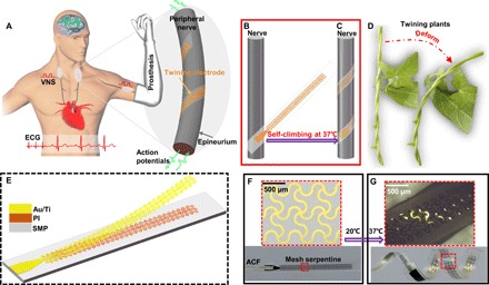
The paper presents a climbing-inspired twining electrode made of a shape memory polymer substrate and mesh serpentine Au/Ti/PI wires that self-climbs onto peripheral nerves when actuated by body temperature (~37°C), forming a conformal interface without additional surgical fixation. In rabbits, the device was used for vagus nerve stimulation with ECG recording and for sciatic nerve evoked action potential recording.
Key findings
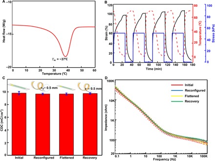
- The synthesized SMP has a transition temperature of ~37°C, shape fixity and recovery ratios above 97% over four cycles, fracture strain ~1100%, and elastic modulus changing from ~100 MPa to ~300 kPa before and after the phase transition.
- Electrochemical characterization showed average CDC ~9.7 mC/cm² and impedance magnitude ~156 ohms at 1 kHz, with little change across initial flat, reconfigured, flattened, and recovery states; fabricated electrodes had ~126 ohms at ~0.5 mm inner radius and ~119 ohms at ~1 mm inner radius.
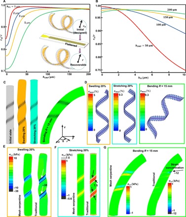
- Structural optimization gave r0/r = 0.98 and maximum Au strain εAumax = 0.24%; the twining electrode had bending stiffness EI ~1.0 × 10⁻¹⁰ N·m² and stretching stiffness EA 0.08 N, and FEA showed Au strain below fracture strain (5%) during 20% swelling and below yield strain (0.3%) during 20% stretching and bending (R = 15 mm).
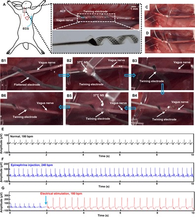
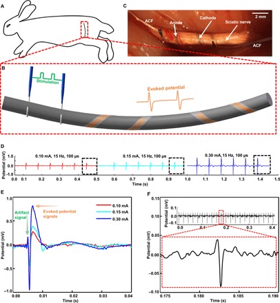
- In vivo, epinephrine increased rabbit heart rate from 180 to 240 bpm; VNS at 0.4 mA, 100 μs, 10 Hz reduced HR to 180 bpm and increased R-wave peak from 100 to 250 mV. Sciatic nerve recording showed evoked CNAP peak potentials of ~275, ~380, and ~820 μV for 0.1, 0.15, and 0.3 mA stimulation, and ~80 μV evoked CNAPs without stimulus artifact at an SNR of 16 dB.
Methods
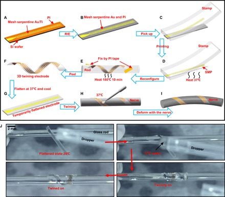
- The electrode was fabricated by depositing/etching Au/Ti mesh serpentine structures on ultrathin PI, transfer printing them onto an SMP substrate, reconfiguring the assembly into a 3D helix at 180°C for 10 min, and temporarily flattening it at 37°C; self-climbing was driven by 37°C normal saline.
- Material and device characterization included DSC, DMA, tensile testing, CV, and EIS in phosphate-buffered saline using a three-electrode setup with six samples in four states; FEA (Abaqus) modeled stress/strain under 20% swelling, 20% stretching, and bending to R = 15 mm.
- In vivo experiments used two New Zealand rabbits (3.2–3.3 kg): one for right vagus nerve stimulation with ECG recording, and one for sciatic nerve evoked potential recording using hooked platinum stimulating electrodes and twining recording electrodes.
Limitations
- The paper does not include a dedicated limitations section; it states that future twining electrodes will benefit from new high-surface-area materials and high-resolution designs for selective stimulation and recording.
- Evident from the text: in vivo validation was acute and limited to two rabbits, with no chronic implantation, long-term stability, or histological safety data reported.
- Evident from the text: biocompatibility of the main SMP precursors is cited from previous work rather than newly demonstrated in this study.
Model-written summary. Check it against the paper before relying on it.
Integrity signals
Registry
Cites
Statistics
Figures
Verification
Claims the paper makes
6 Valid · 2 Not valid · 17 Not checked · 62 Own results and interpretations
None of the sources cited for this claim have been checked yet.
In the paper
“Large mechanical and geometrical mismatches still exist at the neural interfaces.”
§ Climbing-inspired twining electrodes using shape memory for peripheral nerve stimulation and recording > INTRODUCTION
What the sources say
- Not checked yetcites [5] Naples R. G., Mortimer J. T., Scheiner A., Sweeney J. D., A spiral nerve cuff electrode for peripheral nerve stimulation. IEEE Trans. Biomed. Eng. 35, 905– 916 (1988). 3198136 source
- Not checked yetcites [6] Tarver W. B., George R. E., Maschino S. E., Holder L. K., Wernicke J. F., Clinical experience with a helical bipolar stimulating lead. Pacing Clin. Electrophysiol. 15, 1545– 1556 (1992). 1383969 source
- Not checked yetcites [7] Tyler D. J., Durand D. M., Functionally selective peripheral nerve stimulation with a flat interface nerve electrode. IEEE Trans. Neural Syst. Rehabil. Eng. 10, 294– 303 (2002). 12611367 source
- Could not verifycites [8] Foldes E. L., Ackermann D. M., Bhadra N., Kilgore K. L., Bhadra N., Design, fabrication and evaluation of a conforming circumpolar peripheral nerve cuff electrode for acute experimental use. J. Neurosci. Methods 196, 31– 37 (2011). 21187115 PMC3042051 sourceExperimental — accuracy not yet measured
Our copy of Foldes et al. (2011) frames the electrode-nerve interface in terms of achieving a conforming, close-fitting geometry — the Introduction stresses that 'consistent and predictable electrical peripheral nerve stimulation requires a robust and stable physical interface' obtained by a cylindrical cuff conforming to the nerve, and §2.1 sets a cuff-to-nerve diameter ratio of 0.9–1.1 — but it nowhere asserts that large mechanical or geometrical mismatches persist at neural interfaces, and it makes no statement about mechanical/compliance (stiffness) mismatch or about 'first-generation extraneural electrodes'. The Discussion (§4) notes only minor geometric limits of its own design (silicone bowing at IPDs beyond ~4 mm, sheath opening at lengths >2 mm), which is not the general claim being attributed. Searches run: 'mechanical and geometrical mismatch at the neural interface', 'compliance mismatch between electrode and nerve tissue', 'limitations of existing cuff electrodes: size mismatch, stiffness, poor conformal contact', 'first generation extraneural electrode problems'. Nothing in our copy supports or contradicts the claim, so it cannot be judged from this source. (LLM, conf=0.62)
plain- Asserted: passes — The authors put it forward as true.
- Checkable: passes — It can be checked against evidence.
- Load-bearing: passes — The paper’s conclusions depend on it.
- Based: passes — The paper gives a reason why it holds.
The cited sources support only part of this claim — read each source’s verdict below.
In the paper
“To decrease damage to nerves and increase long-term stability, several forms of extraneural electrodes have been proposed to replace intraneural electrodes, which penetrate into the nerve fascicles (3, 4).”
§ Climbing-inspired twining electrodes using shape memory for peripheral nerve stimulation and recording > INTRODUCTION
What the sources say
- Could not verifycites [3] Fattahi P., Yang G., Kim G., Abidian M. R., A review of organic and inorganic biomaterials for neural interfaces. Adv. Mater. 26, 1846– 1885 (2014). 24677434 PMC4373558 sourceExperimental — accuracy not yet measured
Our copy of Fattahi et al. (2014) never addresses extraneural versus intraneural (intrafascicular) electrodes for peripheral nerves; searched for 'extraneural', 'intrafascicular/nerve fascicles', 'cuff/peripheral nerve electrodes' and 'penetrating vs surface electrodes to reduce damage and improve long-term stability'. The closest passages discuss central implants only — e.g., Section 6.3 (Mechanical Stability) that stiff silicon penetrating electrodes 'create mechanical trauma on insertion' while flexible polyimide/parylene 'may improve this issue', and Section 3.1 that flexible surface microelectrodes 'may generate less tissue damage and be functional longer than silicon-based electrodes' — plus Section 6.3 mentioning hydrogel tubes 'for interfacing with the peripheral nerve'. None of these states the proposal of extraneural electrodes to replace fascicle-penetrating ones, so the claim can be neither confirmed nor contradicted from our copy. (LLM, conf=0.70)
- Partly supportedcites [4] Ecker M., Joshi-Imre A., Modi R., Frewin C. L., Garcia-Sandoval A., Maeng J., Gutierrez-Heredia G., Pancrazio J. J., Voit W. E., From softening polymers to multimaterial based bioelectronic devices. Multifunct. Mater. 2, 012001 (2019). sourceExperimental — accuracy not yet measured
Our copy supports the extraneural half of the claim: §2.2 (Nerve cuffs) describes 'several forms' of cuffs wrapping around nerves — first-generation hook-like self-coiling devices [30], newer folding thin-film cuffs in various sizes for acute and chronic studies [68,73], and softening-polymer cuffs — and the Fig. 3 caption explicitly labels them 'extra-neural cuff' devices; §2.2 also ties these to reducing nerve damage ('often causes nerve damage [67,68]', 'safer electrical stimulation') and to chronic/long-term use. What our copy does NOT support is the replacement framing: no passage mentions intraneural/intrafascicular electrodes or devices that penetrate nerve fascicles, and the paper's stated motive is overcoming the limits of silicone cuff electrodes (thickness, hardness, no size adjustment), not supplanting intraneural ones. The 'increase long-term stability' rationale appears only generally (§5.2 on reliability of chronic implants), not as a comparison against intraneural electrodes. (LLM, conf=0.68)
plainstates a cause- Asserted: passes — The authors put it forward as true.
- Checkable: passes — It can be checked against evidence.
- Load-bearing: passes — The paper’s conclusions depend on it.
- Based: passes — The paper gives a reason why it holds.
- Could not verify
The sources with a verdict support this claim; the rest weren’t checked yet, have no full text we could read, or their verdicts aren’t rated yet.
In the paper
“With the introduction of dynamic covalent bonds into the polymer network, the permanent shape of traditional thermoset SMPs can be reconfigured (23, 24).”
§ Climbing-inspired twining electrodes using shape memory for peripheral nerve stimulation and recording > INTRODUCTION
What the sources say
- Supportedcites [23] Zhao Q., Zou W., Luo Y., Xie T., Shape memory polymer network with thermally distinct elasticity and plasticity. Sci. Adv. 2, e1501297 (2016). 26824077 PMC4730863 source
The Introduction (Abstract section) states that permanent reshaping without macroscopic melting 'is achieved by covalent bond exchange in a polymer network, allowing its topography to be rearranged in response to an external force... the permanent reshaping can occur while the material maintains its dynamic crosslinking state,' listing 'malleability of thermoset polymers' among its consequences. The paper's own crosslinked PCL-thiolene network (Fig. 1A right-hand route; Figs. 3-4 discussion) demonstrates exactly this: reversible covalent bonds activated at Tp give nonrecoverable, cumulative redefinition of a permanently crosslinked (thermoset) SMP's permanent shape, with 'numerous cycles of manipulation of a permanent shape.' The paraphrase therefore matches what this source attributes. (LLM, conf=0.85)
- Not checked yetcites [24] Zheng N., Hou J., Xu Y., Fang Z., Zou W., Zhao Q., Xie T., Catalyst-free thermoset polyurethane with permanent shape reconfigurability and highly tunable triple-shape memory performance. ACS Macro Lett. 6, 326– 330 (2017). 35610855 source
plainstates a cause- Asserted: passes — The authors put it forward as true.
- Checkable: passes — It can be checked against evidence.
- Load-bearing: passes — The paper’s conclusions depend on it.
- Based: passes — The paper gives a reason why it holds.
- Supported
A result the paper reports from its own data — it cites no other source for it, so there is none to check. The quote shows it is in the paper.
In the paper
“The differential scanning calorimeter (DSC) curve for the synthetic SMPs (Fig. 3A) shows that the melt transition temperature (Tm) is approximately 37°C (i.e., Ttrans = ~37°C), which means that the SMPs recovering their permanent shapes can be actuated by body temperature (movie S4).”
§ Climbing-inspired twining electrodes using shape memory for peripheral nerve stimulation and recording > RESULTS > Material characterization and structure optimization
plain- Asserted: passes — The authors put it forward as true.
- Checkable: passes — It can be checked against evidence.
- Load-bearing: passes — The paper’s conclusions depend on it.
- Based: passes — The paper gives a reason why it holds.
A result the paper states — it cites no source for it, so there is none to check. The quote shows it is in the paper.
In the paper
“Distinct elastic modulus controllability from ~100 MPa to ~300 kPa before and after the phase transition greatly facilitates the surgical implantation in limited space, where the twining electrodes are handled in a relatively stiff state and soften after climbing on the nerves.”
§ Climbing-inspired twining electrodes using shape memory for peripheral nerve stimulation and recording > INTRODUCTION
plain- Asserted: passes — The authors put it forward as true.
- Checkable: passes — It can be checked against evidence.
- Load-bearing: passes — The paper’s conclusions depend on it.
- Based: passes — The paper gives a reason why it holds.
38 more claims
Create a free account to see all 87 claims and to verify the 54 references still open.
- Probably not a substantial claim. 17
- It may state two separate claims as one. 29
Claims other papers make about this one
Each is checked against this paper’s own text: does it say what the citing paper says it does?
The cited source says what the claim attributes to it.
In the citing paper
“In vivo Vagus nerve stimulation was carried out on a rabbit to demonstrate the validity of the device (Figure 8F).”
From Flexible Electronics and Devices as Human–Machine Interfaces for Medical Robotics (2022) · cites this paper as [358]
Self-check
Percentages, sums and reported differences re-computed from the paper’s own numbers.
Each contradiction between two passages listed here was confirmed by an independent check.
Rated minor: it would not touch a result — a value no result rests on, a p-value that stays on the same side of the significance level, or a conflict that is clerical, resolved elsewhere in the paper, or outside its own findings.
In the paper
§ Climbing-inspired twining electrodes using shape memory for peripheral nerve stimulation and recording > RESULTS > Material characterization and structure optimization
“the selected hSMP and hPI are ~2 and ~100 μm, respectively”
§ Climbing-inspired twining electrodes using shape memory for peripheral nerve stimulation and recording > RESULTS > Fabrication process
“Fabrication of the mesh serpentine starts with spin casting of an ultrathin PI film (~2 μm) (fig. S3) onto silicon wafers coated with a layer of a sacrificial polymethylmethacrylate (PMMA) film.”
Citations
Cites
Pursuing prosthetic electronic skin
Chortos, A., Liu, J., & Bao, Z. (2016) · Nature Materials · doi:10.1038/nmat4671 · Cited by 2,396
The rise of plastic bioelectronics
Someya, T., Bao, Z., & Malliaras, G. G. (2016) · Nature · doi:10.1038/nature21004 · Cited by 1,884
Recent Advances in Flexible and Stretchable Bio‐Electronic Devices Integrated with Nanomaterials
Choi, S., Lee, H., Ghaffari, R., Hyeon, T., & Kim, D. (2016) · Advanced Materials · doi:10.1002/adma.201504150 · Cited by 1,104
Fractal design concepts for stretchable electronics
Fan, J. A., Yeo, W.-H., Su, Y., Hattori, Y., Lee, W., Jung, S.-Y., Zhang, Y., Liu, Z., Cheng, H., Falgout, L., et al. (2014) · Nature Communications · doi:10.1038/ncomms4266 · Cited by 1,088
Self-citation3D Printing of Shape Memory Polymers for Flexible Electronic Devices
Zarek, M., Layani, M., Cooperstein, I., Sachyani, E., Cohn, D., & Magdassi, S. (2016) · Advanced Materials · doi:10.1002/adma.201503132 · Cited by 903
Navarro, X., Krueger, T. B., Lago, N., Micera, S., Stieglitz, T., & Dario, P. (2005) · Journal of the Peripheral Nervous System · doi:10.1111/j.1085-9489.2005.10303.x · Cited by 845
Flexible and Stretchable Electronics for Biointegrated Devices
Kim, D.-H., Ghaffari, R., Lu, N., & Rogers, J. A. (2012) · Annual Review of Biomedical Engineering · doi:10.1146/annurev-bioeng-071811-150018 · Cited by 749
Highly Flexible Silver Nanowire Electrodes for Shape‐Memory Polymer Light‐Emitting Diodes
Yu, Z., Zhang, Q., Li, L., Chen, Q., Niu, X., Liu, J., & Pei, Q. (2011) · Advanced Materials · doi:10.1002/adma.201003398 · Cited by 688
A Review of Organic and Inorganic Biomaterials for Neural Interfaces
Fattahi, P., Yang, G., Kim, G., & Abidian, M. R. (2014) · Advanced Materials · doi:10.1002/adma.201304496 · Cited by 580 · cited 1×
“To decrease damage to nerves and increase long-term stability, several forms of extraneural electrodes have been proposed to replace intraneural electrodes, which penetrate into the nerve fascicles (3, 4).”
BackgroundA jump-start for electroceuticals
Famm, K., Litt, B., Tracey, K. J., Boyden, E. S., & Slaoui, M. (2013) · Nature · doi:10.1038/496159a · Cited by 554
Cited by
Items 201–300 of 329
Intraneural ultramicroelectrode arrays for function-specific interfacing to the vagus nerve
Ghazavi, A., González‐González, M. A., Romero‐Ortega, M. I., & Cogan, S. F. (2020) · Biosensors and Bioelectronics · doi:10.1016/j.bios.2020.112608 · Cited by 14
Cho, Y., Shin, H., Park, J., & Lee, S. (2021) · Micromachines · doi:10.3390/mi12060720 · Cited by 14
“Recently, a neural interface capable of self-implanting into nerves through the SME of SMPs programmed with Tg near body temperature was reported [16].”
BackgroundKim, H. J., Sunwoo, S., Koo, J. H., & Kim, D. (2024) · Korean Journal of Chemical Engineering · doi:10.1007/s11814-024-00318-8 · Cited by 13
Wang, M., Jia, L., Jia, X., Li, H., & Feng, X. (2025) · Science Advances · doi:10.1126/sciadv.ads5634 · Cited by 13
Self-citationStimuli-responsive biomedical polymeric films for tissue regeneration
Xu, Z., Deng, J., Gao, D., Du, Y., Zhang, Y., & Lai, Y. (2025) · Microstructures · doi:10.20517/microstructures.2024.139 · Cited by 13
Self-citationShape memory polymers for neural interfaces: a mini-review
Dai, L., Zheng, C., Song, J., & Xiao, R. (2025) · Smart Materials and Structures · doi:10.1088/1361-665x/adf0b8 · Cited by 13
Flexible and wearable bioelectronics for electrocardiography monitoring: a review
Zheng, C., Wang, Z., Li, L., Ma, Y., & Feng, X. (2025) · Science China Materials · doi:10.1007/s40843-025-3688-9 · Cited by 13
“1), which have broad applications in health monitoring, brain-machine interface, and disease diagnosis and therapy systems [ 8–”
Self-citationMultisite Simultaneous Neural Recording of Motor Pathway in Free-Moving Rats
Lang, Y., Tang, R., Liu, Y., Xi, P., Liu, H., Quan, Z., Song, D. F., Lv, X., Huang, Q., & He, J. (2021) · Biosensors · doi:10.3390/bios11120503 · Cited by 13
“A helix-shaped memory electrode was invented for peripheral nerve stimulation and recording [52].”
BackgroundJi, S., Wu, X., Jiang, Y., Wang, T., Liu, Z., Cao, C., Ji, B., Chi, L., Li, D., & Chen, X. (2022) · ACS Nano · doi:10.1021/acsnano.2c06682 · Cited by 13
Lee, J., Oh, M., Park, J.-H., Kang, S.-H., & Kang, S.-K. (2023) · Polymers · doi:10.3390/polym15092003 · Cited by 13
“In addition, reverse engineering methods [34], stimuli-responsive materials [35], and self-healing materials [36,37] have been incorporated to establish conformal contact with the biostructure.”
MethodWang, X., Wang, H., Sun, K., Li, W., Wang, X., Chen, X., Hu, L. Y., & Fan, Y. (2021) · Applied Materials Today · doi:10.1016/j.apmt.2021.101265 · Cited by 13
Electrical Failure Mechanism in Stretchable Thin-Film Conductors
Zhao, Y., Yu, M., Sun, J., Zhang, S., Li, Q., Teng, L., Tian, Q., Xie, R., Li, G., Liu, L., et al. (2022) · ACS Applied Materials & Interfaces · doi:10.1021/acsami.1c22447 · Cited by 13
Lee, J., Kim, S., Chung, W. G., Kim, E., Song, H., Oh, M., Kim, E.-M., Liu, J., Jang, K., Lee, T., et al. (2024) · Advanced Engineering Materials · doi:10.1002/adem.202400499 · Cited by 12
Kim, S., Kim, T., Kim, H., Kim, J., Rogers, J. A., & Hahn, S. K. (2024) · Advanced Materials · doi:10.1002/adma.202413189 · Cited by 12
Hydrogel‐Based Multifunctional Soft Electronics with Distributed Sensing Units: A Review
Zhang, C. W., Hao, X. P., Zheng, Q., & Wu, Z. L. (2023) · Advanced Sensor Research · doi:10.1002/adsr.202200069 · Cited by 12
Bioelectric Interface Technologies in Cells and Organoids
Xue, Z., & Zhao, J. Z. (2023) · Advanced Materials Interfaces · doi:10.1002/admi.202300550 · Cited by 12
Thermoformed Parylene‐C Cuff Electrodes for Small Nerve Interfacing
Zurita, F., Freko, S., Hiendlmeier, L., Duca, F. D., Groll, T., Seelbach, O., Steiger, K., & Wolfrum, B. L. (2023) · Advanced NanoBiomed Research · doi:10.1002/anbr.202300102 · Cited by 12
Kuang, X. (2021) · Elsevier eBooks · doi:10.1016/b978-0-12-818411-0.00004-5 · Cited by 12
Lee, J. Y., Kim, T., Cho, S., Shin, J. H., Yeo, W., Kim, T. S., & Yu, K. J. (2025) · Bioactive Materials · doi:10.1016/j.bioactmat.2025.02.006 · Cited by 11
“The device is made from a shape memory polymer (SMP), which allows it to initially be deployed in a straight configuration and then conform to the spiral shape once in place around the nerve [118,257].”
Wang, H., Zhao, B., Li, W., Liu, Y., Li, L., Zhao, Y., & Zhang, W. (2025) · Chemical Engineering Journal · doi:10.1016/j.cej.2025.162523 · Cited by 11
Fan, F., Chen, L., Wang, Y., Wang, P., Niu, Y., Lv, Y., Zhou, Y., Shu, Z., Liu, P., Dong, W., et al. (2025) · Soft Science · doi:10.20517/ss.2025.52 · Cited by 11
Self-citationBody-responsive shape-memory polymers for biomedical applications
Tajik, E., Reihani, N., Karamzadeh, V., Tang, G., & Ravanbakhsh, H. (2026) · Bioactive Materials · doi:10.1016/j.bioactmat.2025.12.054 · Cited by 11
“Furthermore, the adaptability and recovery of body-responsive SMPs show promise for the design of twining electrodes for neural stimulation and recording [178,208,268].”
Niu, D., Jiang, W., Li, D., Ye, G., Luo, F. C., & Liu, H. (2021) · Smart Materials and Structures · doi:10.1088/1361-665x/ac3404 · Cited by 11
“Inspired by the smooth and continuous shape transformations in natural systems, a lot of efforts have been tried in the last decades to develop healthcare-and manufacturing-related robots [1–4], artificial muscles [5–7], and deployable aerospace [8–11].”
Mechanoadaptive Bioelectronics for Deep Tissue Sensing
Ren, X., Yuan, Y., Li, J., & Hu, B. (2024) · Chinese Journal of Chemistry · doi:10.1002/cjoc.202400879 · Cited by 11
Photo‐Programmed Deformations in Rigid Liquid Crystalline Polymers Triggered by Body Temperature
Kim, H., Li, J., Hsieh, Y. S. Y., Cho, M., Ahn, S., & Li, C. (2022) · Small · doi:10.1002/smll.202203772 · Cited by 11
Suzuki, Y., Hu, Q., Batchelor, B. L., Voit, W., & Ecker, M. (2022) · Smart Materials and Structures · doi:10.1088/1361-665x/aca576 · Cited by 11
An, D., Gong, G., Xu, D., Zhou, Z., Li, R., & Su, Y. (2024) · Advanced Functional Materials · doi:10.1002/adfm.202412796 · Cited by 10
Zhou, T., Yu, R., Bai, X., Yang, Y., Fan, Z., Du, X., Chen, Y., Wang, Y., Yang, Z., Sun, X., et al. (2026) · Nature Communications · doi:10.1038/s41467-026-71689-1 · Cited by 10
“For example, shape-memory polymerthin-fi lmelectrodesachieveconformaltissuecontactthrough thermally triggered shape recovery of a deformablepolymer substrate carrying metal nano fi lm electrodes 34 .”
Self-citationLong-term implantable flexible neural interfaces for electrophysiological monitoring
Chen, B., Lu, J., Chen, Z., Han, X., Sun, Y., Lin, X., Tang, Z., Jia, S., Xie, G., Huo, F., et al. (2025) · Journal of Materials Chemistry C · doi:10.1039/d4tc04289a · Cited by 10
Yin, Y., Li, M., Yuan, W., Chen, X., & Li, Y. (2019) · Proceedings of the Royal Society A Mathematical Physical and Engineering Sciences · doi:10.1098/rspa.2019.0402 · Cited by 10
Self-citationThermal analysis of the flexible electronics affixed on large curvature myocardium surface
Yin, Y., Li, Y., & Li, M. (2019) · International Journal of Heat and Mass Transfer · doi:10.1016/j.ijheatmasstransfer.2019.118983 · Cited by 10
Self-citationDesign and applications of conductive polymers in wearable electrophysiological sensing
Zhou, N., Zhang, L., Cui, T., Wu, P., & Lei, Z. (2025) · Science China Technological Sciences · doi:10.1007/s11431-025-3114-6 · Cited by 9
“It could spontaneously wrap around a peripheral nerve assisted by a temperature-responsive polymer, achieving excellent compliance (Figure 3(f)) [30].”
3D Printing of Metals with sub‐10 µm Resolution
Wang, J., Shou, J., Liu, D., Yao, Y., Qian, Q., Wang, Z., Ren, J., Zhang, B., Chen, H., Yu, Y., et al. (2024) · Small · doi:10.1002/smll.202406518 · Cited by 9
Quinn, K. N., Tian, Y., Budde, R. B., Irazoqui, P. P., TUFFAHA, S. H., & Thakor, N. V. (2023) · Muscle & Nerve · doi:10.1002/mus.28029 · Cited by 9
“(A 66,67 The polymers naturally unfold at 37 C to match the contours of their target tissue. circumvent many of the problems caused by skin, making them a suitable alternative for improved clinical implementation and long-term use.”
BackgroundTu, K., Bao, R., Wang, L., Guo, Z., Jiang, C., Su, Y., Liu, S., & Liu, J. (2024) · Advanced Functional Materials · doi:10.1002/adfm.202306666 · Cited by 9
Zhang, K., Tang, C., Yu, S., Guan, H., Sun, X. J., Cao, M., Zhang, S., Sun, X., & Peng, H. (2024) · Journal of Materials Chemistry B · doi:10.1039/d4tb00789a · Cited by 9
Miniaturized Designs of Implantable and Wireless Hemodynamic Pressure Monitoring Capsules
Wang, Z., Jia, E., Sun, Z., Zhang, P., Chen, Y., & Feng, X. (2024) · IEEE Transactions on Biomedical Engineering · doi:10.1109/tbme.2024.3513333 · Cited by 9
“Among them, the key role of flexible electronics and their structural design in improving the efficacy and applicability of implantable devices is emphasized [35], [36], [37], [38], [39], [40].”
Self-citationPark, J. Y., Lim, J., Russell, C. R., Chen, P.-H., Eksioglu, D., Hong, S., Mesa, J. C., Ward, M. P., Lee, C. H., & Lee, H. (2025) · Advanced Healthcare Materials · doi:10.1002/adhm.202404189 · Cited by 9
Linghu, C., Mao, W., Jiang, H., Gao, H., & Hsia, K. J. (2025) · International Journal of Solids and Structures · doi:10.1016/j.ijsolstr.2025.113431 · Cited by 9
Flexible and Soft Materials and Devices for Neural Interface
Castagnola, E., Zheng, X. S., & Cui, X. T. (2021) · Handbook of Neuroengineering · doi:10.1007/978-981-15-2848-4_5-1 · Cited by 8
A drug-loaded flexible substrate improves the performance of conformal cortical electrodes
Qin, R., Li, T., Tan, Y., Sun, F., Zhou, Y., Lv, R., You, X., Ji, B., Li, P., & Huang, W. (2024) · Bio-Design and Manufacturing · doi:10.1007/s42242-024-00299-x · Cited by 8
Nano‐ and Microscale Optical and Electrical Biointerfaces and Their Relevance to Energy Research
Hou, K., Yang, C., Shi, J., Kuang, B., & Tian, B. (2021) · Small · doi:10.1002/smll.202100165 · Cited by 8
Zhang, Y., Huang, Q., Huang, W., & Zhang, M. (2021) · Carbon · doi:10.1016/j.carbon.2021.05.065 · Cited by 8
Self-citationFull-field, conformal epiretinal electrode array using hydrogel and polymer hybrid technology
Zhou, M., Young, B. K., della Valle, E., Koo, B., Kim, J., & Weiland, J. D. (2023) · Scientific Reports · doi:10.1038/s41598-023-32976-9 · Cited by 8
Kim, Y., Kim, K., Kim, J., Choi, H., Shin, M., & Son, D. (2026) · Advanced Functional Materials · doi:10.1002/adfm.202523385 · Cited by 7
“Examples of this form factor include rigid-island platforms that employ thin inorganic electrodes in a wavy or buckled structure [18, 19], and intrinsically stretchable composites fabricated by mixing solid-state conductive metal fillers in various polymer matrices (e.g., polydimethylsiloxane,…”
Multifunctional Ferroelectric Bioelectronic Interfaces for Long‐Term Biosafe Vagus Nerve Modulation
Zhu, X., Zhao, Q., Wang, Y., & Du, X. (2026) · Advanced Materials · doi:10.1002/adma.73023 · Cited by 8
Huang, Y., Chen, L., Lai, J., Cao, B., Wang, Y., Xu, Z., Ye, Y., Cai, T., Li, Z., Bi, L., et al. (2026) · Nature Electronics · doi:10.1038/s41928-026-01625-0 · Cited by 7
“40).”
A novel brain–computer interface flexible electrode material with magnetorheological properties
He, G., Tong, Y., Zhang, C., Xue, B., Dong, X., Xuan, S., Sun, P., & Qi, M. (2021) · Materials Advances · doi:10.1039/d1ma00712b · Cited by 7
Water-Responsive 3D Electronics for Smart Biological Interfaces
Cui, Y., Li, L., Liu, C., Wang, Y., Sun, M., Jia, B., Shen, Z., Sheng, X., & Deng, Y. (2023) · Nano Letters · doi:10.1021/acs.nanolett.3c03394 · Cited by 7
Lee, K. K., Celt, N., & Ardoña, H. A. M. (2024) · Biophysics Reviews · doi:10.1063/5.0181222 · Cited by 7
Inspiration of plant-related adhesion for plant wearable sensor interface design
Teng, P., Cai, Y., Liu, X., Tuo, Y., Wu, S., Wang, Q., Li, Y., Zhang, F., & Wang, S. (2025) · Nanoscale · doi:10.1039/d5nr00359h · Cited by 7
Self-citationSelf-Assembly at Curved Biointerfaces
Gao, L., Dai, X., Wu, Y., Wang, Y., Cheng, L., & Yan, L. (2024) · ACS Nano · doi:10.1021/acsnano.4c09675 · Cited by 7
Implantation and Control of Wireless, Battery-free Systems for Peripheral Nerve Interfacing
Wang, H., D’Andrea, D., Choi, Y. S., Bouricha, Y., Wickerson, G., Ahn, H., Guo, H., Huang, Y. Y., Sandhu, M. S., Jordan, S. W., et al. (2021) · Journal of Visualized Experiments · doi:10.3791/63085 · Cited by 7
Neuroprosthesis and Functional Electrical Stimulation (Peripheral)
Popović, D. B., & Popović‐Maneski, L. (2022) · Handbook of Neuroengineering · doi:10.1007/978-981-15-2848-4_51-1 · Cited by 7
Ren, X., Tang, W., Yuan, Y., Chen, S., Lu, F., Mao, J., Fan, J., Wei, X., Chu, M., & Hu, B. (2024) · Advanced Science · doi:10.1002/advs.202412361 · Cited by 6
Water-responsive Contraction for Shape-adaptive Bioelectronics
Ge, M., Xu, S., Tang, Y., Wang, Y., Liao, Y., & Wang, J. (2024) · Innovation Discovery · doi:10.53964/id.2025002 · Cited by 6
Dodda, J. M., Kunrath, M. F., Zhao, L., Bodaghi, M., Yousefi, A., & Ashammakhi, N. A. (2025) · Advanced Materials · doi:10.1002/adma.202510702 · Cited by 6
Structural and Functional Designs of Advanced Neural Electrodes
Cheng, J., Xiao, M., & Li, Z. (2025) · ACS Applied Materials & Interfaces · doi:10.1021/acsami.5c15407 · Cited by 6
Liu, P., Zhou, L., Xu, D., An, D., Lu, Y., Hu, B., Shao, Y., Huang, N., Guo, C., Chen, L., et al. (2026) · Proceedings of the National Academy of Sciences · doi:10.1073/pnas.2521817123 · Cited by 6
“Embodiments range from shape - memory - polymer sheets with temperature - triggered wrapping across sciatic nerve for neuromodulation (16 ), to silicon - membrane - based electrodes as restorative peripheral neural interface (17), and to electrochemically actuated 3D conductive polymers for…”
Bioelectronics hydrogels for implantable cardiac and brain disease medical treatment application
Meng, Y., Xu, L., & Cheng, G. (2025) · International Journal of Biological Macromolecules · doi:10.1016/j.ijbiomac.2025.139945 · Cited by 6
Learning from the brain’s architecture: bioinspired strategies towards implantable neural interfaces
Rommelfanger, N. J., Keck, C. H. C., Chen, Y., & Hong, G. (2021) · Current Opinion in Biotechnology · doi:10.1016/j.copbio.2021.07.020 · Cited by 6
Self-citationVascularized versus Free Nerve Grafts: An Experimental Study on Rats
Giglia, G., Rosatti, F., Giannone, A. G., Gambino, G., Zizzo, M. G., Florena, A. M., Sardo, P., & Toia, F. (2023) · Journal of Personalized Medicine · doi:10.3390/jpm13121682 · Cited by 6
“Although vascularized and free nerve grafts still represent the most advantageous choice in clinical practices to date, research in the field of tissue engineering could represent the future, obtaining similar results while reducing morbidity at the donor site.”
BackgroundSilicon-based transient electronics: principles, devices and applications
Zhao, H., Liu, M., & Guo, Q. (2024) · Nanotechnology · doi:10.1088/1361-6528/ad3ce1 · Cited by 6
“…transient electronic devices allow real-time monitoring of human physiological signals, including biopotentials [ 108 ] , biophysical signals [ 101 ] , and biochemical signals [ 69 ] , and real-time therapy of human diseases, such as nerve regeneration [ 77, 109, 110 ] and drug delivery [ 112 ] .”
BackgroundYang, X., Gao, P., Zhang, H., Zhao, J., Wang, T., & Xu, G. (2024) · IEEE Transactions on Antennas and Propagation · doi:10.1109/tap.2024.3434370 · Cited by 6
“Notably, Zhang et al. [24] designed a bionic 3-”
BackgroundPostbuckling analyses of ribbon-type 3D structures assembled on cylindrical substrates
Jin, T., Zhao, J., & Zhang, Y. (2024) · Acta Mechanica Sinica · doi:10.1007/s10409-024-24130-x · Cited by 5
Self-citationDeveloping Conductive Materials for Peripheral Nerve Interfaces
Zhang, H., Lü, L., Wang, Y., & Zhao, Y. (2025) · Small Methods · doi:10.1002/smtd.202501219 · Cited by 5
Bioadhesive and conformable bioelectronic interfaces for vasomotoricity monitoring and regulation
Wang, X., Fan, W., Liu, Y., Chen, L., Zhou, E., Wei, X., Sun, L., Yu, B., Tao, T. H., Zhou, Z., et al. (2025) · Nature Communications · doi:10.1038/s41467-025-64118-2 · Cited by 5
“The range of current amplitudes for stimulation was partially guided by previous research on peripheral nerve stimulation 47 – 52 .”
3D‐Assembled Bionic Tactile Sensing “Skin” for Soft Machines
Zhang, R., Chen, Y., Wang, Z., Liang, Z., Ma, Y., Chen, Y., & Feng, X. (2024) · Advanced Sensor Research · doi:10.1002/adsr.202400102 · Cited by 5
Self-citationUV curable micro‐structured shape memory epoxy with tunable performance
Ren, H., Xie, L., Xu, Y., Zhao, Q., & Zheng, N. (2021) · Journal of Applied Polymer Science · doi:10.1002/app.51319 · Cited by 5
Self-citationInorganic Flexible Electronics: Materials, Strategies, and Applications
Qinglei, G., & Di, Z. (2022) · Wiley eBooks · doi:10.1002/9783527813933.ch4 · Cited by 5
Chronic CNT Fiber Interface With Median Nerve at Acupoint PC6 for Rat’s Myocardial Ischemia Control
Yu, A., Li, P., Yuan, J., Han, L., Li, M., Song, X., Liu, C., Fu, Q., Ning, S., Chai, Y., et al. (2025) · IEEE Transactions on Neural Systems and Rehabilitation Engineering · doi:10.1109/tnsre.2025.3555405 · Cited by 4
“1 (f), the CSC of CNTF (18.62 ± 3.85 mC/cm 2 , n = 4) exceeded that of the Pb/Ir wires and was comparable to previously reported extraneural electrodes [38], highlighting its great potential for neural stimulation.”
InfluentialStimuli‐Responsive Soft Implants for Minimally Invasive Tissue Repair and Biosensing
Chen, Z., Lu, G., Feng, M., Wang, Z., & Jiang, Y. (2026) · Advanced Materials Technologies · doi:10.1002/admt.202502221 · Cited by 4
A soft, bioinspired artificial lymphatic system for interactive ascites transfer
Wang, P., Fu, J. S., Jin, P., Zeng, J., Miao, X., Wang, H., Ma, Y., & Feng, X. (2023) · Bioengineering & Translational Medicine · doi:10.1002/btm2.10567 · Cited by 4
Self-citationShu, Y., Yang, S., Li, P., Gou, S., Cheng, Y., Jia, Q., & Du, Z. J. (2024) · Frontiers in Bioengineering and Biotechnology · doi:10.3389/fbioe.2024.1476447 · Cited by 4
“Cuff electrodes generally use fl exible silicone resin or polyimide as the substrate, which can change shape as tissue grows after implantation (Zhang et al., 2019).”
Photolithographic Fabrication of Mechanically Adaptive Devices
Monney, B., Kilchoer, C., & Weder, C. (2021) · ACS Polymers Au · doi:10.1021/acspolymersau.1c00037 · Cited by 4
Yang, Q., Wang, X., Wang, B., Liu, Y., Sun, Y., Na, L., Wang, F., Zhang, C., Liu, H., & Zhang, X. (2021) · Macromolecular Chemistry and Physics · doi:10.1002/macp.202000385 · Cited by 4
Self-citationKravtcova, A., Toncheva, A., Rantataro, S., Peltola, E., Raquez, J., Lambert, P., & Zhou, Q. F. (2021) · IEEE Sensors Journal · doi:10.1109/jsen.2021.3078358 · Cited by 4
Chen, H. (2021) · IEEE International Conference on Nano/Micro Engineered and Molecular Systems · doi:10.1109/nems51815.2021.9451422 · Cited by 4
“These methods include designing stimulus-responsive materials with dynamic stiffness to facilitate insertion of the device, intelligent devices with slmpe conversion capabilities, and neuron-like nerve probes [25].”
MethodWater-triggered self-wrapping and immobilization of an all-organic hydrogel-based cuff electrode
Osawa, S., Imamura, F., Nozaki, N., Ko, T., Osaki, S., Tottori, S., Nakagawa, A., & Nishizawa, M. (2025) · Materials Today Bio · doi:10.1016/j.mtbio.2025.102248 · Cited by 3
Criss‐Cross Packaging for Serpentine Interconnects to Improve Stretchability of Flexible Electronics
Jin, Z., Zhang, T., Tu, D., Dai, X., Cheng, Y., Zhang, L., Xu, C., Lu, B., & Zhao, Q. (2025) · Advanced Materials Technologies · doi:10.1002/admt.202402164 · Cited by 3
Self-citationA biodegradable low-voltage soft actuator with exceptional energy density and ultrafast response
Shen, W., Xu, J. Q., Wang, F., Li, Q., Luo, J., Qi, R., Liu, C., Zhong, K., Shao, G., Zhou, J., et al. (2026) · Science Advances · doi:10.1126/sciadv.aec5692 · Cited by 3
“Soft actuators should be responsive to mild and human-compatible stimuli to ensure safe operation within physiological environments ( 7 ).”
InfluentialSelf-citationDynamic biophysical coupling in bioelectronic interfaces for peripheral nerve modulation
Pan, W., Yang, J., Zhao, Y., Gao, Z., Liu, H., & Ma, D. (2026) · Nature Reviews Electrical Engineering · doi:10.1038/s44287-026-00301-x · Cited by 3
“The concept has diversified into bioinspired wrapping electrodes that combine shape-memory behaviour to autonomously lock onto nerve fibres 99 , and spiral formats that relax tubular constraints to enable higher-resolution, minimally invasive bundle interfacing 100–102 .”
Rodrigues, M. Â., Marinho, J. P. N., Vieira, L. A. F., Gomes, D. A., & de Sousa, E. M. B. (2024) · ACS Applied Nano Materials · doi:10.1021/acsanm.4c02467 · Cited by 3
Self-folding graphene cuff electrodes for peripheral nerve stimulation
Goto, T., Sakai, K., Mizuno, Y., Yamaguchi, M., & Teshima, T. (2025) · APL Materials · doi:10.1063/5.0255032 · Cited by 3
Conformal assessment of bioelectronics on tissue
Jiao, Y., Wang, J., Zhang, Y., Ma, Y., & Feng, X. (2025) · Optics & Laser Technology · doi:10.1016/j.optlastec.2025.113482 · Cited by 3
Self-citationNeuroprosthesis and Functional Electrical Stimulation (Peripheral)
Popović, D. B., & Popović‐Maneski, L. (2023) · Handbook of Neuroengineering · doi:10.1007/978-981-16-5540-1_51 · Cited by 3
Flexible Electronics for Wearable Sensing Systems
Fu, Q. (2024) · Wiley eBooks · doi:10.1002/9783527841080.ch6 · Cited by 3
Biotic‐Abiotic Interface Engineering for Peripheral Nerve Modulation and Repair
Jekal, J., Park, J. T., Kim, E., Lee, Y., & Jang, K. (2025) · Small Methods · doi:10.1002/smtd.202501620 · Cited by 2
Ultra‐Thin Soft Pneumatic Actuation for Minimally Invasive Neural Interfacing
Coles, L., Troughton, J. G., Carnicer‐Lombarte, A., Malliaras, G. G., & Proctor, C. M. (2025) · Advanced Materials Technologies · doi:10.1002/admt.202502125 · Cited by 2
“The incorporation of soft robotics into nerve interfaces, propelled by breakthroughs in soft material science and advanced fabrication methods, presents promising avenues for achieving less invasive implantation strategies [ 20–23 ].”
Full‐Stack Architectures for Intelligent Brain‐Computer Interfaces
Lee, H. K., Kim, H. B., Park, S. U., Joo, J., Min, J., Lee, G., Kang, J. Y., Jeong, H., Yoo, J., & Won, S. M. (2026) · Advanced Science · doi:10.1002/advs.75838 · Cited by 2
“…rehabilitation-oriented neuromodulation by enabling conformal and stable coupling to target nerves via self-closing or self-wrapping architectures, supporting functional rehabilitation through targeted activation of peripheral motor pathways and restoration of sensory feedback [ 323, 324 ].”
Poor, M. M., Sohankar, M., Yousefi‐Koma, A., & Hamedi, M. (2026) · Medical & Biological Engineering & Computing · doi:10.1007/s11517-026-03625-w · Cited by 2
Thermal Strain-Induced Self-Rolling Mesh Cuff Electrodes for Non-Linear Peripheral Nerve
Ji, B., Chen, L., Wang, M., Guo, Z., Zhou, Y., Huangfu, S., Zhang, K., Feng, H., Chang, H., & Liu, J. (2021) · IEEE International Conference on Micro Electro Mechanical Systems (MEMS) · doi:10.1109/mems51782.2021.9375225 · Cited by 2
“As the most common form, the split-cylinder cuff electrodes have a tubular structure which is opened along the long axis [3, 4], while the emerging spiral ribbon electrodes are wrapped helically around the nerve [5, 6].”
BackgroundTransfer-printed devices for biomedical applications
Liu, C., & Sheng, X. (2024) · Elsevier eBooks · doi:10.1016/b978-0-443-18845-9.00012-0 · Cited by 2
Flexible sensors enabled by transfer printing techniques
Huang, X., Liu, L., Seo, J., Zhao, Q., & Zhao, H. (2024) · Elsevier eBooks · doi:10.1016/b978-0-443-18845-9.00010-7 · Cited by 1
Critical size and internal force analysis of tendril-inspired spontaneous Helicalization mechanism
Zhang, Z., & Wang, C. (2024) · Thin-Walled Structures · doi:10.1016/j.tws.2024.112036 · Cited by 1
Huang, X., Jin, C., Liu, X., Li, H., & Meng, X. (2025) · Materials Science and Engineering A · doi:10.1016/j.msea.2025.148982 · Cited by 1
Zhang, J., Shen, S. Q., Zhang, N., Wang, P., Liu, S., Han, Y., Sui, M., Pu, C., Lin, R., Xu, F., et al. (2025) · Advanced Functional Materials · doi:10.1002/adfm.202511531 · Cited by 1
Self-citationLi, M., Ly, C. M., Wen, J., Rustogi, P., & Meng, E. F. (2026) · Bioelectronic Medicine · doi:10.1186/s42234-025-00199-0 · Cited by 1
“The thermally induced shape change of SMPs can be used to wrap an electrode array around a nerve at body temperature (Hiendlmeier et al. 2023; Zhang et al. 2019).”
Finite deformation theory for growth-inspired conformal integration with programmable stress state
ZHANG, Y., Jian, W., & Feng, X. (2026) · Journal of the Mechanics and Physics of Solids · doi:10.1016/j.jmps.2026.106619 · Cited by 1
Self-citationRecent Advance for PVDF Based Flexible Piezoelectric Sensors
Tan, C., Yang, A., Ling, Y., Liu, M., & Huang, L. (2026) · EcoEnergy · doi:10.1002/ece2.70085 · Cited by 1
Metadata
- Authors
- Zhang, Y., Zheng, N., Cao, Y., Wang, F., Wang, P., Ma, Y., Lu, B., Hou, G., Fang, Z., Liang, Z., Yue, M., Li, Y., Chen, Y., Fu, J., Wu, J., Xie, T., & Feng, X.
- Affiliations
- Tsinghua University (CN)
- State Key Laboratory of Chemical Engineering (CN)
- Institute of Flexible Electronics Technology of THU, Jiaxing 314000, China.
- Corresponding author
- Feng, Xue
- ORCID
- Venue
- Science Advances
- Published
- 2019-04-05
- Volume
- 5
- Issue
- 4
- Pages
- eaaw1066-eaaw1066
- ISSN
- 2375-2548 (ISSN-L)
- PMC
- PMC6505533
- OpenAlex
- W2939525019
- Semantic Scholar
- b3a40db8c16eaf07ed6df41a31d1c7ee4b486e98
- Cited by
- 314
- References
- 56
- Citations by year
- 2026: 22 · 2025: 53 · 2024: 62 · 2023: 42 · 2022: 31 · 2021: 58 · 2020: 34 · 2019: 12
- FWCI
- 16.12
- Type
- journal article
- Language
- en
- Open access
- gold
- doi.org
- Licence
- cc-by
- Locations
- Science Advances (publishedVersion, cc-by)
- PubMed Central (publishedVersion, cc-by)
- PubMed (publishedVersion)
- Funders
- National Natural Science Foundation of China (11625207, 11320101001, 11222220)
- National Basic Research Program of China (2015CB351900)
- Topics
- Life Sciences › Neuroscience › Cellular and Molecular Neuroscience › Neuroscience and Neural Engineering
- Life Sciences › Neuroscience › Neurology › Vagus Nerve Stimulation Research
- Physical Sciences › Engineering › Biomedical Engineering › Advanced Sensor and Energy Harvesting Materials
- Keywords
- shape memory polymers, peripheral nerve stimulation, peripheral nerve recording, neural interfaces, stretchable electrodes, vagus nerve stimulation
- MeSH terms
- Action Potentials, Animals, Electric Stimulation, Electrodes, Peripheral Nerves, Polymers, Rabbits, Elastic Modulus
About these checks
Registry, retraction, Cites and statistics results are computed, not judged, with one exception: a printed reference that no index lists is read once by a model before it counts as a citation error. Claims are extracted by a model and kept only when their quote appears verbatim in the text; each cited source is then checked, with a model’s help, for whether it says what the claim relies on. Those verdicts can be wrong — read the claim’s quote and the cited source before relying on one.
The claim verdicts this report counts are experimental: their accuracy is not yet measured.
A language model drew the claims from the paper on 2026-10-04, under version 3 of the claim definition (model deepseek-v4.1-flash, prompt extract-v3).
A second model labelled each claim on 2026-10-04 (model jev-1.13-20260917, definition revision gold-guideline-v0.2).
Cited sources are checked with a language model’s help (qwen3.8-omni-flash).
Statistics and arithmetic are recomputed, not judged.
Confidence bands are shown only for fields calibrated on our annotated test set. None is calibrated yet, so no band is shown.
What counts as a claim ›How we measure confidence ›How we check this ›
Are you an author, or do you see a problem with this page? Request a review
Data sources
Bibliographic data: Semantic Scholar, OpenAlex, Crossref.
Contains information from the Semantic Scholar Academic Graph Datasets, made available under the ODC Attribution License (ODC-BY 1.0).
Semantic Scholar’s data is described in: Kinney, R., et al. (2023). The Semantic Scholar Open Data Platform. arXiv:2301.10140.
