

A Modified R-Type Bacteriocin Specifically Targeting Clostridium difficile Prevents Colonization of Mice without Affecting Gut Microbiota Diversity
Open accessThe DOI matches its Crossref record; 64 of the 64 references it cites were found in a registry or an index.
Abstract
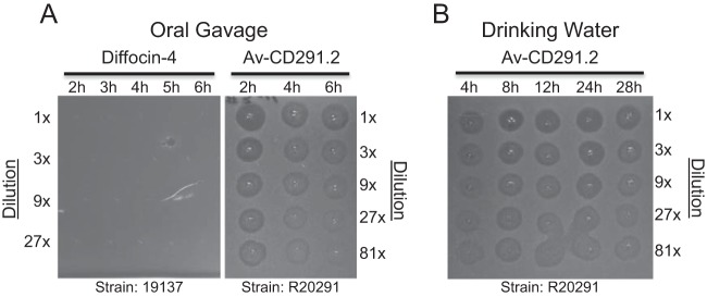
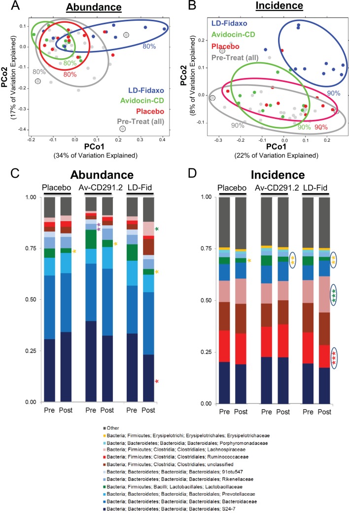
UNLABELLED: Clostridium difficile is a leading cause of nosocomial infections worldwide and has become an urgent public health threat requiring immediate attention. Epidemic lineages of the BI/NAP1/027 strain type have emerged and spread through health care systems across the globe over the past decade. Limiting person-to-person transmission and eradicating C. difficile, especially the BI/NAP1/027 strain type, from health care facilities are difficult due to the abundant shedding of spores that are impervious to most interventions. Effective prophylaxis for C. difficile infection (CDI) is lacking. We have genetically modified a contractile R-type bacteriocin ("diffocin") from C. difficile strain CD4 to kill BI/NAP1/027-type strains for this purpose. The natural receptor binding protein (RBP) responsible for diffocin targeting was replaced with a newly discovered RBP identified within a prophage of a BI/NAP1/027-type target strain by genome mining. The resulting modified diffocins (a.k.a. Avidocin-CDs), Av-CD291.1 and Av-CD291.2, were stable and killed all 16 tested BI/NAP1/027-type strains. Av-CD291.2 administered in drinking water survived passage through the mouse gastrointestinal (GI) tract, did not detectably alter the mouse gut microbiota or disrupt natural colonization resistance to C. difficile or the vancomycin-resistant Enterococcus faecium (VREF), and prevented antibiotic-induced colonization of mice inoculated with BI/NAP1/027-type spores. Given the high incidence and virulence of the pathogen, preventing colonization by BI/NAP1/027-type strains and limiting their transmission could significantly reduce the occurrence of the most severe CDIs. This modified diffocin represents a prototype of an Avidocin-CD platform capable of producing targetable, precision anti-C. difficile agents that can prevent and potentially treat CDIs without disrupting protective indigenous microbiota. IMPORTANCE: Treatment and prevention strategies for bacterial diseases rely heavily on traditional antibiotics, which impose strong selection for resistance and disrupt protective microbiota. One consequence has been an upsurge of opportunistic pathogens, such as Clostridium difficile, that exploit antibiotic-induced disruptions in gut microbiota to proliferate and cause life-threatening diseases. We have developed alternative agents that utilize contractile bactericidal protein complexes (R-type bacteriocins) to kill specific C. difficile pathogens. Efficacy in a preclinical animal study indicates these molecules warrant further development as potential prophylactic agents to prevent C. difficile infections in humans. Since these agents do not detectably alter the indigenous gut microbiota or colonization resistance in mice, we believe they will be safe to administer as a prophylactic to block transmission in high-risk environments without rendering patients susceptible to enteric infection after cessation of treatment.
Overview
The claims and statistics of this paper haven’t been checked yet.
Free: it reads the open-access text and checks the first 5 claims that cite a source we can check. Create a free account to check every claim.
Integrity signals
Registry
Cites
Statistics
Figures


Verification
Claims the paper makes
The claims and statistics of this paper haven’t been checked yet.
Free: it reads the open-access text and checks the first 5 claims that cite a source we can check. Create a free account to check every claim.
Claims other papers make about this one
Each is checked against this paper’s own text: does it say what the citing paper says it does?
The source backs part of the claim, not all of it — or backs it for a different population, condition or comparison.
In the citing paper
“the bacteriocin Avidocin-CDs, engineered to selectively target the clinically relevant C. difficile strain BI/NAP1/027, has been reported to promote pathogen clearance in mice upon oral administration.”
From Antibiotic-Induced Changes in the Intestinal Microbiota and Disease (2016) · cites this paper as [125]
Experimental — accuracy not yet measuredThe cited source says what the claim attributes to it.
In the citing paper
“Avidocin-CDs, unlike antibiotics, was not found to interfere with the ability of the microbiota to provide colonization resistance [125].”
From Antibiotic-Induced Changes in the Intestinal Microbiota and Disease (2016) · cites this paper as [125]
Self-check
Percentages, sums and reported differences re-computed from the paper’s own numbers.
The self-check needs the paper’s full text.
Citations
Cites
QIIME allows analysis of high-throughput community sequencing data
Caporaso, J. G., Kuczynski, J., Stombaugh, J., Bittinger, K., Bushman, F. D., Costello, E. K., Fierer, N., Peña, A. G., Goodrich, J. K., Gordon, J. I., et al. (2010) · Nature Methods · doi:10.1038/nmeth.f.303 · Cited by 34,887 · cited 1×
“QIIME and custom scripts were used to quality filter and demultiplex the sequencing reads (60).”
Enzymatic assembly of DNA molecules up to several hundred kilobases
Gibson, D. G., Young, L., Chuang, R.-Y., Venter, J. C., Hutchison, C. A., & Smith, H. O. (2009) · Nature Methods · doi:10.1038/nmeth.1318 · Cited by 9,996 · cited 1×
“Due to the ~20-kb size, the diffocin gene cluster was cloned into the integration vector using multiple DNA fragments via the Gibson assembly method (57).”
UniFrac: a New Phylogenetic Method for Comparing Microbial Communities
Lozupone, C., & Knight, R. (2005) · Applied and Environmental Microbiology · doi:10.1128/aem.71.12.8228-8235.2005 · Cited by 8,227 · cited 1×
“The unweighted UniFrac score considers only the presence or absence (incidence) of taxa (62, 63).”
Antibiotic resistance threats in the United States, 2019
(U.S.), C. F. D. C. A. P. (2019) · doi:10.15620/cdc:82532 · Cited by 5,782
Antibiotic resistance-the need for global solutions
Laxminarayan, R., Duse, A., Wattal, C., Zaidi, A. K. M., Wertheim, H. F. L., Sumpradit, N., Vlieghe, E., Hara, G. L., Gould, I. M., Goossens, H., et al. (2013) · The Lancet Infectious Diseases · doi:10.1016/s1473-3099(13)70318-9 · Cited by 4,439
The Treatment-Naive Microbiome in New-Onset Crohn’s Disease
Gevers, D., Kugathasan, S., Denson, L. A., Vázquez-Baeza, Y., Van Treuren, W., Ren, B., Schwager, E., Knights, D., Song, S. J., Yassour, M., et al. (2014) · Cell Host & Microbe · doi:10.1016/j.chom.2014.02.005 · Cited by 3,220
Lozupone, C. A., Hamady, M., Kelley, S. T., & Knight, R. (2007) · Applied and Environmental Microbiology · doi:10.1128/aem.01996-06 · Cited by 2,656
Cox, L. M., Yamanishi, S., Sohn, J., Alekseyenko, A. V., Leung, J. M., Cho, I., Kim, S. G., Li, H., Gao, Z., Mahana, D., et al. (2014) · Cell · doi:10.1016/j.cell.2014.05.052 · Cited by 1,859 · cited 1×
“In spite of the extraordinary benefits of traditional antibiotics, the negative consequences of continuing to treat and mistreat bacterial diseases with antibiotics rife with off-target effects are beginning to look dire (9, 51).”
An ecological and evolutionary perspective on human–microbe mutualism and disease
Dethlefsen, L., McFall-Ngai, M., & Relman, D. A. (2007) · Nature · doi:10.1038/nature06245 · Cited by 1,660
Fidaxomicin versus Vancomycin for Clostridium difficile Infection
Louie, T. J., Miller, M. A., Mullane, K. M., Weiss, K., Lentnek, A., Golan, Y., Gorbach, S., Sears, P., & Shue, Y.-K. (2011) · New England Journal of Medicine · doi:10.1056/nejmoa0910812 · Cited by 1,585
Yamamoto, K. R., Alberts, B. M., Benzinger, R., Lawhorne, L., & Treiber, G. (1970) · Virology · doi:10.1016/0042-6822(70)90218-7 · Cited by 1,565
UniFrac – An online tool for comparing microbial community diversity in a phylogenetic context
Lozupone, C., Hamady, M., & Knight, R. (2006) · BMC Bioinformatics · doi:10.1186/1471-2105-7-371 · Cited by 1,453 · cited 1×
“The unweighted UniFrac score considers only the presence or absence (incidence) of taxa (62, 63).”
What are the consequences of the disappearing human microbiota?
Blaser, M. J., & Falkow, S. (2009) · Nature Reviews Microbiology · doi:10.1038/nrmicro2245 · Cited by 855
The Changing Epidemiology of Clostridium difficile Infections
Freeman, J., Bauer, M. P., Baines, S. D., Corver, J., Fawley, W. N., Goorhuis, B., Kuijper, E. J., & Wilcox, M. H. (2010) · Clinical Microbiology Reviews · doi:10.1128/cmr.00082-09 · Cited by 826 · cited 1×
“As a nosocomial infection, CDI should be preventable, but incidence continues to rise (34, 36, 37).”
Dysbiosis of the Gut Microbiota Is Associated with HIV Disease Progression and Tryptophan Catabolism
Vujkovic-Cvijin, I., Dunham, R. M., Iwai, S., Maher, M. C., Albright, R. G., Broadhurst, M. J., Hernandez, R. D., Lederman, M. M., Huang, Y., Somsouk, M., et al. (2013) · Science Translational Medicine · doi:10.1126/scitranslmed.3006438 · Cited by 738 · cited 1×
“With a better understanding of the human gut microbiota has come the knowledge that loss of microbial diversity results in vulnerability to many diseases (48–50).”
Emergence and global spread of epidemic healthcare-associated Clostridium difficile
He, M., Miyajima, F., Roberts, P., Ellison, L., Pickard, D. J., Martin, M. J., Connor, T. R., Harris, S. R., Fairley, D., Bamford, K. B., et al. (2013) · Nature Genetics · doi:10.1038/ng.2478 · Cited by 735 · cited 2×
“Recent studies indicate 30 to 35% of North American CDIs were due to BI/ NAP1/027-type (ribotype 027 [RT027]) strains (2, 3), most of which are clonal and derived from 1 of 2 epidemic lineages (FQR1 and FQR2) that independently acquired fluoroquinolone resistance (4).”
Self-citationBuffie, C. G., Jarchum, I., Equinda, M., Lipuma, L., Gobourne, A., Viale, A., Ubeda, C., Xavier, J., & Pamer, E. G. (2012) · Infection and Immunity · doi:10.1128/iai.05496-11 · Cited by 596 · cited 2×
“2 treatment was not unexpected since the gut microbiota had not had sufficient time to recover after being disrupted by clindamycin (26) and mice can harbor C.”
The pyocins of Pseudomonas aeruginosa
Michel-Briand, Y., & Baysse, C. (2002) · Biochimie · doi:10.1016/s0300-9084(02)01422-0 · Cited by 560 · cited 1×
“S1 in the supplemental material) (14–16).”
Riggs, M. M., Sethi, A. K., Zabarsky, T. F., Eckstein, E. C., Jump, R. L. P., & Donskey, C. J. (2007) · Clinical Infectious Diseases · doi:10.1086/521854 · Cited by 550
Self-citationMicrobial Dysbiosis Is Associated with Human Breast Cancer
Xuan, C., Shamonki, J. M., Chung, A., DiNome, M. L., Chung, M., Sieling, P. A., & Lee, D. J. (2014) · PLoS ONE · doi:10.1371/journal.pone.0083744 · Cited by 543 · cited 1×
“Assignment of taxonomic classification for each resulting sequence was performed as described by Xuan et al. (49).”
Lawley, T. D., Clare, S., Walker, A. W., Stares, M. D., Connor, T. R., Raisen, C., Goulding, D., Rad, R., Schreiber, F., Brandt, C., et al. (2012) · PLoS Pathogens · doi:10.1371/journal.ppat.1002995 · Cited by 540
Self-citationStabler, R. A., He, M., Dawson, L., Martin, M., Valiente, E., Corton, C., Lawley, T. D., Sebaihia, M., Quail, M. A., Rose, G., et al. (2009) · Genome Biology · doi:10.1186/gb-2009-10-9-r102 · Cited by 454 · cited 1×
“The genome sequence for strain R20291 (RT027) contains the phi027 prophage insertion (20).”
Self-citationRole of the Intestinal Microbiota in Resistance to Colonization by Clostridium difficile
Britton, R. A., & Young, V. B. (2014) · Gastroenterology · doi:10.1053/j.gastro.2014.01.059 · Cited by 446
Stop the killing of beneficial bacteria
Blaser, M. (2011) · Nature · doi:10.1038/476393a · Cited by 381 · cited 1×
“In spite of the extraordinary benefits of traditional antibiotics, the negative consequences of continuing to treat and mistreat bacterial diseases with antibiotics rife with off-target effects are beginning to look dire (9, 51).”
The Clostridium difficile spo0A Gene Is a Persistence and Transmission Factor
Deakin, L. J., Clare, S., Fagan, R. P., Dawson, L. F., Pickard, D. J., West, M. R., Wren, B. W., Fairweather, N. F., Dougan, G., & Lawley, T. D. (2012) · Infection and Immunity · doi:10.1128/iai.00147-12 · Cited by 360 · cited 1×
“difficile spores in their fur for prolonged periods (40).”
Self-citationNakayama, K., Takashima, K., Ishihara, H., Shinomiya, T., Kageyama, M., Kanaya, S., Ohnishi, M., Murata, T., Mori, H., & Hayashi, T. (2000) · Molecular Microbiology · doi:10.1046/j.1365-2958.2000.02135.x · Cited by 359 · cited 1×
“Because short genes immediately downstream of RBPs often encode chaperone proteins that assist in tail assembly (15, 21), the two short genes ptsN and ptsO downstream of ptsM were included in a second new construct (Fig.”
Clostridium difficile infection: epidemiology, risk factors and management
Ananthakrishnan, A. N. (2011) · Nature Reviews Gastroenterology & Hepatology · doi:10.1038/nrgastro.2010.190 · Cited by 352 · cited 2×
“difficile is a leading cause of nosocomial infections worldwide (34, 35).”
Lawley, T. D., Clare, S., Walker, A. W., Goulding, D., Stabler, R. A., Croucher, N., Mastroeni, P., Scott, P., Raisen, C., Mottram, L., et al. (2009) · Infection and Immunity · doi:10.1128/iai.00558-09 · Cited by 349 · cited 1×
“2 in preventing BI-7 colonization and shedding during the spore transmission studies demonstrated the prophylactic potency of Avidocin-CD in vivo using an animal model reflective of human environmental exposure (24).”
Self-citationMeasures to Control and Prevent Clostridium difficile Infection
Gerding, D. N., Muto, C. A., & Owens, Jr. (2008) · Clinical Infectious Diseases · doi:10.1086/521861 · Cited by 311 · cited 2×
“Because today’s preventative options are limited to antibiotic stewardship and good hygienic practices (7, 8), new preventative approaches are urgently needed.”
Youngman, P. J., Perkins, J. B., & Losick, R. (1983) · Proceedings of the National Academy of Sciences · doi:10.1073/pnas.80.8.2305 · Cited by 306
Miller, M., Gravel, D., Mulvey, M., Taylor, G., Boyd, D., Simor, A., Gardam, M., McGeer, A., Hutchinson, J., Moore, D., et al. (2010) · Clinical Infectious Diseases · doi:10.1086/649213 · Cited by 278 · cited 1×
“As a nosocomial infection, CDI should be preventable, but incidence continues to rise (34, 36, 37).”
Sweetener Preference of C57BL/6ByJ and 129P3/J Mice
Bachmanov, A. A. (2001) · Chemical Senses · doi:10.1093/chemse/26.7.905 · Cited by 260 · cited 2×
“Sucrose (4% [wt/vol] final concentration) was added to the 1% bicarbonate formulation in order to increase water consumption and reduce diurnal variation in consumption rates (23).”
Clostridium difficile PCR ribotype 027: assessing the risks of further worldwide spread
Clements, A. C., Magalhães, R. J. S., Tatem, A. J., Paterson, D. L., & Riley, T. V. (2010) · The Lancet Infectious Diseases · doi:10.1016/s1473-3099(10)70080-3 · Cited by 206 · cited 1×
“difficile is a leading cause of nosocomial infections worldwide (34, 35).”
Petrella, L. A., Sambol, S. P., Cheknis, A., Nagaro, K., Kean, Y., Sears, P. S., Babakhani, F., Johnson, S., & Gerding, D. N. (2012) · Clinical Infectious Diseases · doi:10.1093/cid/cis430 · Cited by 189 · cited 2×
“Antibiotic treatment options for CDI, particularly those caused by RT027 strains, are plagued by high rates of relapse or recurrence after successful initial treatment (5, 6).”
NAP1 Strain Type Predicts Outcomes From Clostridium difficile Infection
See, I., Mu, Y., Cohen, J., Beldavs, Z. G., Winston, L. G., Dumyati, G., Holzbauer, S., Dunn, J., Farley, M. M., Lyons, C., et al. (2014) · Clinical Infectious Diseases · doi:10.1093/cid/ciu125 · Cited by 170 · cited 4×
“Because of the predominance of RT027 strains in many locations (~30% of CDI cases in the United States during 2011 through 2013) (2, 3) and the fact that this strain type is associated with increased disease severity and the highest rates of recurrence (2, 3, 6, 41), a stand-alone anti-RT027 agent, such as Av-CD291.”
InfluentialEthanol Consumption and Taste Preferences in C57BL/6ByJ and 129/J Mice
Bachmanov, A. A., Tordoff, M. G., & Beauchamp, G. K. (1996) · Alcoholism: Clinical and Experimental Research · doi:10.1111/j.1530-0277.1996.tb01630.x · Cited by 163 · cited 2×
“that replaced the usual water bottle in the cage (22).”
Retargeting R-Type Pyocins To Generate Novel Bactericidal Protein Complexes
Williams, S. R., Gebhart, D., Martin, D. W., & Scholl, D. (2008) · Applied and Environmental Microbiology · doi:10.1128/aem.00141-08 · Cited by 159 · cited 2×
“Because short genes immediately downstream of RBPs often encode chaperone proteins that assist in tail assembly (15, 21), the two short genes ptsN and ptsO downstream of ptsM were included in a second new construct (Fig.”
Self-citationPrevalence and Risk Factors for Asymptomatic Clostridium difficile Carriage
Alasmari, F., Seiler, S. M., Hink, T., Burnham, C.-A. D., & Dubberke, E. R. (2014) · Clinical Infectious Diseases · doi:10.1093/cid/ciu258 · Cited by 154
Figueroa, I., Johnson, S., Sambol, S. P., Goldstein, E. J. C., Citron, D. M., & Gerding, D. N. (2012) · Clinical Infectious Diseases · doi:10.1093/cid/cis357 · Cited by 148 · cited 1×
“Antibiotic treatment options for CDI, particularly those caused by RT027 strains, are plagued by high rates of relapse or recurrence after successful initial treatment (5, 6).”
Association of Relapse of Clostridium difficile Disease with BI/NAP1/027
Marsh, J. W., Arora, R., Schlackman, J. L., Shutt, K. A., Curry, S. R., & Harrison, L. H. (2012) · Journal of Clinical Microbiology · doi:10.1128/jcm.02291-12 · Cited by 141 · cited 1×
“Because of the predominance of RT027 strains in many locations (~30% of CDI cases in the United States during 2011 through 2013) (2, 3) and the fact that this strain type is associated with increased disease severity and the highest rates of recurrence (2, 3, 6, 41), a stand-alone anti-RT027 agent, such as Av-CD291.”
Donskey, C. J., Hanrahan, J. A., Hutton, R. A., & Rice, L. B. (1999) · The Journal of Infectious Diseases · doi:10.1086/314874 · Cited by 139 · cited 2×
“The sample size (n 10) for the colonization resistance studies was based on the effect size from previous published research (52).”
Self-citationAsymptomatic Clostridium difficile Colonisation and Onward Transmission
Eyre, D. W., Griffiths, D., Vaughan, A., Golubchik, T., Acharya, M., O’Connor, L., Crook, D. W., Walker, A. S., & Peto, T. E. A. (2013) · PLoS ONE · doi:10.1371/journal.pone.0078445 · Cited by 136
Bachmanov, A. A., Reed, D. R., Tordoff, M. G., Price, R. A., & Beauchamp, G. K. (1996) · Behavior Genetics · doi:10.1007/bf02361229 · Cited by 130 · cited 1×
“Graduated drinking tubes (22, 59) were used to deliver the drinking solution and measure daily consumption by each animal.”
Lawley, T. D., Clare, S., Deakin, L. J., Goulding, D., Yen, J. L., Raisen, C., Brandt, C., Lovell, J., Cooke, F., Clark, T. G., et al. (2010) · Applied and Environmental Microbiology · doi:10.1128/aem.00718-10 · Cited by 125 · cited 1×
“During CDI, the abundant shedding of spores impervious to most interventions makes it difficult to eradicate the pathogen from health care facilities and to limit person-to-person transmission (38, 39).”
Self-citationTickler, I. A., Goering, R. V., Whitmore, J. D., Lynn, A. N. W., Persing, D. H., & Tenover, F. C. (2014) · Antimicrobial Agents and Chemotherapy · doi:10.1128/aac.02775-13 · Cited by 123
Scholl, D., Cooley, M., Williams, S. R., Gebhart, D., Martin, D., Bates, A., & Mandrell, R. (2009) · Antimicrobial Agents and Chemotherapy · doi:10.1128/aac.01660-08 · Cited by 122
Self-citationAbujamel, T., Cadnum, J. L., Jury, L. A., Sunkesula, V. C. K., Kundrapu, S., Jump, R. L., Stintzi, A. C., & Donskey, C. J. (2013) · PLoS ONE · doi:10.1371/journal.pone.0076269 · Cited by 108 · cited 1×
“Mice with altered microbiota due to prior vancomycin administration became colonized with VA17 and VREF C68 as expected (33), with shedding levels reaching 108 CFU/g of feces on day 5 (Fig.”
Self-citationComparison of Strain Typing Results for Clostridium difficile Isolates from North America
Tenover, F. C., Åkerlund, T., Gerding, D. N., Goering, R. V., Boström, T., Jonsson, A.-M., Wong, E. Y., Wortman, A. T., & Persing, D. H. (2011) · Journal of Clinical Microbiology · doi:10.1128/jcm.02446-10 · Cited by 105
Novel High-Molecular-Weight, R-Type Bacteriocins of Clostridium difficile
Gebhart, D., Williams, S. R., Bishop-Lilly, K. A., Govoni, G. R., Willner, K. M., Butani, A., Sozhamannan, S., Martin, D., Fortier, L.-C., & Scholl, D. (2012) · Journal of Bacteriology · doi:10.1128/jb.01272-12 · Cited by 100 · cited 7×
“First, the entire PBSX prophage was eliminated ( PBSX) to create strain BDG9, as previously described (14).”
InfluentialSelf-citationJump, R., Polinkovsky, A., Hurless, K. N., Sitzlar, B. M., Eckart, K. E., Tomas, M. E., Deshpande, A., Nerandzic, M. M., & Donskey, C. J. (2014) · PLoS ONE · doi:10.1371/journal.pone.0101267 · Cited by 95 · cited 1×
“difficile strain that has been used in previous mouse colonization studies and has MICs of 128 g/ml and 1 g/ml for clindamycin and vancomycin, respectively (30, 31).”
Self-citationMulticenter Study of Clostridium difficile Infection Rates from 2000 to 2006
Dubberke, E. R., Butler, A. M., Yokoe, D. S., Mayer, J., Hota, B., Mangino, J. E., Khan, Y. M., Popovich, K. J., Fraser, V. J., & Prevention, P. E. P. F. T. C. F. D. C. A. (2010) · Infection Control & Hospital Epidemiology · doi:10.1086/656245 · Cited by 92
In vitro and in vivo evaluation of tiacumicins B and C against Clostridium difficile
Swanson, R. N., Hardy, D. J., Shipkowitz, N. L., Hanson, C. W., Ramer, N. C., Fernandes, P. B., & Clement, J. J. (1991) · Antimicrobial Agents and Chemotherapy · doi:10.1128/aac.35.6.1108 · Cited by 91 · cited 2×
“2 mg/kg to 5 mg/kg) (29) and far below the recommended 5-mg/kg human daily dose (28).”
KAGEYAMA, M. (1964) · The Journal of Biochemistry · doi:10.1093/oxfordjournals.jbchem.a127839 · Cited by 90
Pultz, N. J., & Donskey, C. J. (2005) · Antimicrobial Agents and Chemotherapy · doi:10.1128/aac.49.8.3529-3532.2005 · Cited by 77 · cited 1×
“difficile strain that has been used in previous mouse colonization studies and has MICs of 128 g/ml and 1 g/ml for clindamycin and vancomycin, respectively (30, 31).”
Self-citationNagaro, K. J., Phillips, S. T., Cheknis, A. K., Sambol, S. P., Zukowski, W. E., Johnson, S., & Gerding, D. N. (2013) · Antimicrobial Agents and Chemotherapy · doi:10.1128/aac.00580-13 · Cited by 75
van den Berg, R. J., Ameen, H. A., Furusawa, T., Claas, E. C. J., van der Vorm, E. R., & Kuijper, E. J. (2005) · Journal of Medical Microbiology · doi:10.1099/jmm.0.45825-0 · Cited by 62 · cited 1×
“The detection of a mixed infection (~10% of CDIs involve more than a single ribotype) would likely require deploying more than a single targeted Avidocin-CD (43, 44).”
Liu, S., Endo, K., Ara, K., Ozaki, K., & Ogasawara, N. (2008) · Microbiology · doi:10.1099/mic.0.2008/016881-0 · Cited by 58 · cited 1×
“To improve diffocin yield, we removed several genes from BDR11 via a marker-free deletion method published by Liu et al.(56).”
Nucleoid occlusion prevents cell division during replication fork arrest in Bacillus subtilis
Bernard, R., Marquis, K. A., & Rudner, D. Z. (2010) · Molecular Microbiology · doi:10.1111/j.1365-2958.2010.07369.x · Cited by 58 · cited 1×
“1) (54, 55).”
Clostridium difficile Mixed Infection and Reinfection
Eyre, D. W., Walker, A. S., Griffiths, D., Wilcox, M. H., Wyllie, D. H., Dingle, K. E., Crook, D. W., & Peto, T. (2011) · Journal of Clinical Microbiology · doi:10.1128/jcm.05177-11 · Cited by 56 · cited 1×
“The detection of a mixed infection (~10% of CDIs involve more than a single ribotype) would likely require deploying more than a single targeted Avidocin-CD (43, 44).”
STUDIES OF A PYOCIN. III. BIOLOGICAL PROPERTIES OF THE PYOCIN.
KAGEYAMA, M., Ikeda, K., & Egami, F. (1964) · PubMed · doi:10.1093/oxfordjournals.jbchem.a127841 · Cited by 52 · cited 1×
“Although the binding is highly specific, the killing mechanism is generic and extremely potent; a single R-type bacteriocin is sufficient to kill a target bacterium (18).”
Scholl, D., Gebhart, D., Williams, S. R., Bates, A., & Mandrell, R. (2012) · PLoS ONE · doi:10.1371/journal.pone.0033637 · Cited by 46 · cited 3×
“A common mechanism for the emergence of resistance to R-type bacteriocins is loss of the bacterial surface receptor (17, 19).”
InfluentialSelf-citationStiefel, U., Pultz, N. J., Helfand, M. S., & Donskey, C. J. (2004) · Infection Control and Hospital Epidemiology · doi:10.1086/502408 · Cited by 43 · cited 1×
“C68 is a clinical VanB VREF isolate that has also been used previously in mouse colonization studies (32).”
Self-citationFidaxomicin: A novel macrocyclic antibiotic for the treatment of Clostridium difficile infection
Crawford, T., Huesgen, E. C., & Danziger, L. H. (2012) · American Journal of Health-System Pharmacy · doi:10.2146/ajhp110371 · Cited by 38 · cited 2×
“2 mg/kg to 5 mg/kg) (29) and far below the recommended 5-mg/kg human daily dose (28).”
Sporicides for Clostridium difficile: the devil is in the detail
Wilcox, M. H., Fraise, A. P., Bradley, C. R., Walker, J. T., & Finch, R. G. (2011) · Journal of Hospital Infection · doi:10.1016/j.jhin.2010.10.017 · Cited by 21 · cited 1×
“During CDI, the abundant shedding of spores impervious to most interventions makes it difficult to eradicate the pathogen from health care facilities and to limit person-to-person transmission (38, 39).”
Cited by
Clostridium difficile infection
Smits, W. K., Lyras, D., Lacy, D. B., Wilcox, M. H., & Kuijper, E. J. (2016) · Nature Reviews Disease Primers · doi:10.1038/nrdp.2016.20 · Cited by 853
Antibiotic-Induced Changes in the Intestinal Microbiota and Disease
Becattini, S., Taur, Y., & Pamer, E. G. (2016) · Trends in Molecular Medicine · doi:10.1016/j.molmed.2016.04.003 · Cited by 901
“Furthermore, avidocinCDs, unlike antibiotics, was not found to interfere with the ability of the microbiota to provide colonization resistance [125].”
BackgroundMechanisms and consequences of intestinal dysbiosis
Weiss, G. A., & Hennet, T. (2017) · Cellular and Molecular Life Sciences · doi:10.1007/s00018-017-2509-x · Cited by 775
“difficile [187] and methicillin-resistant Staphylococcus aureus [188].”
BackgroundTargeting mechanisms of tailed bacteriophages
Nobrega, F. L., Vlot, M., de Jonge, P. A., Dreesens, L. L., Beaumont, H. J. E., Lavigne, R., Dutilh, B. E., & Brouns, S. J. J. (2018) · Nature Reviews Microbiology · doi:10.1038/s41579-018-0070-8 · Cited by 594
Eleraky, N. E., Allam, A., Hassan, S. B., & Omar, M. M. (2020) · Pharmaceutics · doi:10.3390/pharmaceutics12020142 · Cited by 342
“Avidocin and purocin [223] Gm+ve and Gm−ve bacteria Preclinical Modified R-type bacteriocin Novacta biosystems (NVB-302) [224] C.”
Background
Metadata
- Authors
- Gebhart, D., Lok, S., Clare, S., Tomas, M., Stares, M., Scholl, D., Donskey, C. J., Lawley, T. D., & Govoni, G. R.
- Affiliations
- AvidBiotics (United States) (US)
- Wellcome Sanger Institute (GB)
- Louis Stokes Cleveland VA Medical Center (US)
- Corresponding author
- Govoni, Gregory R.
- Venue
- mBio
- Publisher
- American Society for Microbiology
- Published
- 2015-05
- Volume
- 6
- Issue
- 2
- ISSN
- 2150-7511 (ISSN-L)
- 2161-2129
- PMC
- PMC4453579
- OpenAlex
- W2147684011
- Cited by
- 127
- References
- 64
- Citations by year
- 2026: 7 · 2025: 11 · 2024: 10 · 2023: 11 · 2022: 12 · 2021: 12 · 2020: 9 · 2019: 6 · 2018: 14 · 2017: 12 · 2016: 16 · 2015: 7
- FWCI
- 12.72
- Type
- journal article
- Language
- en
- Open access
- gold
- doi.org
- Licence
- cc-by
- creativecommons.org/licenses/by/3.0/ (vor)
- journals.asm.org/non-commercial-tdm-license (tdm)
- Locations
- mBio (publishedVersion, cc-by)
- Europe PMC (PubMed Central) (publishedVersion, cc-by)
- PubMed Central (publishedVersion, cc-by)
- PubMed (publishedVersion)
- DOAJ (DOAJ: Directory of Open Access Journals) (submittedVersion, cc-by-sa)
- Funders
- Wellcome Trust (WT 098051)
- National Institutes of Health (1r43ai098186-01)
- National Institute of Allergy and Infectious Diseases (1R43 AI098186, R43 AI098186)
- Topics
- Health Sciences › Medicine › Infectious Diseases › Clostridium difficile and Clostridium perfringens research
- Life Sciences › Immunology and Microbiology › Virology › Bacteriophages and microbial interactions
- Life Sciences › Agricultural and Biological Sciences › Food Science › Probiotics and Fermented Foods
- Keywords
- Clostridium difficile, colonization resistance, gut microbiota, vancomycin-resistant Enterococcus faecium, antibiotic-induced dysbiosis, nosocomial infections
- MeSH terms
- Administration, Oral, Animals, Anti-Bacterial Agents, Bacteriocins, Clostridium Infections, Female, Male, Mice, Inbred C57BL, Clostridioides difficile, Gastrointestinal Tract
About these checks
Registry, retraction, Cites and statistics results are computed, not judged, with one exception: a printed reference that no index lists is read once by a model before it counts as a citation error. Claims are extracted by a model and kept only when their quote appears verbatim in the text; each cited source is then checked, with a model’s help, for whether it says what the claim relies on. Those verdicts can be wrong — read the claim’s quote and the cited source before relying on one.
Statistics and arithmetic are recomputed, not judged.
Confidence bands are shown only for fields calibrated on our annotated test set. None is calibrated yet, so no band is shown.
What counts as a claim ›How we measure confidence ›How we check this ›
Are you an author, or do you see a problem with this page? Request a review
Data sources
Bibliographic data: Semantic Scholar, OpenAlex, Crossref.
Contains information from the Semantic Scholar Academic Graph Datasets, made available under the ODC Attribution License (ODC-BY 1.0).
Semantic Scholar’s data is described in: Kinney, R., et al. (2023). The Semantic Scholar Open Data Platform. arXiv:2301.10140.
