

Mitochondrial dysfunction in cell senescence and aging
Open accessThe DOI matches its Crossref record; 145 of the 145 references it cites were found in a registry or an index.
Abstract
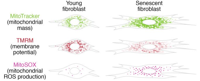
Mitochondrial dysfunction and cell senescence are hallmarks of aging and are closely interconnected. Mitochondrial dysfunction, operationally defined as a decreased respiratory capacity per mitochondrion together with a decreased mitochondrial membrane potential, typically accompanied by increased production of oxygen free radicals, is a cause and a consequence of cellular senescence and figures prominently in multiple feedback loops that induce and maintain the senescent phenotype. Here, we summarize pathways that cause mitochondrial dysfunction in senescence and aging and discuss the major consequences of mitochondrial dysfunction and how these consequences contribute to senescence and aging. We also highlight the potential of senescence-associated mitochondrial dysfunction as an antiaging and antisenescence intervention target, proposing the combination of multiple interventions converging onto mitochondrial dysfunction as novel, potent senolytics.
Overview
The claims and statistics of this paper haven’t been checked yet.
Free: it reads the open-access text and checks the first 5 claims that cite a source we can check. Create a free account to check every claim.
Integrity signals
Registry
Cites
Statistics
Figures

Verification
Claims the paper makes
The claims and statistics of this paper haven’t been checked yet.
Free: it reads the open-access text and checks the first 5 claims that cite a source we can check. Create a free account to check every claim.
Claims other papers make about this one
Each is checked against this paper’s own text: does it say what the citing paper says it does?
The source backs part of the claim, not all of it — or backs it for a different population, condition or comparison.
In the citing paper
“Both proliferation-competent glial cells (which constitute approximately 50% of brain cells) and postmitotic neurons can be positive for markers of cellular senescence in aging, obesity, traumatic injury, and neurodegeneration31,45–49, among other conditions.”
From Guidelines for minimal information on cellular senescence experimentation in vivo (2024) · cites this paper as [45]
Experimental — accuracy not yet measuredThe source backs part of the claim, not all of it — or backs it for a different population, condition or comparison.
In the citing paper
“Therefore, the dysregulation of mitochondrial fusion and fission pathways in senescent astrocytes likely contributes to mitochondrial dysfunction, oxidative stress, and the overall senescence-associated phenotype observed in these cells (Ma et al., 2020; Kleele et al., 2021; Miwa et al., 2022).”
From Aging promotes an increase in mitochondrial fragmentation in astrocytes (2024) · cites this paper as Miwa et al., 2022
Experimental — accuracy not yet measuredThe source backs part of the claim, not all of it — or backs it for a different population, condition or comparison.
In the citing paper
“Dysfunctional mitochondrial dynamics, as observed here, particularly excessive mitochondrial fission, have been associated with oxidative stress, mitochondrial DNA damage, and cellular senescence in various cell types (Twig et al., 2008; Miwa et al., 2022).”
From Aging promotes an increase in mitochondrial fragmentation in astrocytes (2024) · cites this paper as Miwa et al., 2022
Experimental — accuracy not yet measuredThe cited source says what the claim attributes to it.
In the citing paper
“while the presence of senescence markers has been proven even in post-mitotic cells”
From Guidelines for minimal information on cellular senescence experimentation in vivo (2024) · cites this paper as [45]
The check wasn’t confident either way, so it gives no verdict — never disputed.
In the citing paper
“Evidence from the literature indicates that senescent cells exhibit an increase in oxygen consumption rate (Hutter et al., 2004; Son et al., 2017; Miwa et al., 2022; Bang et al., 2023; Diniz et al., 2024a).”
From Aging promotes an increase in mitochondrial fragmentation in astrocytes (2024) · cites this paper as Miwa et al., 2022
Experimental — accuracy not yet measured
2 more claims about this paper
Create a free account to see every claim other papers make about this one.
Self-check
Percentages, sums and reported differences re-computed from the paper’s own numbers.
The self-check needs the paper’s full text.
Citations
Cites
The serial cultivation of human diploid cell strains
Hayflick, L., & Moorhead, P. S. (1961) · Experimental Cell Research · doi:10.1016/0014-4827(61)90192-6 · Cited by 8,508 · cited 1×
“Introduction What is cell senescence? Cell senescence was originally defined as the irreversible loss of replicative potential of primary cells in culture (1), initiated as a persistent DNA damage response to dysfunctional telomeres (2).”
BackgroundTelomeres shorten during ageing of human fibroblasts
Harley, C. B., Futcher, A. B., & Greider, C. W. (1990) · Nature · doi:10.1038/345458a0 · Cited by 5,750 · cited 2×
“Telomere dysfunction might be caused by telomere shortening during DNA replication (3, 4), replication-independent telomere damage (5), or a variety of other stresses (6).”
BackgroundA role for mitochondria in nlrp3 inflammasome activation
Zhou, R., Yazdi, A. S., Menu, P., & Tschopp, J. (2011) · Nature · doi:10.1038/nature09663 · Cited by 5,602
The senescence-associated secretory phenotype: the dark side of tumor suppression
Coppé, J.-P., Desprez, P.-Y., Krtolica, A., & Campisi, J. (2010) · Annual Review of Pathology: Mechanisms of Disease · doi:10.1146/annurev-pathol-121808-102144 · Cited by 5,206
Wu, Z., Puigserver, P., Andersson, U., Zhang, C., Adelmant, G., Mootha, V., Troy, A., Cinti, S., Lowell, B., Scarpulla, R. C., et al. (1999) · Cell · doi:10.1016/s0092-8674(00)80611-x · Cited by 4,139 · cited 1×
“PGC-1α activates the transcription factors Nrf1/2 and ERRα, which further control the TFAM transcription in response to increased mitochondrial biogenesis (65).”
BackgroundClearance of p16Ink4a-positive senescent cells delays ageing-associated disorders
Baker, D. J., Wijshake, T., Tchkonia, T., LeBrasseur, N. K., Childs, B. G., van de Sluis, B., Kirkland, J. L., & van Deursen, J. M. (2011) · Nature · doi:10.1038/nature10600 · Cited by 3,670
AMPK regulates energy expenditure by modulating NAD+ metabolism and SIRT1 activity
Cantó, C., Gerhart-Hines, Z., Feige, J. N., Lagouge, M., Noriega, L., Milne, J. C., Elliott, P. J., Puigserver, P., & Auwerx, J. (2009) · Nature · doi:10.1038/nature07813 · Cited by 3,300
Cellular Senescence: Defining a Path Forward
Gorgoulis, V., Adams, P. D., Alimonti, A., Bennett, D. C., Bischof, O., Bishop, C., Campisi, J., Collado, M., Evangelou, K., Ferbeyre, G., et al. (2019) · Cell · doi:10.1016/j.cell.2019.10.005 · Cited by 3,225 · cited 1×
“Telomere dysfunction might be caused by telomere shortening during DNA replication (3, 4), replication-independent telomere damage (5), or a variety of other stresses (6).”
BackgroundNakahira, K., Haspel, J. A., Rathinam, V. A. K., Lee, S.-J., Dolinay, T., Lam, H. C., Englert, J. A., Rabinovitch, M., Cernadas, M., Kim, H. P., et al. (2011) · Nature Immunology · doi:10.1038/ni.1980 · Cited by 3,049
A DNA damage checkpoint response in telomere-initiated senescence
Fagagna, F. D. D., Reaper, P. M., Clay-Farrace, L., Fiegler, H., Carr, P., von Zglinicki, T., Saretzki, G., Carter, N. P., & Jackson, S. P. (2003) · Nature · doi:10.1038/nature02118 · Cited by 2,917 · cited 1×
“Introduction What is cell senescence? Cell senescence was originally defined as the irreversible loss of replicative potential of primary cells in culture (1), initiated as a persistent DNA damage response to dysfunctional telomeres (2).”
Background
Research works printed but not linked: 2
- von Zglinicki T. Mechanisms of cell senescence in aging. In: Musi N, Hornsby P eds. Handbook of the Biology of Aging. Academic Press; 2021:53–68.
- Miwa S, Johnson TE. Stress and aging. Vijg J, et al., eds. In: The Molecular and Cellular Biology of Aging. Gerontological Society of America; 2016:216-249.
Cited by
Items 901–999 of 999
Li, X., Xu, H. M., Shi, M., Liu, K., & Liu, X. (2026) · Toxicology and Applied Pharmacology · doi:10.1016/j.taap.2026.117772
Characterizing mitochondrial phenotypes and MERCS in aged human skeletal muscle myoblasts
Unten, Y., Takafuji, K., Masukagami, Y., Shiiba, I., Horiuchi, K., Husník, F., Yanagi, S., Tateishi, N., & Suzuki, T. (2026) · PLoS ONE · doi:10.1371/journal.pone.0343604 · Cited by 3
Age-dependent mitochondrial toxicity in renal stem cells: A precise 3D silk matrix model
Yu, P., Ding, H., Ma, J.-X., Duan, Z. J., Chen, Y., Atala, A., & Zhang, Y. (2026) · Genes & Diseases · doi:10.1016/j.gendis.2026.102108
“60 In our study, we discovered that oUSCs exhibited higher levels of β -galactosidase expression compared with yUSCs.”
Energy Metabolic Regulatory Materials Promote Wound Healing in Senescent Environment
Wang, X., Zhang, T., Zhou, H., & Yang, L. (2026) · Progress in molecular and subcellular biology · doi:10.1007/978-3-032-17771-1_1
Molecular drivers of ageing and emerging longevity therapies
Dhinakaran, M. L., Murugesu, S., & Srinivasan, D. K. (2026) · Exploration of Drug Science · doi:10.37349/eds.2026.1008152
Bioactive sesquiterpenes from heartwood of Pterocarpus santalinus as potent SIRT-1 activators
Lone, B. A., Choudhary, S., Dutt, M., Ayoub, N., Bhushan, A., Sharma, S., Gairola, S., Kumar, A., & Gupta, P. (2026) · Phytomedicine Plus · doi:10.1016/j.phyplu.2026.100973
The cellular and molecular pathophysiology of diabetes in aging
Namrata, N., Mundlia, P., Attri, D., Sharma, I., Sharma, A., & Giri, A. (2026) · Elsevier eBooks · doi:10.1016/b978-0-443-44665-8.00008-0
Multi-omics senescence landmarks facilitate pro-senescence and senolytic sequential cancer therapy
Xu, Y., Zhu, L., Jiang, Z., Wei, B., Zhou, G., Liu, H., Chen, X., He, Y., & Li, H. (2026) · Genes & Diseases · doi:10.1016/j.gendis.2026.102113
Doe, D., Vu, A., Ham, J. P., & Perlin, M. H. (2026) · Journal of Fungi · doi:10.3390/jof12030206
“The dysfunction of these organelles is associated with aging, diseases and other biological processes [11,12].”
Disturbed LTB4 Metabolism in ILC2s Aggravates Ulcerative Colitis via Intercellular ATP Accumulation
Li, W., Zheng, M., Wang, Y., Chen, Y., Bai, H., Li, R., Li, S., & Yanqing, L. I. (2026) · Inflammation · doi:10.1007/s10753-026-02486-7
Dilating the aging clock with light
Jing‐Yi, J. C., Ho, I. Y. M., Quah, C. B., Bai, Y. Y., Ren, J., Foo, A. S. C., & Teh, D. B. L. (2026) · The Innovation · doi:10.1016/j.xinn.2026.101368
“70 An important compounding factor is the tumor suppressor p53 .”
Xu, S., Wang, J., Sha, Y., & Ji, Z. (2026) · Gene Reports · doi:10.1016/j.genrep.2026.102501
Cong, W., Jing, H., Li, Z., Zhang, W., Zhang, N., Xie, Y., Gao, S., Huang, Y., & Ning, J. (2026) · Advanced Science · doi:10.1002/advs.202523931
“The process of mitochondrial dysfunction is always accompanied by deregulation of mitochondrial homeostasis [ 13, 38 ].”
Targeting the liver for longevity
Yu, J., Fan, B., Gao, X., Fan, Y., Wang, S., Huang, C., & Fan, S. (2026) · Ageing Research Reviews · doi:10.1016/j.arr.2026.103142
Tomkova, K., Lewis, J. W., Nicholson, T. A., Acharjee, A., Anderson, A., Barlow, J. P., Frost, K., Jackson, T., Magalhaes, J. P., Mondal, S., et al. (2026) · bioRxiv (Cold Spring Harbor Laboratory) · doi:10.64898/2026.04.20.719545
“Mitochondrial dysfunction promotes DNA damage, triggering p53-dependent senescence pathways and amplifying SASP burden 62 .”
Li, W., Liu, J., Fu, X., Liu, Y., Xu, Y., Fang, X., Guo, Q., & Liu, Q. (2026) · Archives of Pharmacal Research · doi:10.1007/s12272-026-01614-z
Hu, Z., Chen, Y., Yao, Y., Yang, X., & Li, G. (2026) · Biochemical and Biophysical Research Communications · doi:10.1016/j.bbrc.2026.153831
Xiao, F., Wang, H., Zhao, L., Li, G., Ma, S.-Y., Yang, L.-Q., Wang, H., Zhang, Y. X., Li, J., & Kong, Q. (2026) · Cell Reports Medicine · doi:10.1016/j.xcrm.2026.102767 · Cited by 1
“31 Taken together, these findings highlight a potential link between ribosome heterogeneity, mitochondrial function, and healthy longevity.”
Overview of hallmarks of aging
Sangeeth, A., Subramanian, M., Singh, A., & Singh, A. (2026) · Elsevier eBooks · doi:10.1016/b978-0-443-33159-6.00004-7
Autophagy as a therapeutic target to enhance reverse aging
Akimov, O., Kostenko, V., Kostenko, H., & Kostenko, V. (2026) · Elsevier eBooks · doi:10.1016/b978-0-443-33159-6.00015-1
Chang, H., Feng, Y., Zhang, B., Xue, Y., Jiao, J., & Guo, Y. (2026) · JACS Au · doi:10.1021/jacsau.6c00378
A Ganoderma lucidum Extract Improves Mitochondrial Function in Aging Skin and Promotes Wound Healing
Tao, L., Tai, M., Zhou, Z., Chen, Z., Zhang, R., Ge, Y., & Ju, Z. (2026) · Clinical Cosmetic and Investigational Dermatology · doi:10.2147/ccid.s596475
“14,15 Specifically, cellular senescence often instigates mitochondrial dysfunction, characterized by diminished oxidative phosphorylation efficiency, reduced ATP synthesis, and a decline in mitochondrial membrane potential, frequently accompanied by an overproduction of ROS.”
Arias, N., Rodríguez‐Fernández, L., Zorzo, C., Pena-León, V., Cordero, L. M., Gutiérrez‐Menéndez, A., Martínez, & Arias, J. D. (2026) · GeroScience · doi:10.1007/s11357-026-02278-9 · Cited by 1
“Mitochondrial dysfunction, driven by impaired mitophagy and declining respiratory capacity, further compromises neuronal survival [18–21], contributing to neuroinflammation and cognitive decline.”
Uranova, N. A., Vikhreva, O. V., & Рахманова, В. И. (2026) · European Archives of Psychiatry and Clinical Neuroscience · doi:10.1007/s00406-026-02263-5
“A cause of cellular senescence is mitochondrial dysfunction defined as a decreased respiratory capacity per mitochon - drion and a decreased mitochondrial membrane potential, typically accompanied by increased production of oxygen free radicals [59].”
ECM-Mediated Regulation of Organelles and Their Function
Malaviya, R., Raut, V., Mazumdar, D., Sherkhane, T., Saha, A., Dalai, A., & Balasubramanian, N. (2026) · Biology of extracellular matrix · doi:10.1007/978-3-032-22618-1_7
Zhang, L., Yang, Q., Xu, C., Hu, Y., Xiao, L., & Yu, J. (2026) · European Archives of Psychiatry and Clinical Neuroscience · doi:10.1007/s00406-026-02258-2
Makalani, F., Ghanbari, A., Tabandeh, M. R., Nasta, T. Z., Souq, A. J., & Jalili, C. (2026) · Research in Pharmaceutical Sciences · doi:10.4103/rps.rps_175_25
“This mitochondrial dysfunction can lead to metabolic alterations, including increased oxidative phosphorylation activity, even in the presence of inefficient electron transport and reduced ATP production, which may contribute to the persistence of the senescent state (50) .”
Puchalska, M., & Witkowska‐Piłaszewicz, O. (2026) · Equine Veterinary Journal · doi:10.1002/evj.70189
Bu, F., Sun, M., Xu, R., Jiang, B., Feng, S., Peng, C., Huang, W., Wang, X., & Zhao, C. (2026) · Journal of Agricultural and Food Chemistry · doi:10.1021/acs.jafc.5c14474
Blue period - features of senescence 30 years after beta-galactosidase
Kuehnemann, C., & Wiley, C. D. (2026) · Aging · doi:10.18632/aging.206380
“Thus, loss of mitochondrial function is not only a feature of senescence, but can also influence senescence and the SASP in multiple ways [91, 92].”
Amino acid metabolism at the senescence–cancer interface
Lori, G., Mancini, C., Pranzini, E., Magherini, F., & Taddei, M. L. (2026) · Trends in cancer · doi:10.1016/j.trecan.2026.04.007
Deletion of TOP2B promoter using CRISPR-Cas9 induces senescence in HEK 293T cells
Saroj, N., Velangani, H. G., Kiran, S., & Dholaniya, P. S. (2026) · Biochemistry and Biophysics Reports · doi:10.1016/j.bbrep.2026.102620
“This alteration in mitochondrial function is consistent with the senescence phenotype, where mitochondrial dysfunction often serves as both a marker and a contributor to cellular aging [52].”
Gut microbiota: The hidden hallmark of aging
Kaushik, K., Sharma, M., Sharma, R., Reza, A., Sharma, G., Panwar, S., Nagraik, R., Negi, P., & Sharma, A. (2026) · Acta Microbiologica et Immunologica Hungarica · doi:10.1556/030.2026.02808
Shao, Q., Chen, D., Shao, W., & Ye, Q. (2026) · Research · doi:10.34133/research.1326 · Cited by 1
“1 ) [ 4 , 48 ].”
Diverse Forms of Autophagy and Their Roles in Liver Disease and Aging: A Comprehensive Review
Heo, S., Lee, M. Y., Jeong, C. Y., Kim, D. J., & Jun, J. H. (2026) · Proteomes · doi:10.3390/proteomes14020028
“A defining feature of senescent hepatocytes is impaired mitochondrial quality control, characterized by reduced mitochondrial membrane potential (MMP), defective mitochondrial respiration, and impaired mitophagy [26].”
Age-related changes to brain energetics revealed by PET/MRI imaging
Bevington, C. W. J., Dhaliwal, S., McKenzie, J., Stoessl, A. J., & Sossi, V. (2026) · NeuroImage Clinical · doi:10.1016/j.nicl.2026.104014
“…of neurons; given the increasing genetic and in vitro evidence for mitochondrial dysfunction with age (Cui et al., 2012; Jang et al., 2018; Miwa et al, 2022; Guo et al., 2023), it is reasonable to hypothesize that energy production mechanisms change with age, which would lead to changes…”
Zhang, H., Wang, Y., Wang, C., Guo, X., Chen, H., Hou, Y., Wu, X., Wu, Z.-J., Pan, W., Ma, R.-J., et al. (2026) · bioRxiv (Cold Spring Harbor Laboratory) · doi:10.64898/2026.05.27.728315
Jiang, W., Lynam, E. G., Delafosse, J., Birdsey, G. M., RANDI, A. M., Matter, K., & Balda, M. S. (2026) · Cells · doi:10.3390/cells15111015 · Cited by 1
“Loss of the mitochondrial network and fragmentation can lead to mitochondrial dysfunction and senescence by excessive production of ROS [45,46].”
Campbell, A. N., Jung, K. N., Doyle, S. J., Lebayle, E., Corneo, B., Feng, J., Ricupero, C. L., Bain, J., & Strich, R. (2026) · bioRxiv (Cold Spring Harbor Laboratory) · doi:10.64898/2026.06.01.729270
HMGA2 identified via m6A-senescence multi-omics in laryngeal squamous cell carcinoma
Jin, M., Qu, L., Xu, Z., Teng, S., Liu, S., Li, Q., Zhang, F., Hao, Q., Wang, P., Zhao, R., et al. (2026) · Scientific Reports · doi:10.1038/s41598-026-55980-1
Chen, L., Hu, S., Tang, X., Wang, Y., Zou, Y., Luo, F., & Wan, H. (2026) · Frontiers in Cell and Developmental Biology · doi:10.3389/fcell.2026.1740661
“Mitochondrial dysfunction is tightly correlated with aging (Miwa et al., 2022).”
Sethuraman, V. (2026) · Medical Hypotheses · doi:10.1016/j.mehy.2026.112044
Nambara, T., Parsons, T. M., Raulin, A., Gaona, A., Roy, B., & Kanekiyo, T. (2026) · Cytotherapy · doi:10.1016/j.jcyt.2026.102940
Son, H.-J., Lee, G., Ryu, Y., Noh, K. B., Hwang, J. S., Oh, S., & Shin, Y. J. (2026) · Translational Vision Science & Technology · doi:10.1167/tvst.15.6.23
“62,63 TGF-β –treated cells exhibited reduced mitochondrial membrane potential and increased levels of parkin and PINK1, which are markers of mitophagy activation.”
Qian, Z., Li, S., Yu-yang, L., Min‐lin, L., Huang, C., Min-hui, L., We, L., & Si-yuan, T. (2026) · Biochemical Pharmacology · doi:10.1016/j.bcp.2026.118172 · Cited by 1
Garg, R. (2026) · Advanced Health Science and Technology Journal · doi:10.4103/ahstj.ahstj_4_26
Wang, J., Wei, M., Song, X., Zhang, J. Y., Zhang, L., Chen, S., Hu, Y., Gao, H., & Li, J. (2026) · Advanced Science · doi:10.1002/advs.202522788 · Cited by 1
“Moreover, mitochondrial dysfunction is a proximal trigger for senescence and SASP activation [ 38 ].”
Ziraoui, N.-E., Mendoza, R., Harrington, V., & Carter, S. (2026) · International journal of agriculture and food sciences research. · doi:10.62051/ijafsr.v4n1.05
“Mitochondrial dysfunction and cellular senescence are closely interconnected, and the senescent phenotype is typically accompanied by lower mitochondrial membrane potential, impaired respiratory capacity, and increased reactive oxygen species (ROS) generation [1].”
The multifaceted inducers of cellular senescence
Gal, H., & Krizhanovsky, V. (2026) · Aging · doi:10.18632/aging.206391
“…as a core mechanism linking senescence, ferroptosis susceptibility, lysosomal remodeling, and retrograde mitochondrial-to-nuclear signaling pathways that extend beyond MiDAS, highlighting the need to distinguish MiDAS from other oxidative stress-and metabolism-driven senescence programs [98].”
Yuan, B., Fu, J., Zhao, Y., Zheng, G., Lin, H., Chen, X., Guo, X., & Shi, J. (2026) · Materials Today Bio · doi:10.1016/j.mtbio.2026.103389 · Cited by 2
“A stable mitochondrial membrane potential (MMP) is fundamental to mitochondrial function [53].”
Chen, S., & Kokkoli, E. (2026) · Journal of Controlled Release · doi:10.1016/j.jconrel.2026.115125
Inhibition of NHE1 induces trophoblast cell senescence via ADGRG6 mediated by CXCL8
Lin, Z., Shi, J., Huang, X., Wang, J., Zheng, Z., Chen, M., 朱晓勇, Li, M., & Shao, J. (2026) · Journal of Reproductive Immunology · doi:10.1016/j.jri.2026.104927
Jiang, Y., Dai, Y., Li, X., Cui, Y., Liu, H., Hasegawa, T., & Li, M. (2026) · Cellular Signalling · doi:10.1016/j.cellsig.2026.112694
Catechin and applications to aging research
Sureda, A., Quetglas‐Llabrés, M. M., Jiménez‐García, M., & Tejada, S. (2026) · Elsevier eBooks · doi:10.1016/b978-0-443-29192-0.00030-3
Li, R. X., Hu, C., Zhou, P., Si, G., Cui, J., He, Y., Han, Y., Zhang, J., Li, W., Wang, L., et al. (2026) · Journal of Translational Medicine · doi:10.1186/s12967-026-08538-8 · Cited by 1
“Mitochondrial dysfunction is a defining characteristic of aging tissues and senescent cells, with changes in mitochondrial mass, mitochondrial DNA (mtDNA), and respiratory capacity playing a pivotal role in the aging process [7].”
Liu, Z., Abulaiti, A., Li, D., Li, J., Wahafu, P., Zhang, Y., Maimaitiming, M., Li, S. Y., Li, G., & Yakufu, M. (2026) · Ageing Research Reviews · doi:10.1016/j.arr.2026.103221 · Cited by 1
Does Aging Affect Diet-Induced Thermogenesis? Revisiting the Evidence from Human Studies
Gonzalez, K. D., Dalamaga, Μ., Hu, L., & Magkos, F. (2026) · Current Obesity Reports · doi:10.1007/s13679-026-00730-7
“These include age-related changes in mitochondrial efficiency, which may influence heat dissipation during later phases of DIT [ 55], and circadian variation, as some studies in young individuals report higher DIT in the morning than later in the day [56, 57 ], while others show no effect [ 58– 60…”
Senescent cells accumulate lipid droplets
Rachmian, N., Shmulevich, R., Akiva, H., Pokorná, Š., Dassa, B., Cherqui, U., Malitsky, S., Itkin, M., Kliger, B. A., Goliand, I., et al. (2026) · Aging · doi:10.18632/aging.206390 · Cited by 1
Ning, N., Wang, C., Gao, T., Ma, J. X., Ding, M., Zhang, F., Liu, Q., Zhang, W., & Gong, L. (2026) · Molecular Carcinogenesis · doi:10.1002/mc.70134
“It was reported that mitochondrial dysfunction, including increased production of oxygen free radicals and decreased mitochondrial membrane potential, is an important cause and consequence of CSEN [27].”
SERPINE1–AP2A1 interplay links substrate stiffness to fibroblast senescencePreprint
Nisa, I. C., Chantachotikul, P., Saito, T., Bertocchi, C., & Deguchi, S. (2026) · bioRxiv (Cold Spring Harbor Laboratory) · doi:10.64898/2026.06.30.735455
“Cellular senescence is a state of irreversible cell cycle arrest triggered by diverse stressors, including telomere shortening [1], [2], [3], mitochondrial dysfunction [4], [5], activation of the DNA damage response [6], and epigenetic modifications [7], [8].”
Exercise-induced mitochondrial remodeling in energy-demanding organs during aging
Zhou, Z., Gao, J., Wang, M., & Zhang, H. (2026) · iScience · doi:10.1016/j.isci.2026.116791 · Cited by 1
“62 Aging and disease onset in tissues and organs are intricately associated with the decline in mitochondrial quality despite the presence of organ-specific aging patterns.”
Mitochondrial dysfunction in energy metabolism: Unraveling cardiometabolic diseases
Gao, J., & Yao, M. (2026) · Elsevier eBooks · doi:10.1016/b978-0-443-34186-1.00011-6
Robinson, Z. R., Fong, S. C., Choi, D. Y., Siopack, S., Bolanos-Campos, A., Morshedian, N., Utke, E., Kanda, M., Kato, C., Conboy, M. J., et al. (2026) · Aging · doi:10.18632/aging.206399
“With respect to this molecular mechanism, senescent and malignant cells have similarities in restructured metabolic state marked by inefficient oxidative phosphorylation, heightened glycolytic flux, and disrupted redox balance [26 – 28].”
Exploring the Anticancer Potential of Monoterpenes
Baloğlu, K. İ., & Erdoğan, A. (2026) · Journal of Anatolian Environmental and Animal Sciences · doi:10.35229/jaes.1926404
Al-Jameel, S. S. (2026) · Brazilian Journal of Biology · doi:10.1590/1519-6984.304624
Wang, K., Hu, X., Bi, C., Zhuang, Y., Zhang, Y., Shao, Z., Chen, L., Xiang, H., Jin, L., Chen, S., et al. (2026) · Advanced Materials · doi:10.1002/adma.74133
Pioger, A., Martin, N., Abbadie, C., & Dehennaut, V. (2026) · Biochimica et Biophysica Acta (BBA) - Reviews on Cancer · doi:10.1016/j.bbcan.2026.189665
Distinct roles of COPI proteins attenuated in cell senescence
Mazan-Mamczarz, K., Wind, E. J., Pal, A., Salamini-Montemurri, M., Tsitsipatis, D., Kim, K. M., Munk, R. B., Leng, J., Shin, C. H., Martindale, J. L., et al. (2026) · Science Advances · doi:10.1126/sciadv.aec2786
“For example, mitochondria in senescent cells produce more reactive oxygen species ( 3 ), and lysosomes display increased β - galactosidase activity ( 1 , 2 ).”
Sodré, A. F. P., Rodrigues, L. G., Sloan, K. P., Sloan, L. A., Tanaka, M., Curi, R., Takeda, L. N., Goulart, R. D. Á., de Souza, A. L. D. M., Detregiachi, C. R. P., et al. (2026) · International Journal of Molecular Sciences · doi:10.3390/ijms27156554
“As pointed out above, mitochondria are cellular organelles present in the cytoplasm of eukaryotic cells and are composed of two membranes, one internal and one exter-nal [124,125].”
Zhou, Y., Wu, Z., Zheng, T., Luo, S., Zhang, L., Li, Y., Liu, S., Liu, T., Zhang, T., & Zhang, X. (2026) · Journal of Controlled Release · doi:10.1016/j.jconrel.2026.115197 · Cited by 1
Bisphenol F triggers nephrotoxicity: The pivotal role of renal tubular epithelial cells senescence
Zhang, K., Du, X., Li, Z., Yin, X., Duan, Y., Li, H., Liang, N., Wang, Y., Su, Y., Wei, Z., et al. (2026) · Journal of Hazardous Materials Advances · doi:10.1016/j.hazadv.2026.101418
Piezo1-mitochondrial calcium coupling in mechanotransduction, mitochondrial stress, and cell fate
Shi, P., Zhao, X., Li, M., Wu, X., Li, Y., & Qiming, Z. (2026) · Cell Calcium · doi:10.1016/j.ceca.2026.103181 · Cited by 1
Weston, M. S., Domínguez-Prieto, M., & Moisoi, N. (2026) · Cellular Signalling · doi:10.1016/j.cellsig.2026.112763
Liang, C., Cheng, Y., Li, Z., Zheng, A., Fu, X., Zhang, W., Cao, L., & Wang, C. (2026) · Advanced Materials · doi:10.1002/adma.74352
“Mitochondria play an essential role in regulating the metabolic processes, especially governing the TCA cycle and OXPHOS process [ 47, 48 ].”
Yu, Z., Liu, Z., Fan, J., Li, Y., He, J., Zhang, J., Fu, P., Gao, D., Xia, F., Mei, Y., et al. (2026) · Immunity & Ageing · doi:10.1186/s12979-026-00575-7
“…pathway to promote cellular senescence in SFs. Mitochondrial dysfunction, characterized by diminished respiratory capacity, loss of mitochondrial membrane potential, and typically increased production of reactive oxygen species, serves as both a cause and a consequence of cellular senescence [49].”
Bioengineering strategies to interrogate and modulate the aging immune system
Nikolich‐Žugich, J., Wolf, M. T., Pompano, R. R., & Maisel, K. (2026) · Nature Aging · doi:10.1038/s43587-026-01187-y
“These signs include replicative arrest, increased cellular p21 and p16 levels 30,40,41 , compromised mitochondrial function and an increase in mitochondrial and cellular reactive oxygen species 42 , and frequent, but not invariable, alterations of the cellular secretome 30,41 .”
Chen, F., Zhang, C., Li, Y., Khan, M. A., Pang, X., Zhu, S., & Tang, B. (2026) · Industrial Crops and Products · doi:10.1016/j.indcrop.2026.124024
A review of podocyte senescence and aging
Shankland, S. J., Ishibe, S., Pippin, J. W., & Wessely, O. (2026) · Elsevier eBooks · doi:10.1016/b978-0-443-34069-7.00006-9 · Cited by 1
Han, L., Ho, C.-T., Ye, Z., Zhang, H., Zong, C., Kou, X., & Lu, M. (2026) · Food Bioscience · doi:10.1016/j.fbio.2026.109641
Lv, W., Zhong, X., Yuan, Q., Gong, X., Zhao, Y., Zhang, S., Wu, A., Wan, M., Li, X., Zheng, Y., et al. (2026) · Aging Cell · doi:10.1111/acel.70652
“Gene Ontology (GO) enrichment analysis indicated that AGGF1 modulates mitochondrial-related pathways (Figure 4b), which are known to contribute to cellular senescence (Miwa et al. 2022).”
SIRT5 regulates bone homeostasis via PINK1/Parkin mediated mitophagy and protects against senescence
Paul, A., Yadav, N., Singh, D. P., Kaushal, S. K., Dixit, M., Singh, N. C., Kaushik, D., Yadav, P. N., Mitra, K., Sarkar, J., et al. (2026) · Genes & Diseases · doi:10.1016/j.gendis.2026.102395
Namkaew, J., Kongpracha, P., & Jaroonwitchawan, T. (2026) · Biology · doi:10.3390/biology15151328 · Cited by 1
“The balance between mitochondrial fission and fusion is therefore tightly linked to cellular senescence, and https://doi.org/10.3390/biology15151328 disturbed dynamics are now recognized as a part of the core senescence mechanism [60,61].”
InfluentialAntioxidant MOF Nanozyme‐Engineered Periodontal Ligament Stem Cells Promote Diabetic Wound Healing
Liu, X., Dong, Q., Sun, Y., Wu, J., Zhao, X., Chu, C., Li, T., Chao, D., & Ding, G. (2026) · Advanced Healthcare Materials · doi:10.1002/adhm.71523
Mitochondria Dictate Progressive Versus Bimodal Aging of the Mammary Gland
Jenkins, E. C., Chattopadhyay, M., Mashaka, T., Polushakov, D., Torres‐Martín, M., Sia, D., Bado, I., & Germain, D. (2026) · Aging Cell · doi:10.1111/acel.70661
“Mitochondrial dysfunction is a hallmark of aging (Balaban et al. 2005; Bratic and Larsson 2013; Guo et al. 2023; Harrington et al. 2023; Miwa et al. 2022).”
Huang, L., Huang, R., Lv, W., Li, Z., Tu, Y., Song, G., & Luo, Q. (2026) · Journal of Cellular Physiology · doi:10.1002/jcp.70220
“Mitochondrial dysfunction, characterized by impaired respiratory capacity and reduced membrane potential along with elevated ROS production, serves as both a cause and a consequence of cell senescence (Miwa et al. 2022).”
Jia, P. A. N., Chuan-Guo, L. I. U., & Liu, H. (2026) · Shengming kexue · doi:10.3724/cbls.2026107
Pytel, B., Márton, Z., Kocsa, J., Berta, G., Kovács, L. Á., Farkas, J. I., Füredi, N., Havasi, Z., Kormos, V., & Gaszner, B. (2026) · GeroScience · doi:10.1007/s11357-026-02475-6
Cheng, Y., Jia, Y., & Zhou, L. (2026) · ACS Food Science & Technology · doi:10.1021/acsfoodscitech.6c00442
Liu, B., Zhang, Z., Zhang, C., Liu, P., Xiong, Z., Li, Y., Zhang, Q., Chen, H., Zhang, H., & Huang, C. (2026) · Research Square · doi:10.21203/rs.3.rs-10146539/v1
Mitochondrial regulation of cellular senescence heterogeneity
Ahn, M., Yoon, S.-J., & Seo, J. H. (2026) · Frontiers in Cell and Developmental Biology · doi:10.3389/fcell.2026.1866640
“Conversely, restoration of NAD + through metabolic intermediates such as NMN or NAM can delay replicative senescence and improve mitochondrial function, highlighting this axis as a key metabolic checkpoint (Miwa et al., 2022; Pi et al., 2019).”
InfluentialXu, X.-Z., Dong, M.-S., Dai, J.-Y., Wang, Q.-Y., Dong, X.-C., & Jin, Y. (2026) · Frontiers in Microbiology · doi:10.3389/fmicb.2026.1865281 · Cited by 2
“IL-18 IL-18 drives IFN-γ production by NK cells and T cells, contributing to the perpetuation of chronic inflammation in autoimmune diseases and inflammatory bowel disease (IBD) (Fu and Wu, 2023).”
InfluentialGender differences and ethnic disparities in cardiac aging
Fatima, R., Rejili, M., Khan, Y., Eltaib, L., Maqbool, M., Rana, A. J., Fatima, S., Hussain, M. S., & Tufail, M. (2026) · Elsevier eBooks · doi:10.1016/b978-0-443-45811-8.00018-0
Khalili, H. S., Abadi, B., & Rezaei, N. (2026) · European Journal of Pharmacology · doi:10.1016/j.ejphar.2026.179250
Cardiac aging beyond senescence: structural, functional, and physiological changes
Aundhia, C. J., Mishra, R., Pandey, S. N., Singh, S. K., Singla, N., Sharma, R., Gupta, G., Dahiya, R., & Dahiya, S. (2026) · Elsevier eBooks · doi:10.1016/b978-0-443-45811-8.00006-4
Future perspectives: personalized medicine and senescence modulation in cardiovascular health
Nissar, I., Uppal, J., Gayaz, M., Chawla, A., Mohi-ud-din, R., Hussain, M. S., Mir, R. H., & Ahmad, A. (2026) · Elsevier eBooks · doi:10.1016/b978-0-443-45811-8.00008-8
Zhang, Z., & Liao, Y. (2026) · Cureus · doi:10.7759/cureus.114591
“This intracellular impairment in perceiving and utilizing metabolites is a fundamental reason for the diminished repair, regeneration, and ECM synthesis capacity of aged skin [48].”
Xu, C., Deng, H., Tian, X., Ma, S., Kong, Y., Zhang, R., Yao, S., & Lan, H. (2026) · Molecular and Cellular Biochemistry · doi:10.1007/s11010-026-05696-7
“Mitochondrial dysregulation can trigger excessive fragmentation, leading to the release of mitochondrial DNA (mtDNA) into the cytoplasm, which in turn activates the cGMP-AMP synthase–interferon gene stimulator (cGAS–STING) innate immune pathway [12 , 13 ].”
Metadata
- Authors
- Miwa, S., Kashyap, S., Chini, E., & von Zglinicki, T.
- Affiliations
- Newcastle University (GB)
- Mayo Clinic (US)
- Venue
- Journal of Clinical Investigation
- Published
- 2022-07-01
- Volume
- 132
- Issue
- 13
- ISSN
- 0021-9738 (ISSN-L)
- 1558-8238
- PMC
- PMC9246372
- OpenAlex
- W4283723802
- Cited by
- 1,061
- References
- 145
- Citations by year
- 2026: 294 · 2025: 348 · 2024: 263 · 2023: 136 · 2022: 20
- FWCI
- 19.97
- Type
- journal article
- Language
- en
- Open access
- gold
- doi.org
- Licence
- Locations
- Journal of Clinical Investigation (publishedVersion, cc-by)
- PubMed Central (publishedVersion, cc-by)
- PubMed (publishedVersion)
- DOAJ (DOAJ: Directory of Open Access Journals) (submittedVersion)
- Newcastle University ePrints (Newcastle Univesity) (cc-by)
- Funders
- Glenn Foundation for Medical Research
- Ted Nash Long Life Foundation
- Cancer Research UK (C12161/A24009)
- Newcastle University
- Fondation pour la Recherche Médicale
- Versus Arthritis
- National Institutes of Health (H2020, AG26094, CA233790, AG58812)
- Directorate for Biological Sciences
- Horizon 2020 Framework Programme (857524)
- Medical Research Council (MR/P020941/1)
- Biotechnology and Biological Sciences Research Council (BB/S006710/1)
- National Institute on Aging (AG-58812, R01AG058812, AG-26094)
- National Cancer Institute (R01CA233790)
- Topics
- Life Sciences › Biochemistry, Genetics and Molecular Biology › Molecular Biology › Mitochondrial Function and Pathology
- Life Sciences › Biochemistry, Genetics and Molecular Biology › Cell Biology › Telomeres, Telomerase, and Senescence
- Life Sciences › Biochemistry, Genetics and Molecular Biology › Aging › Genetics, Aging, and Longevity in Model Organisms
- Keywords
- mitochondrial dysfunction, cellular senescence, aging, mitochondrial membrane potential, respiratory capacity, reactive oxygen species, senolytics
- MeSH terms
- Mitochondria, Phenotype, Cellular Senescence
About these checks
Registry, retraction, Cites and statistics results are computed, not judged, with one exception: a printed reference that no index lists is read once by a model before it counts as a citation error. Claims are extracted by a model and kept only when their quote appears verbatim in the text; each cited source is then checked, with a model’s help, for whether it says what the claim relies on. Those verdicts can be wrong — read the claim’s quote and the cited source before relying on one.
Statistics and arithmetic are recomputed, not judged.
Confidence bands are shown only for fields calibrated on our annotated test set. None is calibrated yet, so no band is shown.
What counts as a claim ›How we measure confidence ›How we check this ›
Are you an author, or do you see a problem with this page? Request a review
Data sources
Bibliographic data: Semantic Scholar, OpenAlex, Crossref.
Contains information from the Semantic Scholar Academic Graph Datasets, made available under the ODC Attribution License (ODC-BY 1.0).
Semantic Scholar’s data is described in: Kinney, R., et al. (2023). The Semantic Scholar Open Data Platform. arXiv:2301.10140.
