

Mitochondrial dysfunction in cell senescence and aging
Open accessThe DOI matches its Crossref record; 145 of the 145 references it cites were found in a registry or an index.
Abstract
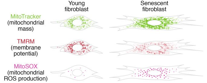
Mitochondrial dysfunction and cell senescence are hallmarks of aging and are closely interconnected. Mitochondrial dysfunction, operationally defined as a decreased respiratory capacity per mitochondrion together with a decreased mitochondrial membrane potential, typically accompanied by increased production of oxygen free radicals, is a cause and a consequence of cellular senescence and figures prominently in multiple feedback loops that induce and maintain the senescent phenotype. Here, we summarize pathways that cause mitochondrial dysfunction in senescence and aging and discuss the major consequences of mitochondrial dysfunction and how these consequences contribute to senescence and aging. We also highlight the potential of senescence-associated mitochondrial dysfunction as an antiaging and antisenescence intervention target, proposing the combination of multiple interventions converging onto mitochondrial dysfunction as novel, potent senolytics.
Overview
The claims and statistics of this paper haven’t been checked yet.
Free: it reads the open-access text and checks the first 5 claims that cite a source we can check. Create a free account to check every claim.
Integrity signals
Registry
Cites
Statistics
Figures

Verification
Claims the paper makes
The claims and statistics of this paper haven’t been checked yet.
Free: it reads the open-access text and checks the first 5 claims that cite a source we can check. Create a free account to check every claim.
Claims other papers make about this one
Each is checked against this paper’s own text: does it say what the citing paper says it does?
The source backs part of the claim, not all of it — or backs it for a different population, condition or comparison.
In the citing paper
“Both proliferation-competent glial cells (which constitute approximately 50% of brain cells) and postmitotic neurons can be positive for markers of cellular senescence in aging, obesity, traumatic injury, and neurodegeneration31,45–49, among other conditions.”
From Guidelines for minimal information on cellular senescence experimentation in vivo (2024) · cites this paper as [45]
Experimental — accuracy not yet measuredThe source backs part of the claim, not all of it — or backs it for a different population, condition or comparison.
In the citing paper
“Therefore, the dysregulation of mitochondrial fusion and fission pathways in senescent astrocytes likely contributes to mitochondrial dysfunction, oxidative stress, and the overall senescence-associated phenotype observed in these cells (Ma et al., 2020; Kleele et al., 2021; Miwa et al., 2022).”
From Aging promotes an increase in mitochondrial fragmentation in astrocytes (2024) · cites this paper as Miwa et al., 2022
Experimental — accuracy not yet measuredThe source backs part of the claim, not all of it — or backs it for a different population, condition or comparison.
In the citing paper
“Dysfunctional mitochondrial dynamics, as observed here, particularly excessive mitochondrial fission, have been associated with oxidative stress, mitochondrial DNA damage, and cellular senescence in various cell types (Twig et al., 2008; Miwa et al., 2022).”
From Aging promotes an increase in mitochondrial fragmentation in astrocytes (2024) · cites this paper as Miwa et al., 2022
Experimental — accuracy not yet measuredThe cited source says what the claim attributes to it.
In the citing paper
“while the presence of senescence markers has been proven even in post-mitotic cells”
From Guidelines for minimal information on cellular senescence experimentation in vivo (2024) · cites this paper as [45]
The check wasn’t confident either way, so it gives no verdict — never disputed.
In the citing paper
“Evidence from the literature indicates that senescent cells exhibit an increase in oxygen consumption rate (Hutter et al., 2004; Son et al., 2017; Miwa et al., 2022; Bang et al., 2023; Diniz et al., 2024a).”
From Aging promotes an increase in mitochondrial fragmentation in astrocytes (2024) · cites this paper as Miwa et al., 2022
Experimental — accuracy not yet measured
2 more claims about this paper
Create a free account to see every claim other papers make about this one.
Self-check
Percentages, sums and reported differences re-computed from the paper’s own numbers.
The self-check needs the paper’s full text.
Citations
Cites
The serial cultivation of human diploid cell strains
Hayflick, L., & Moorhead, P. S. (1961) · Experimental Cell Research · doi:10.1016/0014-4827(61)90192-6 · Cited by 8,508 · cited 1×
“Introduction What is cell senescence? Cell senescence was originally defined as the irreversible loss of replicative potential of primary cells in culture (1), initiated as a persistent DNA damage response to dysfunctional telomeres (2).”
BackgroundTelomeres shorten during ageing of human fibroblasts
Harley, C. B., Futcher, A. B., & Greider, C. W. (1990) · Nature · doi:10.1038/345458a0 · Cited by 5,750 · cited 2×
“Telomere dysfunction might be caused by telomere shortening during DNA replication (3, 4), replication-independent telomere damage (5), or a variety of other stresses (6).”
BackgroundA role for mitochondria in nlrp3 inflammasome activation
Zhou, R., Yazdi, A. S., Menu, P., & Tschopp, J. (2011) · Nature · doi:10.1038/nature09663 · Cited by 5,602
The senescence-associated secretory phenotype: the dark side of tumor suppression
Coppé, J.-P., Desprez, P.-Y., Krtolica, A., & Campisi, J. (2010) · Annual Review of Pathology: Mechanisms of Disease · doi:10.1146/annurev-pathol-121808-102144 · Cited by 5,206
Wu, Z., Puigserver, P., Andersson, U., Zhang, C., Adelmant, G., Mootha, V., Troy, A., Cinti, S., Lowell, B., Scarpulla, R. C., et al. (1999) · Cell · doi:10.1016/s0092-8674(00)80611-x · Cited by 4,139 · cited 1×
“PGC-1α activates the transcription factors Nrf1/2 and ERRα, which further control the TFAM transcription in response to increased mitochondrial biogenesis (65).”
BackgroundClearance of p16Ink4a-positive senescent cells delays ageing-associated disorders
Baker, D. J., Wijshake, T., Tchkonia, T., LeBrasseur, N. K., Childs, B. G., van de Sluis, B., Kirkland, J. L., & van Deursen, J. M. (2011) · Nature · doi:10.1038/nature10600 · Cited by 3,670
AMPK regulates energy expenditure by modulating NAD+ metabolism and SIRT1 activity
Cantó, C., Gerhart-Hines, Z., Feige, J. N., Lagouge, M., Noriega, L., Milne, J. C., Elliott, P. J., Puigserver, P., & Auwerx, J. (2009) · Nature · doi:10.1038/nature07813 · Cited by 3,300
Cellular Senescence: Defining a Path Forward
Gorgoulis, V., Adams, P. D., Alimonti, A., Bennett, D. C., Bischof, O., Bishop, C., Campisi, J., Collado, M., Evangelou, K., Ferbeyre, G., et al. (2019) · Cell · doi:10.1016/j.cell.2019.10.005 · Cited by 3,225 · cited 1×
“Telomere dysfunction might be caused by telomere shortening during DNA replication (3, 4), replication-independent telomere damage (5), or a variety of other stresses (6).”
BackgroundNakahira, K., Haspel, J. A., Rathinam, V. A. K., Lee, S.-J., Dolinay, T., Lam, H. C., Englert, J. A., Rabinovitch, M., Cernadas, M., Kim, H. P., et al. (2011) · Nature Immunology · doi:10.1038/ni.1980 · Cited by 3,049
A DNA damage checkpoint response in telomere-initiated senescence
Fagagna, F. D. D., Reaper, P. M., Clay-Farrace, L., Fiegler, H., Carr, P., von Zglinicki, T., Saretzki, G., Carter, N. P., & Jackson, S. P. (2003) · Nature · doi:10.1038/nature02118 · Cited by 2,917 · cited 1×
“Introduction What is cell senescence? Cell senescence was originally defined as the irreversible loss of replicative potential of primary cells in culture (1), initiated as a persistent DNA damage response to dysfunctional telomeres (2).”
Background
Research works printed but not linked: 2
- von Zglinicki T. Mechanisms of cell senescence in aging. In: Musi N, Hornsby P eds. Handbook of the Biology of Aging. Academic Press; 2021:53–68.
- Miwa S, Johnson TE. Stress and aging. Vijg J, et al., eds. In: The Molecular and Cellular Biology of Aging. Gerontological Society of America; 2016:216-249.
Cited by
Items 501–600 of 999
The drug discovery and therapeutic nano-strategies targeting cellular senescence
Wang, Y., Liu, M., Chen, X., Wang, S., Li, J., Sun, Y., Zheng, X., Zhang, J.-X., & Li, H. (2025) · Materials Today Bio · doi:10.1016/j.mtbio.2025.102480 · Cited by 13
The impact of cellular senescence on aging skeletal muscle
Kamal, M. J., Bevington, R. T., Johnson, A., & Parise, G. (2025) · Frontiers in Cell and Developmental Biology · doi:10.3389/fcell.2025.1719279 · Cited by 8
Arteaga, M. B., Tarasova, K. V., Kidtiwong, A., Gültekin, S., Gerner, I., & Jenner, F. (2026) · Cell Death Discovery · doi:10.1038/s41420-026-02961-y · Cited by 5
Senescent Schwann cells as therapeutic targets in nerve regeneration and peripheral neuropathies
Rebolledo, D. L., Khan, M. A., Melov, S., Höke, A., & Court, F. A. (2026) · Trends in Molecular Medicine · doi:10.1016/j.molmed.2026.01.003 · Cited by 6
Unlock the power of bovine milk-derived exosomes for degenerative diseases associated with aging
Meng, Y., Sun, J. Y., Zhang, G., Yu, T., & Piao, H. (2023) · Journal of Functional Foods · doi:10.1016/j.jff.2023.105788 · Cited by 6
Proteomic analysis reveals the aging-related pathways contribute to pulmonary fibrogenesis
Zhang, T., Yuan, X., Jiang, M., Liu, B., Zhai, N., Zhang, Q., Song, X., Lv, C., Zhang, J., & Li, H. (2023) · Aging · doi:10.18632/aging.205355 · Cited by 6
“Mitochondria are involved in many biological processes including ferroptosis and autophagy, so mitochondrial dysfunction is an important component of different diseases associated with aging [18, 19].”
BackgroundChi, Q., Yu, X., Luo, D., Zhu, L., Yang, X., & Li, S. (2024) · Pesticide Biochemistry and Physiology · doi:10.1016/j.pestbp.2024.105930 · Cited by 6
Yang, Y., Wen, H., Li, Y., Zeng, X., Wei, G., Gu, Z., & Ni, T. (2024) · Life Medicine · doi:10.1093/lifemedi/lnae021 · Cited by 6
“…exhibit and partially induced by some other hallmarks of aging like mitochondrial dysfunction, which include decreased mitochondrial membrane potential, imbalanced energy metabolism homeostasis, increased reactive oxygen species (ROS), and mitochondrial superoxide (mtSOX) productions [4, 7, 12–14].”
Neratinib stimulates senescence of mammary cancer cells by reducing the levels of SIRT1
Li, W., Fu, P., Shi, P., Hu, B., & Li, H. (2024) · Aging · doi:10.18632/aging.205882 · Cited by 6
Zhang, Y., Ma, L., Wang, Z., Gao, C., Yang, L., Li, M., Tang, X., Yuan, H., Pang, D., & Ouyang, H. (2024) · International Journal of Biological Macromolecules · doi:10.1016/j.ijbiomac.2024.134673 · Cited by 11
Sato, M., Kadomatsu, T., Morinaga, J., Kinoshita, Y., Torigoe, D., Horiguchi, H., Ohtsuki, S., Yamamura, S., Kusaba, R., Yamaguchi, T., et al. (2025) · Molecular Metabolism · doi:10.1016/j.molmet.2025.102107 · Cited by 8
Liu, M., & Qiao, H. (2025) · Molecular Medicine Reports · doi:10.3892/mmr.2025.13523 · Cited by 6
“Mitochondria are bioenergetic and metabolic centers extensively involved in various biological processes with complex adaptive mechanisms (10). alterations in the normal morphology and dysfunction of mitochondria are relevant factor pairs affecting the progression of multiple diseases (11).”
Wang, H., Dai, G.-C., Gao, Y., Liu, X., Lu, P., Cao, M., Sheng, R., Shi, L., Cheng, Z., Li, Y., et al. (2025) · Stem Cell Reviews and Reports · doi:10.1007/s12015-025-10928-2 · Cited by 5
“…that mitochondrial dysfunction is a key driver of cellular senescence, with increased oxidative stress and elevated reactive oxygen species (ROS) generation, accelerated DNA and protein damage accumulation, and enhanced secretion of senescence-associated secretory phenotype (SASP) factors [19, 20].”
Berardi, D., Farrell, G., Al-Sultan, A., McCulloch, A., Hall, N. M., Sousa, R. P. P., Jimenez, M., Selman, C., Bellantuono, I., Johnson, C. H., et al. (2025) · Aging Cell · doi:10.1111/acel.70127 · Cited by 7
“…changes through structural enlargement/stiffening (Tilton et al. 2024), telomere shortening (Rossiello et al. 2022), mitochondrial dysfunction (Miwa et al. 2022), impaired autophagy (Fang et al. 2021), and the secretion of a milieu of biomolecules that constitute the inflammatory mediating…”
Yang, R., Zhan, Y., Deng, Z., Zhang, J., Chen, S., Zhang, Y., Fu, H., Meng, X., Wu, J., Gu, Y., et al. (2025) · Redox Biology · doi:10.1016/j.redox.2025.103793 · Cited by 8
“Not only as a consequence of cellular senescence, mitochondrial dysfunction is also a cause of senescence, creating a vicious cycle [34].”
Nejati‐Koshki, K., Mokhtari, B., Badalzadeh, R., Arabzadeh, A., & Mohammadzadeh, A. (2023) · Molecular Biology Reports · doi:10.1007/s11033-023-08499-x · Cited by 5
“Additionally, aging increases the susceptibility of cardiomyocytes to apoptosis/necrosis and diminishes myocardial tolerance to injury induced by I/R [22, 23].”
BackgroundCamacho-Pereira, J., de Souza, L. O. L., Chichierchio, M. S., Rodrigues-Chaves, C., Lomba, L. D. S., Fonseca-Oliveira, M., Carvalho-Mendonça, D., Silva-Rodrigues, T., & Galina, A. (2023) · Free Radical Biology and Medicine · doi:10.1016/j.freeradbiomed.2023.09.035 · Cited by 5
Shin, J. S., Kim, T.-G., Kim, Y. H., Eom, S. Y., Park, S. H., Lee, D. H., Park, T. J., Park, S. S., & Kim, J. (2023) · Journal of Pathology and Translational Medicine · doi:10.4132/jptm.2023.10.09 · Cited by 5
“Since mitochondrial dysfunction is primarily involved in cellular senescence, the efficiency of oxidative phosphorylation (OXPHOS) in senescent cells from normal tissues is significantly decreased [ 12 ] .”
Revitalizing mitochondrial function and empowering spinal cord recovery using copper-catalyzed Pdots
Zhao, B., Wang, F., Sun, J., Gao, J., Zhao, L., Shen, Z., Li, Z., Wang, Y., Wang, Y., Li, D., et al. (2023) · Chemical Engineering Journal · doi:10.1016/j.cej.2023.148264 · Cited by 5
Cellular Senescence in Acute Liver Injury: What Happens to the Young Liver?
He, K., Zhou, D., Pu, Z., Chen, S., Shen, Y., Zhao, S., Qian, X., Hu, Q., Wu, X., Xie, Z., et al. (2024) · Aging and Disease · doi:10.14336/ad.2024.0586 · Cited by 8
“In addition, senescent cells typically have an increased number of dysfunctional mitochondria [29]; however, mitochondrial dysfunction is involved in other cellular processes; therefore, it is also not a common marker of senescence [7].”
Dhavarasa, P., Sack, T., Cerrato, C. P., Cheng, A. P., Zhang, Y. Y., Chen, K., & Kelley, S. O. (2024) · ACS Chemical Biology · doi:10.1021/acschembio.4c00287 · Cited by 6
Wang, X., Hu, J., Xie, S., Li, W., Zhang, H., Huang, L., Qian, Z., Zhao, C., & Hu, J. (2024) · International Immunopharmacology · doi:10.1016/j.intimp.2024.113024 · Cited by 6
Impact of Aging and Cellular Senescence in the Pathophysiology of Preeclampsia
Suvakov, S. R., Kattah, A. G., Gojković, T., Enninga, E. A. L., Pruett, J. E., Jayachandran, M., Sousa, C., Santos, J., Hassan, C. A., Suarez, M. L. G., et al. (2023) · Comprehensive physiology · doi:10.1002/cphy.c230003 · Cited by 6
Spermidine toxicity in Saccharomyces cerevisiae due to mitochondrial complex III deficiency
Su, W.-H., Smith, J. J., Cheng, E., Nishitani, M. S., Choi, C. Y., Lee, K. R., Salzano, A. P., & Schriner, S. E. (2025) · Biogerontology · doi:10.1007/s10522-025-10233-y · Cited by 4
“Depending on the genotype of the individual, spermidine could potentially harm the very individuals it is intended to help.”
The emerging role of cellular senescence in amyotrophic lateral sclerosis
Tan, X., & Gao, N. (2025) · Frontiers in Neuroscience · doi:10.3389/fnins.2025.1599492 · Cited by 8
“…direct and persistent DNA damage (Ou and Schumacher, 2018), activation of tumor suppressors (Liu et al., 2023) and/or oncogenes (Zhu et al., 2020), mitochondrial dysfunction (Miwa et al., 2022), epigenetic dynamics (Zhu et al., 2021), and therapy-induced senescence (TIS; Prasanna et al., 2021).”
Mitochondrial dysfunction drives age-related degeneration of the thoracic aorta
Dhanekula, A., Harrison, B. R., Pharaoh, G. A., Mattson-Hughes, A., Tarantini, S., Stuppard, R. S., DeRoo, S. C., Burke, C. R., Hwang, B., Pal, J. D., et al. (2025) · GeroScience · doi:10.1007/s11357-025-01937-7 · Cited by 6
“Impairments in oxidative phosphorylation with age result in altered bioenergetics and redox homeostasis in multiple tissues [7–13].”
Liu, Y., Huang, D., Pan, M., Li, X., Qin, G., Yu, X., Zhang, Z., Mai, K., & Zhang, W. (2025) · Fish & Shellfish Immunology · doi:10.1016/j.fsi.2025.111050 · Cited by 4
Guo, H., Wang, J., Yuan, Y., Qi, J., Li, S., Chen, H. D., Huang, Z., Liu, H., Yu, S., Yu, Z., et al. (2026) · Carbohydrate Polymers · doi:10.1016/j.carbpol.2026.124901 · Cited by 6
Premature aging in serious mental illness
Diniz, B. S., Fries, G. R., Kuo, C.-L., Xu, M., & Lenze, E. J. (2026) · Neuropsychopharmacology · doi:10.1038/s41386-026-02322-4 · Cited by 5
Parab, A. R., Salazar, A., Bark, S. J., Divenko, M., Putluri, V., Lieu, D. J., Singh, A. S., Putluri, N., & Mysorekar, I. U. (2026) · Aging Cell · doi:10.1111/acel.70391 · Cited by 7
“Previous reports show accumulation of mitochondria in aged cells can be attributed to impaired mitophagy (Miwa et al. 2022).”
The aging cornea: From mechanisms to clinical applications
Zheng, X., Wu, Y., Feng, Y., Cai, M., Zhang, M., Zhang, L., Yang, D., Mao, T., Gu, H., & Ou, S. (2026) · Ageing Research Reviews · doi:10.1016/j.arr.2026.103103 · Cited by 6
Su, D., Ma, R., Su, H., Tan, C., Zhu, Y., Liu, Y., Jiang, X., Sun, X., Lei, T., Zhang, M., et al. (2026) · npj Aging · doi:10.1038/s41514-026-00359-9 · Cited by 5
Correale, J. D. (2026) · Journal of Neurochemistry · doi:10.1111/jnc.70414 · Cited by 4
“Mitochondrial depolarisation increases ROS production, activating DNA damage responses that stabilize p53 and promote senescence (Miwa et al. 2022).”
Upregulation of YPEL3 expression and induction of human breast cancer cell death by microRNAs
Lee, B., Kwon, Y., Shin, S., Kwon, T.-U., Park, H., Lee, H. G., Kwak, J.-H., & Chun, Y. (2024) · Toxicological Research · doi:10.1007/s43188-024-00251-2 · Cited by 5
“Cell senescence and early stages of apoptosis are strongly associated with mitochondrial dysfunction [30, 31].”
BackgroundLiu, Y., Xu, M., Wang, L., Li, M., Li, M., & Mu, S. (2025) · Lipids in Health and Disease · doi:10.1186/s12944-025-02446-4 · Cited by 9
“Various pathological processes are associated with ageing, such as cellular senescence, low-grade systemic inflammation, immunosenescence, mitochondrial dysfunction, and shifts in body composition and hormone levels [9–11].”
Inhibition of miMOMP-induced SASP to combat age-related disease
Liao, X., Guo, Z., He, M., & Zhang, Y. (2025) · Frontiers in Aging · doi:10.3389/fragi.2025.1505063 · Cited by 5
Shboul, S. A., Karsaneh, O. A. A., Alrjoub, M., Alqudah, M. A. Y., El‐Sadoni, M., Alhesa, A., Ramadan, M., Barukba, M., Al-Quran, E. F., Masaadeh, A. H., et al. (2025) · International Journal of Immunopathology and Pharmacology · doi:10.1177/03946320251324821 · Cited by 4
The Science of Aging: Therapeutic Approaches to Extend Lifespan and Healthspan
Praveena, M. M. (2025) · SBV Journal of Basic Clinical and Applied Health Science · doi:10.4103/sbvj.sbvj_11_25 · Cited by 5
Grapentine, S., Agarwal, P., Dolinsky, V. W., & Bakovic, M. (2025) · PLoS ONE · doi:10.1371/journal.pone.0320510 · Cited by 4
“…[52] and age-related diseases including obesity, T2DM, and fibrotic diseases [53,54], and triggered by various stressors shown in Pcyt2 + /- NASH (mitochondrial dysfunction, oxidative stress, inflammation, perturbed proteostasis and autophagy, epigenetic changes, and genomic instability) [55–57].”
Zhu, L., Huang, R., Feng, J., Zhang, M., Huang, X., Chen, Z., Wang, W., & Chen, Y. (2025) · Journal of Ethnopharmacology · doi:10.1016/j.jep.2025.119664 · Cited by 7
The role of glutamate dehydrogenase in the ageing brain
Zhou, T., & Wang, H. (2025) · Frontiers in Pharmacology · doi:10.3389/fphar.2025.1586655 · Cited by 6
“The cellular dysfunction observed during ageing has been partially attributed to impaired mitochondrial activity (Javadpour et al., 2024; Miwa et al., 2022).”
The advances in acetylation modification in senescence and aging-related diseases
Xu, M., Wang, W., Lu, S., Xiong, M., Zhao, T., Yu, Y., Song, C., Yang, J., Zhang, N., Cao, L., et al. (2025) · Frontiers in Physiology · doi:10.3389/fphys.2025.1553646 · Cited by 6
“…cellular dysfunction through compromised electron transport chain efficiency, impaired calcium homeostasis, and defective mitochondrial quality control mechanisms, ultimately manifesting as multi-tiered physiological decline across tissues and organ systems (Miwa et al., 2022; Guo et al., 2023a).”
Zhang, S., Cui, H., Wang, J., Zhou, J., Chen, H., Wang, X., Yang, Q., Cao, J., Hao, K., Fu, S., et al. (2025) · Bioactive Materials · doi:10.1016/j.bioactmat.2025.05.007 · Cited by 11
“Strategies that improve mitochondrial function have shown promise in reducing SASP production and alleviating senescence [17].”
New insights into the role of cellular senescence and rheumatic diseases
Wen, J., Li, J., Wan, L., & Wang, F. (2025) · Frontiers in Immunology · doi:10.3389/fimmu.2025.1557402 · Cited by 8
“Additionally, changes in mitochondrial function during senescence are considered to have a controlling role in cell fate (90, 91).”
NIPSNAP1 and NIPSNAP2 facilitate healthy aging independent of mitophagy
Lv, J., Wang, J., Chen, Q., Zhong, Q., Zhan, H., Guo, Q., Li, J., Guo, N., Yu, F., Tong, J., et al. (2025) · Metabolism · doi:10.1016/j.metabol.2025.156324 · Cited by 5
CD47-mediated regulation of glucose and lipid metabolism: implications for the pathogenesis of MASLD
Jiang, X., Zhao, W., Shen, B., Han, Y., & Chen, K. (2025) · Frontiers in Endocrinology · doi:10.3389/fendo.2025.1535382 · Cited by 7
“As cells age, mitochondria may exhibit reduced energy production, weakened antioxidant capacity, and impaired autophagy, leading to abnormal lipid accumulation within cells (88, 89).”
Kalantar, M.-S., Mahdavi, M. R. V., Nasiri, L., Hassanpour, H., & Kaboudanian-Ardestani, S. (2025) · Chemico-Biological Interactions · doi:10.1016/j.cbi.2025.111666 · Cited by 5
Chen, S., Yuan, A., Fan, S., Ding, X., Zhang, S. Q., Li, Y., & Dai, J. (2025) · International Immunopharmacology · doi:10.1016/j.intimp.2025.115287 · Cited by 4
Innate lymphoid cells prevent graft-versus-host disease via IL-9–driven T-cell senescence
Tufa, D. M., Hoffmeyer, E., Schaller, K., Kooiman, B., Woods, E., Wang, D., Jones, D., Hall, S. C., Cruz, J. C., & Verneris, M. R. (2025) · Blood · doi:10.1182/blood.2025028504 · Cited by 4
Trivalent and Pentavalent Antimonials Impair Cardiac Mitochondrial Function in Mice
de Souza, I. I. A., Pavarino, M. E. M. F., da Rosa, C. F. M., Marinho, L. E., Moraes, C. D. S., Nascimento, J. H. M., de Carvalho, A. C. C., & Maciel, L. (2025) · International Journal of Molecular Sciences · doi:10.3390/ijms26189073 · Cited by 4
Chen, H., Chen, J., Huang, L., Lai, X., Xia, K., Lu, Q., Xie, B., Huang, Y., Qiu, Y., Wang, T., et al. (2025) · Advanced Science · doi:10.1002/advs.202507759 · Cited by 5
Kai-Xin-San improves aging and associated neuroinflammation through mitochondrial autophagy
Gao, Y., Zhang, J., Zhang, X., Xing, C., Xie, P., Zhou, Z., Zhan, T., Zeng, H., Wang, Y., Liu, Y., et al. (2025) · Journal of Ethnopharmacology · doi:10.1016/j.jep.2025.120721 · Cited by 4
Unveiling the importance of SIRT5 for cardiac health and disease in an era of increasing longevity
Grzeczka, A., Graczyk, S., & Kordowitzki, P. (2025) · GeroScience · doi:10.1007/s11357-025-02021-w · Cited by 6
“Specifically, the depletion of mitochondrial sirtuins, including SIRT5, has been demonstrably linked to senescence responses and the secretion of distinct senescence-associated secretory phenotype factors [33].”
Feng, Y. R., Zhi, X., Xiao, T., & Feng, L. (2026) · Mechanisms of Ageing and Development · doi:10.1016/j.mad.2026.112158 · Cited by 6
Savić, A., Beribaka, M., Novaković, A. R., Vasiljević, L., & Palangetić, M. (2026) · Journal of Insect Science · doi:10.1093/jisesa/ieag020 · Cited by 6
Yue, C., Wu, Y., Xia, Y., Xin, T., Gong, Y., Tao, L., Shen, C., Zhu, Y., Shen, M., Wang, D., et al. (2024) · American Journal of Physiology-Cell Physiology · doi:10.1152/ajpcell.00126.2024 · Cited by 4
Safety of embryo cryopreservation: insights from mid-term placental transcriptional changes
Luo, Q.-Y., Zhang, S., Wu, H., Mo, J.-Y., Yu, J., He, R.-K., Jiang, Z., Zhu, K.-J., Liu, X., Lin, Z., et al. (2024) · Reproductive Biology and Endocrinology · doi:10.1186/s12958-024-01241-7 · Cited by 4
“Upon comparing previous animal experiments related to embryo cryopreservation, we found that the DEGs between frozen and fresh embryos in many of the experiments were also enriched on the OXPHOS pathway [44, 50, 51].”
MethodYu, J., Guo, M., Liu, Y., Li, S., Ni, J., Feng, Y., & Ding, J. (2024) · Analytical Chemistry · doi:10.1021/acs.analchem.4c02113 · Cited by 4
Félix, N. Q., Tornquist, L., Sehn, A. P., D’avila, H. F., Todendi, P. F., Valim, A. R. D. M., & Reuter, C. P. (2024) · BMC Pediatrics · doi:10.1186/s12887-024-05111-4 · Cited by 4
“Therefore, telomeres are recognized as intrinsic biomarkers of the cellular aging process [3] and are susceptible to oxidative stress and systemic inflammation caused by external factors, which can accelerate the reduction of telomere length (TL) [4].”
BackgroundCytoophidium complexes resonate with cell fates
Li, Y., & Liu, J. (2025) · Cellular and Molecular Life Sciences · doi:10.1007/s00018-025-05578-z · Cited by 6
Maria, M. K. M., Moniem, E. M. A., Hanafy, A. K., Farag, D. B. E., Radwan, I. A., Abbass, M. M. S., Moshy, S. E., Rady, D., Dörfer, C. E., & El‐Sayed, K. M. F. (2025) · Dentistry Journal · doi:10.3390/dj13030106 · Cited by 7
“Mitochondrial dysfunction, marked by decreased respiratory capacity per mitochondrion, is a defining feature in aging tissues and senescent cells, increasing ROS production and causing further cell damage and aging [2,9].”
Xie, J., Shu, X., Xie, Z., Tang, J., & Guo, W. (2025) · European Journal of Pharmacology · doi:10.1016/j.ejphar.2025.177475 · Cited by 6
Nagar, L., Saini, A., Vishwas, S., Singh, S. K., Gupta, G., MacLoughlin, R. J., Löbenberg, R., Davies, N. M., Chellappan, D. K., Paudel, K. R., et al. (2025) · Archives of Toxicology · doi:10.1007/s00204-025-04116-x · Cited by 6
“The dysregulation or imbalance of this mTOR pathway results into disruption of mitochondrial functions which further exacerbating the cellular senescence via different pathways, described in below image (Sun et al. 2023; Miwa et al. 2022).”
Bang, E., Hwangbo, H., Kim, M. Y., Ji, S. Y., Kim, D. H., Shim, J., Moon, S., Kim, G., Cheong, J., & Choi, Y. H. (2023) · Environmental Toxicology and Pharmacology · doi:10.1016/j.etap.2023.104211 · Cited by 4
Xu, D., Chen, X., Wu, M., Bi, J., Xue, H., & Chen, H. (2024) · Heliyon · doi:10.1016/j.heliyon.2024.e28278 · Cited by 6
Viana, J. V. D. S., de Oliveira, L. R. M., Rodrigues, L. L. V., Silva, Y. L. F. E., Rodrigues, A. L. R., Silva, A. R., Alves, P. V., Silva, H. V. R., & Pereira, A. F. (2024) · Cell Biology International · doi:10.1002/cbin.12244 · Cited by 4
“The increase in ΔΨ m can be explained by the increase in mitochondria mass, and these modifications can decrease the ROS levels by the increase in electron transport (Miwa et al., 2022).”
Remodeling of Mitochondria in Cancer and Other Diseases
Teng, Y. (2023) · International Journal of Molecular Sciences · doi:10.3390/ijms24097693 · Cited by 4
Shee, S., Martinez-Martinez, Y. B., Koleske, B. N., Yabaji, S. M., Kobzik, L., Kramnik, I., & Bishai, W. R. (2025) · bioRxiv (Cold Spring Harbor Laboratory) · doi:10.1101/2025.03.28.645957 · Cited by 4
Yang, J., Xu, Z., Zheng, W., Li, Y., Wei, Q., & Yang, L. (2024) · BMC Cancer · doi:10.1186/s12885-024-12492-3 · Cited by 6
“This impairment of mitochondrial function acts as both a cause and a consequence of cellular senescence, prompting the per-meabilization of mitochondrial membranes and initiating either apoptosis or necrosis [46, 47].”
BackgroundSun, C., Xu, Y., Du, H., Chen, Y., Qu, W., He, M., Liu, Z., Huang, J. J., Huo, J., Yin, J., et al. (2025) · Antioxidants · doi:10.3390/antiox14030315 · Cited by 3
“Regarding the sources of ROS, Choi and Nicolas et al. [46,47] demonstrated that PA is a significant contributor, as it can elevate oxidative stress levels in C2C12 myotube cells.”
Guan, X., Dong, X., Wang, Y.-X., Xu, B., & Wu, X. (2025) · Molecular Pain · doi:10.1177/17448069251332100 · Cited by 4
Ren, X., Jin, C., Li, Q., Fu, C., Yu, F., Xu, Z., Liang, Z., & Wang, T. (2025) · International Journal of Biological Macromolecules · doi:10.1016/j.ijbiomac.2025.142913 · Cited by 9
Genetic targets related to aging for the treatment of coronary artery disease
Huang, K., Chen, Z., Wang, R., Ying, H., Duan, J., Zhang, Y., Shi, Q., Yang, C., & Yang, L. (2025) · BMC Medical Genomics · doi:10.1186/s12920-025-02137-0 · Cited by 3
“Similarly, dysfunction of mitochondrial oxidative phosphorylation leads to oxidative damage caused by ROS production, which is an important molecular basis for the pathological and physiological conditions of aging [31].”
Revealing the Landscape Crosstalk Between Reproductive System and Organs Aging
Wu, J., Wu, P., Wang, S., Guan, Y., & Wang, J. (2025) · The FASEB Journal · doi:10.1096/fj.202403410r · Cited by 4
Senolytic effects of a modified Gingerenone A
Moaddel, R., Sanehira, C., Keyes, G. S., Cui, C., Ahmadkhaniha, R., Candia, J., Price, N. L., Eckroth, S., Middleton, B., Khadeer, M., et al. (2025) · npj Aging · doi:10.1038/s41514-025-00230-3 · Cited by 3
“The increase in fi cial in senescence as defects in oxidative phosphorylation function have been observed in various models of senescence 43 , and may protect the mitochondria from functional deterioration 44 .”
Gao, H., Shi, J., Li, G., Lai, Z., Liu, Y., Yuan, C., & Mei, W. (2025) · Antioxidants · doi:10.3390/antiox14080968 · Cited by 5
“…the expression of elastin and Collagen III was significantly reduced in DOX-induced HSF senescent cells compared to the blank control group (*** p 0.001), suggesting that the loss of extracellular matrix (ECM) components in the senescent cells resulted in impaired skin barrier function [45].”
InfluentialLv, P., Ren, B., & Cheng, G. (2025) · Letters in Drug Design & Discovery · doi:10.1016/j.lddd.2025.100082 · Cited by 4
Shi, Y., Yang, L., Chen, Y., & Li, Z. (2025) · Prostaglandins & Other Lipid Mediators · doi:10.1016/j.prostaglandins.2025.107026 · Cited by 4
Ibarra-Sánchez, A., Madera-Salcedo, I. K., Esparza‐Reyes, D., Mendoza‐Montiel, P., Padilla, J., & González‐Espinosa, C. (2025) · Aging Cell · doi:10.1111/acel.70186 · Cited by 3
“It has been proposed that those characteristics observable in senescent cells could be attributable to an increase in the number of mitochondria per cell, since higher numbers of this organelle (although dysfunctional) have been observed in some senescent cells (reviewed in Miwa et al. 2022).”
Ministrini, S., Liberale, L., Erle, H.-E., Percoco, G., Tfayli, A. M., Assi, A., Kapitonov, I., Greiner, I., & Camici, G. G. (2025) · Current Issues in Molecular Biology · doi:10.3390/cimb47090722 · Cited by 4
Potential connections between senescence and chemobrain: An early overview of literature
Melhem, N. Y., Qutifan, S., Alsalem, M., & Saleh, T. (2025) · Ageing Research Reviews · doi:10.1016/j.arr.2025.102896 · Cited by 5
Multifactorial Imaging Analysis as a Platform for Studying Cellular Senescence Phenotypes
Shatalova, R. O., Larin, I. I., & Shevyrev, D. (2025) · Journal of Imaging · doi:10.3390/jimaging11100351 · Cited by 4
“These processes form positive feedback loops that maintain the senescent phenotype [28,29].”
HMGB1 and mitochondrial dysfunction: a double-edged sword in ageing
Wahid, M., & Cunningham, M. (2025) · Open Biology · doi:10.1098/rsob.240376 · Cited by 3
“However, dysfunctional mitochondria link cellular senescence to inflammation through the release of DAMPs [89]. mtDNA is released from damaged mitochondria where it can activate key inflammatory pathways, such as both the NLRP3 inflammasome and the cGAS/STING pathway, resulting in the secretion of…”
Tang, X., Hu, J., Fan, X., Su, H., Li, M., Zhang, L., & Ma, D. (2025) · Immunity & Ageing · doi:10.1186/s12979-025-00541-9 · Cited by 4
“…shortened lifespan, xerostomia), and soluble Klotho protein modulates mTOR pathway components, implicating mTOR in SG senescence [75]. mTOR signaling activation alters cellular morphology and function [76]; while organelles such as mitochondria and lysosomes [77, 78] contribute to senescence.”
Nutrient-Sensing Pathways: Gatekeepers of Longevity
Goel, F., Garg, V. K., Singh, P., & Tripathi, P. N. (2025) · Nutritional neurosciences · doi:10.1007/978-981-95-2790-8_8 · Cited by 3
Sun, Z., Zheng, Y., Wang, T., Wei, S., Wu, Z., Zhang, J., Ge, J., An, Q., Zhu, J., & Qian, H. (2025) · Cellular and Molecular Life Sciences · doi:10.1007/s00018-025-05899-z · Cited by 5
“We hypothesized that aberrant upregulation of GRP75 may lead to mitochondrial Ca²⁺ overload, resulting in mitochondrial dysfunction and ultimately accelerating the photoaging process [27].”
Prussian Blue Nanoparticles Promoting Diabetic Bone Regeneration via Mitochondrial Recovery
Gu, A., Lao, A., Li, W., Liu, Z., Zhou, C., Cai, J., Chen, Q., Lin, K., Song, L., Wu, X., et al. (2025) · BME Frontiers · doi:10.34133/bmef.0204 · Cited by 5
Zhang, Y., Sun, Z., Meng, W., Lei, S., Sun, J., Tang, Y., & Mu, X. (2025) · Frontiers in Aging · doi:10.3389/fragi.2025.1657947 · Cited by 4
Liu, L., Li, L., Lu, L., Fan, S., Liu, L.-Z., He, R.-B., Li, H., Xian, X., & Li, W. (2025) · Alzheimer s Research & Therapy · doi:10.1186/s13195-025-01900-w · Cited by 4
“MMP loss is an important change indicating mitochondrial damage [55].”
Russo, L., Babboni, S., Turco, S. D., & Basta, G. (2025) · Cells · doi:10.3390/cells14231908 · Cited by 3
“Prolonged hypoxia promotes ROS accumulation and activates stress-response pathways that accelerate cellular senescence [17,18].”
Impaired glycolysis-derived serine metabolism as a key driver of podocyte injury with senescence
Hu, H., Zhu, Z., Li, L., Hu, J., Yang, Q., Peng, Z., Li, W., Cui, X., Fan, Y., Chen, W., et al. (2025) · Nature Communications · doi:10.1038/s41467-025-66850-1 · Cited by 8
“Cellular senescence is typically characterized by irreversible proliferative arrest and is triggered by various stressors, including DNA damage, mitochondrial dysfunction, and in fl ammation 34,35 .”
Yu, W., Li, Y., Jiang, S., Tian, J., Sun, S.-W., Zhang, L., & Xiao, D. (2025) · iScience · doi:10.1016/j.isci.2025.114311 · Cited by 4
“45 Apoptosis is another key determinant of cardiac cell turnover and homeostasis.”
HK1 and HK2 Beyond Glycolysis: Mitochondrial Interactions and Dual Roles in Metabolism and Cell Fate
Pesce, N. A., Seminara, G., Giarrusso, G., & Tomasello, M. F. (2025) · Advanced Biology · doi:10.1002/adbi.202500472 · Cited by 7
“In contrast, aging is often accompanied by a weakening of this interaction, leading to mitochondrial alterations, impaired nutrient sensing, and reduced metabolic efficiency [74].”
Jung, M., Han, S., Ryu, S., Cha, S., Sim, Y. E., Jung, S. H., Tak, H., Kim, W., & Lee, E. K. (2025) · Cells · doi:10.3390/cells15010048 · Cited by 3
“Cellular senescence is a stress-responsive state characterized by growth arrest, chromatin remodeling, mitochondrial dysfunction, and the acquisition of senescence-associated secretory phenotype (SASP) [1–3].”
Lee, W., Song, G., & Bae, H. (2026) · Biochemical Pharmacology · doi:10.1016/j.bcp.2026.117672 · Cited by 4
(孙义), Y. S., Lin, Y., Xing, S., Huang, W., Wang, Y., Guo, G., Yan, H., Zhao, D., Cheng, W., Wu, Z., et al. (2026) · EBioMedicine · doi:10.1016/j.ebiom.2026.106146 · Cited by 4
Chen, X., Wang, D., Tao, J., & Zhang, A. (2026) · Ageing Research Reviews · doi:10.1016/j.arr.2026.103067 · Cited by 3
Zhang, X., Li, L., Liu, H., Zhang, Y., Wu, Y., Sun, C., Guo, J., & Li, M. (2026) · Cellular Signalling · doi:10.1016/j.cellsig.2026.112442 · Cited by 4
Li, T., Zhao, Q., Gao, Y., Zhang, Y., Song, Z., Huang, W., & Zhu, X. (2026) · Free Radical Biology and Medicine · doi:10.1016/j.freeradbiomed.2026.02.058 · Cited by 3
Miao, J., Li, J., Meng, P., Li, X., Huang, J., Liang, Y., Ling, X., Wang, X., Shen, W., Zhou, S. Y., et al. (2026) · Nature Communications · doi:10.1038/s41467-026-71553-2 · Cited by 3
“Mitochondrial injury contributes to cell fate conversion, particularly senescence fate transition 14,15 .”
Retracted
Metadata
- Authors
- Miwa, S., Kashyap, S., Chini, E., & von Zglinicki, T.
- Affiliations
- Newcastle University (GB)
- Mayo Clinic (US)
- Venue
- Journal of Clinical Investigation
- Published
- 2022-07-01
- Volume
- 132
- Issue
- 13
- ISSN
- 0021-9738 (ISSN-L)
- 1558-8238
- PMC
- PMC9246372
- OpenAlex
- W4283723802
- Cited by
- 1,061
- References
- 145
- Citations by year
- 2026: 294 · 2025: 348 · 2024: 263 · 2023: 136 · 2022: 20
- FWCI
- 19.97
- Type
- journal article
- Language
- en
- Open access
- gold
- doi.org
- Licence
- Locations
- Journal of Clinical Investigation (publishedVersion, cc-by)
- PubMed Central (publishedVersion, cc-by)
- PubMed (publishedVersion)
- DOAJ (DOAJ: Directory of Open Access Journals) (submittedVersion)
- Newcastle University ePrints (Newcastle Univesity) (cc-by)
- Funders
- Glenn Foundation for Medical Research
- Ted Nash Long Life Foundation
- Cancer Research UK (C12161/A24009)
- Newcastle University
- Fondation pour la Recherche Médicale
- Versus Arthritis
- National Institutes of Health (H2020, AG26094, CA233790, AG58812)
- Directorate for Biological Sciences
- Horizon 2020 Framework Programme (857524)
- Medical Research Council (MR/P020941/1)
- Biotechnology and Biological Sciences Research Council (BB/S006710/1)
- National Institute on Aging (AG-58812, R01AG058812, AG-26094)
- National Cancer Institute (R01CA233790)
- Topics
- Life Sciences › Biochemistry, Genetics and Molecular Biology › Molecular Biology › Mitochondrial Function and Pathology
- Life Sciences › Biochemistry, Genetics and Molecular Biology › Cell Biology › Telomeres, Telomerase, and Senescence
- Life Sciences › Biochemistry, Genetics and Molecular Biology › Aging › Genetics, Aging, and Longevity in Model Organisms
- Keywords
- mitochondrial dysfunction, cellular senescence, aging, mitochondrial membrane potential, respiratory capacity, reactive oxygen species, senolytics
- MeSH terms
- Mitochondria, Phenotype, Cellular Senescence
About these checks
Registry, retraction, Cites and statistics results are computed, not judged, with one exception: a printed reference that no index lists is read once by a model before it counts as a citation error. Claims are extracted by a model and kept only when their quote appears verbatim in the text; each cited source is then checked, with a model’s help, for whether it says what the claim relies on. Those verdicts can be wrong — read the claim’s quote and the cited source before relying on one.
Statistics and arithmetic are recomputed, not judged.
Confidence bands are shown only for fields calibrated on our annotated test set. None is calibrated yet, so no band is shown.
What counts as a claim ›How we measure confidence ›How we check this ›
Are you an author, or do you see a problem with this page? Request a review
Data sources
Bibliographic data: Semantic Scholar, OpenAlex, Crossref.
Contains information from the Semantic Scholar Academic Graph Datasets, made available under the ODC Attribution License (ODC-BY 1.0).
Semantic Scholar’s data is described in: Kinney, R., et al. (2023). The Semantic Scholar Open Data Platform. arXiv:2301.10140.
