

Mitochondrial dysfunction in cell senescence and aging
Open accessThe DOI matches its Crossref record; 145 of the 145 references it cites were found in a registry or an index.
Abstract
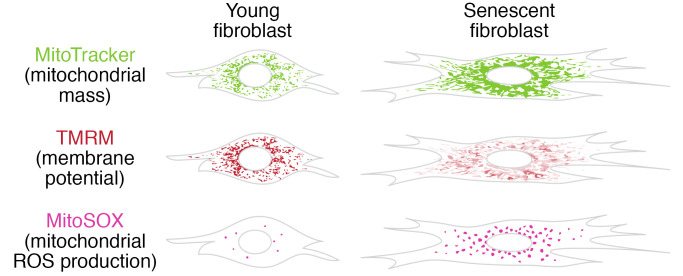
Mitochondrial dysfunction and cell senescence are hallmarks of aging and are closely interconnected. Mitochondrial dysfunction, operationally defined as a decreased respiratory capacity per mitochondrion together with a decreased mitochondrial membrane potential, typically accompanied by increased production of oxygen free radicals, is a cause and a consequence of cellular senescence and figures prominently in multiple feedback loops that induce and maintain the senescent phenotype. Here, we summarize pathways that cause mitochondrial dysfunction in senescence and aging and discuss the major consequences of mitochondrial dysfunction and how these consequences contribute to senescence and aging. We also highlight the potential of senescence-associated mitochondrial dysfunction as an antiaging and antisenescence intervention target, proposing the combination of multiple interventions converging onto mitochondrial dysfunction as novel, potent senolytics.
Overview
The claims and statistics of this paper haven’t been checked yet.
Free: it reads the open-access text and checks the first 5 claims that cite a source we can check. Create a free account to check every claim.
Integrity signals
Registry
Cites
Statistics
Figures

Verification
Claims the paper makes
The claims and statistics of this paper haven’t been checked yet.
Free: it reads the open-access text and checks the first 5 claims that cite a source we can check. Create a free account to check every claim.
Claims other papers make about this one
Each is checked against this paper’s own text: does it say what the citing paper says it does?
The source backs part of the claim, not all of it — or backs it for a different population, condition or comparison.
In the citing paper
“Both proliferation-competent glial cells (which constitute approximately 50% of brain cells) and postmitotic neurons can be positive for markers of cellular senescence in aging, obesity, traumatic injury, and neurodegeneration31,45–49, among other conditions.”
From Guidelines for minimal information on cellular senescence experimentation in vivo (2024) · cites this paper as [45]
Experimental — accuracy not yet measuredThe source backs part of the claim, not all of it — or backs it for a different population, condition or comparison.
In the citing paper
“Therefore, the dysregulation of mitochondrial fusion and fission pathways in senescent astrocytes likely contributes to mitochondrial dysfunction, oxidative stress, and the overall senescence-associated phenotype observed in these cells (Ma et al., 2020; Kleele et al., 2021; Miwa et al., 2022).”
From Aging promotes an increase in mitochondrial fragmentation in astrocytes (2024) · cites this paper as Miwa et al., 2022
Experimental — accuracy not yet measuredThe source backs part of the claim, not all of it — or backs it for a different population, condition or comparison.
In the citing paper
“Dysfunctional mitochondrial dynamics, as observed here, particularly excessive mitochondrial fission, have been associated with oxidative stress, mitochondrial DNA damage, and cellular senescence in various cell types (Twig et al., 2008; Miwa et al., 2022).”
From Aging promotes an increase in mitochondrial fragmentation in astrocytes (2024) · cites this paper as Miwa et al., 2022
Experimental — accuracy not yet measuredThe cited source says what the claim attributes to it.
In the citing paper
“while the presence of senescence markers has been proven even in post-mitotic cells”
From Guidelines for minimal information on cellular senescence experimentation in vivo (2024) · cites this paper as [45]
The check wasn’t confident either way, so it gives no verdict — never disputed.
In the citing paper
“Evidence from the literature indicates that senescent cells exhibit an increase in oxygen consumption rate (Hutter et al., 2004; Son et al., 2017; Miwa et al., 2022; Bang et al., 2023; Diniz et al., 2024a).”
From Aging promotes an increase in mitochondrial fragmentation in astrocytes (2024) · cites this paper as Miwa et al., 2022
Experimental — accuracy not yet measured
2 more claims about this paper
Create a free account to see every claim other papers make about this one.
Self-check
Percentages, sums and reported differences re-computed from the paper’s own numbers.
The self-check needs the paper’s full text.
Citations
Cites
The serial cultivation of human diploid cell strains
Hayflick, L., & Moorhead, P. S. (1961) · Experimental Cell Research · doi:10.1016/0014-4827(61)90192-6 · Cited by 8,508 · cited 1×
“Introduction What is cell senescence? Cell senescence was originally defined as the irreversible loss of replicative potential of primary cells in culture (1), initiated as a persistent DNA damage response to dysfunctional telomeres (2).”
BackgroundTelomeres shorten during ageing of human fibroblasts
Harley, C. B., Futcher, A. B., & Greider, C. W. (1990) · Nature · doi:10.1038/345458a0 · Cited by 5,750 · cited 2×
“Telomere dysfunction might be caused by telomere shortening during DNA replication (3, 4), replication-independent telomere damage (5), or a variety of other stresses (6).”
BackgroundA role for mitochondria in nlrp3 inflammasome activation
Zhou, R., Yazdi, A. S., Menu, P., & Tschopp, J. (2011) · Nature · doi:10.1038/nature09663 · Cited by 5,602
The senescence-associated secretory phenotype: the dark side of tumor suppression
Coppé, J.-P., Desprez, P.-Y., Krtolica, A., & Campisi, J. (2010) · Annual Review of Pathology: Mechanisms of Disease · doi:10.1146/annurev-pathol-121808-102144 · Cited by 5,206
Wu, Z., Puigserver, P., Andersson, U., Zhang, C., Adelmant, G., Mootha, V., Troy, A., Cinti, S., Lowell, B., Scarpulla, R. C., et al. (1999) · Cell · doi:10.1016/s0092-8674(00)80611-x · Cited by 4,139 · cited 1×
“PGC-1α activates the transcription factors Nrf1/2 and ERRα, which further control the TFAM transcription in response to increased mitochondrial biogenesis (65).”
BackgroundClearance of p16Ink4a-positive senescent cells delays ageing-associated disorders
Baker, D. J., Wijshake, T., Tchkonia, T., LeBrasseur, N. K., Childs, B. G., van de Sluis, B., Kirkland, J. L., & van Deursen, J. M. (2011) · Nature · doi:10.1038/nature10600 · Cited by 3,670
AMPK regulates energy expenditure by modulating NAD+ metabolism and SIRT1 activity
Cantó, C., Gerhart-Hines, Z., Feige, J. N., Lagouge, M., Noriega, L., Milne, J. C., Elliott, P. J., Puigserver, P., & Auwerx, J. (2009) · Nature · doi:10.1038/nature07813 · Cited by 3,300
Cellular Senescence: Defining a Path Forward
Gorgoulis, V., Adams, P. D., Alimonti, A., Bennett, D. C., Bischof, O., Bishop, C., Campisi, J., Collado, M., Evangelou, K., Ferbeyre, G., et al. (2019) · Cell · doi:10.1016/j.cell.2019.10.005 · Cited by 3,225 · cited 1×
“Telomere dysfunction might be caused by telomere shortening during DNA replication (3, 4), replication-independent telomere damage (5), or a variety of other stresses (6).”
BackgroundNakahira, K., Haspel, J. A., Rathinam, V. A. K., Lee, S.-J., Dolinay, T., Lam, H. C., Englert, J. A., Rabinovitch, M., Cernadas, M., Kim, H. P., et al. (2011) · Nature Immunology · doi:10.1038/ni.1980 · Cited by 3,049
A DNA damage checkpoint response in telomere-initiated senescence
Fagagna, F. D. D., Reaper, P. M., Clay-Farrace, L., Fiegler, H., Carr, P., von Zglinicki, T., Saretzki, G., Carter, N. P., & Jackson, S. P. (2003) · Nature · doi:10.1038/nature02118 · Cited by 2,917 · cited 1×
“Introduction What is cell senescence? Cell senescence was originally defined as the irreversible loss of replicative potential of primary cells in culture (1), initiated as a persistent DNA damage response to dysfunctional telomeres (2).”
Background
Research works printed but not linked: 2
- von Zglinicki T. Mechanisms of cell senescence in aging. In: Musi N, Hornsby P eds. Handbook of the Biology of Aging. Academic Press; 2021:53–68.
- Miwa S, Johnson TE. Stress and aging. Vijg J, et al., eds. In: The Molecular and Cellular Biology of Aging. Gerontological Society of America; 2016:216-249.
Cited by
Items 101–200 of 999
de Luzy, I. R., Lee, M. K., Mobley, W. C., & Studer, L. (2024) · Nature Aging · doi:10.1038/s43587-024-00586-3 · Cited by 41
“For example, several pathways associated with mitochondrial dysfunction have been identified in aging and senescence, as reviewed by Miwa et al 11 .”
Metabolic remodeling in cancer and senescence and its therapeutic implications
Kim, Y., Jang, Y., Kim, M.-S., & Kang, C. (2024) · Trends in Endocrinology & Metabolism · doi:10.1016/j.tem.2024.02.008 · Cited by 36
Mitophagy in relation to chronic inflammation/ROS in aging
Kong, L., Li, S., Fu, Y., Cai, Q., Du, X., Liang, J., & Ma, T. (2024) · Molecular and Cellular Biochemistry · doi:10.1007/s11010-024-05042-9 · Cited by 43
The mechanisms, hallmarks, and therapies for brain aging and age-related dementia
Jin, S., Lü, W., Zhang, J., Zhang, L., Tao, F., Zhang, Y., Hu, X., & Liu, Q. (2024) · 中国科学通报:英文版 · doi:10.1016/j.scib.2024.09.005 · Cited by 44
Cellular senescence in liver diseases: From mechanisms to therapies
Ge, T., Shao, Y., Bao, X., Xu, W., & Lu, C. (2023) · International Immunopharmacology · doi:10.1016/j.intimp.2023.110522 · Cited by 38
The Role of Estrogen in Mitochondrial Disease
Huang, X., Pan, C.-H., Yin, F., Peng, J., & Yang, L. (2025) · Cellular and Molecular Neurobiology · doi:10.1007/s10571-025-01592-8 · Cited by 43
“1) (Kasapoğlu and Seli 2020; Miwa et al. 2022).”
Tang, R., Harasymowicz, N. S., Wu, C., Choi, Y., Lenz, K. L., Oswald, S. J., & Guilak, F. (2024) · Proceedings of the National Academy of Sciences · doi:10.1073/pnas.2402954121 · Cited by 35
“The accumulation of damaged mitochondria and the excessive ROS production are involved in chronic inflammation, cellular senescence, and aging ( 59 , 60 ).”
Mitochondria: one of the vital hubs for molecular hydrogen’s biological functions
Zhang, X., Xie, F., Ma, S., Ma, C., Jiang, X., Yang, Y., Song, Y., Liu, M., Zhao, P., & Ma, X. (2023) · Frontiers in Cell and Developmental Biology · doi:10.3389/fcell.2023.1283820 · Cited by 39
Zheng, B., Long, W. S., Zheng, W., Zeng, Y., Guo, X., Chan, K., She, M., Leung, A. S., Lu, Y., & Wong, W. (2024) · Journal of Medicinal Chemistry · doi:10.1021/acs.jmedchem.3c02240 · Cited by 34
Photobiomodulation in the aging brain: a systematic review from animal models to humans
Rodríguez‐Fernández, L., Zorzo, C., & Arias, J. L. (2024) · GeroScience · doi:10.1007/s11357-024-01231-y · Cited by 38
“Thus, mitochondrial dysfunction can be caused by alterations in mitochondrial autophagy—that is, mitophagy—, resulting in alterations in the respiratory capacity, reducing the mitochondrial membrane potential [12–15].”
BackgroundBang, E., Hwangbo, H., Lee, H., Park, C., Hong, S. H., Kim, H. S., Jung, Y., Hyun, Y., Hyun, J. W., Kim, G., et al. (2024) · Toxicology · doi:10.1016/j.tox.2024.154002 · Cited by 34
The emerging role of miRNAs in biological aging and age-related diseases
Turko, R., Hajja, A., Magableh, A. M., Omer, M. H., Shafqat, A., Khan, M. I., & Yaqinuddin, A. (2025) · Non-coding RNA Research · doi:10.1016/j.ncrna.2025.05.002 · Cited by 43
“Mitochondrial dysfunction with elevated ROS production is also one of the consequences of cellular senescence [82].”
Neuroinflammation and Natural Antidepressants: Balancing Fire with Flora
Figueiredo Godoy, A. C., Frota, F. F., Araújo, L. P., Valenti, V. E., Pereira, E. D. S. B. M., Detregiachi, C. R. P., Galhardi, C. M., Caracio, F. C., Haber, R. S. A., Fornari Laurindo, L., et al. (2025) · Biomedicines · doi:10.3390/biomedicines13051129 · Cited by 32
“Oxidative damage to mtDNA impairs the synthesis of ETC components, while dysregulated biogenesis reduces the formation of healthy mitochondria [132].”
Neuroinflammation and Natural Antidepressants: Balancing Fire with Flora
Figueiredo Godoy, A. C., Frota, F. F., Araújo, L. P., Valenti, V. E., Pereira, E. D. S. B. M., Detregiachi, C. R. P., Galhardo, C. M., Caracio, F. C., Haber, R. S. A., Fornari Laurindo, L., et al. (2025) · doi:10.20944/preprints202504.0573.v2 · Cited by 33
MAVS Antagonizes Human Stem Cell Senescence as a Mitochondrial Stabilizer
Wang, C., Yang, K., Liu, X., Wang, S., Song, M., Belmonte, J. C. I., Qu, J. T., Liu, G., & Zhang, W. (2023) · Research · doi:10.34133/research.0192 · Cited by 37
Sun, J., Gui, Y., Zhou, S., & Zheng, X. (2023) · Journal of Advanced Research · doi:10.1016/j.jare.2023.11.006 · Cited by 42
“Mitochondria dysfunction is a defining characteristic of aging [93].”
Molecular hallmarks of ageing in amyotrophic lateral sclerosis
Jagaraj, C. J., Shadfar, S., Kashani, S. A., Saravanabavan, S., Farzana, F., & Atkin, J. D. (2024) · Cellular and Molecular Life Sciences · doi:10.1007/s00018-024-05164-9 · Cited by 43
Exploration of the mechanism and therapy of ovarian aging by targeting cellular senescence
Tang, W., Wang, K., Feng, Y., Tsui, K., Singh, K. K., Stout, M. B., Wang, S., & Wu, M. (2025) · Life Medicine · doi:10.1093/lifemedi/lnaf004 · Cited by 37
“Mitochondria maintain their quantity, size, shape, and distribution through fission and fusion, also known as mitochondrial dynamics, thereby regulating mitochondrial function [58].”
Cancer stem cell fate determination: mito-nuclear communication
Fan, M., Shi, Y., Zhao, J., & Li, L. (2023) · Cell Communication and Signaling · doi:10.1186/s12964-023-01160-x · Cited by 39
Scarpetta, V., Bodaleo, F. J., Salio, C., Agarwal, A., Sassoé‐Pognetto, M., & Patrizi, A. (2023) · Fluids and Barriers of the CNS · doi:10.1186/s12987-023-00420-9 · Cited by 33
“The significant reduction of TMRM accumulation in aged ChP mitochondria indicates a disruption of the mitochondrial membrane potential [50], indirectly suggesting that the ability to synthetize ATP through oxidative phosphorylation may be impaired at baseline.”
BackgroundZhang, H., Ran, M., Jiang, L., Sun, X., Qiu, T., Li, J., Wang, N., Yao, X., Zhang, C., Deng, H., et al. (2023) · Ecotoxicology and Environmental Safety · doi:10.1016/j.ecoenv.2023.115711 · Cited by 34
Brief guide to senescence assays using cultured mammalian cells
Kang, E. S., Kang, C., Lee, Y., & Lee, S. (2024) · Molecules and Cells · doi:10.1016/j.mocell.2024.100102 · Cited by 43
Qu, X., Zhou, X., Zhang, J., Huang, Y., Zhao, R., Li, N., Wang, J., Chen, L., Cui, W., & Luo, X. (2024) · Small · doi:10.1002/smll.202409936 · Cited by 38
Epigenetic Mechanisms in Aging: Extrinsic Factors and Gut Microbiome
Borrego-Ruiz, A., & Borrego, J. J. (2024) · Genes · doi:10.3390/genes15121599 · Cited by 36
Dietary polyphenols as geroprotective compounds: From Blue Zones to hallmarks of ageing
Davinelli, S., Medoro, A., Hu, F. B., & Scapagnini, G. (2025) · Ageing Research Reviews · doi:10.1016/j.arr.2025.102733 · Cited by 35
Hwang, E. S., & Song, S. B. (2023) · Molecules and Cells · doi:10.14348/molcells.2023.0121 · Cited by 32
“In cells undergoing replicative senescence, the mitochondria are elongated and also resist mitophagy (Kang and Hwang, 2009; Miwa et al., 2022).”
Aging Triggers Mitochondrial Dysfunction in Mice
Rosa, F. L. L., de Souza, I. I. A., Monnerat, G., de Carvalho, A. C. C., & Maciel, L. (2023) · International Journal of Molecular Sciences · doi:10.3390/ijms241310591 · Cited by 35
“Besides increased ROS production, mitochondria of aged or senescent cells show genomic instability, decompensated mitochondrial turnover, unbalanced NAD+/NADH ratio, and a Ca2+ overload [13].”
BackgroundFang, C., Du, L., Gao, S., Chen, Y., Chen, Z., Wu, Z., Li, L., Li, J., Zeng, X., Li, M., et al. (2024) · Annals of the Rheumatic Diseases · doi:10.1136/ard-2024-225630 · Cited by 35
Baliou, S., Ιωάννου, Π., Apetroaei, M.-M., Vakonaki, E., Fragkiadaki, P., Kirithras, E., Tzatzarakis, M. N., Arsene, A. L., Docea, A. O., & Tsatsakis, A. M. (2024) · Nutrients · doi:10.3390/nu16152525 · Cited by 34
“…to the mitochondrial free radical theory of aging, as people age, their mitochondria start to undergo defects, resulting in an increase in the generation of reactive oxygen species (ROS), which exacerbates damage to mitochondria and triggers additional destruction of cells throughout the body [5].”
BackgroundCai, J., Chen, Y., She, Y., He, X., Feng, H., Sun, H., Yin, M., Gao, J., Sheng, C., Li, Q., et al. (2025) · Brain Pathology · doi:10.1111/bpa.13316 · Cited by 30
“Only a minute fraction of mitochondria undergoes senescence under physiological conditions [5].”
Metabolic reprogramming in T cell senescence: a novel strategy for cancer immunotherapy
Liu, L., Hao, Z., Yang, X., Li, Y., Wang, S., & Li, L. (2025) · Cell Death Discovery · doi:10.1038/s41420-025-02468-y · Cited by 35
“This imbalance subsequently affects changes in mitochondrial membrane potential (MMP) [53].”
Age-Related Alterations in Immune Function and Inflammation: Focus on Ischemic Stroke
Chen, Q., Wu, M., Tang, Q., Yan, P., & Zhu, L. (2023) · Aging and Disease · doi:10.14336/ad.2023.0721-1 · Cited by 43
“Mechanisms contributing to mitochondrial dysfunction during aging and senescence include genomic instability (mitochondrial DNA mutations), calcium overload, dysregulated nutrient-sensing pathways, imbalanced NAD+/NADH ratios, and impaired mechanisms involved in mitochondrial mass homeostasis [167].”
BackgroundYang, X., Zhou, P., Zhao, Z., Li, J., Fan, Z., Li, X., Cui, Z., & Fu, A. (2023) · Antioxidants · doi:10.3390/antiox12112006 · Cited by 33
“MMP is an important indicator to denote mitochondrial integrity and the ability of oxidative phosphorylation (OXPHOS) [41,42].”
Ke, H., Zhang, X.-J., Liang, S., Zhou, C., Hu, Y., Huang, Q., & Wu, J. (2024) · Journal of Ethnopharmacology · doi:10.1016/j.jep.2024.118421 · Cited by 30
Zheng, G., Yu, W., Zeng, X., Yang, C., Wang, Y., Yue, Z., Xiao, Q., Zhang, W., Wu, X., Zang, F., et al. (2024) · Journal of Nanobiotechnology · doi:10.1186/s12951-024-02594-2 · Cited by 37
Luo, Y., Zhang, L., & Zhao, T. (2023) · Frontiers in Endocrinology · doi:10.3389/fendo.2023.1193228 · Cited by 29
“Previous studies have emphasized that mitochondrial OXPHOS dysfunction is a common feature of senescent cells (40), and our study confirms this finding again in the DKD model.”
ResultUnlocking the Power: New Insights into the Anti-Aging Properties of Mushrooms
Luo, J., Ganesan, K., & Xu, B. (2024) · Journal of Fungi · doi:10.3390/jof10030215 · Cited by 37
“However, as we age, the efficiency of these repair mechanisms can decline, leading to a higher accumulation of unrepaired DNA damage [234–236].”
BackgroundLiao, Y., Zhang, Z., Hu, W., Zhang, S., Zhao, Y., Ouyang, L., Yu, C., Liu, M., Mi, B., & Liu, G. (2025) · Biomaterials · doi:10.1016/j.biomaterials.2025.123182 · Cited by 33
Ma, Y., Jiang, J., Zhao, C., Wei, B., & Gao, J. (2025) · Hepatology Communications · doi:10.1097/hc9.0000000000000802 · Cited by 35
“[106] COX-2 is a key mediator of cellular senescence.”
Xia, H., Ye, Y., Li, J., Gong, Z.-Z., Meng, R.-Y., Chen, A., Wang, S., & Kankala, R. K. (2025) · ACS Applied Materials & Interfaces · doi:10.1021/acsami.5c15412 · Cited by 29
Reliable Hallmarks and Biomarkers of Senescent Lymphocytes
Сергеевна, М. Ю., Tereshchenko, V., Bogdanova, D. A., & Александрович, Р. С. (2023) · International Journal of Molecular Sciences · doi:10.3390/ijms242115653 · Cited by 32
“Selective damage to mitochondria or inhibition of autophagy accelerate cellular senescence [140,141].”
BackgroundCinat, D., Souza, A. L. D., Soto-Gamez, A., Bruin, A. L. J.-D., Coppes, R. P., & Barazzuol, L. (2023) · Radiotherapy and Oncology · doi:10.1016/j.radonc.2023.110028 · Cited by 30
Mitochondrial-related microRNAs and their roles in cellular senescence
Luo, L., An, X., Xiao, Y., Sun, X., Li, S., Wang, Y., Sun, W., & Yu, D. (2024) · Frontiers in Physiology · doi:10.3389/fphys.2023.1279548 · Cited by 34
“…energy metabolism, particularly the altered NAD+/NADH ratio, has a profound impact, not only on mitochondrial ef fi ciency but also on cellular health, as it has been implicated in triggering cellular senescence (Stein and Imai, 2014; Wiley et al., 2016; Bakalova et al., 2022; Miwa et al., 2022).”
BackgroundBai, J., Wang, X., Chen, Y., Yuan, Q., Yang, Z., Mi, Y., & Zhang, C. (2024) · Cells · doi:10.3390/cells13050415 · Cited by 31
“Ovarian aging Meanwhile, mitochondrial dysfunction is an essential indicator of cellular senescence [27]. is characterized by delayed follicular development decreased number of oocytes, and In this investigation, we discovered that Nob supplementation extensively promoted proliferation and cell…”
A review on polyamines as promising next-generation neuroprotective and anti-aging therapy
Arthur, R., Jamwal, S., & Kumar, P. (2024) · European Journal of Pharmacology · doi:10.1016/j.ejphar.2024.176804 · Cited by 39
Pei, J., Chen, S., Li, L., Wang, K., Pang, A. N., Niu, M., Peng, X., Li, N., Wu, H., & Nie, P. (2024) · The Science of The Total Environment · doi:10.1016/j.scitotenv.2024.174737 · Cited by 33
Pathophysiology of the Effects of Oxidative Stress on the Skeletal System
Shinohara, I., Morita, M., Chow, S. K., Murayama, M., Sususki, Y., Gao, Q., & Goodman, S. B. (2025) · Journal of Orthopaedic Research® · doi:10.1002/jor.26075 · Cited by 32
“Mitochondrial dysfunction causes cellular senescence by damaging DNA, proteins, and lipids through excessive ROS production, impairing the ability of tissues to regenerate [11].”
Dong, Z., Liu, X., Li, S., & Fu, X. (2025) · Advanced Science · doi:10.1002/advs.202501612 · Cited by 40
Chemical constituents and nutritional health functions of Dendrobium nobile: a review
Guopan, W. A. N. G., Wang, J., Yidan, D. E. N. G., Qin, L., He, Y., & Tan, D. (2022) · Food Science and Technology · doi:10.1590/fst.84522 · Cited by 27
Tobacco toxins trigger bone marrow mesenchymal stem cells aging by inhibiting mitophagy
Xiang, K., Ren, M., Liu, F., Li, Y., He, P., Gong, X., Chen, T., Wu, T., Huang, Z., She, H., et al. (2024) · Ecotoxicology and Environmental Safety · doi:10.1016/j.ecoenv.2024.116392 · Cited by 32
Xiong, Y., Huang, W., Ling, X., Zhou, S. Y., Wang, X., Li, X., & Zhou, L. (2024) · Acta Pharmacologica Sinica · doi:10.1038/s41401-024-01298-5 · Cited by 31
“MiDAs is also accompanied with decreased activity of mitochondrial metabolic enzymes (e.g., citrate synthase), respiratory chain dysfunction, and decreased production of ATP, all of accelerators of aging [17, 22].”
Understanding and targeting senescence in kidney disease
Chanvillard, L., Mason, T., & Ferenbach, D. A. (2025) · Clinical Kidney Journal · doi:10.1093/ckj/sfaf190 · Cited by 34
Perspectives on mitochondrial dysfunction in the regeneration of aging skeletal muscle
Wang, K., Gan, M., Lei, Y., Liao, T., Li, J., Niu, L., Zhao, Y., Chen, L., Wang, Y., Zhu, L., et al. (2025) · Cellular & Molecular Biology Letters · doi:10.1186/s11658-025-00771-1 · Cited by 40
“Notably, this mitochondrial dysfunction reciprocally reinforces the cellular senescence phenotype via feedback regulation mechanisms [99].”
Sutanto, H., Fetarayani, D., Narendra, M. R., & Nasution, S. A. (2025) · European Journal of Internal Medicine · doi:10.1016/j.ejim.2025.106488 · Cited by 39
“Among the most well-characterized initiators are persistent DNA damage, telomere attrition, and mitochondrial dysfunction — each of which contributes to a chronic intracellular stress response that culminates in SASP expression [18,37].”
Wang, L., Wang, M., Niu, H., Zhi, Y., Li, S., He, X., Ren, Z., Wen, S., Wu, L., Wen, S., et al. (2024) · Journal of Molecular and Cellular Cardiology · doi:10.1016/j.yjmcc.2023.12.007 · Cited by 27
Gu, X., Tai, Y., Liu, Z., Zhang, X., Liu, K., Zhou, L., Yin, W., Deng, Y., Kong, D., Midgley, A. C., et al. (2024) · Advanced Healthcare Materials · doi:10.1002/adhm.202304675 · Cited by 29
Transcription stress at telomeres leads to cytosolic DNA release and paracrine senescence
Siametis, A., Stratigi, K., Giamaki, D., Chatzinikolaou, G., Akalestou-Clocher, A., Goulielmaki, E., Luke, B., Schumacher, B., & Garinis, G. A. (2024) · Nature Communications · doi:10.1038/s41467-024-48443-6 · Cited by 29
“The higher levels of oxidative DNA damage in Tcea1 − / − MEFs were in line with the increased mitochondrial abundance associated with senescence 21,22 and, consistently, were reduced when the cells were treated with the antioxidant N-acetyl cysteine (NAC) (Fig.”
AQP1 differentially orchestrates endothelial cell senescence
Shabanian, K., Shabanian, T., Karsai, G., Pontiggia, L., Paneni, F., Ruschitzka, F. T., Beer, J. H., & Saravi, S. S. S. (2024) · Redox Biology · doi:10.1016/j.redox.2024.103317 · Cited by 27
“It has become clear that disrupted mitochondrial respiration and energy metabolism may drive cellular senescence and dysfunction in these cells [26].”
BackgroundOzdemir, S. A., Faizan, M. I., Kaur, G., Shaikh, S. B., Islam, K. U., & Rahman, I. (2025) · International Journal of Molecular Sciences · doi:10.3390/ijms26199687 · Cited by 35
Kulovic‐Sissawo, A., Tocantins, C., Diniz, M. S., Weiß, E., Steiner, A. J., Tokic, S., Madreiter‐Sokolowski, C. T., Pereira, S. P., & Hiden, U. (2024) · Biology · doi:10.3390/biology13020070 · Cited by 32
“Mitochondrial dysfunction is a characteristic of aging [16] and various chronic diseases [17–19] and is closely linked to endothelial dysfunction and cardiovascular disease (CVD) development [20].”
BackgroundLi, S., Sun, D., Chen, S., Zhang, S., Gu, Q., Shen, Y., Li, X., Xu, X., Wei, F., & Wang, N. (2024) · Investigative Ophthalmology & Visual Science · doi:10.1167/iovs.65.1.14 · Cited by 29
Shi, J., Lai, Z., Zhou, W., Yang, H., Zhang, T., Sun, J., Zhao, J., & Li, M. (2024) · The EMBO Journal · doi:10.1038/s44318-024-00220-3 · Cited by 33
“…such as that of nicotinamide adenine dinucleotide (NAD + ) and branched chain amino acid (BCAA: leucine, valine, and isoleucine) metabolism, is considered directly and indirectly involved in cell senescence (Igelmann et al, 2021; Le Couteur et al, 2020; Miwa et al, 2022; Moller et al, 2022).”
The interplay between cell death and senescence in cancer
Shimizu, K., Inuzuka, H., & Tokunaga, F. (2024) · Seminars in Cancer Biology · doi:10.1016/j.semcancer.2024.11.001 · Cited by 30
SLC29A1 and SLC29A2 are human nicotinamide cell membrane transporters
Chen, M., Yuan, L., Chen, B., Chang, H., Luo, J., Zhang, H., Chen, Z., Kong, J., Yi, Y., Bai, M., et al. (2025) · Nature Communications · doi:10.1038/s41467-025-56402-y · Cited by 30
“Because mitochondrial damage contributes to cellular senescence progress 69 , the inductionof mitochondrial impairment by ENT1/ 2 de fi ciency (Fig.”
Yang, L. C., You, J., Yang, X., Jiao, R., Xu, J., Zhang, Y., Mi, W., Zhu, L., Ye, Y., Ren, R., et al. (2025) · Nature Communications · doi:10.1038/s41467-025-57334-3 · Cited by 30
“Senescent cells show increased glycolysis and mitochondrial dysfunction, among other metabolic abnormalities 23,25 – 28 .”
Endometrial Aging and Reproductive Decline: The Central Role of Mitochondrial Dysfunction
Kobayashi, H., Nishio, M., Umetani, M., Shigetomi, H., Imanaka, S., & Hashimoto, H. (2025) · International Journal of Molecular Sciences · doi:10.3390/ijms26115060 · Cited by 28
“Genomic instability is characterized by the accumulation of DNA mutations, chromosomal abnormalities, and copy number variations, which progressively increase with age and ultimately lead to irreversible growth arrest [92,161,162].”
Time-resolved proteomic analyses of senescence highlight metabolic rewiring of mitochondria
Kim, J. Y., Atanassov, I., Dethloff, F., Kroczek, L., & Langer, T. (2023) · Life Science Alliance · doi:10.26508/lsa.202302127 · Cited by 25
“Together, these studies establish a central role of mitochondria in CS and the SASP and posit mitochondria as an attractive target for senotherapy (17).”
BackgroundResearch progress on interferon and cellular senescence
Wang, D., Chen, K., Wang, Z., Wu, H., & Li, Y. (2024) · The FASEB Journal · doi:10.1096/fj.202400808rr · Cited by 23
“5 In addition, aging cells produce various pro-inflammatory chemokines and cytokines, collectively known as the senescence-associated secretory phenotype (SASP).”
Sung, J. Y., Kim, S. G., Kang, Y. J., Park, S., & Choi, H. C. (2024) · Biochimica et Biophysica Acta (BBA) - Molecular and Cell Biology of Lipids · doi:10.1016/j.bbalip.2024.159453 · Cited by 40
Wang, Z., Zhang, Y., Chen, S., Qu, Y., Tang, M., Wang, W., Li, W., & Gu, L. (2024) · Chemical Engineering Journal · doi:10.1016/j.cej.2024.149842 · Cited by 28
Liu, S., Li, K., He, Y., Chen, S., Yang, W., Chen, X., Feng, S., Xiong, L., Peng, Y., & Shao, Z. (2024) · Advanced Science · doi:10.1002/advs.202400749 · Cited by 33
Therapy-Induced Senescence: Novel Approaches for Markers Identification
Pacifico, F., Magni, F., Leonardi, A., & Crescenzi, E. (2024) · International Journal of Molecular Sciences · doi:10.3390/ijms25158448 · Cited by 30
“Mitochondrial volume also appears to increase proportionally with cellular volume in senescent cells, a change accompanied by extensive reprogramming of mitochondrial proteome [15] and by mitochondrial dysfunctions [16].”
BackgroundWang, Z., Zhang, Y., Wu, F., Qiu, B., Yin, Y., Wang, X., Huang, R., Zhang, X., Sun, Z., Tang, W., et al. (2025) · Neuro-Oncology · doi:10.1093/neuonc/noaf107 · Cited by 29
“46,47 Mitochondrial damage promotes mtdsRNA efflux, activating DDX58-mediated innate immune response in cellular senescence.”
Recent advances in omics and the integration of multi-omics in osteoarthritis research
Liu, Y., Molchanov, V., Brass, D., & Yang, T. (2025) · Arthritis Research & Therapy · doi:10.1186/s13075-025-03563-2 · Cited by 29
“These changes can significantly affect various metabolic pathways [47].”
Reprogramming cellular senescence and aging clocks for advanced cancer immunotherapy
Yang, H., Liu, D., Qiu, L., Wang, R., Zhang, C., Yu, D., Zhong, Q., Yuki, N., Song, Z., Zhu, Τ., et al. (2025) · Molecular Cancer · doi:10.1186/s12943-025-02459-8 · Cited by 38
“Mitochondrial dysfunction-associated senescence(midas) is defined as senescence triggered by mitochondrial dysfunction [38].”
Moselhy, O. A., Abdel-Aziz, N., El-Bahkery, A. M., Moselhy, S. S., & Ibrahim, E. A. (2025) · Scientific Reports · doi:10.1038/s41598-025-87635-y · Cited by 28
The fate of mitochondrial respiratory complexes in aging
Salmonowicz, H., & Szczepanowska, K. (2025) · Trends in Cell Biology · doi:10.1016/j.tcb.2025.02.008 · Cited by 25
Antioxidant Effect of Curcumin and Its Impact on Mitochondria: Evidence from Biological Models
Avendaño-Briseño, K. A., Escutia-Martínez, J., Cruz, E. Y. H., & Pedraza‐Chaverrí, J. (2025) · Journal of Xenobiotics · doi:10.3390/jox15050139 · Cited by 33
“Such mitochondrial dysfunction contributes to a range of pathological conditions, including aging and metabolic diseases [48].”
de Lange, P., Lombardi, A., Silvestri, E., Cioffi, F., Giacco, A., Iervolino, S., Petito, G., Senese, R., Lanni, A., & Moreno, M. (2023) · International Journal of Molecular Sciences · doi:10.3390/ijms241411676 · Cited by 27
Hou, F., Huang, J., Qing, F., Guo, T., Ouyang, S., Xie, L., Ding, Y., Yu, J., Li, Y. X., Liu, X., et al. (2023) · Ecotoxicology and Environmental Safety · doi:10.1016/j.ecoenv.2023.115262 · Cited by 22
Zhang, H., Zhou, H., Shen, X. M., Lin, X., Zhang, Y., Sun, Y., Zhou, Y., Zhang, L., & Zhang, D. (2023) · Frontiers in Cell and Developmental Biology · doi:10.3389/fcell.2023.1276707 · Cited by 28
“…as persistent DNA damage response in pancreatic β -cells, vascular endothelial cells (Zhao et al., 2020), senescence-associated mitochondrial dysfunction (Miwa et al., 2022) and ultimately promoting metabolic disorders such as T2DM, MetS, osteoporosis, and atherosclerotic cardiovascular diseases.”
BackgroundTargeting vascular senescence in cardiovascular disease with aging
Hall, S. A., & Lesniewski, L. A. (2024) · The Journal of Cardiovascular Aging · doi:10.20517/jca.2023.45 · Cited by 32
“Mitochondria in senescent cells become hyperfused and elongated, suggesting mitochondrial dysfunction is an indicator of senescence and a possible target for senescent cell clearance [52] .”
BackgroundAdvances in the study of mitophagy in osteoarthritis
Cao, H., Zhou, X., Xu, B., Hu, H., Guo, J., Wang, M., Li, N., & Zou, J. (2024) · Journal of Zhejiang University SCIENCE B · doi:10.1631/jzus.b2300402 · Cited by 24
“During pathogenesis, chondrocytes inevitably develop reduced MMP and mitochondrial dysfunction (Miwa et al., 2022).”
Huang, J., Hao, J., Wang, P., & Xu, Y. (2024) · Kidney International Reports · doi:10.1016/j.ekir.2024.05.005 · Cited by 24
“10 Mitochondrial dysfunction has been linked to the senescence of VSMCs. 81-83 Circulating Klotho levels are often decreased in CKD, and Klotho de fi ciency is a model of premature aging.”
BackgroundPeng, Y., Zhao, T., Rong, S., Yang, S., Teng, W., Xie, Y., & Wang, Y. (2024) · Journal of Nanobiotechnology · doi:10.1186/s12951-024-02818-5 · Cited by 25
“Ambiguities plague the quality control field and substantially restrict the therapeutic use of MSCs. Mitochondrial dysfunction and cellular senescence are closely related hallmarks of aging [10–12].”
BackgroundTargeting NAMPT‐OPA1 for treatment of senile osteoporosis
Bai, C., Tian, B., Zhang, M., Qin, Q., Shi, X., Yang, X., Gao, X., Zhou, X., Shan, H., & Bai, J. (2024) · Aging Cell · doi:10.1111/acel.14400 · Cited by 27
“Mitochondrial dysfunction and cell senescence are hallmarks of aging and are closely interconnected (Miwa et al., 2022 ).”
Tian, R.-C., Zhang, R., & Ma, C. (2025) · Biomolecules · doi:10.3390/biom15020276 · Cited by 26
“Mitochondrial function declines with age, which is the primary reason for the production of ROS during the aging process [33,34].”
The role of chondrocyte senescence in osteoarthritis pathogenesis and therapeutic implications
Huang, K., & Cai, H. (2025) · Experimental Gerontology · doi:10.1016/j.exger.2025.112828 · Cited by 33
Saleh, T., Khasawneh, A. I., Himsawi, N. M., Abu-Raideh, J. A., Ejeilat, V., Elshazly, A. M., & Gewirtz, D. A. (2022) · International Journal of Molecular Sciences · doi:10.3390/ijms232415512 · Cited by 24
“Senescent cells often accumulate damaged mitochondria characterized by (i) reduced oxidative potential, (ii) decreased mitochondrial membrane potential, (iii) structural changes, and (iv) build-up in free radicals [93,94].”
BackgroundProtein-Bound Uremic Toxins in Senescence and Kidney Fibrosis
Yang, Y., Mihajlovic, M., & Masereeuw, R. (2023) · Biomedicines · doi:10.3390/biomedicines11092408 · Cited by 24
“Different types of senescence have been reported to increase ROS and mitochondrial dysfunction [68], which influences the intrinsic apoptosis pathway by the abnormal expression of Bcl-2 family and caspase family markers, which in turn maintain and reinforce senescence [133].”
BackgroundToo big not to fail: emerging evidence for size‐induced senescence
Manohar, S., & Neurohr, G. E. (2023) · FEBS Journal · doi:10.1111/febs.16983 · Cited by 22
“Low mitochondrial membrane potential is correlated with an increase in reactive oxygen species (ROS) production [40 – 42], which has been implicated in cell cycle exit and aging since the 1950s [43].”
BackgroundZhou, X., Tan, B., Gui, W., Zhou, C., Zhao, H., Lin, X., & Li, H. (2023) · Molecular Medicine · doi:10.1186/s10020-023-00752-0 · Cited by 22
“Meanwhile, mitochondrial dysfunction contributes to irreversible cell-cycle arrest and the development of senescence-associated secretory phenotypes (SASPs) (Miwa et al. 2022).”
BackgroundYang, Y., Yu, L., Zhu, T., Xu, S., He, J., Mao, N., Liu, Z., & Wang, D. (2024) · International Journal of Biological Macromolecules · doi:10.1016/j.ijbiomac.2024.129813 · Cited by 21
The miR-30-5p/TIA-1 axis directs cellular senescence by regulating mitochondrial dynamics
Tak, H., Cha, S., Hong, Y., Jung, M., Ryu, S., Han, S., Jeong, S. M., Kim, W., & Lee, E. K. (2024) · Cell Death and Disease · doi:10.1038/s41419-024-06797-1 · Cited by 26
Cellular senescence in aging: Molecular basis, implications and therapeutic interventions
Princilly, J., Veerabhadrappa, B., Rao, N. N., & Dyavaiah, M. (2023) · Advances in protein chemistry and structural biology · doi:10.1016/bs.apcsb.2023.02.021 · Cited by 28
He, Y., He, T., Li, H., Chen, W., Zhong, B., Wu, Y., Chen, R., Hu, Y., Ma, H., Wu, B., et al. (2024) · Biomedicine & Pharmacotherapy · doi:10.1016/j.biopha.2024.116428 · Cited by 29
Qian, B., Li, T., Zheng, Z.-X., Zhang, H., Xu, W., Mo, S.-M., Cui, J., Chen, W.-J., Lin, Y., & Lin, Z. (2024) · Journal of Hazardous Materials · doi:10.1016/j.jhazmat.2024.134466 · Cited by 22
Richardson, K. K., Adam, G. O., Ling, W., Warren, A. D., Marques-Carvalho, A., Thostenson, J. D., Krager, K. J., Aykin‐Burns, N., Byrum, S. D., Almeida, M., et al. (2024) · Molecular Metabolism · doi:10.1016/j.molmet.2024.102012 · Cited by 24
“These features are commonly observed when damaged mitochondria accumulate due to lack of mitophagy [29 e 31].”
BackgroundXie, W., Deng, L., Zhang, X., Huang, X., Ding, J., Liu, W. Q., & Tang, S. (2024) · Journal of Agricultural and Food Chemistry · doi:10.1021/acs.jafc.4c04887 · Cited by 20
Role of Mitochondrial Dynamics in Skin Homeostasis: An Update
Quan, T., Li, R., & Gao, T. (2025) · International Journal of Molecular Sciences · doi:10.3390/ijms26051803 · Cited by 25
“Mitochondrial damage and cell aging are two signs of aging and are linked to each other [87].”
Metadata
- Authors
- Miwa, S., Kashyap, S., Chini, E., & von Zglinicki, T.
- Affiliations
- Newcastle University (GB)
- Mayo Clinic (US)
- Venue
- Journal of Clinical Investigation
- Published
- 2022-07-01
- Volume
- 132
- Issue
- 13
- ISSN
- 0021-9738 (ISSN-L)
- 1558-8238
- PMC
- PMC9246372
- OpenAlex
- W4283723802
- Cited by
- 1,061
- References
- 145
- Citations by year
- 2026: 294 · 2025: 348 · 2024: 263 · 2023: 136 · 2022: 20
- FWCI
- 19.97
- Type
- journal article
- Language
- en
- Open access
- gold
- doi.org
- Licence
- Locations
- Journal of Clinical Investigation (publishedVersion, cc-by)
- PubMed Central (publishedVersion, cc-by)
- PubMed (publishedVersion)
- DOAJ (DOAJ: Directory of Open Access Journals) (submittedVersion)
- Newcastle University ePrints (Newcastle Univesity) (cc-by)
- Funders
- Glenn Foundation for Medical Research
- Ted Nash Long Life Foundation
- Cancer Research UK (C12161/A24009)
- Newcastle University
- Fondation pour la Recherche Médicale
- Versus Arthritis
- National Institutes of Health (H2020, AG26094, CA233790, AG58812)
- Directorate for Biological Sciences
- Horizon 2020 Framework Programme (857524)
- Medical Research Council (MR/P020941/1)
- Biotechnology and Biological Sciences Research Council (BB/S006710/1)
- National Institute on Aging (AG-58812, R01AG058812, AG-26094)
- National Cancer Institute (R01CA233790)
- Topics
- Life Sciences › Biochemistry, Genetics and Molecular Biology › Molecular Biology › Mitochondrial Function and Pathology
- Life Sciences › Biochemistry, Genetics and Molecular Biology › Cell Biology › Telomeres, Telomerase, and Senescence
- Life Sciences › Biochemistry, Genetics and Molecular Biology › Aging › Genetics, Aging, and Longevity in Model Organisms
- Keywords
- mitochondrial dysfunction, cellular senescence, aging, mitochondrial membrane potential, respiratory capacity, reactive oxygen species, senolytics
- MeSH terms
- Mitochondria, Phenotype, Cellular Senescence
About these checks
Registry, retraction, Cites and statistics results are computed, not judged, with one exception: a printed reference that no index lists is read once by a model before it counts as a citation error. Claims are extracted by a model and kept only when their quote appears verbatim in the text; each cited source is then checked, with a model’s help, for whether it says what the claim relies on. Those verdicts can be wrong — read the claim’s quote and the cited source before relying on one.
Statistics and arithmetic are recomputed, not judged.
Confidence bands are shown only for fields calibrated on our annotated test set. None is calibrated yet, so no band is shown.
What counts as a claim ›How we measure confidence ›How we check this ›
Are you an author, or do you see a problem with this page? Request a review
Data sources
Bibliographic data: Semantic Scholar, OpenAlex, Crossref.
Contains information from the Semantic Scholar Academic Graph Datasets, made available under the ODC Attribution License (ODC-BY 1.0).
Semantic Scholar’s data is described in: Kinney, R., et al. (2023). The Semantic Scholar Open Data Platform. arXiv:2301.10140.
