

Mitochondrial dysfunction in cell senescence and aging
Open accessThe DOI matches its Crossref record; 145 of the 145 references it cites were found in a registry or an index.
Abstract
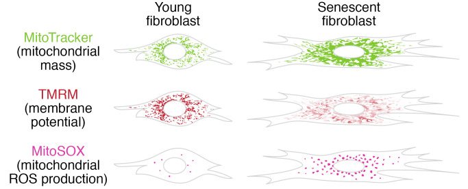
Mitochondrial dysfunction and cell senescence are hallmarks of aging and are closely interconnected. Mitochondrial dysfunction, operationally defined as a decreased respiratory capacity per mitochondrion together with a decreased mitochondrial membrane potential, typically accompanied by increased production of oxygen free radicals, is a cause and a consequence of cellular senescence and figures prominently in multiple feedback loops that induce and maintain the senescent phenotype. Here, we summarize pathways that cause mitochondrial dysfunction in senescence and aging and discuss the major consequences of mitochondrial dysfunction and how these consequences contribute to senescence and aging. We also highlight the potential of senescence-associated mitochondrial dysfunction as an antiaging and antisenescence intervention target, proposing the combination of multiple interventions converging onto mitochondrial dysfunction as novel, potent senolytics.
Overview
The claims and statistics of this paper haven’t been checked yet.
Free: it reads the open-access text and checks the first 5 claims that cite a source we can check. Create a free account to check every claim.
Integrity signals
Registry
Cites
Statistics
Figures

Verification
Claims the paper makes
The claims and statistics of this paper haven’t been checked yet.
Free: it reads the open-access text and checks the first 5 claims that cite a source we can check. Create a free account to check every claim.
Claims other papers make about this one
Each is checked against this paper’s own text: does it say what the citing paper says it does?
The source backs part of the claim, not all of it — or backs it for a different population, condition or comparison.
In the citing paper
“Both proliferation-competent glial cells (which constitute approximately 50% of brain cells) and postmitotic neurons can be positive for markers of cellular senescence in aging, obesity, traumatic injury, and neurodegeneration31,45–49, among other conditions.”
From Guidelines for minimal information on cellular senescence experimentation in vivo (2024) · cites this paper as [45]
Experimental — accuracy not yet measuredThe source backs part of the claim, not all of it — or backs it for a different population, condition or comparison.
In the citing paper
“Therefore, the dysregulation of mitochondrial fusion and fission pathways in senescent astrocytes likely contributes to mitochondrial dysfunction, oxidative stress, and the overall senescence-associated phenotype observed in these cells (Ma et al., 2020; Kleele et al., 2021; Miwa et al., 2022).”
From Aging promotes an increase in mitochondrial fragmentation in astrocytes (2024) · cites this paper as Miwa et al., 2022
Experimental — accuracy not yet measuredThe source backs part of the claim, not all of it — or backs it for a different population, condition or comparison.
In the citing paper
“Dysfunctional mitochondrial dynamics, as observed here, particularly excessive mitochondrial fission, have been associated with oxidative stress, mitochondrial DNA damage, and cellular senescence in various cell types (Twig et al., 2008; Miwa et al., 2022).”
From Aging promotes an increase in mitochondrial fragmentation in astrocytes (2024) · cites this paper as Miwa et al., 2022
Experimental — accuracy not yet measuredThe cited source says what the claim attributes to it.
In the citing paper
“while the presence of senescence markers has been proven even in post-mitotic cells”
From Guidelines for minimal information on cellular senescence experimentation in vivo (2024) · cites this paper as [45]
The check wasn’t confident either way, so it gives no verdict — never disputed.
In the citing paper
“Evidence from the literature indicates that senescent cells exhibit an increase in oxygen consumption rate (Hutter et al., 2004; Son et al., 2017; Miwa et al., 2022; Bang et al., 2023; Diniz et al., 2024a).”
From Aging promotes an increase in mitochondrial fragmentation in astrocytes (2024) · cites this paper as Miwa et al., 2022
Experimental — accuracy not yet measured
2 more claims about this paper
Create a free account to see every claim other papers make about this one.
Self-check
Percentages, sums and reported differences re-computed from the paper’s own numbers.
The self-check needs the paper’s full text.
Citations
Cites
The serial cultivation of human diploid cell strains
Hayflick, L., & Moorhead, P. S. (1961) · Experimental Cell Research · doi:10.1016/0014-4827(61)90192-6 · Cited by 8,508 · cited 1×
“Introduction What is cell senescence? Cell senescence was originally defined as the irreversible loss of replicative potential of primary cells in culture (1), initiated as a persistent DNA damage response to dysfunctional telomeres (2).”
BackgroundTelomeres shorten during ageing of human fibroblasts
Harley, C. B., Futcher, A. B., & Greider, C. W. (1990) · Nature · doi:10.1038/345458a0 · Cited by 5,750 · cited 2×
“Telomere dysfunction might be caused by telomere shortening during DNA replication (3, 4), replication-independent telomere damage (5), or a variety of other stresses (6).”
BackgroundA role for mitochondria in nlrp3 inflammasome activation
Zhou, R., Yazdi, A. S., Menu, P., & Tschopp, J. (2011) · Nature · doi:10.1038/nature09663 · Cited by 5,602
The senescence-associated secretory phenotype: the dark side of tumor suppression
Coppé, J.-P., Desprez, P.-Y., Krtolica, A., & Campisi, J. (2010) · Annual Review of Pathology: Mechanisms of Disease · doi:10.1146/annurev-pathol-121808-102144 · Cited by 5,206
Wu, Z., Puigserver, P., Andersson, U., Zhang, C., Adelmant, G., Mootha, V., Troy, A., Cinti, S., Lowell, B., Scarpulla, R. C., et al. (1999) · Cell · doi:10.1016/s0092-8674(00)80611-x · Cited by 4,139 · cited 1×
“PGC-1α activates the transcription factors Nrf1/2 and ERRα, which further control the TFAM transcription in response to increased mitochondrial biogenesis (65).”
BackgroundClearance of p16Ink4a-positive senescent cells delays ageing-associated disorders
Baker, D. J., Wijshake, T., Tchkonia, T., LeBrasseur, N. K., Childs, B. G., van de Sluis, B., Kirkland, J. L., & van Deursen, J. M. (2011) · Nature · doi:10.1038/nature10600 · Cited by 3,670
AMPK regulates energy expenditure by modulating NAD+ metabolism and SIRT1 activity
Cantó, C., Gerhart-Hines, Z., Feige, J. N., Lagouge, M., Noriega, L., Milne, J. C., Elliott, P. J., Puigserver, P., & Auwerx, J. (2009) · Nature · doi:10.1038/nature07813 · Cited by 3,300
Cellular Senescence: Defining a Path Forward
Gorgoulis, V., Adams, P. D., Alimonti, A., Bennett, D. C., Bischof, O., Bishop, C., Campisi, J., Collado, M., Evangelou, K., Ferbeyre, G., et al. (2019) · Cell · doi:10.1016/j.cell.2019.10.005 · Cited by 3,225 · cited 1×
“Telomere dysfunction might be caused by telomere shortening during DNA replication (3, 4), replication-independent telomere damage (5), or a variety of other stresses (6).”
BackgroundNakahira, K., Haspel, J. A., Rathinam, V. A. K., Lee, S.-J., Dolinay, T., Lam, H. C., Englert, J. A., Rabinovitch, M., Cernadas, M., Kim, H. P., et al. (2011) · Nature Immunology · doi:10.1038/ni.1980 · Cited by 3,049
A DNA damage checkpoint response in telomere-initiated senescence
Fagagna, F. D. D., Reaper, P. M., Clay-Farrace, L., Fiegler, H., Carr, P., von Zglinicki, T., Saretzki, G., Carter, N. P., & Jackson, S. P. (2003) · Nature · doi:10.1038/nature02118 · Cited by 2,917 · cited 1×
“Introduction What is cell senescence? Cell senescence was originally defined as the irreversible loss of replicative potential of primary cells in culture (1), initiated as a persistent DNA damage response to dysfunctional telomeres (2).”
Background
Research works printed but not linked: 2
- von Zglinicki T. Mechanisms of cell senescence in aging. In: Musi N, Hornsby P eds. Handbook of the Biology of Aging. Academic Press; 2021:53–68.
- Miwa S, Johnson TE. Stress and aging. Vijg J, et al., eds. In: The Molecular and Cellular Biology of Aging. Gerontological Society of America; 2016:216-249.
Cited by
Items 301–400 of 999
Song, Z., Wang, L., Ouyang, M., Xing, S., Liu, S., Sun, L., & Yu, H. (2024) · PLoS ONE · doi:10.1371/journal.pone.0306969 · Cited by 17
Jeong, D., Park, S. H., Kim, J., Kim, H., Jang, Y., Koh, J., Jeon, Y. K., Tasaki, T., Kwon, Y. T., Han, D., et al. (2025) · Proceedings of the National Academy of Sciences · doi:10.1073/pnas.2425015122 · Cited by 14
Li, J., Wang, Y., Shrestha, S., Gewirtz, A. T., Ding, Y., & Zou, J. (2025) · Nutrients · doi:10.3390/nu17172887 · Cited by 13
“Aging cells also exhibit high mtDNA mutation rates, which drive increased ROS production, apoptosis, and senescence [26–28].”
Interplay between aging and metabolic diseases: from molecular mechanisms to therapeutic horizons
Zhong, D., Sun, Y., Zhao, L., Hu, Z., Li, G., Li, H., & Xie, Z. (2025) · Medical Review · doi:10.1515/mr-2025-0047 · Cited by 20
“With advancing age, mitochondria exhibit declining e ffi ciency due to accumulated DNA mutations, reduced oxidative phosphorylation capacity, and impaired mitophagy – the process responsible for clearing damaged mitochondria [60].”
Senescent Cells: Dual Implications on the Retinal Vascular System
Habibi-Kavashkohie, M. R., Scorza, T., & Oubaha, M. (2023) · Cells · doi:10.3390/cells12192341 · Cited by 13
“Reactive oxygen species (ROS) are a by-product of mitochondrial respiration and increase strikingly in SCs due to low mitochondrial membrane potential [28,29].”
BackgroundInfluentialZhang, M., Wei, J., Sun, Y., He, C., Ma, S., Pan, X., & Zhu, X. (2024) · Journal of Advanced Research · doi:10.1016/j.jare.2024.03.008 · Cited by 16
Iron retardation in lysosome protects senescent cells from ferroptosis
Feng, Y., Wei, H., Lyu, M., Yu, Z., Chen, J., Lyu, X., & Zhuang, F. (2024) · Aging · doi:10.18632/aging.205777 · Cited by 34
Yang, Y., Wang, B., Dong, H., Lin, H., Ho, M. Y.-M., Hu, K., Zhang, N., Ma, J., Xie, R., Cheng, K. K. Y., et al. (2024) · Proceedings of the National Academy of Sciences · doi:10.1073/pnas.2401218121 · Cited by 16
“Mitochondrial dysfunction, such as increased mitochondrial ROS and dysregulated nutrient sensing pathways is known to cause senescence ( 62 ), but the role of mitochondrial enzymes in senescence is less explored.”
The Impact of HIV on Early Brain Aging—A Pathophysiological (Re)View
Lazăr, M., Moroti, R., Barbu, E. C., Chițu, C. E., Tilişcan, C., Erculescu, T. M., Rosca, R. R., Frasila, S., Schmilevschi, E. T., Simion, V., et al. (2024) · Journal of Clinical Medicine · doi:10.3390/jcm13237031 · Cited by 13
Xiao, S. J., Peng, K., Chen, R., Liu, X., Zhou, B., He, R., Yan, Y., Wan, R., Yin, Y., Lu, S., et al. (2024) · Theriogenology · doi:10.1016/j.theriogenology.2024.11.015 · Cited by 12
Shi, Y., Tong, X., Weng, Y., Xiao, Y., Wu, J., Wang, J., Wang, W., Yan, M., Yan, M., Li, Z., et al. (2024) · Journal of Translational Medicine · doi:10.1186/s12967-024-05953-7 · Cited by 14
Cellular Senescence in Glial Cells: Implications for Multiple Sclerosis
Maupin, E. A., & Adams, K. L. (2025) · Journal of Neurochemistry · doi:10.1111/jnc.16301 · Cited by 12
Raman Spectroscopy in Cellular and Tissue Aging Research
Kim, J. H., Yang, D., & Park, S. (2025) · Aging Cell · doi:10.1111/acel.14494 · Cited by 14
“Mitochondria's phenotypes and activity were also changed in aging cells compared to normal cells (Miwa et al. 2022; Sun, Youle, and Finkel 2016).”
Selective BCL-2 inhibitor triggers STING-dependent antitumor immunity via inducing mtDNA release
Zhang, W., Pan, X., Wang, L., Li, W., Dai, X., Zheng, M., Guo, H., Chen, X., Xu, Y., Wu, H., et al. (2025) · Journal for ImmunoTherapy of Cancer · doi:10.1136/jitc-2024-010889 · Cited by 12
“This observation is consistent with numerous previous studies showing that solid cancer cells that are less dependent on BCL-2 are usually not sensitive to ABT-199 (online supplemental figure 5B).”
Koner, S., Seenivasan, R., & Chandrasekaran, N. (2025) · Toxicology · doi:10.1016/j.tox.2025.154239 · Cited by 14
Age-Associated Loss in Renal Nestin-Positive Progenitor Cells
Buyan, M. I., Andrianova, N., Popkov, V., Zorova, L., Pevzner, I., Silachev, D., Zorov, D., & Plotnikov, E. (2022) · International Journal of Molecular Sciences · doi:10.3390/ijms231911015 · Cited by 7
“Impaired mitochondrial functioning and resulting changes in mitochondrial membrane potential being a typical feature of cells from old organisms, which may also cause and regulate the level of oxidative stress [61].”
BackgroundCao, Y., Wang, Y., Zhao, N., Yuan, Z., Zhang, L. H., & Jin, P. (2025) · Metabolism · doi:10.1016/j.metabol.2025.156356 · Cited by 11
Nowacka, A., Śniegocki, M., Śniegocka, M., & Ziółkowska, E. (2025) · Antioxidants · doi:10.3390/antiox14080911 · Cited by 11
“For example, chronic activation of mTORC1 in aged muscle promotes anabolic resistance, while declining SIRT1 activity exacerbates oxidative stress and inflammation [7,18,38,54,75,79–87].”
Mitochondrial-encoded peptide MOTS-c prevents pancreatic islet cell senescence to delay diabetes
Kong, B. S., Lee, H., L’Yi, S., Hong, S., & Cho, Y. M. (2025) · Experimental & Molecular Medicine · doi:10.1038/s12276-025-01521-1 · Cited by 16
“6h).”
Chen, K., Wang, W., Shu, H.-Y., Lyu, J., Cui, W., & Fang, H. (2025) · Interdisciplinary medicine · doi:10.1002/inmd.70054 · Cited by 16
Mitochondrial Dynamics in Aging Heart
Patyal, P., Azhar, G., Verma, A., Sharma, S., Shrivastava, J., Abdi, S. A. H., Zhang, X., & Wei, J. Y. (2025) · Biomedicines · doi:10.3390/biomedicines13112603 · Cited by 21
“For example, low-dose rotenone (Complex I inhibitor) or antimycin A (Complex III inhibitor) can exacerbate oxidative damage and activate maladaptive stress responses resembling those seen in senescent myocardium [157,158].”
Cáceres-Palomo, L., Sánchez‐Mejías, E., Trujillo‐Estrada, L., Pérez-Moreno, J. J., Lopez-Oliva, E., Lim, T. E., DeFlitch, L., Chang, S. H., Kampman, L., Corces, M. R., et al. (2025) · Journal of Neuroinflammation · doi:10.1186/s12974-025-03607-z · Cited by 12
“…oxidative metabolism, decreased mitochondrial fission, altered mitophagy and higher production of mitochondrial ROS are features of senescent cells [38, 39], we wanted to evaluate whether AD astrocytes may present a senescent phenotype which may explain these mitochondrial alterations.”
InfluentialVenz, R., Goyala, A., Soto-Gamez, A., Yenice, T., Demaria, M., & Ewald, C. Y. (2023) · GeroScience · doi:10.1007/s11357-023-00909-z · Cited by 14
“…in mammals, mitochondrial dysfunction can drive cells into senescence (MiDAS) [75], and senolytic inhibition of Bcl2 changes mitochondrial membrane potential and thereby promoting necrosis in addition to apoptosis [76], suggesting a conserved non-canonical link to our findings in C. elegans .”
BackgroundPremature senescence and cardiovascular disease following cancer treatments: mechanistic insights
Jain, A., Casanova, D., Padilla, A. V., Paniagua-Bojorges, A., Kotla, S., Ko, K. A., Samanthapudi, V. S. K., Chau, K. M., Nguyen, M. T., Wen, J. J., et al. (2023) · Frontiers in Cardiovascular Medicine · doi:10.3389/fcvm.2023.1212174 · Cited by 13
“The interplay between aging and mitochondrial dysfunction contributes to a senescent phenotype (175, 176).”
BackgroundSelf-citationLiang, J., Yao, F., Fang, D., Chen, L., Zou, Z., Feng, L., Zhuang, Y., Xie, T., Wei, P., Li, P., et al. (2023) · The FASEB Journal · doi:10.1096/fj.202301273rr · Cited by 11
Chen, Y., Zhu, Z., Yan, Y., Sun, H., Wang, G., Du, X., Li, F., Yuan, S., Wang, W., Wang, M., et al. (2024) · CNS Neuroscience & Therapeutics · doi:10.1111/cns.14819 · Cited by 11
“29,30 In our previous study, P7C3 protected mitochondrial function.”
BackgroundTan, S., Zhang, X., Guo, X., Pan, G., Yan, L., Ding, Z., Li, R., Wang, D., Yan, Y., Dong, Z., et al. (2024) · Cell Death and Disease · doi:10.1038/s41419-024-06912-2 · Cited by 15
“A systematic functional evaluation”
Wu, Z., Zeng, Y., Chen, H.-M., Xiao, Z., Guo, J., Abubakar, M. N., Shen, M., Xiao, H., Song, X., & Cai, Y. (2024) · ACS Applied Materials & Interfaces · doi:10.1021/acsami.4c09734 · Cited by 11
Unraveling the metabolic heterogeneity and commonality in senescent cells using systems modeling
Li, G., Li, Y., Yu, Q., Zhou, Q.-Q., Zhang, R., Weng, C.-J., Ge, M., & Kong, Q.-P. (2025) · Life Medicine · doi:10.1093/lifemedi/lnaf003 · Cited by 12
“Some studies have reported that senescent cells show reduced mitochondrial function and increased ROS production, which cause protein and lipid damage [15, 16].”
Yang, S., Wang, L., Liang, X., Pei, T., Zeng, Y., Xie, B., Wang, Y., Yang, M., Wei, D., & Cheng, W. (2025) · International Journal of Biological Macromolecules · doi:10.1016/j.ijbiomac.2025.141715 · Cited by 15
Mitochondrial and microtubule defects in Exfoliation Glaucoma
Venkatesan, A., Ridilla, M., Castro, N., Wolosin, J. M., Henty-Ridilla, J. L., Knox, B. E., Ganapathy, P. S., Brown, J. S., DeVincentis, A. F., Sieminski, S. F., et al. (2025) · Free Radical Biology and Medicine · doi:10.1016/j.freeradbiomed.2025.03.046 · Cited by 12
Zeng, S., Liu, L., Cheng, Q., Chen, W., Wang, M., Wu, M., Wang, Z., Du, J., Xiang, R., Qi, Q., et al. (2025) · Journal of Medicinal Chemistry · doi:10.1021/acs.jmedchem.4c02879 · Cited by 16
Mitochondrial Dysfunction and Atherosclerosis: The Problem and the Search for Its Solution
Невойт, Г. В., Jaruševičius, G., Potyazhenko, M., Mintser, O. P., Bumblytė, I. A., & Vainoras, A. (2025) · Biomedicines · doi:10.3390/biomedicines13040963 · Cited by 15
“In this case, vicious circles are formed from the molecular mechanisms of MD [255].”
Ren, Y., Sun, T., Yu, L., Liu, D., Gao, R., Li, T., Guo, M., Liu, Q., Wang, H.-L., & Pang, M. (2025) · Journal of Cellular and Molecular Medicine · doi:10.1111/jcmm.70566 · Cited by 13
“Moreover, the activation of AMPK increased the concentration of intracellular NAD+, thereby promoting the expression of Sirt1, which was consistent with previous reports [55].”
Huang, Y., Mao, J., Li, Z., Wang, W., Ni, Z., Cai, F., Tang, J., Wang, W., Zhang, L., Zhou, L., et al. (2025) · Advanced Materials · doi:10.1002/adma.202500156 · Cited by 24
Ünal, Ö., Akgun-Unal, N., & Baltacı, A. K. (2025) · Biogerontology · doi:10.1007/s10522-025-10254-7 · Cited by 13
“These complexes are essential for the electron transport chain and ATP production, as noted by Miwa et al. (2022).”
Lu, J.-J., Zhang, Q., Chen, Y., Yuan, G.-C., Huang, Y.-K., Wu, T., Zhang, Q., Dong, J., Jiang, L., & Li, X. (2025) · Phytomedicine · doi:10.1016/j.phymed.2025.157021 · Cited by 15
Building the case for mitochondrial transplantation as an anti-aging cardiovascular therapy
Headley, C. A., & Tsao, P. S. (2023) · Frontiers in Cardiovascular Medicine · doi:10.3389/fcvm.2023.1141124 · Cited by 11
“Cells affected by aging-associated mito-dysfunction undergo concurrent cellular events that continually catalyze mito-dysfunction and cell dysregulations (44).”
BackgroundMukem, S., Sayoh, I., Maungchanburi, S., & Thongbuakaew, T. (2023) · Advances in Pharmacological and Pharmaceutical Sciences · doi:10.1155/2023/6641347 · Cited by 19
“Tis is apparently distinct in the accumulation of senescent cells and secretory phenotype [53, 54].”
BackgroundSaleh, T., Naffa, R. G., Barakat, N. A., Ismail, M. A., Alotaibi, M. R., & Alsalem, M. (2024) · Neurotoxicity Research · doi:10.1007/s12640-024-00690-7 · Cited by 11
“For instance, mitochondrial dysfunction, an established feature of the senescent phenotype (Wiley et al. 2016; Miwa et al. 2022), has been linked to the development of neuronal dysfunction (Pradeepkiran et al. 2023).”
Zhou, L., Zhai, G., & Tian, G. (2024) · Aging · doi:10.18632/aging.205664 · Cited by 12
Ling, S., Zhang, Y., Chen, Y., He, W., Wu, T., Wang, J., Wei, Y., Lu, M., Zhao, W., Li, Q., et al. (2025) · Advanced Materials · doi:10.1002/adma.202513720 · Cited by 14
Cellular senescence in cancer: Friend or fraud?
Chen, Q., Liu, J., Wu, J., Xiang, X., Zhou, X., Mi, J., Wei, J., Zou, X., & Wu, A. (2026) · Drug Resistance Updates · doi:10.1016/j.drup.2026.101363 · Cited by 20
Qian, X., Wang, Y., Kuang, P., Li, B., Jiang, K., Wang, A., Xiong, K., Hong, W., Yu, B., Qin, G., et al. (2026) · Redox Biology · doi:10.1016/j.redox.2026.104091 · Cited by 13
“Although this study did not directly validate the relationship between mitochondria and senescence, it is well-established that mitochondrial functional decline is a hallmark of cellular aging [35].”
Clutton, G., Weideman, A. M., Goonetilleke, N. P., & Maurer, T. A. (2022) · Frontiers in Cell and Developmental Biology · doi:10.3389/fcell.2022.961021 · Cited by 14
“This was not due to a loss of mitochondrial mass, as the frequency of MitoTracker® Greenhigh cells was higher in CD8dim than CD8bright cells (Supplementary Figure S4).”
BackgroundInfluentialMitochondrial disorders as a mechanism for the development of obese Sarcopenia
Liao, T., Xiong, L., Wang, X., Yang, S., & Liang, Z. (2023) · Diabetology & Metabolic Syndrome · doi:10.1186/s13098-023-01192-w · Cited by 10
“As aging progresses, the number of damaged mitochondria increases in cells and cannot be reduced through mitochondrial division and mitoph-agy [82, 83].”
BackgroundMasuda, K., Sakurai, T., & Hirano, A. (2024) · Scientific Reports · doi:10.1038/s41598-024-66347-9 · Cited by 11
Liu, C., Wu, C., Zhang, S., & Lv, Z. (2024) · Experimental Gerontology · doi:10.1016/j.exger.2024.112528 · Cited by 11
Tagliafico, L., da Costa, R. T., Boccia, L., Kavehmoghaddam, S., Ramirez, B., Tokarska-Schlattner, M., Scoma, E. R., Hambardikar, V. D., Bonfiglio, T., Caffa, I., et al. (2024) · Aging Cell · doi:10.1111/acel.14289 · Cited by 9
“While the mechanisms driving mammalian cells to senescence are complex and still remain incompletely described, mitochondrial dysfunction, including bioenergetic dysregulation, seems to be crucially involved in these mechanisms (Miwa et al., 2022).”
Batitucci, G., Abud, G. F., Ortiz, G. U., Belisário, L. F., Travieso, S. G., Viliod, M. C. D. L., Venturini, A. C. R., & de Freitas, E. C. (2024) · Ageing Research Reviews · doi:10.1016/j.arr.2024.102460 · Cited by 13
Phospholipid Phosphatase 3 (PLPP3) Induces Oxidative Stress to Accelerate Ovarian Aging in Pigs
Quan, H., Guo, Y., Li, S., Jiang, Y., Shen, Q., He, Y., Zhou, X., Yuan, X., & Li, J. (2024) · Cells · doi:10.3390/cells13171421 · Cited by 12
“…that the differentially expressed genes were mainly enriched in the regulation of the reproductive process and membrane potential, and previous studies have suggested that the decreased membrane potential brought by mitochondrial dysfunction contributes to cellular senescence [37] (Figure 2E).”
BackgroundNeuronal Cell Rearrangement During Aging: Antioxidant Compounds as a Potential Therapeutic Approach
Bej, E., Cesare, P., d’Angelo, M., Volpe, A. R., & Castelli, V. (2024) · Cells · doi:10.3390/cells13231945 · Cited by 13
Chewing-Activated TRPV4/PIEZO1– HIF-1α –Zn Axes in a Rat Periodontal Complex
Wang, Y., Lee, B. H., Yang, Z., Ho, T. J., Ci, H., Jackson, B. P., Punshon, T., Wang, B., Levy, J., & Ho, S. P. (2025) · Journal of Dental Research · doi:10.1177/00220345241294001 · Cited by 14
“Mitochondrial function (MFN2) and hypoxia ( HIF-1α ) relate to cell senescence (p16) (Xing et al. 2018; Miwa et al. 2022).”
Simvastatin ameliorates senescence-induced mitochondrial dysfunction in vascular smooth muscle cells
Rossi, C., Macchi, C., D'Alonzo, C., Venturin, M., Ruscica, M., Corsini, A., Battaglia, C., & Bellosta, S. (2025) · Atherosclerosis · doi:10.1016/j.atherosclerosis.2025.119176 · Cited by 14
Li, C., Ji, H., Zhuang, S., Xie, X., Cui, D., & Zhang, C. (2025) · Redox Report · doi:10.1080/13510002.2025.2491846 · Cited by 12
“Moreover, the accumulation of such defective mitochondria in the body can accelerate inflammation and amplify oxidative stress [126].”
Anti-aging effect of tea and its phytochemicals
Liang, H., Mei, Q., Ang, S., Li, D., & He, C. (2025) · Food Research International · doi:10.1016/j.foodres.2025.116572 · Cited by 15
He, J., Zhang, B., Zhang, H., Tu, Q., Chen, X., Qiu, Y., Liu, Z., Xia, W., Wu, X., & Tao, J. Y. (2025) · Human Mutation · doi:10.1155/humu/6771390 · Cited by 10
“Cellular senescence is characterized by an irreversible arrest of the cell cycle in the G1 phase [23, 24] and an increased production of ROS, primarily attributed to mitochondria dysfunction [22].”
Gu, C., Guo, T., Chen, X., Zhou, X., & Sun, Y. (2025) · Cellular and Molecular Neurobiology · doi:10.1007/s10571-025-01580-y · Cited by 11
“Mitochondria are vulnerable to inflammatory insult, and mitochondrial dysfunction is one of the major consequences of cellular senescence which is typically characterized by the morphological fragmentation (Miwa et al. 2022; Chapman et al. 2019).”
Wang, T., Zhou, X., Yin, X., Zhang, A., Fan, Y., Chen, K., Tao, H., Tang, Z., Zhang, P., He, X., et al. (2025) · International Journal of Oncology · doi:10.3892/ijo.2025.5781 · Cited by 14
“Mitochondrial involvement in ferroptosis extends beyond regulating redox imbalance, as they are also crucial in ROS accumulation (14).”
Popa‐Wagner, A., Hermann, D. M., Doeppner, T. R., Surugiu, R., & Pîrșcoveanu, D. F. V. (2025) · Journal of Cerebral Blood Flow & Metabolism · doi:10.1177/0271678x251365020 · Cited by 19
“In a study using a rat model of focal ischemia targeting the right middle cerebral artery, researchers induced comparable lesion volumes in rats aged 10, 63, and 180 days.”
Developmental origins of Parkinson disease: Improving the rodent models
Jiménez-Salvador, I., Meade, P., Iglesias, E., Bayona‐Bafaluy, M. P., & Ruiz‐Pesini, E. (2023) · Ageing Research Reviews · doi:10.1016/j.arr.2023.101880 · Cited by 12
“Therefore, ageing greatly affects OXPHOS function (Amorim et al., 2022; Lesnefsky and Hoppel, 2006; Miwa et al., 2022).”
Hebert, J. F., & Myatt, L. (2023) · Antioxidants · doi:10.3390/antiox12030719 · Cited by 15
“MitoTracker identifies intact mitochondria, including those in senescent cells, while MitoSox identifies mitochondria actively generating superoxide [48].”
BackgroundEndogenous Biological Drivers in Diabetic Lower Limb Wounds Recurrence: Hypothetical Reflections
Berlanga‐Acosta, J., Ojalvo, A. G., Guillén, G. E., & Ayala‐Ávila, M. (2023) · International Journal of Molecular Sciences · doi:10.3390/ijms241210170 · Cited by 11
Bioimaging tools reveal copper processing in fish cells by mitophagy
Xia, Y., & Wang, W. (2023) · Aquatic Toxicology · doi:10.1016/j.aquatox.2023.106633 · Cited by 9
Li, S., Zhuang, Y., Ji, Y. R., Chen, X., He, L., Chen, S., Luo, Y., Shen, L., Xiao, J., Wang, H., et al. (2024) · Free Radical Biology and Medicine · doi:10.1016/j.freeradbiomed.2024.01.054 · Cited by 12
Seydi, E., Rahemi, M., Esmaily, H., Arjmand, A., & Pourahmad, J. (2023) · Journal of Pharmacy and Pharmacology · doi:10.1093/jpp/rgad079 · Cited by 11
Duan, C., Wang, Z., Wu, J., Tan, C., Fang, F., Qian, W., Han, C., & Hou, X. (2024) · Research · doi:10.34133/research.0343 · Cited by 12
“Fut2 seemed to mainly affect respiratory chain complexes and mitophagy as described in our study, which are closely associated with aging-related diseases as previously reported [50].”
ResultThe mitochondrial multi-omic response to exercise training across tissuesPreprint
Amar, D., Gay, N. R., Jimenez‐Morales, D., Beltran, P. M. J., Ramaker, M. E., Raja, A. N., Zhao, B., Sun, Y., Marwaha, S., Gaul, D. A., et al. (2023) · bioRxiv (Cold Spring Harbor Laboratory) · doi:10.1101/2023.01.13.523698 · Cited by 10
“Maintaining mitochondrial function is critical for prolonged healthspan, as mitochondrial dysfunction is a hallmark of aging and is associated with cardiovascular, metabolic and neurodegenerative diseases 85 .”
SA-β-Gal in Kidney Tubules as a Predictor of Renal Outcome in Patients with Chronic Kidney Disease
Esposito, P. B., Picciotto, D., Verzola, D., Garibotto, G., Parodi, E. L., Sofia, A., Costigliolo, F., Gaggero, G., Zanetti, V., Saio, M., et al. (2024) · Journal of Clinical Medicine · doi:10.3390/jcm13020322 · Cited by 10
“In contrast, in chronic diseases, senescent cells accumulate in the kidney in response to a variety of stressors such as metabolic stress, telomere shortening [22], oncogenic mutations, inflammation, and mitochondrial dysfunction [23].”
BackgroundSizek, H., Deritei, D., Fleig, K., Harris, M., Regan, P., Glass, K., & Regan, E. R. (2024) · Translational Oncology · doi:10.1016/j.tranon.2024.102084 · Cited by 11
“Briefly, the omitted process involves high p21-mediated p38 MAPK activation, leading to increased cytosolic ROS that locks in a p21 → p38 → ROS & p21 feedback loop [13].”
Selective autophagy: a therapeutic target for healthy aging?
Manastireanu, D. M., Salazar, N. A., Bejarano, E., & Nieto-Torres, J. L. (2024) · Aging advances. · doi:10.4103/agingadv.agingadv-d-24-00002 · Cited by 10
Is Senolytic Therapy in Cardiovascular Diseases Ready for Translation to Clinics?
Yang, Z., Potenza, D. M., & Ming, X. (2025) · Biomolecules · doi:10.3390/biom15040545 · Cited by 13
Current strategies for senescence treatment: Focused on theranostic performance of nanomaterials
Cong, Y., Li, X., & Hong, H. (2025) · Journal of Controlled Release · doi:10.1016/j.jconrel.2025.113710 · Cited by 10
Guo, J., Wang, Y., Liu, Q., Luo, Z., Tian, X., Wang, Y., Liu, Y., Ning, Z., Guo, Y., Gao, H., et al. (2025) · Advanced Science · doi:10.1002/advs.202501956 · Cited by 11
Polzer, O., Kinloch, E., & Fitzsimons, C. P. (2025) · Aging Brain · doi:10.1016/j.nbas.2025.100141 · Cited by 11
“Mitochondria dysfunction, in particular of the mitochondrial complex I (NADH: ubiquinone oxidoreductase) in the brain, is associated with cell senescence and ageing [87].”
Senescence of dental pulp stem cells: phenotypes, underlying mechanisms and regulatory molecules
Zhang, P., Cui, Y., Li, Z., Liu, L., Liu, X., Ding, X. H., & Ding, G. (2025) · Human Cell · doi:10.1007/s13577-025-01259-y · Cited by 11
“However, several approaches, such as mitochondria-specific antioxidants and gene repair technologies aimed at repairing damaged mitochondria, have shown promise in improving mitochondrial function and alleviating cellular aging [71].”
Fan, Z., Wang, X., Wu, Y., Li, S., Kong, L., Li, S., Ma, T., & Pan, C. (2026) · Environmental Pollution · doi:10.1016/j.envpol.2026.127778 · Cited by 9
Endothelial cell senescence and mitochondrial dysfunction in vascular ageing
Walczak, I., Tarnawska, M., Stawarska, K., Braczko, A., Paterek, A., Rolski, F., Leszek, P., Smolarek, D., Kapusta, M., Narajczyk, M., et al. (2026) · Ageing Research Reviews · doi:10.1016/j.arr.2026.103119 · Cited by 14
Talk to Me—Interplay between Mitochondria and Microbiota in Aging
Endres, K., & Friedland, K. (2023) · International Journal of Molecular Sciences · doi:10.3390/ijms241310818 · Cited by 13
“This decline is specific for different tissues with reduced complex III and IV activity in the heart muscle and diminished complex I and III function in brain, liver, and skeletal muscle, as well as parts of our immune system such as CD4+ T-helper cells [17,29,30].”
BackgroundSingle‐cell landscape analysis reveals systematic senescence in mammalian Down syndrome
Chen, Y., Xiao, Y., Zhang, Y., Wang, R., Wang, F., Gao, H., Liu, Y., Zhang, R., Sun, H., Zhou, Z., et al. (2023) · Clinical and Translational Medicine · doi:10.1002/ctm2.1310 · Cited by 9
Alsalem, M., Ellaithy, A., Bloukh, S., Haddad, M., & Saleh, T. (2024) · Supportive Care in Cancer · doi:10.1007/s00520-023-08287-0 · Cited by 13
“Senescent cells accumulate dysfunctional mitochondria characterized by reduced mitochondrial membrane potential and structural changes [118, 119].”
Zhicheng, J., Li, Y., Peixuan, W., Yang, K., Mengyu, S., Chen, W. L., Qihui, L., & Ying, G. (2024) · Journal of Ovarian Research · doi:10.1186/s13048-023-01341-9 · Cited by 12
“Mitochondrial dysfunction is reflected in many aspects, including the decrease of MMP, the increase of mtDNA mutations, and the accumulation of ROS, etc [26, 27].”
BackgroundUrolithin A prevents age-related hearing loss in C57BL/6J mice likely by inducing mitophagy
Cho, S. I., Jo, E.-R., & Jang, H. S. (2024) · Experimental Gerontology · doi:10.1016/j.exger.2024.112589 · Cited by 13
“Mitochondria generate ATP and supply cellular energy.”
InfluentialDecline of stress resilience in aging rats: Focus on choroid plexus-cerebrospinal fluid-hippocampus
Liu, K., Li, H., Zeng, N.-X., Lu, W., Wu, X., Xu, H., Yan, C., & Wu, L. (2022) · The World Journal of Biological Psychiatry · doi:10.1080/15622975.2022.2151044 · Cited by 10
The role of mitochondria and mitophagy in cell senescence
Ali, T., Hussain, F., Kayani, H. U. R., Naeem, M., & Anjum, F. (2023) · Advances in protein chemistry and structural biology · doi:10.1016/bs.apcsb.2023.03.001 · Cited by 10
Targeting mitochondrial dysfunction to salvage cellular senescence for managing neurodegeneration
Sharma, K., Sarkar, J., Trisal, A., Ghosh, R., Dixit, A., & Singh, A. K. (2023) · Advances in protein chemistry and structural biology · doi:10.1016/bs.apcsb.2023.02.016 · Cited by 8
Hu, S., Wang, T.-Y., Ni, L., Hu, F.-X., Yue, B.-W., Zheng, Y., Wang, T.-L., Kumar, A., Wang, Y., Wang, J.-E., et al. (2024) · Current Medical Science · doi:10.1007/s11596-024-2892-0 · Cited by 10
He, B., Zeng, Q., Tian, Y., Luo, Y., Liao, M., Huang, W., Wu, B., Luo, Z., Huang, X., Liu, W. Q., et al. (2024) · Biomedicines · doi:10.3390/biomedicines12071436 · Cited by 10
“Moreover, mitochondrial dysfunction in senescent cells leads to increased ROS production, which may further contribute to the progression of cellular senescence and fibrosis [38].”
BackgroundXiao, R., Hu, S., Du, X., Wang, Y., Fang, K., Zhu, Y., Lou, N., Yuan, C., & Yang, J. (2024) · Aging and Disease · doi:10.14336/ad.202.0565 · Cited by 12
“Mitochondrial dysfunction, indicated by reduced respiratory capacity and increased mitochondrial mass, is a hallmark of cellular aging [36].”
Gu, Y., Qiu, Y., Li, Y., & Wen, W. (2024) · Toxicology Research · doi:10.1093/toxres/tfae136 · Cited by 10
Nanoparticle–Protein Corona-Based Tissue Proteomics for the Aging Mouse Proteome Atlas
Wang, L., Xu, Y., Xie, Y., Xu, C., Dai, X., Wang, M., & Liu, Y. (2024) · Analytical Chemistry · doi:10.1021/acs.analchem.4c00932 · Cited by 10
Tang, H., Gao, P., Peng, W., Wang, X., Wang, Z., Deng, W., Yin, K., & Zhu, X. (2024) · Cell Communication and Signaling · doi:10.1186/s12964-024-01859-5 · Cited by 9
“Together with the increased production of free oxygen radicals, which are a cause and consequence of cellular senescence, mitochondrial dysfunction plays a major role in multiple feedback loops that induce and preserve the senescent phenotype [38].”
BackgroundНевойт, Г. В., Jaruševičius, G., Potyazhenko, M., Mintser, O. P., Bumblytė, I. A., & Vainoras, A. (2024) · Diseases · doi:10.3390/diseases12110277 · Cited by 14
Deng, Z., Li, C., Hu, S., Zhong, Y., Li, W., Lin, Z., Mo, X., Li, M., Xu, D., Long, D., et al. (2025) · Cell Communication and Signaling · doi:10.1186/s12964-024-01975-2 · Cited by 11
“One of the hallmarks of aging is mitochondrial dysfunction, which has recently been recognized as both a cause and consequence of cellular senescence [45].”
Sauchinone Ameliorates Senescence Through Reducing Mitochondrial ROS Production
Kuk, M. U., Lee, Y. H., Kim, D., Lee, K. S., Park, J. H., Yoon, J. H., Lee, Y. J., So, B., Kim, M., Kwon, H. W., et al. (2025) · Antioxidants · doi:10.3390/antiox14030259 · Cited by 8
“Inefficient electron transfer increases ROS production and decreases MMP, whereas efficient electron transfer decreases ROS production and increases MMP [47].”
Regulatory Role of Bioactive Compounds from Natural Spices on Mitochondrial Function
Han, L., Ho, C., & Lu, M. (2025) · Journal of Agricultural and Food Chemistry · doi:10.1021/acs.jafc.4c12341 · Cited by 10
Lactylation: A Novel Epigenetic Regulator of Cellular Senescence
Sun, C., Li, J., Dong, L., Mou, Y., Zhang, B., & Song, X. (2025) · Aging and Disease · doi:10.14336/ad.2025.0277 · Cited by 14
“The progression of mitochondrial dysfunction, exacerbates cellular senescence, thereby establishing a self-perpetuating cycle [90].”
The Role of the Exposome in Aging and Age-Related Diseases: A Comprehensive Review
Kareem, M. A., Ashwini, A., & Sunil, T. (2025) · Journal of Pharmacy And Bioallied Sciences · doi:10.4103/jpbs.jpbs_599_25 · Cited by 8
“[15] Cellular decline also relies on mitochondrial dysfunction, [19] whereas chronic inflammation is a key pathway for environmental exposures to cause age‑related diseases.”
Endogenous double-stranded RNA: Bridging immune activation and cancer therapeutics
Liu, X., Che, L., & Weng, H. (2025) · Cell investigation. · doi:10.1016/j.clnves.2025.100018 · Cited by 10
Sonar, S. A., Bhat, R., Thompson, H. L., Coplen, C. P., Uhrlaub, J. L., Jergović, M., & Nikolich‐Žugich, J. (2025) · Aging Cell · doi:10.1111/acel.70100 · Cited by 9
Metadata
- Authors
- Miwa, S., Kashyap, S., Chini, E., & von Zglinicki, T.
- Affiliations
- Newcastle University (GB)
- Mayo Clinic (US)
- Venue
- Journal of Clinical Investigation
- Published
- 2022-07-01
- Volume
- 132
- Issue
- 13
- ISSN
- 0021-9738 (ISSN-L)
- 1558-8238
- PMC
- PMC9246372
- OpenAlex
- W4283723802
- Cited by
- 1,061
- References
- 145
- Citations by year
- 2026: 294 · 2025: 348 · 2024: 263 · 2023: 136 · 2022: 20
- FWCI
- 19.97
- Type
- journal article
- Language
- en
- Open access
- gold
- doi.org
- Licence
- Locations
- Journal of Clinical Investigation (publishedVersion, cc-by)
- PubMed Central (publishedVersion, cc-by)
- PubMed (publishedVersion)
- DOAJ (DOAJ: Directory of Open Access Journals) (submittedVersion)
- Newcastle University ePrints (Newcastle Univesity) (cc-by)
- Funders
- Glenn Foundation for Medical Research
- Ted Nash Long Life Foundation
- Cancer Research UK (C12161/A24009)
- Newcastle University
- Fondation pour la Recherche Médicale
- Versus Arthritis
- National Institutes of Health (H2020, AG26094, CA233790, AG58812)
- Directorate for Biological Sciences
- Horizon 2020 Framework Programme (857524)
- Medical Research Council (MR/P020941/1)
- Biotechnology and Biological Sciences Research Council (BB/S006710/1)
- National Institute on Aging (AG-58812, R01AG058812, AG-26094)
- National Cancer Institute (R01CA233790)
- Topics
- Life Sciences › Biochemistry, Genetics and Molecular Biology › Molecular Biology › Mitochondrial Function and Pathology
- Life Sciences › Biochemistry, Genetics and Molecular Biology › Cell Biology › Telomeres, Telomerase, and Senescence
- Life Sciences › Biochemistry, Genetics and Molecular Biology › Aging › Genetics, Aging, and Longevity in Model Organisms
- Keywords
- mitochondrial dysfunction, cellular senescence, aging, mitochondrial membrane potential, respiratory capacity, reactive oxygen species, senolytics
- MeSH terms
- Mitochondria, Phenotype, Cellular Senescence
About these checks
Registry, retraction, Cites and statistics results are computed, not judged, with one exception: a printed reference that no index lists is read once by a model before it counts as a citation error. Claims are extracted by a model and kept only when their quote appears verbatim in the text; each cited source is then checked, with a model’s help, for whether it says what the claim relies on. Those verdicts can be wrong — read the claim’s quote and the cited source before relying on one.
Statistics and arithmetic are recomputed, not judged.
Confidence bands are shown only for fields calibrated on our annotated test set. None is calibrated yet, so no band is shown.
What counts as a claim ›How we measure confidence ›How we check this ›
Are you an author, or do you see a problem with this page? Request a review
Data sources
Bibliographic data: Semantic Scholar, OpenAlex, Crossref.
Contains information from the Semantic Scholar Academic Graph Datasets, made available under the ODC Attribution License (ODC-BY 1.0).
Semantic Scholar’s data is described in: Kinney, R., et al. (2023). The Semantic Scholar Open Data Platform. arXiv:2301.10140.
