

Mitochondrial dysfunction in cell senescence and aging
Open accessThe DOI matches its Crossref record; 145 of the 145 references it cites were found in a registry or an index.
Abstract
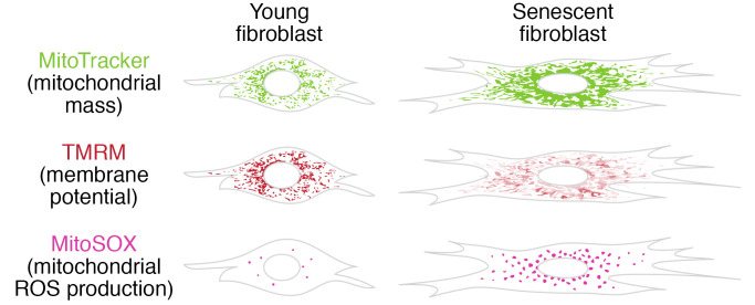
Mitochondrial dysfunction and cell senescence are hallmarks of aging and are closely interconnected. Mitochondrial dysfunction, operationally defined as a decreased respiratory capacity per mitochondrion together with a decreased mitochondrial membrane potential, typically accompanied by increased production of oxygen free radicals, is a cause and a consequence of cellular senescence and figures prominently in multiple feedback loops that induce and maintain the senescent phenotype. Here, we summarize pathways that cause mitochondrial dysfunction in senescence and aging and discuss the major consequences of mitochondrial dysfunction and how these consequences contribute to senescence and aging. We also highlight the potential of senescence-associated mitochondrial dysfunction as an antiaging and antisenescence intervention target, proposing the combination of multiple interventions converging onto mitochondrial dysfunction as novel, potent senolytics.
Overview
The claims and statistics of this paper haven’t been checked yet.
Free: it reads the open-access text and checks the first 5 claims that cite a source we can check. Create a free account to check every claim.
Integrity signals
Registry
Cites
Statistics
Figures

Verification
Claims the paper makes
The claims and statistics of this paper haven’t been checked yet.
Free: it reads the open-access text and checks the first 5 claims that cite a source we can check. Create a free account to check every claim.
Claims other papers make about this one
Each is checked against this paper’s own text: does it say what the citing paper says it does?
The source backs part of the claim, not all of it — or backs it for a different population, condition or comparison.
In the citing paper
“Both proliferation-competent glial cells (which constitute approximately 50% of brain cells) and postmitotic neurons can be positive for markers of cellular senescence in aging, obesity, traumatic injury, and neurodegeneration31,45–49, among other conditions.”
From Guidelines for minimal information on cellular senescence experimentation in vivo (2024) · cites this paper as [45]
Experimental — accuracy not yet measuredThe source backs part of the claim, not all of it — or backs it for a different population, condition or comparison.
In the citing paper
“Therefore, the dysregulation of mitochondrial fusion and fission pathways in senescent astrocytes likely contributes to mitochondrial dysfunction, oxidative stress, and the overall senescence-associated phenotype observed in these cells (Ma et al., 2020; Kleele et al., 2021; Miwa et al., 2022).”
From Aging promotes an increase in mitochondrial fragmentation in astrocytes (2024) · cites this paper as Miwa et al., 2022
Experimental — accuracy not yet measuredThe source backs part of the claim, not all of it — or backs it for a different population, condition or comparison.
In the citing paper
“Dysfunctional mitochondrial dynamics, as observed here, particularly excessive mitochondrial fission, have been associated with oxidative stress, mitochondrial DNA damage, and cellular senescence in various cell types (Twig et al., 2008; Miwa et al., 2022).”
From Aging promotes an increase in mitochondrial fragmentation in astrocytes (2024) · cites this paper as Miwa et al., 2022
Experimental — accuracy not yet measuredThe cited source says what the claim attributes to it.
In the citing paper
“while the presence of senescence markers has been proven even in post-mitotic cells”
From Guidelines for minimal information on cellular senescence experimentation in vivo (2024) · cites this paper as [45]
The check wasn’t confident either way, so it gives no verdict — never disputed.
In the citing paper
“Evidence from the literature indicates that senescent cells exhibit an increase in oxygen consumption rate (Hutter et al., 2004; Son et al., 2017; Miwa et al., 2022; Bang et al., 2023; Diniz et al., 2024a).”
From Aging promotes an increase in mitochondrial fragmentation in astrocytes (2024) · cites this paper as Miwa et al., 2022
Experimental — accuracy not yet measured
2 more claims about this paper
Create a free account to see every claim other papers make about this one.
Self-check
Percentages, sums and reported differences re-computed from the paper’s own numbers.
The self-check needs the paper’s full text.
Citations
Cites
The serial cultivation of human diploid cell strains
Hayflick, L., & Moorhead, P. S. (1961) · Experimental Cell Research · doi:10.1016/0014-4827(61)90192-6 · Cited by 8,508 · cited 1×
“Introduction What is cell senescence? Cell senescence was originally defined as the irreversible loss of replicative potential of primary cells in culture (1), initiated as a persistent DNA damage response to dysfunctional telomeres (2).”
BackgroundTelomeres shorten during ageing of human fibroblasts
Harley, C. B., Futcher, A. B., & Greider, C. W. (1990) · Nature · doi:10.1038/345458a0 · Cited by 5,750 · cited 2×
“Telomere dysfunction might be caused by telomere shortening during DNA replication (3, 4), replication-independent telomere damage (5), or a variety of other stresses (6).”
BackgroundA role for mitochondria in nlrp3 inflammasome activation
Zhou, R., Yazdi, A. S., Menu, P., & Tschopp, J. (2011) · Nature · doi:10.1038/nature09663 · Cited by 5,602
The senescence-associated secretory phenotype: the dark side of tumor suppression
Coppé, J.-P., Desprez, P.-Y., Krtolica, A., & Campisi, J. (2010) · Annual Review of Pathology: Mechanisms of Disease · doi:10.1146/annurev-pathol-121808-102144 · Cited by 5,206
Wu, Z., Puigserver, P., Andersson, U., Zhang, C., Adelmant, G., Mootha, V., Troy, A., Cinti, S., Lowell, B., Scarpulla, R. C., et al. (1999) · Cell · doi:10.1016/s0092-8674(00)80611-x · Cited by 4,139 · cited 1×
“PGC-1α activates the transcription factors Nrf1/2 and ERRα, which further control the TFAM transcription in response to increased mitochondrial biogenesis (65).”
BackgroundClearance of p16Ink4a-positive senescent cells delays ageing-associated disorders
Baker, D. J., Wijshake, T., Tchkonia, T., LeBrasseur, N. K., Childs, B. G., van de Sluis, B., Kirkland, J. L., & van Deursen, J. M. (2011) · Nature · doi:10.1038/nature10600 · Cited by 3,670
AMPK regulates energy expenditure by modulating NAD+ metabolism and SIRT1 activity
Cantó, C., Gerhart-Hines, Z., Feige, J. N., Lagouge, M., Noriega, L., Milne, J. C., Elliott, P. J., Puigserver, P., & Auwerx, J. (2009) · Nature · doi:10.1038/nature07813 · Cited by 3,300
Cellular Senescence: Defining a Path Forward
Gorgoulis, V., Adams, P. D., Alimonti, A., Bennett, D. C., Bischof, O., Bishop, C., Campisi, J., Collado, M., Evangelou, K., Ferbeyre, G., et al. (2019) · Cell · doi:10.1016/j.cell.2019.10.005 · Cited by 3,225 · cited 1×
“Telomere dysfunction might be caused by telomere shortening during DNA replication (3, 4), replication-independent telomere damage (5), or a variety of other stresses (6).”
BackgroundNakahira, K., Haspel, J. A., Rathinam, V. A. K., Lee, S.-J., Dolinay, T., Lam, H. C., Englert, J. A., Rabinovitch, M., Cernadas, M., Kim, H. P., et al. (2011) · Nature Immunology · doi:10.1038/ni.1980 · Cited by 3,049
A DNA damage checkpoint response in telomere-initiated senescence
Fagagna, F. D. D., Reaper, P. M., Clay-Farrace, L., Fiegler, H., Carr, P., von Zglinicki, T., Saretzki, G., Carter, N. P., & Jackson, S. P. (2003) · Nature · doi:10.1038/nature02118 · Cited by 2,917 · cited 1×
“Introduction What is cell senescence? Cell senescence was originally defined as the irreversible loss of replicative potential of primary cells in culture (1), initiated as a persistent DNA damage response to dysfunctional telomeres (2).”
Background
Research works printed but not linked: 2
- von Zglinicki T. Mechanisms of cell senescence in aging. In: Musi N, Hornsby P eds. Handbook of the Biology of Aging. Academic Press; 2021:53–68.
- Miwa S, Johnson TE. Stress and aging. Vijg J, et al., eds. In: The Molecular and Cellular Biology of Aging. Gerontological Society of America; 2016:216-249.
Cited by
Items 401–500 of 999
González, L., Fukada, S. Y., Hernández‐Cáceres, M. P., Luz‐Crawford, P., Cortéz, C., Rojas, C., Carvajal, P., Sierra‐Cristancho, A., & Vernal, R. (2025) · Immunity & Ageing · doi:10.1186/s12979-025-00526-8 · Cited by 9
“3b and d, and 3e), which contribute to establishing the senescent phenotype [52].”
Apoptotic cells promote circulating tumor cell survival and metastasis
Hagan, C. E., Sheehan, V. M., Rezanka, C. M., Li, M. A., Martínez‐Escardó, L., Campbell, K. J., Snyder, A. G., Headley, M. B., & Oberst, A. (2025) · Communications Biology · doi:10.1038/s42003-025-08541-7 · Cited by 9
Wu, L., Zhu, X., Liu, Y., Zhao, D., Yu, B., Wei, Z., Lin, X., & Qi, L. S. (2025) · Genome biology · doi:10.1186/s13059-025-03683-7 · Cited by 10
“Mitochondrial dysfunction is closely associated with cellular senescence, although the underlying mechanisms remain incomplete [21].”
Lin, W., Gu, S., Zhang, X., Li, K., Zhao, D., Ma, B., Pan, C., Xu, Z., Liu, T., Wang, H., et al. (2025) · Nature Communications · doi:10.1038/s41467-025-63376-4 · Cited by 11
“Moreover, studies have demonstrated that disruption of the homeostatic balance of mitochondria is an important cause of cellular senescence 10,11 .”
Wang, Z., Zhang, X., Li, W., & Yang, B. (2025) · Science China Life Sciences · doi:10.1007/s11427-024-2967-8 · Cited by 9
“Key aspects include mitochondrial aging (mitochondrial DNA damage, OS) (Bratic and Larsson, 2013; Miwa et al., 2022; Wang and Yang, 2025), lysosomal aging (decline in autophagy and lysosomal dysfunction) (Tan and Finkel, 2023), endoplasmic reticulum (ER) aging (protein folding stress, calcium…”
Singh, I., & Singh, A. K. (2025) · Molecular Neurobiology · doi:10.1007/s12035-025-05504-1 · Cited by 11
“Cellular senescence is caused by dysfunctional mitochondria, which can be explained by a decrease in respiratory ability per mitochondrion as well as a decrease in the mitochondrial membrane potential, along with an increase in the concentration of reactive oxygen species (ROS) [9].”
Bao, W., Liu, S., Du, C., Liu, J., Luo, X., he, Y., Huang, D., Xu, W., Lei, Y., Huang, W., et al. (2026) · Journal of Controlled Release · doi:10.1016/j.jconrel.2025.114595 · Cited by 10
Zhao, J., Bu, F., Wu, H., He, J., & Liu, J. (2026) · Translational Psychiatry · doi:10.1038/s41398-026-03941-7 · Cited by 9
Metabolic Priming as a Tool in Redox and Mitochondrial Theragnostics
Pinho, S., Anjo, S. I., & Cunha‐Oliveira, T. (2023) · Antioxidants · doi:10.3390/antiox12051072 · Cited by 12
“Decreased respiratory capacity and/or loss of mitochondrial transmembrane potential [59]”
BackgroundPeripheral blood mononuclear cell mitochondrial dysfunction in acute alcohol‐associated hepatitis
Bellar, A., Welch, N., Dasarathy, J., Attaway, A. H., Musich, R. J., Kumar, A., Sekar, J., Mishra, S., Sandlers, Y. I., Streem, D. W., et al. (2023) · Clinical and Translational Medicine · doi:10.1002/ctm2.1276 · Cited by 10
Wang, S., Lin, D., Cao, J., & Wang, L. (2023) · International Journal of Molecular Sciences · doi:10.3390/ijms241813682 · Cited by 8
Recent Advancement in Elimination Strategies and Potential Rejuvenation Targets of Senescence
Kureel, S. K., Blair, B., & Sheetz, M. P. (2023) · Advanced Biology · doi:10.1002/adbi.202300461 · Cited by 10
Sfera, A., Imran, H., Sfera, D. O., Anton, J., Kozlakidis, Z., & Hazan, S. (2024) · International Journal of Molecular Sciences · doi:10.3390/ijms25115904 · Cited by 10
Targeting metabolism to influence cellular senescence a promising anti-cancer therapeutic strategy
Wang, Z., Gao, J., & Xu, C. (2024) · Biomedicine & Pharmacotherapy · doi:10.1016/j.biopha.2024.116962 · Cited by 9
Sun, M., Wei, C., Gao, Y., Chen, X., Zhong, K., Li, Y., Zhou, Y., Gao, Y., & Wang, H. (2024) · Antioxidants · doi:10.3390/antiox13091086 · Cited by 8
Liu, D., Song, Y., Chen, X., Xie, J., Chen, Y., Hu, X., Xie, J., & Yu, Q. (2024) · Food Bioscience · doi:10.1016/j.fbio.2024.105188 · Cited by 8
Aging-associated mechanisms of atrial fibrillation progression and their therapeutic potential
Zhao, S., Johnston, A. M., Yiu, C. H. K., Moreira, L. M., Reilly, S. N., & Wehrens, X. H. T. (2024) · The Journal of Cardiovascular Aging · doi:10.20517/jca.2024.12 · Cited by 11
“Therefore, mitochondrial dysfunction has been established as one of the key hallmarks of aging [87] and is linked to the development of numerous age-related disorders, in both humans and experimental models of AF [46] .”
Zhu, L., Wang, L., Liu, D., Chen, C., Mo, K., Lan, X., Liu, J., Huang, Y., Guo, D., Huang, H., et al. (2023) · Cell Proliferation · doi:10.1111/cpr.13433 · Cited by 7
“However, the mitochondria in shFEZ1- and shDKK1-transfected LSC showed dispersed distributions and were in the fission state (Figure 5K), similar to senescent cells.(27) Taken together, these data indicate that FEZ1 and DKK1 depletion induced multiple senescence phenotypes, demonstrating the participation of both gene…”
ResultChen, X., Jiang, Z., Wang, Z., He, F., Fu, M., Xie, Z., & Hu, J. (2024) · RSC Advances · doi:10.1039/d4ra00959b · Cited by 7
A Mitochondrial Perspective on the Demands of Reproduction
Hood, W. R. (2024) · Integrative and Comparative Biology · doi:10.1093/icb/icae049 · Cited by 9
Nppb contributes to Sepsis-Induced myocardial injury by regulating Senescence-Related genes
Yang, H., Jiang, Z., Feng, L., Wang, C., Xu, H., Wu, X., Lin, C., & Zeng, K. (2024) · International Immunopharmacology · doi:10.1016/j.intimp.2024.113461 · Cited by 13
Assessing the Effects of Dasatinib on Mesenchymal Stem/Stromal Cells
Heinrichs, D. P., Maldonado, V. V., Ardana, I. K. K., Porter, R. M., & Samsonraj, R. M. (2024) · Cellular and Molecular Bioengineering · doi:10.1007/s12195-024-00830-1 · Cited by 8
“Investigation into mitochondrial dysfunction due to senescence could also provide a more full picture of dasatinib, as this dysfunction is another hallmark of the senescent phenotype [33, 34].”
Zhang, R., Zhang, M., Wang, G., Wu, D., Kong, Y., Deng, H., Li, J., Lan, H., & Wu, M. (2025) · Journal of Environmental Management · doi:10.1016/j.jenvman.2025.124166 · Cited by 9
Komleva, Y. K., Shpiliukova, K. A., Bondar, N., Salmina, A. B., Хилажева, Е. Д., Illarioshkin, S. N., & Пирадов, М. А. (2025) · Frontiers in Aging Neuroscience · doi:10.3389/fnagi.2025.1562453 · Cited by 9
“Furthermore, mitochondria play a crucial role in regulating stem cell function as well as perturbations in mitochondrial dynamics are closely associated with cellular senescence (Miwa et al., 2022).”
The mitochondrial intermembrane space – a permanently proteostasis-challenged compartment
Weith, M., Weiss, K., Stobbe, D., & Riemer, J. (2025) · Biological Chemistry · doi:10.1515/hsz-2025-0108 · Cited by 10
“This vicious cycle contributes to conditions including cellular senescence, chronic diseases and ageing (Haynes and Hekimi 2022; Jomova et al. 2023; Miwa et al. 2022).”
Kuboki, T., & Kidoaki, S. (2025) · Scientific Reports · doi:10.1038/s41598-025-04779-7 · Cited by 8
Recent advances and applications of mitochondria in tumors and inflammation
Hui, L., Pan, M., Li, Y., Huang, Z. Y., Li, H., Zhang, C., Guo, C., & Wang, H. (2025) · Journal of Translational Medicine · doi:10.1186/s12967-025-06722-w · Cited by 10
“These findings emphasize that mitochondrial OXPHOS dysfunction is common in aging cells [81].”
Montilla, M., Pavas-Escobar, N., Guatibonza-Arévalo, I. M., Múnera, A., Rivera-Melo, R. E., & Ruiz, F. A. (2025) · Biomolecules · doi:10.3390/biom15081060 · Cited by 7
“…been identified as a key driver of neuronal senescence, particularly in neurodegenerative diseases such as Alzheimer’s disease and Parkinson’s disease, through mechanisms including impaired oxidative phosphorylation, increased ROS levels, and a loss of mitochondrial membrane potential [142–145].”
Maejima, Y., Yokota, S., Yamachi, M., Hidema, S., Ono, T., Taira, S., Nishimori, K., de Wet, H., & Shimomura, K. (2025) · Aging Cell · doi:10.1111/acel.70198 · Cited by 8
“DNA methylation, mitochondrial dysfunction, inflammation, and senescence interact to form a vicious cycle (Miwa et al. 2022; Walker et al. 2022).”
Lee, C. H., & Lee, S. (2025) · Biomolecules · doi:10.3390/biom15101384 · Cited by 8
“At the cellular and systemic levels, aging manifests through hallmarks such as oxidative stress, chronic low-grade inflammation, mitochondrial dysfunction, and cellular senescence [1,2].”
Chen, Z., Feng, Z., Wang, S., & Zhang, J. (2025) · Advanced Science · doi:10.1002/advs.202515006 · Cited by 12
Guo, Y., He, Y., Zhu, Y., Zhang, W., Lin, H., Zhao, M., Zhang, J., Bai, Y., Wei, Z., Xia, Y., et al. (2025) · Redox Biology · doi:10.1016/j.redox.2025.103919 · Cited by 8
Progress in anti-ageing drug research for age-related diseases: A review
Zhou, Z., Duan, H.-T., Xue, S.-T., & Li, Z. (2025) · Ageing Research Reviews · doi:10.1016/j.arr.2025.102982 · Cited by 15
Integrating Senescence and Oxidative Stress in Cardiac Disease
Yun, H. R., Singh, M. K., Han, S., Ranbhise, J. S., Ha, J., Kim, S. S., & Kang, I. (2025) · International Journal of Molecular Sciences · doi:10.3390/ijms262411917 · Cited by 8
“In the cardiac system, maladaptive mPTP opening promotes loss of ∆Ψ m, ATP depletion, and cardiomyocyte death in models of ischemia–reperfusion injury and chronic remodeling, suggesting that mPTP activity may provide an opportunity to selectively target senescent cardiac cells [123–126].”
Incompatibilities between common mtDNA variants in human disease
Pu, W., & Gu, Z. (2022) · Proceedings of the National Academy of Sciences · doi:10.1073/pnas.2217452119 · Cited by 7
The Footprints of Mitochondrial Fission and Apoptosis in Fluoride-Induced Renal Dysfunction
Zuo, Q., Lin, L., Zhang, Y., Ommati, M. M., Wang, H., & Zhao, J. (2023) · Biological Trace Element Research · doi:10.1007/s12011-023-03994-5 · Cited by 7
“Fis1, Mff, MiD49, and MiD51, located on the surface of mammalian mitochondria, are vital proteins that control and regulate mitochondrial fission [48].”
Renal aging and mitochondrial quality control
Guo, X., Wang, J., Wu, Y., Zhu, X., & Xu, L. (2024) · Biogerontology · doi:10.1007/s10522-023-10091-6 · Cited by 8
“Therefore, cellular senescence and disturbances in mitochondrial homeostasis have been shown to be the main hallmarks of organ senescence(Miwa et al. 2022).”
InfluentialZhou, S., Li, X., Liang, F., Ji, G., Lv, K., Yuan, Y., Zhao, Y., Yan, N., Zhang, C., Cai, S., et al. (2024) · International Journal of Molecular Sciences · doi:10.3390/ijms25094853 · Cited by 8
“Mitochondrial mass is controlled by the equilibrium between the mitochondria biogenesis and the degradation of existing mitochondria via mitophagy [32].”
BackgroundWelch, N., Mishra, S., Bellar, A., Kannan, P., Gopan, A., Goudarzi, M., King, J., Luknis, M., Musich, R. J., Agrawal, V., et al. (2024) · The Journal of Physiology · doi:10.1113/jp285339 · Cited by 8
“Sirtuins, adenine nucleotides and mitochondrial oxidative dysfunction regulate senescence (Mishra et al., 2023; Miwa et al., 2022).”
BackgroundZeng, Q., Luo, Y., Sang, X., Liao, M., Wen, B., Hu, Z., Sun, M., Luo, Z., Huang, X., Liu, W. Q., et al. (2024) · Antioxidants · doi:10.3390/antiox13060675 · Cited by 10
“For another, mitochondrial dysfunction occurring in senescent cells leads to more ROS production, which may further contribute to the progression of cellular senescence and fibrosis [34].”
BackgroundChoi, J., Beroncal, E. L., Chernega, T., Brooks, H., Kennedy, J. L., Fisher, C. E., Flint, A. J., Herrmann, N., Lanctôt, K. L., Mah, L., et al. (2024) · Translational Psychiatry · doi:10.1038/s41398-024-03155-9 · Cited by 9
Zhuang, K., Wang, W., Zheng, X., Guo, X., Xu, C., Ren, X., Shen, W., Han, Q., Feng, Z., Chen, X., et al. (2024) · Stem Cell Research & Therapy · doi:10.1186/s13287-024-04041-3 · Cited by 7
“Nota-bly, Mitochondrial dysfunction has been found to play a crucial role in the development of post-AKI [6] and cellular senescence [7].”
Yousef, A., Fang, L., Heidari, M., Kranrod, J. W., & Seubert, J. M. (2024) · Frontiers in Pharmacology · doi:10.3389/fphar.2024.1486717 · Cited by 10
Salarvandian, S., Digaleh, H., Khodagholi, F., Javadpour, P., Asadi, S., Zaman, A. A. O., & Dargahi, L. (2024) · Behavioural Brain Research · doi:10.1016/j.bbr.2024.115399 · Cited by 8
A review of pharmacological mechanisms, challenges and prospects of macromolecular glycopeptides
Gao, Y., Chen, X., Duan, J., & Xiao, P. (2025) · International Journal of Biological Macromolecules · doi:10.1016/j.ijbiomac.2025.140294 · Cited by 7
Mokhtari, B., Delkhah, M., Badalzadeh, R., & Ghaffari, S. (2025) · Experimental Physiology · doi:10.1113/ep092292 · Cited by 7
“Consequently, in aged hearts, mitochondria are not in an ideal state (Guo et al., 2023; Miwa et al., 2022).”
Hu, H., Liu, J., Yin, J., Hu, G., Huang, B., Tu, L., Liu, X., Xu, B., Cao, Y., Guo, W., et al. (2025) · Journal of Dairy Science · doi:10.3168/jds.2025-26746 · Cited by 9
Mitochondrial homeostasis: a potential target for delaying renal aging
Yang, M., Liu, C., Jiang, N., Liu, Y., Luo, S., Li, C., Zhao, H., Han, Y., Chen, W., Li, L., et al. (2023) · Frontiers in Pharmacology · doi:10.3389/fphar.2023.1191517 · Cited by 14
“Cell senescence was originally defined as the loss of replication potential in primary cells during culture and the development of DNA damage due to telomere dysfunction (Miwa et al., 2022).”
BackgroundGłuchowska, A., Kalenik, B., Kulawiak, B., Wrzosek, A., Szewczyk, A. M., Bednarczyk, P., & Mosieniak, G. (2023) · Mechanisms of Ageing and Development · doi:10.1016/j.mad.2023.111871 · Cited by 10
“Generally, dysfunctional mitochondria are considered hallmarks of both aging and senescence (Miwa et al., 2022).”
Loss of fragile WWOX gene leads to senescence escape and genome instability
Cheng, H., Huang, P.-H., Lai, F., Jan, M., Chen, Y., Chen, S.-Y., Chen, W., Hsu, C., Huang, W., & Hsu, L. (2023) · Cellular and Molecular Life Sciences · doi:10.1007/s00018-023-04950-1 · Cited by 8
“Mitochondria play a vital role to orchestrate cellular energy production through oxidative phosphorylation and are a major source of intracellular ROS [74].”
BackgroundYang, H., Li, Z.-W., Dong, X.-X., Zhang, J., Shan, J., Wang, M., Yang, J., Li, M., Wang, J., & Zhao, H. (2024) · Acta Pharmacologica Sinica · doi:10.1038/s41401-024-01358-w · Cited by 7
“Cellular senescence refers to a stable form of cell cycle typically induced by DNA damage [65].”
Understanding the impact of ER stress on lung physiology
Fu, Z., Wang, W., & Gao, Y. (2024) · Frontiers in Cell and Developmental Biology · doi:10.3389/fcell.2024.1466997 · Cited by 8
Zhao, Y., Hou, W., Yang, L., Chen, K., Lang, Q., Sun, W., & Gao, L. (2025) · Journal of Proteomics · doi:10.1016/j.jprot.2025.105400 · Cited by 9
Dynamic changes in gene expression through aging in Drosophila melanogaster heads
Hanson, K. M., & Macdonald, S. J. (2025) · G3 Genes Genomes Genetics · doi:10.1093/g3journal/jkaf039 · Cited by 5
“2014; Miwa et al .”
Magnesium promoting OVX rats’ rotator cuff tear repair with relieving stem cell senescence effect
Sun, M., Zhang, W., Sun, X., Yu, K., He, X., Zheng, L., Ling, Z., Duan, K., Qi, X., Liu, Y., et al. (2025) · Experimental Cell Research · doi:10.1016/j.yexcr.2025.114593 · Cited by 6
Mesenchymal Stem Cell Senescence and Biomaterial-Based Next-Generation Rejuvenation Strategy
ZHANG, Y. A., Judeh, R., Aravind, S., Zreiqat, H., & Lu, Z. (2025) · Tissue Engineering Part B Reviews · doi:10.1177/19373341251372962 · Cited by 6
Zhou, Y., Xu, Y., Ye, M., Xing, J., Chen, D., Ahmed, A.-A. W. H., Ali, A.-R. R. A. M., Pan, C., Yang, X., & Shen, Z. (2025) · Journal of Translational Medicine · doi:10.1186/s12967-025-07071-4 · Cited by 10
“Increased oxidative stress leads to DNA damage, such as base oxidation, single-strand breaks, double-strand breaks, and telomere shortening, activating p53 and pRb pathways, resulting in cell cycle arrest and senescence [38].”
Alghamdi, E., & Kono, K. (2025) · Cancer Science · doi:10.1111/cas.70223 · Cited by 10
“Mitochondria are central to maintaining cellular metabolism and signaling homeostasis, yet their function declines with age and in senescent cells [69].”
Zhou, X., Wang, N., Wei, J., Li, G., Lue, Z., Liu, C., Bao, Q., Feng, Z., Zhang, M., Hu, H., et al. (2025) · Frontiers in Pharmacology · doi:10.3389/fphar.2025.1732154 · Cited by 11
“Senescent dermal fi broblasts harbour hyper-elongated, functionally static mitochondria that generate excessive ROS, accumulate mtDNA and precipitate bioenergetic failure (Guo et al., 2023; Miwa et al., 2022; Tak et al., 2024).”
Xin, X., Dong, L., Li, J., Zhang, J., & Wu, H. (2025) · Annals of Medicine · doi:10.1080/07853890.2025.2565437 · Cited by 8
“These conditions subsequently drive an increase in oxidative metabolism to sustain cellular survival [76,77].”
Liu, Z., Shen, Z., Lu, H., Lu, B., Zhang, P., Chen, W., Huang, G., Qu, X., Yu, Z., Chen, F., et al. (2025) · ACS Nano · doi:10.1021/acsnano.5c18226 · Cited by 8
Mitochondrial dynamics in skin health and disease: energy, aging, and therapeutic perspectives
Zhang, R., Li, T., Du, F., Js, H., Yu, N., & Long, X. (2026) · Burns & Trauma · doi:10.1093/burnst/tkag008 · Cited by 8
“These changes not only affect the cellular energy supply but also increase intracellular oxidative stress, thereby accelerating the skin aging process [32].”
Passanisi, V. J., & Spencer, S. L. (2026) · iScience · doi:10.1016/j.isci.2026.114801 · Cited by 7
da Costa, M. D. A., de Rezende, V. L., Bolan, S. J., Ebs, M. F. P., da Silva, G., Pellegrini, L. V., dos Santos, L. S., dos Santos, J. C. C., & Gonçalves, C. L. (2026) · Neurodegenerative Disease Management · doi:10.1080/17582024.2026.2623966 · Cited by 5
Deruxtecan-based antibody–drug-conjugates induce senescence in HER2-positive breast cancer
Vezzoli, E., Pinos, R., Galbiati, S., Zambroni, D., Dall’Ara, C., Locatelli, A., Colombo, E., Galbardi, B., Viganò, L., Lavagnino, Z., et al. (2026) · Scientific Reports · doi:10.1038/s41598-026-47488-5 · Cited by 6
“In aging or in oncogene-induced senescent cells, mitochondria usually form a hyper-fused network 30,31 , whereas here we report organelle fragmentation and alteration of their cristae density, which is associated with dysfunctional mitochondria 35 .”
Wang, R., Chan, S. Y., & Finkel, T. (2026) · Circulation · doi:10.1161/circulationaha.125.075567 · Cited by 11
RNA-binding proteins in cellular senescence
Koh, D., Jeon, H. B., Oh, C., Noh, J. H., & Kim, K. M. (2023) · Mechanisms of Ageing and Development · doi:10.1016/j.mad.2023.111853 · Cited by 7
Zhang, Y., Li, X., Liu, R., Huang, X., Yang, Y., Yuan, J., Zhang, Y., Sun, J., & Bai, W. (2024) · Journal of the Science of Food and Agriculture · doi:10.1002/jsfa.13330 · Cited by 6
“25 Apoptotic factors move from the mitochondria to the cytosol through the over-opened mPTP, eventually contributing to apoptosis.”
Haryanti, S., Budiarti, M., Farida, S. N., Dewi, A. P. K., Supriyati, N., Jokopriyambodo, W., Wahyono, S., & Widowati, L. (2024) · IOP Conference Series Earth and Environmental Science · doi:10.1088/1755-1315/1312/1/012041 · Cited by 6
The biomedical application of inorganic metal nanoparticles in aging and aging-associated diseases
Meng, Y., Zhang, J., Liu, Y., Zhu, Y., Lv, H., Xia, F., Guo, Q., Shi, Q., Qiu, C., & Wang, J. (2024) · Journal of Advanced Research · doi:10.1016/j.jare.2024.05.023 · Cited by 9
“Mitochondrial dysfunction was mainly reflected in the decrease of mitochondrial membrane potential (MMP) and the increase of mitochondrial mass in senescence, which was initially related to the overexpression of ROS and decreased mito-phagy, respectively [39].”
Basel, A., Bhadsavle, S. S., Scaturro, K. Z., Parkey, G. K., Gaytan, M. N., Patel, J. J., Thomas, K. N., & Golding, M. C. (2024) · Aging and Disease · doi:10.14336/ad.2024.0722 · Cited by 9
“Aging is a natural phenomenon characterized by a progressive decline in cellular function, leading to an increased risk of disease and death [1, 2].”
Xiao, M., Chi, X., Zhu, X., Xu, Z., Zou, Y., Peng, Y., Luan, S., Dong, J., Dai, Y., & Yin, L. (2024) · Scientific Reports · doi:10.1038/s41598-024-70209-9 · Cited by 6
The multifaceted effects of mitochondria in kidney diseases
Xue, J.-L., Ji, J., Zhou, Y., Zhang, Y., Liu, B., Ma, R., & Li, Z. (2024) · Mitochondrion · doi:10.1016/j.mito.2024.101957 · Cited by 6
Chen, J., Liu, Z., Zhu, Y., Li, Z., Wen, Y., Chen, D., Liang, J., Xiao, Y., Leng, Y., & Zhuo, Y. (2024) · Experimental Eye Research · doi:10.1016/j.exer.2024.110141 · Cited by 7
Zhang, M., Zhang, R., Kong, Y., Li, J., Wang, G., Wu, D., Wu, M., & Lan, H. (2024) · International Immunopharmacology · doi:10.1016/j.intimp.2024.113471 · Cited by 6
Yang, W., Wen, W., Chen, H., Zhang, H., Lu, Y., Wang, P., & Xu, S. (2025) · Chinese Journal of Natural Medicines · doi:10.1016/s1875-5364(25)60808-9 · Cited by 11
Cellular Senescence in the Regenerative Niche Hampers Skeletal Muscle Repair
Qiu, M., Li, Y., Wang, Q., Jian, X., Huang, J.-W., Gui, W., Hu, J., & Liao, H. (2025) · Aging and Disease · doi:10.14336/ad.2024.1501 · Cited by 10
“Subsequently, the accumulation of damaged DNA with aging triggers PARP1 activation, which depletes the NAD pool in a PARP1-dependent manner and restricts the NAD availability for sirtuins, contributing to mitochondrial dysfunction [42].”
Exploring the Mechanisms of Testicular Aging: Advances in Biomarker Research
Chen, W., Zou, H., Xu, H., Cao, R., Zhang, Y., Ma, Y., Wei, L., Zhang, H., & Zhao, J. (2025) · Aging and Disease · doi:10.14336/ad.2025.0070 · Cited by 10
“Mitochondrial dysfunction, characterized by reduced respiratory capacity and membrane potential, is a key cause and marker of cellular aging, playing a crucial role in inducing and maintaining aging phenotypes [37].”
Dai, Z., Lu, S., Shen, Z., Li, Q., Han, G., Liu, J., Huang, Y. H., Zheng, H., Zhang, Y., Chen, G., et al. (2025) · Advanced Healthcare Materials · doi:10.1002/adhm.202501030 · Cited by 7
Yao, S., Zhang, X., Jin, X., Yang, M., Li, Y., Yang, L., Xu, J., & Lei, B. (2022) · Biomolecules · doi:10.3390/biom12111568 · Cited by 7
“Concurrently, mitochondrial damage also result in a variety of changes of cellular activities such as apoptosis [32,33], autophagy [34], metabolism [35,36], senescence [37,38], ion regulation [39,40] and signal transduction [41,42], which seriously affect cell survival.”
BackgroundYan, F., Lu, Q., Wang, C., & Liu, R. (2023) · Food Science and Human Wellness · doi:10.26599/fshw.2022.9250124 · Cited by 6
Zhao, J., Xu, X., Wei, X., Zhang, S., Xu, H., Wei, X., Zhang, Y., & Zhang, J. (2023) · Biomedicines · doi:10.3390/biomedicines11092416 · Cited by 6
“Moreover, targeted elimination of mitochondria within aging cells has been shown to successfully reverse many features of the aging phenotype, including metabolic disorders [40].”
Sfera, A., Thomas, K. A., Ogunjale, I. A., Jafri, N., & Bota, P. G. (2024) · Journal of Clinical Medicine · doi:10.3390/jcm13061691 · Cited by 6
“Mitochondrial dysfunction and loss drive cellular senescence, a phenotype found in insomnia, severe mental illness and frontotemporal lobar degeneration (FTLD) [29–32].”
BackgroundTolstik, T. V., Kirichenko, T. V., Markin, A. M., Bogatyreva, A. I., Markina, Y. V., Kiseleva, D. G., Shaposhnikova, N. N., Starodubova, A. V., & Orekhov, A. N. (2024) · Frontiers in Molecular Biosciences · doi:10.3389/fmolb.2024.1362955 · Cited by 9
“Another important factor that can affect the in fl ammatory status of immune cells and mitochondrial functions is aging (Miwa et al., 2022).”
BackgroundTargeting Mitochondria: Influence of Metabolites on Mitochondrial Heterogeneity
Joof, A. N., Ren, F., Zhou, Y., Wang, M., Li, J., & Tan, Y. (2024) · Cell Biochemistry and Function · doi:10.1002/cbf.4131 · Cited by 6
“Researchers have found that mutations in mtDNA can cause OXPHOS dysfunction, hindering mitochondrial activities and contributing to the progression of various diseases, such as metabolic and neurodegenerative diseases [8, 9].”
Emerging strategies in senotherapeutics: from broad-spectrum senolysis to precision reprogramming
Zhang, W., Song, S., Zhang, Y., Pan, Y., Hu, D., & Wang, Y. (2026) · npj Aging · doi:10.1038/s41514-026-00355-z · Cited by 6
“It can be induced by various triggers, including telomere shortening, oncogenic signaling, oxidative stress 3 , genomic instability,DNAdamage 4 ,andmitochondrialdysfunction 5 .”
Zheng, H., Liang, G., Guan, C., Liu, L., Dong, J., Zhao, J., Tang, M. X., & Kong, L. (2024) · Nanomaterials · doi:10.3390/nano14080689 · Cited by 6
“…[23]. dysfunction is mainly manifested by the overproduction of ROS Mitochondrial dysfunction is mainly manifested by the overproduction of ROS, the depletion of ATP, the destruction of MMPs, and increased apoptosis [24]. the depletion the destruction of MMPs, and increased apoptosis [24].”
BackgroundJiang, C., Song, C., Chen, C., Shen, B., Yang, L., Zhang, C., Liu, F., Wu, X., & Chen, F. (2025) · JOR Spine · doi:10.1002/jsp2.70055 · Cited by 5
“As an organism ages, its tissues and organs experience irreversible functional decline, a steady propensity toward death, a deterioration of the physiological stress response, and a weakening of the homeostatic regulation of the internal environment [22, 23].”
Zhao, G., Li, H., Miao, Y., Cheng, L., Chen, Y., Huang, Y., Zhao, H., Liu, Y., Jing, Y., Wang, S., et al. (2025) · Frontiers in Molecular Biosciences · doi:10.3389/fmolb.2025.1585847 · Cited by 7
Zheng, G., Hu, F., Wu, M., Zhao, Y., Li, F., Fu, J., Wang, Z., Yuan, B., Lin, H., Guo, X., et al. (2025) · Advanced Healthcare Materials · doi:10.1002/adhm.202501411 · Cited by 5
Jiang, Q. Y., Shang, Y., Cheng, H., Zhao, J., Yang, L., He, Z., Wei, J., Liang, R., Su, W., Zheng, L. X., et al. (2025) · Materials Today Bio · doi:10.1016/j.mtbio.2025.102056 · Cited by 10
“0M + NIR group was reduced by 35 The overproduction of ROS causes BMSCs to undergo apoptosis and mitochondrial malfunction, ultimately inhibiting progression in chondrogenic differentiation of BMSCs [36].”
Xiang, Q., Yang, J., Liu, Q., Fang, J., Li, X., Fu, X., Gao, X., & Fu, Z. (2025) · Scientific Reports · doi:10.1038/s41598-025-12800-2 · Cited by 5
“Continuous attenuation of mitochondrial membrane potential is a characteristic of mitochondrial dysfunction, also is an early sign of apoptosis, and low mitochondrial membrane potential is associated with increased ROS production 41 .”
Schilling-Tóth, B. M., Ondrašovičová, S., Vámos, E., Radnai, B., Alymbaeva, D., Bartha, T., Tóth, I., & Kiss, D. S. (2025) · Nanomaterials · doi:10.3390/nano15161291 · Cited by 5
“This idea aligns with a similar effect seen during senescence-associated changes, where the disturbance initially causes increased mitochondrial activity, followed by a decline in ATP production and a mitochondrial stress response [42].”
An age-related decrease in leptin contributes to CD8+ T cell aging in the tumor microenvironment
Wang, F., Bao, R., Xu, S., Li, W., Huang, H., Li, R., Ding, X., Zhang, Y., Yu, X., Han, Q., et al. (2025) · Cell Reports Medicine · doi:10.1016/j.xcrm.2025.102310 · Cited by 11
The potential of mitochondrial transfer as the modifying therapy for osteoarthritis
He, Y., Wang, X., Xu, B., Chen, S., Li, H., Chang, B., Hu, C., Lan, X., Li, S., & Li, G. (2025) · Frontiers in Cell and Developmental Biology · doi:10.3389/fcell.2025.1643141 · Cited by 6
The Therapeutic Potential of Dietary Phytochemicals in Age-Related Neurodegenerative Disorders
Dareowolabi, B. O., Moon, E., & Kim, J. H. (2025) · Pharmaceuticals · doi:10.3390/ph18091268 · Cited by 9
“Senescent cells resist apoptosis and accumulate dysfunctional mitochondria, retaining an active, but dysfunctional metabolic profile [34,35] that promotes increased reactive oxygen species (ROS) production [36].”
Pyruvate Dehydrogenase and Cellular Metabolism in Calcineurin Inhibitor–Induced Kidney Fibrosis
Oda, Y., Nishi, H., Hamano, F., Yoshida, T., Kita, Y., Kopp, J. B., & Nangaku, M. (2025) · Journal of the American Society of Nephrology · doi:10.1681/asn.0000000855 · Cited by 6
Gebhardt, K., Hebecker, A., Sommer, N., Ringseis, R., Eder, K., Huber, M., Raifer, H., Krüger, K., & Weyh, C. (2025) · Journal of Cellular Physiology · doi:10.1002/jcp.70096 · Cited by 5
“…changes in the functionality of immune cells, particularly in subtypes of PBMCs, and their link to chronic inflammation and decreased immunity has already been re-searched from different perspectives, but the metabolic consequences for mitochondrial respiration remained unclear (Miwa et al. 2022).”
Cai, X., Cui, Y.-Y., Ding, C.-Y., Liu, H.-B., Yu, M., Chen, L.-Y., Ding, C.-Y., Wu, X., Zhang, H., Li, C., et al. (2025) · Biomaterials · doi:10.1016/j.biomaterials.2025.123747 · Cited by 11
Ding, X.-Q., Li, K.-X., Liu, J., Zhang, R.-E., Lei, C.-R., Saqirile, Yi, Z., Li, K., Liu, P.-X., Yang, R. L., et al. (2025) · Phytomedicine · doi:10.1016/j.phymed.2025.157466 · Cited by 6
Metadata
- Authors
- Miwa, S., Kashyap, S., Chini, E., & von Zglinicki, T.
- Affiliations
- Newcastle University (GB)
- Mayo Clinic (US)
- Venue
- Journal of Clinical Investigation
- Published
- 2022-07-01
- Volume
- 132
- Issue
- 13
- ISSN
- 0021-9738 (ISSN-L)
- 1558-8238
- PMC
- PMC9246372
- OpenAlex
- W4283723802
- Cited by
- 1,061
- References
- 145
- Citations by year
- 2026: 294 · 2025: 348 · 2024: 263 · 2023: 136 · 2022: 20
- FWCI
- 19.97
- Type
- journal article
- Language
- en
- Open access
- gold
- doi.org
- Licence
- Locations
- Journal of Clinical Investigation (publishedVersion, cc-by)
- PubMed Central (publishedVersion, cc-by)
- PubMed (publishedVersion)
- DOAJ (DOAJ: Directory of Open Access Journals) (submittedVersion)
- Newcastle University ePrints (Newcastle Univesity) (cc-by)
- Funders
- Glenn Foundation for Medical Research
- Ted Nash Long Life Foundation
- Cancer Research UK (C12161/A24009)
- Newcastle University
- Fondation pour la Recherche Médicale
- Versus Arthritis
- National Institutes of Health (H2020, AG26094, CA233790, AG58812)
- Directorate for Biological Sciences
- Horizon 2020 Framework Programme (857524)
- Medical Research Council (MR/P020941/1)
- Biotechnology and Biological Sciences Research Council (BB/S006710/1)
- National Institute on Aging (AG-58812, R01AG058812, AG-26094)
- National Cancer Institute (R01CA233790)
- Topics
- Life Sciences › Biochemistry, Genetics and Molecular Biology › Molecular Biology › Mitochondrial Function and Pathology
- Life Sciences › Biochemistry, Genetics and Molecular Biology › Cell Biology › Telomeres, Telomerase, and Senescence
- Life Sciences › Biochemistry, Genetics and Molecular Biology › Aging › Genetics, Aging, and Longevity in Model Organisms
- Keywords
- mitochondrial dysfunction, cellular senescence, aging, mitochondrial membrane potential, respiratory capacity, reactive oxygen species, senolytics
- MeSH terms
- Mitochondria, Phenotype, Cellular Senescence
About these checks
Registry, retraction, Cites and statistics results are computed, not judged, with one exception: a printed reference that no index lists is read once by a model before it counts as a citation error. Claims are extracted by a model and kept only when their quote appears verbatim in the text; each cited source is then checked, with a model’s help, for whether it says what the claim relies on. Those verdicts can be wrong — read the claim’s quote and the cited source before relying on one.
Statistics and arithmetic are recomputed, not judged.
Confidence bands are shown only for fields calibrated on our annotated test set. None is calibrated yet, so no band is shown.
What counts as a claim ›How we measure confidence ›How we check this ›
Are you an author, or do you see a problem with this page? Request a review
Data sources
Bibliographic data: Semantic Scholar, OpenAlex, Crossref.
Contains information from the Semantic Scholar Academic Graph Datasets, made available under the ODC Attribution License (ODC-BY 1.0).
Semantic Scholar’s data is described in: Kinney, R., et al. (2023). The Semantic Scholar Open Data Platform. arXiv:2301.10140.
