

The DOI matches its Crossref record; 145 of the 145 references it cites were found in a registry or an index.
Mitochondrial dysfunction in cell senescence and aging
Open accessAbstract
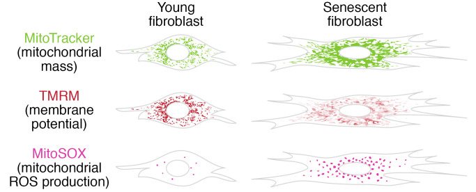
Mitochondrial dysfunction and cell senescence are hallmarks of aging and are closely interconnected. Mitochondrial dysfunction, operationally defined as a decreased respiratory capacity per mitochondrion together with a decreased mitochondrial membrane potential, typically accompanied by increased production of oxygen free radicals, is a cause and a consequence of cellular senescence and figures prominently in multiple feedback loops that induce and maintain the senescent phenotype. Here, we summarize pathways that cause mitochondrial dysfunction in senescence and aging and discuss the major consequences of mitochondrial dysfunction and how these consequences contribute to senescence and aging. We also highlight the potential of senescence-associated mitochondrial dysfunction as an antiaging and antisenescence intervention target, proposing the combination of multiple interventions converging onto mitochondrial dysfunction as novel, potent senolytics.
Overview
The claims and statistics of this paper haven’t been checked yet.
Free: it reads the open-access text and checks the first 5 claims that cite a source we can check. Create a free account to check every claim.
Integrity signals
Registry
Cites
Statistics
Figures

Verification
Claims the paper makes
The claims and statistics of this paper haven’t been checked yet.
Free: it reads the open-access text and checks the first 5 claims that cite a source we can check. Create a free account to check every claim.
Claims other papers make about this one
Each is checked against this paper’s own text: does it say what the citing paper says it does?
The source backs part of the claim, not all of it — or backs it for a different population, condition or comparison.
In the citing paper
“Both proliferation-competent glial cells (which constitute approximately 50% of brain cells) and postmitotic neurons can be positive for markers of cellular senescence in aging, obesity, traumatic injury, and neurodegeneration31,45–49, among other conditions.”
From Guidelines for minimal information on cellular senescence experimentation in vivo (2024) · cites this paper as [45]
Experimental — accuracy not yet measuredThe source backs part of the claim, not all of it — or backs it for a different population, condition or comparison.
In the citing paper
“Therefore, the dysregulation of mitochondrial fusion and fission pathways in senescent astrocytes likely contributes to mitochondrial dysfunction, oxidative stress, and the overall senescence-associated phenotype observed in these cells (Ma et al., 2020; Kleele et al., 2021; Miwa et al., 2022).”
From Aging promotes an increase in mitochondrial fragmentation in astrocytes (2024) · cites this paper as Miwa et al., 2022
Experimental — accuracy not yet measuredThe source backs part of the claim, not all of it — or backs it for a different population, condition or comparison.
In the citing paper
“Dysfunctional mitochondrial dynamics, as observed here, particularly excessive mitochondrial fission, have been associated with oxidative stress, mitochondrial DNA damage, and cellular senescence in various cell types (Twig et al., 2008; Miwa et al., 2022).”
From Aging promotes an increase in mitochondrial fragmentation in astrocytes (2024) · cites this paper as Miwa et al., 2022
Experimental — accuracy not yet measuredThe cited source says what the claim attributes to it.
In the citing paper
“while the presence of senescence markers has been proven even in post-mitotic cells”
From Guidelines for minimal information on cellular senescence experimentation in vivo (2024) · cites this paper as [45]
The check wasn’t confident either way, so it gives no verdict — never disputed.
In the citing paper
“Evidence from the literature indicates that senescent cells exhibit an increase in oxygen consumption rate (Hutter et al., 2004; Son et al., 2017; Miwa et al., 2022; Bang et al., 2023; Diniz et al., 2024a).”
From Aging promotes an increase in mitochondrial fragmentation in astrocytes (2024) · cites this paper as Miwa et al., 2022
Experimental — accuracy not yet measured
2 more claims about this paper
Create a free account to see every claim other papers make about this one.
Self-check
Percentages, sums and reported differences re-computed from the paper’s own numbers.
The self-check needs the paper’s full text.
Citations
Cites
The serial cultivation of human diploid cell strains
Hayflick, L., & Moorhead, P. S. (1961) · Experimental Cell Research · doi:10.1016/0014-4827(61)90192-6 · Cited by 8,508 · cited 1×
“Introduction What is cell senescence? Cell senescence was originally defined as the irreversible loss of replicative potential of primary cells in culture (1), initiated as a persistent DNA damage response to dysfunctional telomeres (2).”
BackgroundTelomeres shorten during ageing of human fibroblasts
Harley, C. B., Futcher, A. B., & Greider, C. W. (1990) · Nature · doi:10.1038/345458a0 · Cited by 5,750 · cited 2×
“Telomere dysfunction might be caused by telomere shortening during DNA replication (3, 4), replication-independent telomere damage (5), or a variety of other stresses (6).”
BackgroundA role for mitochondria in nlrp3 inflammasome activation
Zhou, R., Yazdi, A. S., Menu, P., & Tschopp, J. (2011) · Nature · doi:10.1038/nature09663 · Cited by 5,602
The senescence-associated secretory phenotype: the dark side of tumor suppression
Coppé, J.-P., Desprez, P.-Y., Krtolica, A., & Campisi, J. (2010) · Annual Review of Pathology: Mechanisms of Disease · doi:10.1146/annurev-pathol-121808-102144 · Cited by 5,206
Wu, Z., Puigserver, P., Andersson, U., Zhang, C., Adelmant, G., Mootha, V., Troy, A., Cinti, S., Lowell, B., Scarpulla, R. C., et al. (1999) · Cell · doi:10.1016/s0092-8674(00)80611-x · Cited by 4,139 · cited 1×
“PGC-1α activates the transcription factors Nrf1/2 and ERRα, which further control the TFAM transcription in response to increased mitochondrial biogenesis (65).”
BackgroundClearance of p16Ink4a-positive senescent cells delays ageing-associated disorders
Baker, D. J., Wijshake, T., Tchkonia, T., LeBrasseur, N. K., Childs, B. G., van de Sluis, B., Kirkland, J. L., & van Deursen, J. M. (2011) · Nature · doi:10.1038/nature10600 · Cited by 3,670
AMPK regulates energy expenditure by modulating NAD+ metabolism and SIRT1 activity
Cantó, C., Gerhart-Hines, Z., Feige, J. N., Lagouge, M., Noriega, L., Milne, J. C., Elliott, P. J., Puigserver, P., & Auwerx, J. (2009) · Nature · doi:10.1038/nature07813 · Cited by 3,300
Cellular Senescence: Defining a Path Forward
Gorgoulis, V., Adams, P. D., Alimonti, A., Bennett, D. C., Bischof, O., Bishop, C., Campisi, J., Collado, M., Evangelou, K., Ferbeyre, G., et al. (2019) · Cell · doi:10.1016/j.cell.2019.10.005 · Cited by 3,225 · cited 1×
“Telomere dysfunction might be caused by telomere shortening during DNA replication (3, 4), replication-independent telomere damage (5), or a variety of other stresses (6).”
BackgroundNakahira, K., Haspel, J. A., Rathinam, V. A. K., Lee, S.-J., Dolinay, T., Lam, H. C., Englert, J. A., Rabinovitch, M., Cernadas, M., Kim, H. P., et al. (2011) · Nature Immunology · doi:10.1038/ni.1980 · Cited by 3,049
A DNA damage checkpoint response in telomere-initiated senescence
Fagagna, F. D. D., Reaper, P. M., Clay-Farrace, L., Fiegler, H., Carr, P., von Zglinicki, T., Saretzki, G., Carter, N. P., & Jackson, S. P. (2003) · Nature · doi:10.1038/nature02118 · Cited by 2,917 · cited 1×
“Introduction What is cell senescence? Cell senescence was originally defined as the irreversible loss of replicative potential of primary cells in culture (1), initiated as a persistent DNA damage response to dysfunctional telomeres (2).”
BackgroundNaturally occurring p16Ink4a-positive cells shorten healthy lifespan
Baker, D. J., Childs, B. G., Durik, M., Wijers, M. E., Sieben, C. J., Zhong, J., A. Saltness, R., Jeganathan, K. B., Verzosa, G. C., Pezeshki, A., et al. (2016) · Nature · doi:10.1038/nature16932 · Cited by 2,876
The Achilles' heel of senescent cells: from transcriptome to senolytic drugs
Zhu, Y., Tchkonia, T., Pirtskhalava, T., Gower, A. C., Ding, H., Giorgadze, N., Palmer, A. K., Ikeno, Y., Hubbard, G. B., Lenburg, M., et al. (2015) · Aging Cell · doi:10.1111/acel.12344 · Cited by 2,584 · cited 2×
“This list is not exhaustive; for instance, apoptosis resistance might also be regarded as an essential building block of the senescent phenotype (11).”
BackgroundCalcium, ATP, and ROS: a mitochondrial love-hate triangle
Brookes, P. S., Yoon, Y., Robotham, J. L., Anders, M. W., & Sheu, S.-S. (2004) · American Journal of Physiology-Cell Physiology · doi:10.1152/ajpcell.00139.2004 · Cited by 2,575
Oxidative stress shortens telomeres
von Zglinicki, T. (2002) · Trends in Biochemical Sciences · doi:10.1016/s0968-0004(02)02110-2 · Cited by 2,524 · cited 2×
“This increase in oxidative stress leads to DNA damage, such as oxidized bases, single-strand breaks, double-strand breaks, and telomere shortening (4), activating p53 and pRb pathways and causing cell cycle arrest and senescence (28, 108).”
BackgroundAssessing mitochondrial dysfunction in cells
Brand, M. D., & Nicholls, D. G. (2011) · Biochemical Journal · doi:10.1042/bj20110162 · Cited by 2,405 · cited 1×
“For measurement of MMP in intact cells using potentiometric fluorescent dyes like tetramethylrhodamine methyl ester (TMRM), tetramethylrhodamine ethyl ester (TMRE), and rhodamine 123, careful experimental methodologies need to be employed (45).”
MethodMitochondrial DNA mutations, oxidative stress, and apoptosis in mammalian aging
Kujoth, G. C., Hiona, A., Pugh, T. D., Someya, S., Panzer, K., Wohlgemuth, S. E., Hofer, T., Seo, A. Y., Sullivan, R., Jobling, W. A., et al. (2005) · Science · doi:10.1126/science.1112125 · Cited by 2,165 · cited 2×
“These studies showed that at high density, mtDNA mutations cause physiologically relevant mitochondrial dysfunction and premature aging (55, 56).”
BackgroundThe Respiratory Chain and Oxidative Phosphorylation
Chance, B., & Williams, G. R. (1956) · Advances in Enzymology - and Related Areas of Molecular Biology · doi:10.1002/9780470122624.ch2 · Cited by 2,081
Mitochondrial DNA mutations in human disease
Taylor, R. W., & Turnbull, D. M. (2005) · Nature Reviews Genetics · doi:10.1038/nrg1606 · Cited by 1,852 · cited 1×
“mtDNA mutations or deletions causing mtDNA damage have been implicated in several mitochondrial diseases (51, 52).”
Oncogene-induced senescence is a DNA damage response triggered by DNA hyper-replication
Di Micco, R., Fumagalli, M., Cicalese, A., Piccinin, S., Gasparini, P., Luise, C., Schurra, C., Garre’, M., Giovanni Nuciforo, P., Bensimon, A., et al. (2006) · Nature · doi:10.1038/nature05327 · Cited by 1,821 · cited 2×
“Such stresses often, but not always, involve a persistent DNA damage response (7).”
BackgroundAMP-activated protein kinase: an energy sensor that regulates all aspects of cell function
Hardie, D. G. (2011) · Genes & Development · doi:10.1101/gad.17420111 · Cited by 1,638 · cited 9×
“Canto C, et al. AMPK regulates energy expenditure 9J Clin Invest.”
BackgroundInfluentialMechanisms of Cellular Senescence: Cell Cycle Arrest and Senescence Associated Secretory Phenotype
Kumari, R., & Jat, P. (2021) · Frontiers in Cell and Developmental Biology · doi:10.3389/fcell.2021.645593 · Cited by 1,622
The mitochondrial basis of aging
Sun, N., Youle, R. J., & Finkel, T. (2016) · Molecular Cell · doi:10.1016/j.molcel.2016.01.028 · Cited by 1,441 · cited 3×
“During mitochondrial dysfunction, low MMP is typically associated with increased production of reactive oxygen species (ROS) (32, 33); however, empirical determination of ROS is required for each case to confirm this.”
BackgroundAutophagy maintains stemness by preventing senescence
García-Prat, L., Martínez-Vicente, M., Perdiguero, E., Ortet, L., Rodríguez-Ubreva, J., Rebollo, E., Ruiz-Bonilla, V., Gutarra, S., Ballestar, E., Serrano, A. L., et al. (2016) · Nature · doi:10.1038/nature16187 · Cited by 1,355 · cited 1×
“In contrast, other studies show that impairment in autophagy pathways can induce premature senescence, possibly due to generation of ROS by mitochondrial dysfunction, which in turn activates p53 signaling in primary human fibroblasts (73) and in muscle stem cells (74).”
Cytoplasmic chromatin triggers inflammation in senescence and cancer
Dou, Z., Ghosh, K., Vizioli, M. G., Zhu, J., Sen, P., Wangensteen, K. J., Simithy, J., Lan, Y., Lin, Y., Zhou, Z., et al. (2017) · Nature · doi:10.1038/nature24050 · Cited by 1,335
Telomere dysfunction induces metabolic and mitochondrial compromise
Sahin, E., Colla, S., Liesa, M., Moslehi, J., Müller, F. L., Guo, M., Cooper, M., Kotton, D., Fabian, A. J., Walkey, C., et al. (2011) · Nature · doi:10.1038/nature09787 · Cited by 1,319
Metabolic control of mitochondrial biogenesis through the PGC-1 family regulatory network
Scarpulla, R. C. (2011) · Biochimica et Biophysica Acta (BBA) - Molecular Cell Research · doi:10.1016/j.bbamcr.2010.09.019 · Cited by 1,281 · cited 1×
“The PPARγ coactivator 1 (PGC-1) family, comprising PGC-1α, PGC-1β, and PGC-1–related coactivator, are main regulators of mitochondrial biogenesis (63).”
BackgroundMitochondrial Dysfunction Induces Senescence with a Distinct Secretory Phenotype
Wiley, C. D., Velarde, M. C., Lecot, P., Liu, S., Sarnoski, E. A., Freund, A., Shirakawa, K., Lim, H. W., Davis, S. S., Ramanathan, A., et al. (2016) · Cell Metabolism · doi:10.1016/j.cmet.2015.11.011 · Cited by 1,275 · cited 4×
“While neither increased ROS nor cell senescence was observed in the original publications, this was later rectified (58), and a specific type of cell senescence missing the proinflammatory arm of the SASP was found in adipose tissue of PolG mutator mice, which rapidly accumulate mtDNA mutations (59) (see below).”
BackgroundInfluentialSenolytic drugs: from discovery to translation
Kirkland, J. L., & Tchkonia, T. (2020) · Journal of Internal Medicine · doi:10.1111/joim.13141 · Cited by 1,227
Mammalian Sir2 homolog SIRT3 regulates global mitochondrial lysine acetylation
Lombard, D. B., Alt, F. W., Cheng, H.-L., Bunkenborg, J., Streeper, R. S., Mostoslavsky, R., Kim, J., Yancopoulos, G., Valenzuela, D., Murphy, A., et al. (2007) · Molecular and Cellular Biology · doi:10.1128/mcb.01636-07 · Cited by 1,216 · cited 1×
“SIRT3 plays a unique role as the main mitochondrial deacetylase (89).”
BackgroundInnate immune sensing of cytosolic chromatin fragments through cGAS promotes senescence
Glück, S., Guey, B., Gulen, M. F., Wolter, K., Kang, T.-W., Schmacke, N. A., Bridgeman, A., Rehwinkel, J., Zender, L., & Ablasser, A. (2017) · Nature Cell Biology · doi:10.1038/ncb3586 · Cited by 1,196
Senescence and the SASP: many therapeutic avenues
Birch, J., & Gil, J. (2020) · Genes & Development · doi:10.1101/gad.343129.120 · Cited by 1,177
Zhu, Y., Tchkonia, T., Fuhrmann‐Stroissnigg, H., Dai, H. M., Ling, Y. Y., Stout, M. B., Pirtskhalava, T., Giorgadze, N., Johnson, K. O., Giles, C. B., et al. (2016) · Aging Cell · doi:10.1111/acel.12445 · Cited by 1,148
AMP-activated protein kinase mediates mitochondrial fission in response to energy stress
Toyama, E. Q., Herzig, S., Courchet, J., Lewis, T. L., Losón, O. C., Hellberg, K., Young, N. P., Chen, H., Polleux, F., Chan, D. C., et al. (2016) · Science · doi:10.1126/science.aab4138 · Cited by 1,085 · cited 1×
“AMPK activation promotes mitochondrial biogenesis and regulates mitochondrial dynamics and mitophagy (86), partially by promoting SIRT1-dependent PGC-1α deacetylation by increasing intracellular NAD+/NADH ratio (87).”
BackgroundAutophagy mediates the mitotic senescence transition
Young, A. R. J., Narita, M., Ferreira, M., Kirschner, K., Sadaie, M., Darot, J. F. J., Tavaré, S., Arakawa, S., Shimizu, S., Watt, F. M., et al. (2009) · Genes & Development · doi:10.1101/gad.519709 · Cited by 1,078
Feedback between p21 and reactive oxygen production is necessary for cell senescence
Passos, J. F., Nelson, G., Wang, C., Richter, T., Simillion, C., Proctor, C. J., Miwa, S., Olijslagers, S., Hallinan, J., Wipat, A., et al. (2010) · Molecular Systems Biology · doi:10.1038/msb.2010.5 · Cited by 1,019 · cited 4×
“During mitochondrial dysfunction, low MMP is typically associated with increased production of reactive oxygen species (ROS) (32, 33); however, empirical determination of ROS is required for each case to confirm this.”
BackgroundInfluentialSelf-citationEndoplasmic reticulum-mitochondria contacts: Function of the junction
Rowland, A. A., & Voeltz, G. K. (2012) · Nature Reviews Molecular Cell Biology · doi:10.1038/nrm3440 · Cited by 1,018
Cellular senescence: a translational perspective
Kirkland, J. L., & Tchkonia, T. (2017) · EBioMedicine · doi:10.1016/j.ebiom.2017.04.013 · Cited by 1,013
SIRT5-mediated lysine desuccinylation impacts diverse metabolic pathways
Park, J., Chen, Y., Tishkoff, D. X., Peng, C., Tan, M., Dai, L., Xie, Z., Zhang, Y., Zwaans, B. M. M., Skinner, M. E., et al. (2013) · Molecular Cell · doi:10.1016/j.molcel.2013.06.001 · Cited by 1,000 · cited 1×
“In addition, SIRT4 and SIRT5 also regulate mitochondrial signaling and are involved in rewiring various metabolic pathways (92, 93).”
BackgroundApoptotic Caspases Suppress mtDNA-Induced STING-Mediated Type I IFN Production
White, M. J., McArthur, K., Metcalf, D., Lane, R. M., Cambier, J. C., Herold, M. J., van Delft, M. F., Bedoui, S., Lessene, G., Ritchie, M. E., et al. (2014) · Cell · doi:10.1016/j.cell.2014.11.036 · Cited by 983
The DNA damage response induces inflammation and senescence by inhibiting autophagy of GATA4.
Kang, C., Xu, Q., Martin, T. D., Li, M. Z., Demaria, M., Aron, L., Lu, T., Yankner, B. A., Campisi, J., & Elledge, S. J. (2015) · Science · doi:10.1126/science.aaa5612 · Cited by 982 · cited 1×
“Furthermore, inhibition of selective autophagy for the transcription factor GATA4 was associated with induction of senescence (77).”
BackgroundEpigenetic mechanisms of longevity and aging
Sen, P., Shah, P. P., Nativio, R., & Berger, S. L. (2016) · Cell · doi:10.1016/j.cell.2016.07.050 · Cited by 956 · cited 1×
“These include deregulated nutrient signaling (14, 15), autophagy/mitophagy dysfunction (16), and massive epigenetic reprogramming (17, 18), including activation of (proinflammatory) transcription factors driving the senescence-associated secretory phenotype (SASP) (19–22).”
BackgroundHewitt, G., Jurk, D., Marques, F. D. M., Correia-Melo, C., Hardy, T., Gackowska, A., Anderson, R., Taschuk, M., Mann, J., & Passos, J. F. (2012) · Nature Communications · doi:10.1038/ncomms1708 · Cited by 908 · cited 1×
“Telomere dysfunction might be caused by telomere shortening during DNA replication (3, 4), replication-independent telomere damage (5), or a variety of other stresses (6).”
BackgroundCamacho-Pereira, J., Tarragó, M. G., Chini, C. C. S., Nin, V., Escande, C., Warner, G. M., Puranik, A. S., Schoon, R. A., Reid, J. M., Galina, A., et al. (2016) · Cell Metabolism · doi:10.1016/j.cmet.2016.05.006 · Cited by 901 · cited 11×
“In mammals, three members of the SIRT family, SIRT3, SIRT4, and SIRT5, are localized in mitochondria and modulate mitochondrial metabolism (88).”
BackgroundInfluentialSelf-citationThe secret life of NAD+: an old metabolite controlling new metabolic signaling pathways
Houtkooper, R. H., Cantó, C., Wanders, R. J., & Auwerx, J. (2010) · Endocrine Reviews · doi:10.1210/er.2009-0026 · Cited by 899
AMP-Activated Protein Kinase (AMPK) Controls the Aging Process via an Integrated Signaling Network
Salminen, A., & Kaarniranta, K. (2012) · Ageing Research Reviews · doi:10.1016/j.arr.2011.12.005 · Cited by 860 · cited 1×
“The various mechanisms involved in Ca2+ overload–induced mitochondrial dysfunction include Ca2+-induced nitric oxide production; Ca2+-enhanced cytochrome c dissociation from the IMM; opening of the mitochondrial permeability transition pore (mPTP), which subsequently causes the release of cytochrome c and GSHand NADPH…”
BackgroundChronic inflammation induces telomere dysfunction and accelerates ageing in mice
Jurk, D., Wilson, C., Passos, J. F., Oakley, F., Correia-Melo, C., Greaves, L., Saretzki, G., Fox, C., Lawless, C., Anderson, R., et al. (2014) · Nature Communications · doi:10.1038/ncomms5172 · Cited by 819 · cited 1×
“Senescent cells accumulate with aging in many mammalian and non-mammalian tissues (8), and intervention experiments (9–11) have proven that this accumulation is a physiologically important driver of age-associated functional decline, (multi-)morbidity, and mortality (for review, see ref.”
BackgroundMitochondrial dysfunction accounts for the stochastic heterogeneity in telomere-dependent senescence
Passos, J. F., Saretzki, G., Ahmed, S., Nelson, G., Richter, T., Peters, H., Wappler, I., Birket, M. J., Harold, G., Schaeuble, K., et al. (2007) · PLoS Biology · doi:10.1371/journal.pbio.0050110 · Cited by 796 · cited 2×
“This increase in oxidative stress leads to DNA damage, such as oxidized bases, single-strand breaks, double-strand breaks, and telomere shortening (4), activating p53 and pRb pathways and causing cell cycle arrest and senescence (28, 108).”
BackgroundMitochondria are required for pro-ageing features of the senescent phenotype
Correia‐Melo, C., Marques, F. D., Anderson, R., Hewitt, G., Hewitt, R., Cole, J., Carroll, B. M., Miwa, S., Birch, J., Merz, A., et al. (2016) · The EMBO Journal · doi:10.15252/embj.201592862 · Cited by 796 · cited 5×
“Mitochondrial mass is often increased in senescence in vitro (28–30) and in vivo (31).”
BackgroundInfluentialSelf-citationZhu, Y., Doornebal, E. J., Pirtskhalava, T., Giorgadze, N., Wentworth, M., Fuhrmann-Stroissnigg, H., Niedernhofer, L. J., Robbins, P. D., Tchkonia, T., & Kirkland, J. L. (2017) · Aging · doi:10.18632/aging.101202 · Cited by 757
Caloric restriction reduces age-related and all-cause mortality in rhesus monkeys
Colman, R. J., Beasley, T. M., Kemnitz, J. W., Johnson, S. C., Weindruch, R., & Anderson, R. M. (2014) · Nature Communications · doi:10.1038/ncomms4557 · Cited by 711
The metabolic roots of senescence: mechanisms and opportunities for intervention
Wiley, C. D., & Campisi, J. (2021) · Nature Metabolism · doi:10.1038/s42255-021-00483-8 · Cited by 691
Canonical Nlrp3 Inflammasome Links Systemic Low-Grade Inflammation to Functional Decline in Aging
Youm, Y.-H., Grant, R. W., McCabe, L. R., Albarado, D. C., Nguyen, K. Y., Ravussin, A., Pistell, P., Newman, S., Carter, R., Laque, A., et al. (2013) · Cell Metabolism · doi:10.1016/j.cmet.2013.09.010 · Cited by 674
Moiseeva, O., Deschênes‐Simard, X., St‐Germain, E., Igelmann, S., Huot, G., Cadar, A. E., Bourdeau, V., Pollak, M. N., & Ferbeyre, G. (2013) · Aging Cell · doi:10.1111/acel.12075 · Cited by 654
Jurk, D., Wang, C., Miwa, S., Maddick, M., Korolchuk, V., Tsolou, A., Gonos, E. S., Thrasivoulou, C., Jill Saffrey, M., Cameron, K., et al. (2012) · Aging Cell · doi:10.1111/j.1474-9726.2012.00870.x · Cited by 598 · cited 1×
“Markers for activation of the same building blocks were also found in aged postmitotic cells (23), indicating that a senescence-like phenotype is a general stress response for both proliferation-competent and differentiated postmitotic cells during aging (24).”
BackgroundSelf-citationOxidative stress caused by mitochondrial calcium overload
Peng, T., & Jou, M. (2010) · Annals of the New York Academy of Sciences · doi:10.1111/j.1749-6632.2010.05634.x · Cited by 566
Quantitative Analysis of NAD Synthesis-Breakdown Fluxes
Liu, L., Su, X., Quinn, W. J., Hui, S., Krukenberg, K., Frederick, D. W., Redpath, P., Zhan, L., Chellappa, K., White, E., et al. (2018) · Cell Metabolism · doi:10.1016/j.cmet.2018.03.018 · Cited by 564 · cited 1×
“NAD+ can be synthesized via multiple pathways: the de novo synthesis pathway, the NAD+ salvage pathway, and the Preiss-Handler pathway (94, 95).”
BackgroundSomatic mtDNA mutations cause aging phenotypes without affecting reactive oxygen species production
Trifunovic, A., Hansson, A., Wredenberg, A., Rovio, A. T., Dufour, E., Khvorostov, I., Spelbrink, J. N., Wibom, R., Jacobs, H. T., & Larsson, N.-G. (2005) · Proceedings of the National Academy of Sciences · doi:10.1073/pnas.0508886102 · Cited by 555 · cited 2×
“These studies showed that at high density, mtDNA mutations cause physiologically relevant mitochondrial dysfunction and premature aging (55, 56).”
BackgroundThe dynamic regulation of NAD metabolism in mitochondria
Stein, L. R., & Imai, S.-I. (2012) · Trends in Endocrinology & Metabolism · doi:10.1016/j.tem.2012.06.005 · Cited by 535
NOTCH1 mediates a switch between two distinct secretomes during senescence
Hoare, M., Ito, Y., Kang, T.-W., Weekes, M. P., Matheson, N. J., Patten, D. A., Shetty, S., Parry, A. J., Menon, S., Salama, R., et al. (2016) · Nature Cell Biology · doi:10.1038/ncb3397 · Cited by 522
Booth, L. N., & Brunet, A. (2016) · Molecular Cell · doi:10.1016/j.molcel.2016.05.013 · Cited by 499 · cited 1×
“These include deregulated nutrient signaling (14, 15), autophagy/mitophagy dysfunction (16), and massive epigenetic reprogramming (17, 18), including activation of (proinflammatory) transcription factors driving the senescence-associated secretory phenotype (SASP) (19–22).”
BackgroundReciprocal regulation of p53 and malic enzymes modulates metabolism and senescence
Jiang, P., Du, W., Mancuso, A., Wellen, K. E., & Yang, X. (2013) · Nature · doi:10.1038/nature11776 · Cited by 491
Mitochondrial dysfunction and mitophagy in parkinson’s disease: from mechanism to therapy
Malpartida, A. B., Williamson, M., Narendra, D. P., Wade-Martins, R., & Ryan, B. J. (2021) · Trends in Biochemical Sciences · doi:10.1016/j.tibs.2020.11.007 · Cited by 482 · cited 1×
“Several studies have revealed a role for PGC-1α in mitochondrial dysfunction associated with pathological states (66, 67).”
BackgroundIchim, G., Lopez, J., Ahmed, S. U., Muthalagu, N., Giampazolias, E., Delgado, M. E., Haller, M., Riley, J. S., Mason, S. M., Athineos, D., et al. (2015) · Molecular Cell · doi:10.1016/j.molcel.2015.01.018 · Cited by 479
Jeong, S. M., Xiao, C., Finley, L. W. S., Lahusen, T., Souza, A. L., Pierce, K., Li, Y.-H., Wang, X., Laurent, G., German, N. J., et al. (2013) · Cancer Cell · doi:10.1016/j.ccr.2013.02.024 · Cited by 445 · cited 1×
“In addition, SIRT4 and SIRT5 also regulate mitochondrial signaling and are involved in rewiring various metabolic pathways (92, 93).”
BackgroundMitochondrial homeostasis: The interplay between mitophagy and mitochondrial biogenesis
Palikaras, K., & Tavernarakis, N. (2014) · Experimental Gerontology · doi:10.1016/j.exger.2014.01.021 · Cited by 429 · cited 1×
“Mitochondrial mass homeostasis involves various mechanisms, such as mitochondrial biogenesis, clearance of damaged mitochondria by mitochondria-specific autophagy (mitophagy), and maintenance of mitochondrial dynamics (62).”
BackgroundMitochondria and ageing: Role in heart, skeletal muscle and adipose tissue
Boengler, K., Kosiol, M., Mayr, M., Schulz, R., & Rohrbach, S. (2017) · Journal of Cachexia Sarcopenia and Muscle · doi:10.1002/jcsm.12178 · Cited by 421 · cited 1×
“For instance, in heart muscle (especially in interfibrillar mitochondria), it is primarily complexes III and IV that lose activity with age, while in skeletal muscle, liver, and brain, complex I often appears more sensitive to age-associated loss of function (37, 38).”
BackgroundSenescent cells promote tissue NAD+ decline during ageing via the activation of CD38+ macrophages
Covarrubias, A. J., Kale, A., Perrone, R., Lopez-Dominguez, J. A., Pisco, A. O., Kasler, H. G., Schmidt, M. S., Heckenbach, I., Kwok, R., Wiley, C. D., et al. (2020) · Nature Metabolism · doi:10.1038/s42255-020-00305-3 · Cited by 420
Unraveling the Truth About Antioxidants: Mitohormesis explains ROS-induced health benefits
Ristow, M. (2014) · Nature Medicine · doi:10.1038/nm.3624 · Cited by 417
DNA deletions and clonal mutations drive premature aging in mitochondrial mutator mice
Vermulst, M., Wanagat, J., Kujoth, G. C., Bielas, J. H., Rabinovitch, P. S., Prolla, T. A., & Loeb, L. A. (2008) · Nature Genetics · doi:10.1038/ng.95 · Cited by 411 · cited 1×
“On the other hand, AMPK and sirtuins are activated in later identified as driving premature aging in these mice (57).”
BackgroundPinti, M., Cevenini, E., Nasi, M., De Biasi, S., Salvioli, S., Monti, D., Benatti, S., Gibellini, L., Cotichini, R., Stazi, M. A., et al. (2014) · European Journal of Immunology · doi:10.1002/eji.201343921 · Cited by 409
Mitochondria in Cell Senescence: Is Mitophagy the Weakest Link?
Korolchuk, V. I., Miwa, S., Carroll, B., & von Zglinicki, T. (2017) · EBioMedicine · doi:10.1016/j.ebiom.2017.03.020 · Cited by 409
Self-citationMitochondrial Dysfunction Contributes to Oncogene-Induced Senescence
Moiseeva, O., Bourdeau, V., Roux, A., Deschênes-Simard, X., & Ferbeyre, G. (2009) · Molecular and Cellular Biology · doi:10.1128/mcb.01868-08 · Cited by 408 · cited 3×
“The same type of mitochondrial dysfunction was also found in oncogene- (RAS-)induced senescence (69).”
BackgroundNAD+ metabolism governs the proinflammatory senescence-associated secretome
Nacarelli, T., Lau, L., Fukumoto, T., Zundell, J., Fatkhutdinov, N., Wu, S., Aird, K. M., Iwasaki, O., Kossenkov, A. V., Schultz, D., et al. (2019) · Nature Cell Biology · doi:10.1038/s41556-019-0287-4 · Cited by 402 · cited 2×
“In contrast, in oncogene-induced senescence, the NAD+ salvage pathway was upregulated, leading to enhanced SASP production via AMPK suppression (34).”
BackgroundMechanism of parkin activation by PINK1
Gladkova, C., Maslen, S. L., Skehel, J. M., & Komander, D. (2018) · Nature · doi:10.1038/s41586-018-0224-x · Cited by 393 · cited 1×
“Among different mitophagy pathways, PINK1/parkin is the most studied stress-induced mitophagy pathway (71), and its impairment has been associated with several pathological conditions.”
BackgroundAlano, C. C., Ying, W., & Swanson, R. A. (2004) · Journal of Biological Chemistry · doi:10.1074/jbc.m313329200 · Cited by 374 · cited 1×
“In fact, NAD+ depletion causes mitochondrial membrane depolarization and mitochondrial permeability transition, which are involved in PARP1-mediated cell death (96).”
BackgroundInhibition of p21‐mediated ROS accumulation can rescue p21‐induced senescence
Macip, S., Igarashi, M., Fang, L., Chen, A., Pan, Z., Lee, S. W., & Aaronson, S. A. (2002) · The EMBO Journal · doi:10.1093/emboj/21.9.2180 · Cited by 374
Vizioli, M. G., Liu, T., Miller, K. N., Robertson, N. A., Gilroy, K., Lagnado, A. B., Perez-Garcia, A., Kiourtis, C., Dasgupta, N., Lei, X., et al. (2020) · Genes & Development · doi:10.1101/gad.331272.119 · Cited by 359
CD38 ecto-enzyme in immune cells is induced during aging and regulates NAD+ and NMN levels
Chini, C. C. S., Peclat, T. R., Warner, G. M., Kashyap, S., Espindola-Netto, J. M., de Oliveira, G. C., Gomez, L. S., Hogan, K. A., Tarragó, M. G., Puranik, A. S., et al. (2020) · Nature Metabolism · doi:10.1038/s42255-020-00298-z · Cited by 349 · cited 1×
“In addition, recent studies have reported a role for the NAD+-consuming enzyme CD38 in NAD+ decline during aging (91, 97).”
BackgroundSelf-citationYang, W., Nagasawa, K., Münch, C., Xu, Y., Satterstrom, K., Jeong, S., Hayes, S. D., Jedrychowski, M. P., Vyas, F. S., Zaganjor, E., et al. (2016) · Cell · doi:10.1016/j.cell.2016.10.016 · Cited by 338 · cited 1×
“In mammals, three members of the SIRT family, SIRT3, SIRT4, and SIRT5, are localized in mitochondria and modulate mitochondrial metabolism (88).”
BackgroundMitochondrial fission and fusion mediators, hFis1 and OPA1, modulate cellular senescence
Lee, S., Jeong, S.-Y., Lim, W.-C., Kim, S., Park, Y.-Y., Sun, X., Youle, R. J., & Cho, H. (2007) · Journal of Biological Chemistry · doi:10.1074/jbc.m700679200 · Cited by 298 · cited 1×
“Mitochondrial elongation has been associated with induction of senescence-associated pathways (79, 80).”
BackgroundLee, H.-C., Yin, P.-H., Chi, C.-W., & Wei, Y.-H. (2002) · Journal of Biomedical Science · doi:10.1007/bf02254978 · Cited by 293 · cited 1×
“Increased mitochondrial mass and PGC-1α and Nrf1 and/or Nrf2 induction were associated with replicative (68) and oncogene-induced (69) senescence.”
BackgroundRegulation of mitochondrial biogenesis through TFAM–mitochondrial DNA interactions
Picca, A., & Lezza, A. M. S. (2015) · Mitochondrion · doi:10.1016/j.mito.2015.10.001 · Cited by 283 · cited 1×
“Nuclear-encoded mitochondrial transcription factor A (TFAM) is another important factor that regulates mitochondrial biogenesis and plays a fundamental role in mtDNA transcription (64).”
BackgroundYoon, Y., Yoon, D., Lim, I. K., Yoon, S., Chung, H., Rojo, M., Malka, F., Jou, M., Martinou, J., & Yoon, G. (2006) · Journal of Cellular Physiology · doi:10.1002/jcp.20753 · Cited by 277 · cited 2×
“Increased oxidative damage due to accumulated dysfunctional mitochondria reduces FIS1, leading to formation of giant mitochondria (79).”
BackgroundThe path from mitochondrial ROS to aging runs through the mitochondrial permeability transition pore
Rottenberg, H., & Hoek, J. B. (2017) · Aging Cell · doi:10.1111/acel.12650 · Cited by 275
The senescent bystander effect is caused by ROS-activated NF-κB signalling
Nelson, G., Kucheryavenko, O., Wordsworth, J., & von Zglinicki, T. (2018) · Mechanisms of Ageing and Development · doi:10.1016/j.mad.2017.08.005 · Cited by 272 · cited 2×
“Mitochondrial dysfunction and associated ROS production has also been found in stress-induced senescence (28, 32), replicative senescence (50), oncogene-induced senescence (7, 50), and senescence triggered by genetic telomere uncapping (32).”
BackgroundAhmad, T., Sundar, I. K., Lerner, C. A., Gerloff, J., Tormos, A. M., Yao, H., & Rahman, I. (2015) · The FASEB Journal · doi:10.1096/fj.14-268276 · Cited by 264 · cited 1×
“Cigarette smoke has been shown to induce senescence in lung fibroblasts and small airway epithelial cells by inhibiting mitophagy (75).”
BackgroundAutophagy Impairment Induces Premature Senescence in Primary Human Fibroblasts
Kang, H. T., Lee, K. B., Kim, S. Y., Choi, H. R., & Park, S. C. (2011) · PLoS ONE · doi:10.1371/journal.pone.0023367 · Cited by 254 · cited 1×
“In contrast, other studies show that impairment in autophagy pathways can induce premature senescence, possibly due to generation of ROS by mitochondrial dysfunction, which in turn activates p53 signaling in primary human fibroblasts (73) and in muscle stem cells (74).”
Calcium Transport and Signaling in Mitochondria
Bravo‐Sagua, R., Parra, V., López‐Crisosto, C., Díaz, P., Quest, A. F. G., & Lavandero, S. (2017) · Comprehensive Physiology · doi:10.1002/cphy.c160013 · Cited by 248
The bystander effect contributes to the accumulation of senescent cells in vivo
da Silva, P. F. L., Ogrodnik, M., Kucheryavenko, O., Glibert, J., Miwa, S., Cameron, K., Ishaq, A., Saretzki, G., Nagaraja‐Grellscheid, S., Nelson, G., et al. (2019) · Aging Cell · doi:10.1111/acel.12848 · Cited by 247
Self-citationHUTTER, E., RENNER, K., PFISTER, G., STÖCKL, P., JANSEN-DÜRR, P., & GNAIGER, E. (2004) · Biochemical Journal · doi:10.1042/bj20040095 · Cited by 235 · cited 1×
“Mitochondria in cultured cells under contact inhibition (confluent state) are more coupled than mitochondria in cells that are replicating (41).”
BackgroundWiel, C., Lallet-Daher, H., Gitenay, D., Gras, B., Le Calvé, B., Augert, A., Ferrand, M., Prevarskaya, N., Simonnet, H., Vindrieux, D., et al. (2014) · Nature Communications · doi:10.1038/ncomms4792 · Cited by 231
SIRT3 regulates progression and development of diseases of aging
McDonnell, E., Peterson, B. S., Bomze, H. M., & Hirschey, M. D. (2015) · Trends in Endocrinology & Metabolism · doi:10.1016/j.tem.2015.06.001 · Cited by 217 · cited 1×
“Loss of mitochondrial SIRT function, specifically loss of SIRT3, has been linked to many age-related diseases, including insulin resistance, cancer, cardiovascular diseases, and neurodegeneration (90).”
BackgroundCalorie restriction: Decelerating mTOR-driven aging from cells to organisms (including humans)
Blagosklonny, M. V. (2010) · Cell Cycle · doi:10.4161/cc.9.4.10766 · Cited by 211 · cited 1×
“Induction of mitophagy via inhibition of the mTOR pathway postpones senescence (16, 29, 31, 76).”
BackgroundCellular senescence: the sought or the unwanted?
Sun, Y., Coppé, J.-P., & Lam, E. W.-F. (2018) · Trends in Molecular Medicine · doi:10.1016/j.molmed.2018.08.002 · Cited by 208
Low abundance of the matrix arm of complex I in mitochondria predicts longevity in mice
Miwa, S., Jow, H., Baty, K., Johnson, A., Czapiewski, R., Saretzki, G., Treumann, A., & von Zglinicki, T. (2014) · Nature Communications · doi:10.1038/ncomms4837 · Cited by 206 · cited 2×
“For instance, in heart muscle (especially in interfibrillar mitochondria), it is primarily complexes III and IV that lose activity with age, while in skeletal muscle, liver, and brain, complex I often appears more sensitive to age-associated loss of function (37, 38).”
Self-citationThe role of mitochondrial dysfunction in age-related diseases
Lane, R. K., Hilsabeck, T., & Rea, S. L. (2015) · Biochimica et Biophysica Acta (BBA) - Bioenergetics · doi:10.1016/j.bbabio.2015.05.021 · Cited by 205 · cited 1×
“Senescence drives mitochondrial dysfunction The impairment of mitochondrial function, as well as its morphology, has been associated with aging and many aging-associated pathological conditions, including cancer, neurodegenerative diseases, metabolic diseases, kidney diseases, and others (33, 48, 49).”
BackgroundSenolytics and senostatics as adjuvant tumour therapy
Short, S., Fielder, E., Miwa, S., & von Zglinicki, T. (2019) · EBioMedicine · doi:10.1016/j.ebiom.2019.01.056 · Cited by 202
Self-citationRizza, S., Cardaci, S., Montagna, C., Di Giacomo, G., De Zio, D., Bordi, M., Maiani, E., Campello, S., Borreca, A., Puca, A. A., et al. (2018) · Proceedings of the National Academy of Sciences · doi:10.1073/pnas.1722452115 · Cited by 201 · cited 1×
“Recent studies revealed that excessive S-nitrosylation of parkin resulting in decreased fission was responsible for altered mitophagy in senescence (30).”
BackgroundYoon, Y.-S., Lee, J.-H., Hwang, S.-C., Choi, K. S., & Yoon, G. (2005) · Oncogene · doi:10.1038/sj.onc.1208262 · Cited by 193 · cited 1×
“Similarly, TGF-β1–induced senescence of Mv1Lu mink lung epithelial cells involved the inhibition of complex IV, which caused mitochondrial ROS generation and the persistent disruption of MMP, triggering senescence (36).”
BackgroundCellular Senescence in Postmitotic Cells: Beyond Growth Arrest
Sapieha, P., & Mallette, F. A. (2018) · Trends in Cell Biology · doi:10.1016/j.tcb.2018.03.003 · Cited by 193
Carroll, B., Nelson, G., Rabanal-Ruiz, Y., Kucheryavenko, O., Dunhill-Turner, N. A., Chesterman, C. C., Zahari, Q., Zhang, T., Conduit, S. E., Mitchell, C. A., et al. (2017) · Journal of Cell Biology · doi:10.1083/jcb.201610113 · Cited by 187 · cited 2×
“These include deregulated nutrient signaling (14, 15), autophagy/mitophagy dysfunction (16), and massive epigenetic reprogramming (17, 18), including activation of (proinflammatory) transcription factors driving the senescence-associated secretory phenotype (SASP) (19–22).”
BackgroundBishop, D. J., Botella, J., Genders, A. J., Lee, M. J.-C., Saner, N. J., Kuang, J., Yan, X., & Granata, C. (2019) · Physiology · doi:10.1152/physiol.00038.2018 · Cited by 180 · cited 1×
“Exercise rapidly increases biogenesis of key mitochondrial enzymes in the ETC in skeletal muscle (42).”
BackgroundGreaves, L. C., Nooteboom, M., Elson, J. L., Tuppen, H. A. L., Taylor, G. A., Commane, D. M., Arasaradnam, R. P., Khrapko, K., Taylor, R. W., Kirkwood, T. B. L., et al. (2014) · PLoS Genetics · doi:10.1371/journal.pgen.1004620 · Cited by 172 · cited 1×
“mtDNA mutations accumulate with age in postmitotic tissues (53) and expand clonally in stem cell compartments (54).”
BackgroundSenescence in Post-Mitotic Cells: A Driver of Aging?
von Zglinicki, T., Wan, T., & Miwa, S. (2021) · Antioxidants & Redox Signaling · doi:10.1089/ars.2020.8048 · Cited by 170 · cited 1×
“Markers for activation of the same building blocks were also found in aged postmitotic cells (23), indicating that a senescence-like phenotype is a general stress response for both proliferation-competent and differentiated postmitotic cells during aging (24).”
BackgroundSelf-citationDynamic Modelling of Pathways to Cellular Senescence Reveals Strategies for Targeted Interventions
Dalle Pezze, P., Nelson, G., Otten, E. G., Korolchuk, V. I., Kirkwood, T. B. L., von Zglinicki, T., & Shanley, D. P. (2014) · PLoS Computational Biology · doi:10.1371/journal.pcbi.1003728 · Cited by 166 · cited 2×
“However, the increase in mitochondrial mass results mainly from the accumulation of dysfunctional mitochondria due to alterations in the mitophagy degradation process (29, 70).”
BackgroundMitochondria and aging: A role for the mitochondrial transition pore?
Panel, M., Ghaleh, B., & Morin, D. (2018) · Aging Cell · doi:10.1111/acel.12793 · Cited by 162
Mitochondrial peptides modulate mitochondrial function during cellular senescence
Kim, S., Mehta, H. H., Wan, J., Kuehnemann, C., Chen, J., Hu, J., Hoffman, A. R., & Cohen, P. (2018) · Aging · doi:10.18632/aging.101463 · Cited by 147
Bartolomé, A., García-Aguilar, A., Asahara, S.-I., Kido, Y., Guillén, C., Pajvani, U. B., & Benito, M. (2017) · Molecular and Cellular Biology · doi:10.1128/mcb.00441-17 · Cited by 141 · cited 1×
“For example, higher expression of mTORC1 is associated with reduced mitophagy, resulting in accumulation of defective mitochondria (83).”
BackgroundHansson, M. J., Månsson, R., Morota, S., Uchino, H., Kallur, T., Sumi, T., Ishii, N., Shimazu, M., Keep, M. F., Jegorov, A., et al. (2008) · Free Radical Biology and Medicine · doi:10.1016/j.freeradbiomed.2008.04.021 · Cited by 140
Chini, C., Hogan, K. A., Warner, G. M., Tarragó, M. G., Peclat, T. R., Tchkonia, T., Kirkland, J. L., & Chini, E. (2019) · Biochemical and Biophysical Research Communications · doi:10.1016/j.bbrc.2019.03.199 · Cited by 138
Self-citationYin, Y., Zhou, Z., Liu, W., Chang, Q., Sun, G., & Dai, Y. (2017) · The International Journal of Biochemistry & Cell Biology · doi:10.1016/j.biocel.2017.01.001 · Cited by 136
Mitochondria and Calcium Regulation as Basis of Neurodegeneration Associated With Aging
Müller, M., Ahumada-Castro, U., Sanhueza, M., González-Billault, C. E., Court, F. A., & Cárdenas, C. (2018) · Frontiers in Neuroscience · doi:10.3389/fnins.2018.00470 · Cited by 133
Han, X., Guan, N., Ren, Y.-L., Wei, Q., Tao, Y., Yang, G., Liu, X.-Y., Bu, D., Zhang, Y., & Zhu, S. (2018) · BMC Nephrology · doi:10.1186/s12882-018-0940-3 · Cited by 126
Mitochondrial reactive oxygen species: Do they extend or shorten animal lifespan?
Sanz, A. (2016) · Biochimica et Biophysica Acta (BBA) - Bioenergetics · doi:10.1016/j.bbabio.2016.03.018 · Cited by 122
Somatic mitochondrial DNA mutations in cortex and substantia nigra in aging and Parkinson’s disease
Simon, D. K., Lin, M. T., Zheng, L., Liu, G.-J., Ahn, C. H., Kim, L. M., Mauck, W. M., Twu, F., Beal, M. F., & Johns, D. R. (2004) · Neurobiology of Aging · doi:10.1016/s0197-4580(03)00037-x · Cited by 118 · cited 1×
“mtDNA mutations accumulate with age in postmitotic tissues (53) and expand clonally in stem cell compartments (54).”
BackgroundYang, B., Dan, X., Hou, Y., Lee, J., Wechter, N., Krishnamurthy, S., Kimura, R., Babbar, M., Demarest, T. G., McDevitt, R. A., et al. (2021) · Aging Cell · doi:10.1111/acel.13329 · Cited by 117
Nutrient Sensing, Signaling and Ageing: The Role of IGF-1 and mTOR in Ageing and Age-Related Disease
Johnson, S. C. (2018) · Sub-cellular biochemistry/Subcellular biochemistry · doi:10.1007/978-981-13-2835-0_3 · Cited by 110 · cited 1×
“Excessive cellular nutrients activate the insulin/IGF-1 and mTOR pathways, leading to the induction of anabolic processes and inhibition of autophagy (81).”
BackgroundKornfeld, O. S., Qvit, N., Haileselassie, B., Shamloo, M., Bernardi, P., & Mochly‐Rosen, D. (2018) · Scientific Reports · doi:10.1038/s41598-018-32228-1 · Cited by 108 · cited 1×
“Interference with Mff-Drp1 interactions is associated with mitochondrial dysfunction (78).”
BackgroundProteome Alterations in Rat Mitochondria Caused by Aging
Dencher, N. A., Frenzel, M., Reifschneider, N. H., Sugawa, M. D., & Krause, F. (2007) · Annals of the New York Academy of Sciences · doi:10.1196/annals.1395.030 · Cited by 102 · cited 1×
“Their stability appears to decrease with aging (39), and it has been speculated that this may contribute to increased ROS production (40).”
BackgroundRoelofs, B. A., Ge, S. X., Studlack, P. E., & Polster, B. M. (2015) · Free Radical Biology and Medicine · doi:10.1016/j.freeradbiomed.2015.05.032 · Cited by 98 · cited 1×
“MMP indicator dyes such as JC-1 and DiOC6(3) (46) and indicator dyes for mitochondrial superoxide (MitoSOX, Thermo Fisher Scientific) (47) are subject to artifacts and should be used only with extreme caution.”
BackgroundAberrant mTOR activation in senescence and aging: A mitochondrial stress response?
Nacarelli, T., Azar, A., & Sell, C. (2014) · Experimental Gerontology · doi:10.1016/j.exger.2014.11.004 · Cited by 97 · cited 1×
“These include deregulated nutrient signaling (14, 15), autophagy/mitophagy dysfunction (16), and massive epigenetic reprogramming (17, 18), including activation of (proinflammatory) transcription factors driving the senescence-associated secretory phenotype (SASP) (19–22).”
BackgroundMitochondrial diseases caused by mtDNA mutations: a mini-review
Ryzhkova, A. I., Sazonova, M. A., Sinyov, V. V., Валерьевна, Г. Е., Chicheva, M. M., Melnichenko, A. А., Grechko, A. V., Postnov, A. Y., Orekhov, A. N., & Shkurat, T. P. (2018) · Therapeutics and Clinical Risk Management · doi:10.2147/tcrm.s154863 · Cited by 88 · cited 1×
“mtDNA mutations or deletions causing mtDNA damage have been implicated in several mitochondrial diseases (51, 52).”
BackgroundMitochondria as determinant of nucleotide pools and chromosomal stability
Desler, C., Munch‐Petersen, B., Stevnsner, T., Matsui, S., Kulawiec, M., Singh, K. K., & Rasmussen, L. J. (2007) · Mutation research. Fundamental and molecular mechanisms of mutagenesis · doi:10.1016/j.mrfmmm.2007.06.002 · Cited by 85
The Role of Mitochondrial Dysfunction in Vascular Disease, Tumorigenesis, and Diabetes
Zhunina, O. A., Yabbarov, N. G., Grechko, A. V., Starodubova, A. V., Ivanova, E. A., Nikiforov, N. G., & Orekhov, A. N. (2021) · Frontiers in Molecular Biosciences · doi:10.3389/fmolb.2021.671908 · Cited by 82 · cited 1×
“Senescence drives mitochondrial dysfunction The impairment of mitochondrial function, as well as its morphology, has been associated with aging and many aging-associated pathological conditions, including cancer, neurodegenerative diseases, metabolic diseases, kidney diseases, and others (33, 48, 49).”
BackgroundFielder, E., Wan, T., Alimohammadiha, G., Ishaq, A., Low, E., Weigand, B. M., Kelly, G., Parker, C., Griffin, B., Jurk, D., et al. (2022) · eLife · doi:10.7554/elife.75492 · Cited by 79
Self-citationFluorescence Measurement of Mitochondrial Membrane Potential Changes in Cultured Cells
Nicholls, D. G. (2011) · Methods in molecular biology · doi:10.1007/978-1-61779-382-0_8 · Cited by 77
Yoon, G., Kim, H. J., Yoon, Y., Cho, H., Lim, I. K., & Lee, J. (2002) · Biochemical Journal · doi:10.1042/bj20011445 · Cited by 77
Romero, A., Dongil, P., Valencia, I., Vallejo, S., Hipólito‐Luengo, Á. S., Díaz-Araya, G., Bartha, J. L., González-Arlanzón, M. M., Rivilla, F., de la Cuesta, F., et al. (2022) · Aging and Disease · doi:10.14336/ad.2021.0617 · Cited by 77
Pathway analysis of NAD+ metabolism
de Figueiredo, L. F., Gossmann, T. I., Ziegler, M., & Schuster, S. (2011) · Biochemical Journal · doi:10.1042/bj20110320 · Cited by 74 · cited 1×
“NAD+ can be synthesized via multiple pathways: the de novo synthesis pathway, the NAD+ salvage pathway, and the Preiss-Handler pathway (94, 95).”
BackgroundCellular Compartmentation and the Redox/Nonredox Functions of NAD +
Kulkarni, C. A., & Brookes, P. S. (2019) · Antioxidants and Redox Signaling · doi:10.1089/ars.2018.7722 · Cited by 72
Oxidized but not native cardiolipin has pro-inflammatory effects, which are inhibited by Annexin A5
Wan, M., Hua, X., Su, J., Thiagarajan, D., Frostegård, A. G., Haeggström, J. Z., & Frostegård, J. (2014) · Atherosclerosis · doi:10.1016/j.atherosclerosis.2014.05.913 · Cited by 70
Cytosolic malate dehydrogenase regulates senescence in human fibroblasts
Lee, S.-M., Dho, S. H., Ju, S.-K., Maeng, J.-S., Kim, J.-Y., & Kwon, K.-S. (2012) · Biogerontology · doi:10.1007/s10522-012-9397-0 · Cited by 70
Mitochondrial stress induces cellular senescence in an mTORC1-dependent manner
Nacarelli, T., Azar, A., & Sell, C. (2016) · Free Radical Biology and Medicine · doi:10.1016/j.freeradbiomed.2016.03.008 · Cited by 62 · cited 1×
“In senescence, mTOR is persistently activated (15), which may be a consequence of elevated ROS produced from dysfunctional mitochondria (84).”
BackgroundDefects in mitochondrial DNA replication and oxidative damage in muscle of mtDNA mutator mice
Kolesar, J. E., Safdar, A., Abadi, A., MacNeil, L., Crane, J. D., Tarnopolsky, M. A., & Kaufman, B. A. (2014) · Free Radical Biology and Medicine · doi:10.1016/j.freeradbiomed.2014.07.038 · Cited by 61 · cited 1×
“While neither increased ROS nor cell senescence was observed in the original publications, this was later rectified (58), and a specific type of cell senescence missing the proinflammatory arm of the SASP was found in adipose tissue of PolG mutator mice, which rapidly accumulate mtDNA mutations (59) (see below).”
BackgroundMitophagy: An Emerging Target in Ocular Pathology
Skeie, J. M., Nishimura, D. Y., Wang, C. L., Schmidt, G. A., Aldrich, B. T., & Greiner, M. A. (2021) · Investigative Ophthalmology & Visual Science · doi:10.1167/iovs.62.3.22 · Cited by 56 · cited 1×
“Several studies have revealed a role for PGC-1α in mitochondrial dysfunction associated with pathological states (66, 67).”
BackgroundCui, Y., Pan, M., Ma, J., Song, X., Cao, W., & Zhang, P. (2020) · Molecular and Cellular Biochemistry · doi:10.1007/s11010-020-03926-0 · Cited by 52
Patel, P., Mendoza, A., Robichaux, D. J., Wang, M. C., Wehrens, X. H. T., & Karch, J. M. (2021) · Frontiers in Cell and Developmental Biology · doi:10.3389/fcell.2021.765973 · Cited by 47
Cellular aging of mitochondrial DNA-depleted cells
Park, S. Y., Choi, B., Cheon, H., Pak, Y. K., Kulawiec, M., Singh, K. K., & Lee, M. (2004) · Biochemical and Biophysical Research Communications · doi:10.1016/j.bbrc.2004.10.182 · Cited by 42 · cited 1×
“, ρ0 cells) can cause cellular senescence, specifically if associated with high ROS levels (60).”
BackgroundSenoptosis: non-lethal DNA cleavage as a route to deep senescence
Studencka‐Turski, M., & Schaber, J. (2017) · Oncotarget · doi:10.18632/oncotarget.15693 · Cited by 35
González‐Dominguez, A., Montañez, R., Castejón‐Vega, B., Nuñez‐Vasco, J., Lendines‐Cordero, D., Wang, C., Mbalaviele, G., Navarro‐Pando, J. M., Alcocer‐Gómez, E., & Cordero, M. D. (2021) · EMBO Molecular Medicine · doi:10.15252/emmm.202114012 · Cited by 32
Ramírez-Camacho, I., Flores-Herrera, Ó., & Zazueta, C. (2019) · Mitochondrion · doi:10.1016/j.mito.2019.01.001 · Cited by 28 · cited 1×
“Their stability appears to decrease with aging (39), and it has been speculated that this may contribute to increased ROS production (40).”
BackgroundFluorescence Measurement of Mitochondrial Membrane Potential Changes in Cultured Cells
Nicholls, D. G. (2018) · Methods in molecular biology · doi:10.1007/978-1-4939-7831-1_7 · Cited by 28 · cited 1×
“MMP indicator dyes such as JC-1 and DiOC6(3) (46) and indicator dyes for mitochondrial superoxide (MitoSOX, Thermo Fisher Scientific) (47) are subject to artifacts and should be used only with extreme caution.”
BackgroundGnaiger E and the MitoEAGLE task group. Mitochondrial physiology
Gnaiger, E., Aasander Frostner, E., Abdul Karim, N., Abdelrahman, E., Abumrad, N. A., Acuña-Castroviejo, D., Adiele, R., Ahn, B., Alencar, M. B., Ali, S. S., et al. (2020) · ORBi (University of Liège) · doi:10.26124/bec:2020-0001.v1 · Cited by 26
Birket, M. J., Passos, J. F., von Zglinicki, T., & Birch-Machin, M. A. (2009) · Journal of Investigative Dermatology · doi:10.1038/jid.2008.373 · Cited by 24 · cited 1×
“A specific point mutation in the control region of mtDNA (T414G) that accumulates in aging skin was not associated with skin fibroblast senescence (61).”
BackgroundCardiolipin induces premature senescence in normal human fibroblasts
Palaniyappan, A., Mizutani, E., Fujii, M., & Ayusawa, D. (2004) · Biochemical and Biophysical Research Communications · doi:10.1016/j.bbrc.2004.08.177 · Cited by 24
Cited by
Aging Hallmarks and the Role of Oxidative Stress
Maldonado, E., Morales-Pison, S., Urbina, F., & Solari, A. (2023) · Antioxidants · doi:10.3390/antiox12030651 · Cited by 425
“Mitochondrial dysfunction has been considered one of the primary mechanisms contributing to aging and functional decline [210,211].”
BackgroundSenNet recommendations for detecting senescent cells in different tissues
Suryadevara, V., Hudgins, A. D., Rajesh, A., Pappalardo, A., Karpova, A., Dey, A. K., Hertzel, A., Agudelo, A., Rocha, A., Soygur, B., et al. (2024) · Nature Reviews Molecular Cell Biology · doi:10.1038/s41580-024-00738-8 · Cited by 408
“These drivers include mitochondrial DNA (mtDNA) depletion and mutations, inhibitors of the electron transport chain, loss of HSPA9, loss of SIRT3 and SIRT5, and disruptions in complex I assembly 47 .”
Guidelines for minimal information on cellular senescence experimentation in vivo
Ogrodnik, M., Carlos Acosta, J., Adams, P. D., d’Adda di Fagagna, F., Baker, D. J., Bishop, C. L., Chandra, T., Collado, M., Gil, J., Gorgoulis, V., et al. (2024) · Cell · doi:10.1016/j.cell.2024.05.059 · Cited by 376
STAT proteins in cancer: orchestration of metabolism
Li, Y.-J., Zhang, C., Martincuks, A., Herrmann, A., & Yu, H. (2023) · Nature Reviews Cancer · doi:10.1038/s41568-022-00537-3 · Cited by 282
Iron accumulation drives fibrosis, senescence and the senescence-associated secretory phenotype
Maus, M., López-Polo, V., Mateo, L., Lafarga, M., Aguilera, M., De Lama, E., Meyer, K., Sola, A., Lopez-Martinez, C., López-Alonso, I., et al. (2023) · Nature Metabolism · doi:10.1038/s42255-023-00928-2 · Cited by 215
“Previous reports have connected ROS with the secretory phenotype of senescent cells 51,52 .”
Background
Metadata
- Authors
- Miwa, S., Kashyap, S., Chini, E., & von Zglinicki, T.
- Affiliations
- Newcastle University (GB)
- Mayo Clinic (US)
- Venue
- Journal of Clinical Investigation
- Published
- 2022-07-01
- Volume
- 132
- Issue
- 13
- ISSN
- 0021-9738 (ISSN-L)
- 1558-8238
- PMC
- PMC9246372
- OpenAlex
- W4283723802
- Cited by
- 1,061
- References
- 145
- Citations by year
- 2026: 294 · 2025: 348 · 2024: 263 · 2023: 136 · 2022: 20
- FWCI
- 19.97
- Type
- journal article
- Language
- en
- Open access
- gold
- doi.org
- Licence
- Locations
- Journal of Clinical Investigation (publishedVersion, cc-by)
- PubMed Central (publishedVersion, cc-by)
- PubMed (publishedVersion)
- DOAJ (DOAJ: Directory of Open Access Journals) (submittedVersion)
- Newcastle University ePrints (Newcastle Univesity) (cc-by)
- Funders
- Glenn Foundation for Medical Research
- Ted Nash Long Life Foundation
- Cancer Research UK (C12161/A24009)
- Newcastle University
- Fondation pour la Recherche Médicale
- Versus Arthritis
- National Institutes of Health (H2020, AG26094, CA233790, AG58812)
- Directorate for Biological Sciences
- Horizon 2020 Framework Programme (857524)
- Medical Research Council (MR/P020941/1)
- Biotechnology and Biological Sciences Research Council (BB/S006710/1)
- National Institute on Aging (AG-58812, R01AG058812, AG-26094)
- National Cancer Institute (R01CA233790)
- Topics
- Life Sciences › Biochemistry, Genetics and Molecular Biology › Molecular Biology › Mitochondrial Function and Pathology
- Life Sciences › Biochemistry, Genetics and Molecular Biology › Cell Biology › Telomeres, Telomerase, and Senescence
- Life Sciences › Biochemistry, Genetics and Molecular Biology › Aging › Genetics, Aging, and Longevity in Model Organisms
- Keywords
- mitochondrial dysfunction, cellular senescence, aging, mitochondrial membrane potential, respiratory capacity, reactive oxygen species, senolytics
- MeSH terms
- Mitochondria, Phenotype, Cellular Senescence
About these checks
Registry, retraction, Cites and statistics results are computed, not judged, with one exception: a printed reference that no index lists is read once by a model before it counts as a citation error. Claims are extracted by a model and kept only when their quote appears verbatim in the text; each cited source is then checked, with a model’s help, for whether it says what the claim relies on. Those verdicts can be wrong — read the claim’s quote and the cited source before relying on one.
Statistics and arithmetic are recomputed, not judged.
Confidence bands are shown only for fields calibrated on our annotated test set. None is calibrated yet, so no band is shown.
What counts as a claim ›How we measure confidence ›How we check this ›
Are you an author, or do you see a problem with this page? Request a review
Data sources
Bibliographic data: Semantic Scholar, OpenAlex, Crossref.
Contains information from the Semantic Scholar Academic Graph Datasets, made available under the ODC Attribution License (ODC-BY 1.0).
Semantic Scholar’s data is described in: Kinney, R., et al. (2023). The Semantic Scholar Open Data Platform. arXiv:2301.10140.
