

The DOI matches its Crossref record; 145 of the 145 references it cites were found in a registry or an index.
Mitochondrial dysfunction in cell senescence and aging
Open accessAbstract
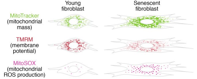
Mitochondrial dysfunction and cell senescence are hallmarks of aging and are closely interconnected. Mitochondrial dysfunction, operationally defined as a decreased respiratory capacity per mitochondrion together with a decreased mitochondrial membrane potential, typically accompanied by increased production of oxygen free radicals, is a cause and a consequence of cellular senescence and figures prominently in multiple feedback loops that induce and maintain the senescent phenotype. Here, we summarize pathways that cause mitochondrial dysfunction in senescence and aging and discuss the major consequences of mitochondrial dysfunction and how these consequences contribute to senescence and aging. We also highlight the potential of senescence-associated mitochondrial dysfunction as an antiaging and antisenescence intervention target, proposing the combination of multiple interventions converging onto mitochondrial dysfunction as novel, potent senolytics.
Overview
The claims and statistics of this paper haven’t been checked yet.
Free: it reads the open-access text and checks the first 5 claims that cite a source we can check. Create a free account to check every claim.
Integrity signals
Registry
Cites
Statistics
Figures

Verification
Claims the paper makes
The claims and statistics of this paper haven’t been checked yet.
Free: it reads the open-access text and checks the first 5 claims that cite a source we can check. Create a free account to check every claim.
Claims other papers make about this one
Each is checked against this paper’s own text: does it say what the citing paper says it does?
The source backs part of the claim, not all of it — or backs it for a different population, condition or comparison.
In the citing paper
“Both proliferation-competent glial cells (which constitute approximately 50% of brain cells) and postmitotic neurons can be positive for markers of cellular senescence in aging, obesity, traumatic injury, and neurodegeneration31,45–49, among other conditions.”
From Guidelines for minimal information on cellular senescence experimentation in vivo (2024) · cites this paper as [45]
Experimental — accuracy not yet measuredThe source backs part of the claim, not all of it — or backs it for a different population, condition or comparison.
In the citing paper
“Therefore, the dysregulation of mitochondrial fusion and fission pathways in senescent astrocytes likely contributes to mitochondrial dysfunction, oxidative stress, and the overall senescence-associated phenotype observed in these cells (Ma et al., 2020; Kleele et al., 2021; Miwa et al., 2022).”
From Aging promotes an increase in mitochondrial fragmentation in astrocytes (2024) · cites this paper as Miwa et al., 2022
Experimental — accuracy not yet measuredThe source backs part of the claim, not all of it — or backs it for a different population, condition or comparison.
In the citing paper
“Dysfunctional mitochondrial dynamics, as observed here, particularly excessive mitochondrial fission, have been associated with oxidative stress, mitochondrial DNA damage, and cellular senescence in various cell types (Twig et al., 2008; Miwa et al., 2022).”
From Aging promotes an increase in mitochondrial fragmentation in astrocytes (2024) · cites this paper as Miwa et al., 2022
Experimental — accuracy not yet measuredThe cited source says what the claim attributes to it.
In the citing paper
“while the presence of senescence markers has been proven even in post-mitotic cells”
From Guidelines for minimal information on cellular senescence experimentation in vivo (2024) · cites this paper as [45]
The check wasn’t confident either way, so it gives no verdict — never disputed.
In the citing paper
“Evidence from the literature indicates that senescent cells exhibit an increase in oxygen consumption rate (Hutter et al., 2004; Son et al., 2017; Miwa et al., 2022; Bang et al., 2023; Diniz et al., 2024a).”
From Aging promotes an increase in mitochondrial fragmentation in astrocytes (2024) · cites this paper as Miwa et al., 2022
Experimental — accuracy not yet measured
2 more claims about this paper
Create a free account to see every claim other papers make about this one.
Self-check
Percentages, sums and reported differences re-computed from the paper’s own numbers.
The self-check needs the paper’s full text.
Citations
Cites
The serial cultivation of human diploid cell strains
Hayflick, L., & Moorhead, P. S. (1961) · Experimental Cell Research · doi:10.1016/0014-4827(61)90192-6 · Cited by 8,508 · cited 1×
“Introduction What is cell senescence? Cell senescence was originally defined as the irreversible loss of replicative potential of primary cells in culture (1), initiated as a persistent DNA damage response to dysfunctional telomeres (2).”
BackgroundTelomeres shorten during ageing of human fibroblasts
Harley, C. B., Futcher, A. B., & Greider, C. W. (1990) · Nature · doi:10.1038/345458a0 · Cited by 5,750 · cited 2×
“Telomere dysfunction might be caused by telomere shortening during DNA replication (3, 4), replication-independent telomere damage (5), or a variety of other stresses (6).”
BackgroundA role for mitochondria in nlrp3 inflammasome activation
Zhou, R., Yazdi, A. S., Menu, P., & Tschopp, J. (2011) · Nature · doi:10.1038/nature09663 · Cited by 5,602
The senescence-associated secretory phenotype: the dark side of tumor suppression
Coppé, J.-P., Desprez, P.-Y., Krtolica, A., & Campisi, J. (2010) · Annual Review of Pathology: Mechanisms of Disease · doi:10.1146/annurev-pathol-121808-102144 · Cited by 5,206
Wu, Z., Puigserver, P., Andersson, U., Zhang, C., Adelmant, G., Mootha, V., Troy, A., Cinti, S., Lowell, B., Scarpulla, R. C., et al. (1999) · Cell · doi:10.1016/s0092-8674(00)80611-x · Cited by 4,139 · cited 1×
“PGC-1α activates the transcription factors Nrf1/2 and ERRα, which further control the TFAM transcription in response to increased mitochondrial biogenesis (65).”
BackgroundClearance of p16Ink4a-positive senescent cells delays ageing-associated disorders
Baker, D. J., Wijshake, T., Tchkonia, T., LeBrasseur, N. K., Childs, B. G., van de Sluis, B., Kirkland, J. L., & van Deursen, J. M. (2011) · Nature · doi:10.1038/nature10600 · Cited by 3,670
AMPK regulates energy expenditure by modulating NAD+ metabolism and SIRT1 activity
Cantó, C., Gerhart-Hines, Z., Feige, J. N., Lagouge, M., Noriega, L., Milne, J. C., Elliott, P. J., Puigserver, P., & Auwerx, J. (2009) · Nature · doi:10.1038/nature07813 · Cited by 3,300
Cellular Senescence: Defining a Path Forward
Gorgoulis, V., Adams, P. D., Alimonti, A., Bennett, D. C., Bischof, O., Bishop, C., Campisi, J., Collado, M., Evangelou, K., Ferbeyre, G., et al. (2019) · Cell · doi:10.1016/j.cell.2019.10.005 · Cited by 3,225 · cited 1×
“Telomere dysfunction might be caused by telomere shortening during DNA replication (3, 4), replication-independent telomere damage (5), or a variety of other stresses (6).”
BackgroundNakahira, K., Haspel, J. A., Rathinam, V. A. K., Lee, S.-J., Dolinay, T., Lam, H. C., Englert, J. A., Rabinovitch, M., Cernadas, M., Kim, H. P., et al. (2011) · Nature Immunology · doi:10.1038/ni.1980 · Cited by 3,049
A DNA damage checkpoint response in telomere-initiated senescence
Fagagna, F. D. D., Reaper, P. M., Clay-Farrace, L., Fiegler, H., Carr, P., von Zglinicki, T., Saretzki, G., Carter, N. P., & Jackson, S. P. (2003) · Nature · doi:10.1038/nature02118 · Cited by 2,917 · cited 1×
“Introduction What is cell senescence? Cell senescence was originally defined as the irreversible loss of replicative potential of primary cells in culture (1), initiated as a persistent DNA damage response to dysfunctional telomeres (2).”
Background
Cited by
Aging Hallmarks and the Role of Oxidative Stress
Maldonado, E., Morales-Pison, S., Urbina, F., & Solari, A. (2023) · Antioxidants · doi:10.3390/antiox12030651 · Cited by 425
“Mitochondrial dysfunction has been considered one of the primary mechanisms contributing to aging and functional decline [210,211].”
BackgroundSenNet recommendations for detecting senescent cells in different tissues
Suryadevara, V., Hudgins, A. D., Rajesh, A., Pappalardo, A., Karpova, A., Dey, A. K., Hertzel, A., Agudelo, A., Rocha, A., Soygur, B., et al. (2024) · Nature Reviews Molecular Cell Biology · doi:10.1038/s41580-024-00738-8 · Cited by 408
“These drivers include mitochondrial DNA (mtDNA) depletion and mutations, inhibitors of the electron transport chain, loss of HSPA9, loss of SIRT3 and SIRT5, and disruptions in complex I assembly 47 .”
Guidelines for minimal information on cellular senescence experimentation in vivo
Ogrodnik, M., Carlos Acosta, J., Adams, P. D., d’Adda di Fagagna, F., Baker, D. J., Bishop, C. L., Chandra, T., Collado, M., Gil, J., Gorgoulis, V., et al. (2024) · Cell · doi:10.1016/j.cell.2024.05.059 · Cited by 376
STAT proteins in cancer: orchestration of metabolism
Li, Y.-J., Zhang, C., Martincuks, A., Herrmann, A., & Yu, H. (2023) · Nature Reviews Cancer · doi:10.1038/s41568-022-00537-3 · Cited by 282
Iron accumulation drives fibrosis, senescence and the senescence-associated secretory phenotype
Maus, M., López-Polo, V., Mateo, L., Lafarga, M., Aguilera, M., De Lama, E., Meyer, K., Sola, A., Lopez-Martinez, C., López-Alonso, I., et al. (2023) · Nature Metabolism · doi:10.1038/s42255-023-00928-2 · Cited by 215
“Previous reports have connected ROS with the secretory phenotype of senescent cells 51,52 .”
BackgroundInflammation and DNA damage: cause, effect or both
Pezone, A., Olivieri, F., Napoli, M. V., Procopio, A., Avvedimento, E. V., & Gabrielli, A. (2023) · Nature Reviews Rheumatology · doi:10.1038/s41584-022-00905-1 · Cited by 212
Sarcopenic obesity: Emerging mechanisms and therapeutic potential
Axelrod, C. L., Dantas, W. S., & Kirwan, J. P. (2023) · Metabolism · doi:10.1016/j.metabol.2023.155639 · Cited by 204
Age-associated remodeling of T cell immunity and metabolism
Han, S., Georgiev, P., Ringel, A. E., Sharpe, A. H., & Haigis, M. C. (2023) · Cell Metabolism · doi:10.1016/j.cmet.2022.11.005 · Cited by 193
Lipids and lipid metabolism in cellular senescence: Emerging targets for age-related diseases
Zeng, Q., Gong, Y., Zhu, N., Shi, Y., Zhang, C., & Qin, L. (2024) · Ageing Research Reviews · doi:10.1016/j.arr.2024.102294 · Cited by 175
Lai, P., Liu, L., Bancaro, N., Troiani, M., Calì, B., Li, Y., Chen, J., Singh, P. K., Arzola, R. A., Attanasio, G., et al. (2025) · Immunity · doi:10.1016/j.immuni.2025.03.005 · Cited by 174
Antiageing strategy for neurodegenerative diseases: from mechanisms to clinical advances
Jiang, Q., Liu, J., Huang, S., Wang, X.-Y., Chen, X., Liu, G.-H., Ye, K., Song, W., Masters, C. L., Wang, J., et al. (2025) · Signal Transduction and Targeted Therapy · doi:10.1038/s41392-025-02145-7 · Cited by 165
Mitochondrial dysfunction in the regulation of aging and aging-related diseases
Zhang, X., Gao, Y., Zhang, S., Wang, Y., Pei, X., Chen, Y., Zhang, J., Zhang, Y., Du, Y., Hao, S., et al. (2025) · Cell Communication and Signaling · doi:10.1186/s12964-025-02308-7 · Cited by 211
“Together, these features reflect changes in the assembly, availability, and functional integrity of the OXPHOS complexes [163].”
Lan, X., Wang, Q., Liu, Y., You, Q., Wei, W., Zhu, C., Hai, D., Cai, Z., Yu, J., Zhang, J., et al. (2024) · Redox Biology · doi:10.1016/j.redox.2024.103406 · Cited by 174
“In this study, the MMP level and MPTP permeability in PC12 cells were assessed after OGD injury at 24 h, which was considered a crucial indicator in evaluating mitochondrial integrity and function [53,54].”
MethodValotto Neto, L. J., Reverete de Araujo, M., Moretti Junior, R. C., Mendes Machado, N., Joshi, R. K., dos Santos Buglio, D., Barbalho Lamas, C., Direito, R., Fornari Laurindo, L., Tanaka, M., et al. (2024) · Antioxidants · doi:10.3390/antiox13040393 · Cited by 146
“Through the execution of these diverse functions, they play essential roles in the development and progression of neurodegenerative diseases such as Alzheimer’s disease and Parkinson’s disease [163,164].”
Li, Y., Sundquist, K., Zhang, N., Wang, X., Sundquist, J., & Memon, A. A. (2023) · EBioMedicine · doi:10.1016/j.ebiom.2022.104432 · Cited by 135
Epigenetic Mechanisms of Aging and Aging-Associated Diseases
la Torre, A., Vecchio, F. L., & Greco, A. (2023) · Cells · doi:10.3390/cells12081163 · Cited by 149
Relaxometry with Nitrogen Vacancy (NV) Centers in Diamond
Mzyk, A., Sigaeva, A., & Schirhagl, R. (2022) · Accounts of Chemical Research · doi:10.1021/acs.accounts.2c00520 · Cited by 109
Age-related epithelial defects limit thymic function and regeneration
Kousa, A. I., Jahn, L., Zhao, K., Flores, A. E., Acenas, D. D., II, Lederer, E. E., Argyropoulos, K. V., Lemarquis, A. L., Granadier, D., Cooper, K., et al. (2024) · Nature Immunology · doi:10.1038/s41590-024-01915-9 · Cited by 121
“Consistent with reports demonstrating a link between mitochondrial function, aging and senescence 42,43 , we found a broad decrease in the transcription of genes within pathways associated with mitochondrial function and metabolism across most cell populations and in fibroblasts upregulation of…”
BackgroundRole of iron in brain development, aging, and neurodegenerative diseases
Gao, Q., Zhou, Y., Chen, Y., Hu, W., Jin, W., Zhou, C., Yuan, H., Li, J., Lin, Z., & Lin, W. (2025) · Annals of Medicine · doi:10.1080/07853890.2025.2472871 · Cited by 117
“Mitochondria undergo morphological changes with aging, characterized by reduced respiratory capacity, decreased steady-state membrane potential, and impaired OXPHOS function [117].”
Shen, Q., Jv, X.-H., Ma, X., Li, C., Liu, L., Jia, W., Qu, L., Chen, L., & Xie, J. (2023) · Acta Pharmacologica Sinica · doi:10.1038/s41401-023-01153-z · Cited by 99
“Cell senescence, fi rst proposed half a century ago, is a homeostatic biological process that prevents uncontrolled cell proliferation [4, 5].”
Epigenetic regulations of cellular senescence in osteoporosis
Huo, S., Tang, X., Chen, W., Gan, D., Guo, H., Yao, Q., Liao, R., Huang, T., Wu, J., Yang, J., et al. (2024) · Ageing Research Reviews · doi:10.1016/j.arr.2024.102235 · Cited by 98
Pathological mechanisms of kidney disease in ageing
Yamamoto, T., & Isaka, Y. (2024) · Nature Reviews Nephrology · doi:10.1038/s41581-024-00868-4 · Cited by 112
“These effects, in turn, induce the release of inflammatory cytokines and contribute to kidney ageing and CKD pathogenesis 69,73,74 .”
Current senolytics: Mode of action, efficacy and limitations, and their future
Rad, A. N., & Grillari, J. (2023) · Mechanisms of Ageing and Development · doi:10.1016/j.mad.2023.111888 · Cited by 96
“The exit of mitochondria from efficient oxidative phosphorylation and consequently the increased reactive oxygen species (ROS) generation have been known as one of the hallmarks of senescent cells (Martini and Passos, 2022; Miwa et al., 2022).”
BackgroundEmerging insights in senescence: pathways from preclinical models to therapeutic innovations
Mansfield, L., Ramponi, V., Gupta, K., Stevenson, T., Mathew, A. B., Barinda, A. J., Herbstein, F., & Morsli, S. (2024) · npj Aging · doi:10.1038/s41514-024-00181-1 · Cited by 88
Insulin and aging – a disappointing relationship
Kolb, H., Kempf, K., & Martin, S. (2023) · Frontiers in Endocrinology · doi:10.3389/fendo.2023.1261298 · Cited by 101
“These changes are associated with mitochondrial dysfunction and increased levels of radical oxygen species (117, 118).”
BackgroundMultifunctional Hydrogel Microneedle Patches Modulating Oxi‐inflamm‐aging for Diabetic Wound Healing
Tian, S., Mei, J., Zhang, L., Wang, S., Yuan, Y., Li, J., Liu, H., Zhu, W., & Xu, D. (2024) · Small · doi:10.1002/smll.202407340 · Cited by 93
The mitochondrial multi-omic response to exercise training across rat tissues
Amar, D., Gay, N. R., Jimenez‐Morales, D., Beltran, P. M. J., Ramaker, M. E., Raja, A. N., Zhao, B., Sun, Y., Marwaha, S., Gaul, D. A., et al. (2024) · Cell Metabolism · doi:10.1016/j.cmet.2023.12.021 · Cited by 101
Redox changes and cellular senescence in Alzheimer's disease
Yu, N., Pasha, M., & Chua, J. J. E. (2024) · Redox Biology · doi:10.1016/j.redox.2024.103048 · Cited by 80
“In turn, damaged mitochondria, characterized by decreased mitochondrial membrane potential and reduced respiratory capacity, further exacerbates ROS levels and trigger cellular senescence, marked by telomere shortening [95 – 97].”
BackgroundSalerno, N., Marino, F., Scalise, M., Salerno, L., Molinaro, C., Filardo, A., Chiefalo, A., Panuccio, G., Angelis, A. D., Urbanek, K., et al. (2022) · Mechanisms of Ageing and Development · doi:10.1016/j.mad.2022.111740 · Cited by 87
Calcium’s Role and Signaling in Aging Muscle, Cellular Senescence, and Mineral Interactions
Terrell, K., Choi, S., & Choi, S. (2023) · International Journal of Molecular Sciences · doi:10.3390/ijms242317034 · Cited by 93
“During cellular senescence, these processes are altered, with the mitochondria of senescent cells typically increasing in size, volume, and number of dysfunctional mitochondria [71].”
BackgroundOxidative Stress-Driven Cellular Senescence: Mechanistic Crosstalk and Therapeutic Horizons
Stojanović, B., Jovanovic, I., Stojanovic, M. D., Stojanović, B., Kovačević, V., Radosavljević, I., Jovanović, D., Kovačević, M. M., Zornić, N., Arsić, A. A., et al. (2025) · Antioxidants · doi:10.3390/antiox14080987 · Cited by 113
“Mitochondria are central hubs in regulating redox homeostasis, and their dysfunction is intimately linked to the onset and progression of cellular senescence [163].”
InfluentialRepurposing SGLT-2 Inhibitors to Target Aging: Available Evidence and Molecular Mechanisms
Grotta, R. L., Frigé, C., Matacchione, G., Olivieri, F., de Candia, P., Ceriello, A., & Prattichizzo, F. (2022) · International Journal of Molecular Sciences · doi:10.3390/ijms232012325 · Cited by 79
“Many age‐related conditions, such as neurodegenerative, metabolic and renal diseases, among others, are indeed also charac‐ terized by a mitochondrial impairment [50].”
BackgroundAging conundrum: A perspective for ovarian aging
Wu, J., Liu, Y., Song, Y., Wang, L., Ai, J., & Li, K. (2022) · Frontiers in Endocrinology · doi:10.3389/fendo.2022.952471 · Cited by 73
Wang, M., Tian, T., Zhou, H., Jiang, S., Jiao, Y., Zhu, Z., Xia, J., Ma, J., & Du, R. (2024) · Journal of Neuroinflammation · doi:10.1186/s12974-024-03072-0 · Cited by 82
“Thus, targeting senescent astrocytes provides insights into therapeutic strategy for neurodegenerative diseases such as PD. Mitochondrial dysfunction is a hallmark of aging and PD [10–12].”
BackgroundCellular senescence in brain aging and cognitive decline
Shafqat, A., Khan, S., Omer, M. H., Niaz, M., Albalkhi, I., AlKattan, K. M., Yaqinuddin, A., Tchkonia, T., Kirkland, J. L., & Hashmi, S. K. (2023) · Frontiers in Aging Neuroscience · doi:10.3389/fnagi.2023.1281581 · Cited by 84
“…in the brain (Area-Gomez et al., 2020; Junxiang et al., 2020; Schmukler et al., 2020; 10.3389/fnagi.2023.1281581 Frontiers in Aging Neuroscience 08 frontiersin.org Wynne et al., 2023), which is implicated in AD pathogenesis and in cellular senescence (Misrani et al., 2021; Miwa et al., 2022).”
BackgroundInfluentialMitochondrial fatty acid oxidation drives senescence
Yamauchi, S., Sugiura, Y., Yamaguchi, J., Zhou, X., Takenaka, S., Odawara, T., Fukaya, S., Fujisawa, T., Naguro, I., Uchiyama, Y., et al. (2024) · Science Advances · doi:10.1126/sciadv.ado5887 · Cited by 82
“Mitochondrial dysfunction is a feature of senescence ( 1 , 12 , 13 ).”
Exploring the Relationship between the Gut Microbiota and Ageing: A Possible Age Modulator
Salazar, J., Durán, P., Díaz, M. P., Chacín, M., Santeliz, R., Mengual, E., Gutiérrez, E., León, X., Díaz, A., Bernal, M., et al. (2023) · International Journal of Environmental Research and Public Health · doi:10.3390/ijerph20105845 · Cited by 71
Zhang, Y., Zou, M., Wu, H., Zhu, J., & Jin, T. (2024) · Neurobiology of Disease · doi:10.1016/j.nbd.2024.106710 · Cited by 78
Hallmarks and Biomarkers of Skin Senescence: An Updated Review of Skin Senotherapeutics
Bulbiankova, D., Díaz‐Puertas, R., Álvarez‐Martínez, F. J., Herranz‐López, M., Barrajón‐Catalán, E., & Micol, V. (2023) · Antioxidants · doi:10.3390/antiox12020444 · Cited by 74
Liu, W., Wang, J., Wang, M., Hou, H., Ding, X., Ma, L., & Liu, M. (2023) · The Journals of Gerontology Series A · doi:10.1093/gerona/glad240 · Cited by 80
Li, H., Wei, T., Zhuang, M., Tan, C., Xie, T., Cai, J., Yao, Y., & Zhu, L. (2023) · Cell Death Discovery · doi:10.1038/s41420-023-01712-7 · Cited by 77
“Information of reagents and antibodies is provided in Supplementary Tables 1 and 2.”
Vascular senescence and aging: mechanisms, clinical implications, and therapeutic prospects
Picos, A., Seoane, N., Campos‐Toimil, M., & Viña, D. (2025) · Biogerontology · doi:10.1007/s10522-025-10256-5 · Cited by 90
“…a central role in these processes by reducing nitric oxide availability, increasing vascular oxidative stress, and sustaining pro-inflammatory signaling, thereby contributing to the pathogenesis of hypertension and atherosclerosis (Chen et al. 2020; Miwa et al. 2022; Ya and Bayrak-tutan 2024).”
InfluentialDuan, Y., Li, Q., Wu, J., Zhou, C., Liu, X., Yue, J., Chen, X., Liu, J., Zhang, Q., Zhang, Y., et al. (2024) · Redox Biology · doi:10.1016/j.redox.2024.103244 · Cited by 75
“Activation of ROCK1 by phosphorylation can promote DRP1 activity and thus elicit mitochondrial hyperfission [63].”
Cellular senescence in Alzheimer’s disease: from physiology to pathology
Zhu, J., Wu, C., & Yang, L. (2024) · Translational Neurodegeneration · doi:10.1186/s40035-024-00447-4 · Cited by 77
Cilleros-Holgado, P., Gómez-Fernández, D., Piñero-Pérez, R., Romero-Domínguez, J. M., Reche-López, D., López-Cabrera, A., Álvarez-Córdoba, M., Munuera-Cabeza, M., Talaverón-Rey, M., Suárez-Carrillo, A., et al. (2023) · Biomolecules · doi:10.3390/biom13121789 · Cited by 71
“…function, which is responsible for primary mitochondrial diseases [12] and, secondarily, for other illnesses and physiological processes related to mitochondrial dysfunction, such as neurodegenerative diseases [13], ageing [14], metabolic diseases [15], cardiac disorders [16], and cancer [17].”
BackgroundFan, J., Fan, J., & Khanzada, Z. (2025) · Muscles · doi:10.3390/muscles4030026 · Cited by 66
“…including muscle stem cell (satellite cell) dysfunction, neuromuscular junction (NMJ) instability, and chronic low-grade inflammaging (also known as inflammation)—all of which contribute to the initiation and progression of muscle deterioration with age and muscle related diseases [13,15–19].”
Dai, Z., Xia, C., Zhao, T., Wang, H., Tian, H., Xu, O., Zhu, X., Zhang, J., & Chen, P. (2022) · Materials Today Bio · doi:10.1016/j.mtbio.2022.100512 · Cited by 72
Liu, S., Cheng, S., Chen, B., Xiao, P., Zhan, J., Liu, J., Chen, Z., Liu, J., Zhang, T., Lei, Y., et al. (2023) · Journal of Nanobiotechnology · doi:10.1186/s12951-023-02211-8 · Cited by 68
“Therefore, the restoration of mitochondrial function has become an important potential approach in anti-ageing therapy [14].”
BackgroundLi, Y., Berliocchi, L., Li, Z., & Rasmussen, L. J. (2023) · Aging Cell · doi:10.1111/acel.13942 · Cited by 73
“Created with BioRe nder.com. protein response (UPRmt)- mediated releasing of IL-6, and impaired mitophagy (Miwa et al., 2022).”
BackgroundLi, H., Cai, R., Zhou, Y., Jiang, Y., & Tan, S. (2025) · Journal of Neuroinflammation · doi:10.1186/s12974-025-03563-8 · Cited by 76
“These spilled mtDNAs are specifically recognized by the cGAS as a danger-associated molecular pattern and bind to the mitochondrial matrix, activates the cGAS to trigger inflammation, and this occurrence becomes more prevalent with age [86, 87].”
The Role of Glial Cell Senescence in Alzheimer's Disease
Alshaebi, F., Sciortino, A., & Kayed, R. (2025) · Journal of Neurochemistry · doi:10.1111/jnc.70051 · Cited by 60
“In turn, several senescence-related mechanisms can impair mitochondrial function (Miwa et al. 2022), creating a feedback loop that drives tissue dysfunction.”
Terao, R., Lee, T. J., Colasanti, J. J., Pfeifer, C. W., Lin, J. B., Santeford, A. C., Hase, K., Yamaguchi, S., Du, D., Sohn, B. S., et al. (2024) · Cell Reports · doi:10.1016/j.celrep.2024.114102 · Cited by 67
Cellular senescence in cancer: from mechanism paradoxes to precision therapeutics
Feng, T., Xie, F., Lee, L. M.-Y., Lin, Z., Tu, Y., Lyu, Y., Yu, P., Wu, J., Chen, B., Zhang, G., et al. (2025) · Molecular Cancer · doi:10.1186/s12943-025-02419-2 · Cited by 82
“Metabolic dysregulation, including mitochondrial dysfunction and altered nutrient sensing pathways such as mTOR and AMPK, also influences both aging and oncogenesis [7, 8].”
Guo, Q., Kawahata, K., Cheng, A., Wang, H., Jia, W., Yoshino, H., & Fukunaga, K. (2022) · Redox Biology · doi:10.1016/j.redox.2022.102547 · Cited by 58
Cheng, X. Y., Tan, X., Wang, W., Zhang, Z., Zhu, R., Wu, M., Li, M., Chen, Y., Liang, Z., Zhu, P., et al. (2022) · Cancer Research · doi:10.1158/0008-5472.can-22-2273 · Cited by 63
Targeting Cellular Senescence for Healthy Aging: Advances in Senolytics and Senomorphics
Alum, E., Izah, S., Uti, D., Ugwu, O., Betiang, P., Basajja, M., & Ejemot-Nwadiaro, R. (2025) · Drug Design, Development and Therapy · doi:10.2147/dddt.s543211 · Cited by 63
“57 FOXO4-DRI, a synthetic peptide, disrupts the”
Mitochondria during T cell aging
Escrig-Larena, J. I., Delgado-Pulido, S., & Mittelbrunn, M. (2023) · Seminars in Immunology · doi:10.1016/j.smim.2023.101808 · Cited by 65
Mitochondrial dynamics and metabolism across skin cells: implications for skin homeostasis and aging
Martic, I., Papaccio, F., Bellei, B., & Cavinato, M. S. (2023) · Frontiers in Physiology · doi:10.3389/fphys.2023.1284410 · Cited by 63
“Mitochondrial dysfunction and cellular senescence are hallmarks of aging and are intricately interconnected (Miwa et al., 2022).”
BackgroundProline restores mitochondrial function and reverses aging hallmarks in senescent cells
Choudhury, D., Rong, N., Kumar, H. V. S., Swedick, S., Samuel, R. Z., Mehrotra, P., Toftegaard, J., Rajabian, N., Thiyagarajan, R., Podder, A. K., et al. (2024) · Cell Reports · doi:10.1016/j.celrep.2024.113738 · Cited by 62
“41 Interestingly, we observed that 7 days of proline treatment decreased mitochondrial content in senescent cells, as evidenced by decreased mitochondrial DNA content (Figure S2G) as well as decreased MitoTracker green intensity (Figures S2H and S2I).”
Mitochondrial dysfunction and therapeutic perspectives in osteoporosis
Liu, J., Gao, Z., & Liu, X. (2024) · Frontiers in Endocrinology · doi:10.3389/fendo.2024.1325317 · Cited by 66
Age-related testosterone decline: mechanisms and intervention strategies
Cheng, H., Zhang, X., Li, Y., Cao, D., Luo, C., Zhang, Q., Zhang, S., & Jiao, Y. (2024) · Reproductive Biology and Endocrinology · doi:10.1186/s12958-024-01316-5 · Cited by 77
Kaur, G., Muthumalage, T., & Rahman, I. (2023) · Aging Cell · doi:10.1111/acel.13850 · Cited by 53
“Mitochondrial functional defects have been known to induce senescent phenotype termed as mitochondrial dysfunction- associated senescence (MiDAS) (Gallage & Gil, 2016; Miwa et al., 2022).”
BackgroundDong, Q., Qiu, H., Piao, C., Li, Z., & Cui, X. (2023) · Journal of Experimental & Clinical Cancer Research · doi:10.1186/s13046-023-02774-2 · Cited by 59
“Cell senescence is a biological phenomenon that commonly occurs in tumor cells after treatment with targeted therapy or chemotherapy and is termed therapy-induced senescence (TIS) [25, 26].”
BackgroundCardiovascular aging: spotlight on mitochondria
Ali, M. A., Gioscia‐Ryan, R. A., Yang, D., Sutton, N. R., & Tyrrell, D. J. (2023) · American Journal of Physiology-Heart and Circulatory Physiology · doi:10.1152/ajpheart.00632.2023 · Cited by 58
Angiogenesis and targeted therapy in the tumour microenvironment: From basic to clinical practice
Yang, S., Fang, Y., Ma, Y., Wang, F., Wang, Y., Jia, J., Yang, Y., Sun, W., Zhou, Q., & Li, Z. (2025) · Clinical and Translational Medicine · doi:10.1002/ctm2.70313 · Cited by 70
Chen, L., Fan, Y., Jiang, N., Huang, X., Yu, M., Zhang, H., Xu, Z., He, D., Wang, Y., Ding, C., et al. (2025) · Nature Nanotechnology · doi:10.1038/s41565-025-01972-7 · Cited by 68
“Senescent MSCs exhibit disrupted mitochondrial homeostasis, including impaired mitophagy, accumulated dysfunctional mitochondria, reduced fission and giant mitochondria formation, ultimately activating ageing-related pathways 8,9 .”
Emerging strategies, applications and challenges of targeting NAD+ in the clinic
Zhang, J., Wang, H., Lautrup, S., Nilsen, H. L., Treebak, J. T., Watne, L. O., Selbæk, G., Wu, L. E., Omland, T., Pirinen, E., et al. (2025) · Nature Aging · doi:10.1038/s43587-025-00947-6 · Cited by 66
“…243,244 , disabled macroautophagy (including defective mitophagy) 6,37,149,245 , deregulated nutrient sensing 246,247 , mitochondrial dysfunction 37,248,249 , cellular senescence 250,251 , stem cell exhaustion 152,252 , altered intercellular communication 253,254 , chronic inflammation 255,256 and…”
Zhou, D., Ran, Y., Yu, R., Liu, Z., Ran, D., & Liu, Z. (2023) · Chemico-Biological Interactions · doi:10.1016/j.cbi.2023.110632 · Cited by 55
Fan, Y., Zhang, W., Huang, X., Fan, M., Shi, C., Zhao, L., Pi, G., Zhang, H., & Ni, S. (2024) · Nature Communications · doi:10.1038/s41467-024-47317-1 · Cited by 56
“…and/or stabilizes p53 and p16INK4 mRNA, 3) dysfunctional mitochondria contribute to innate immune response activation to produce in fl ammation, 4) multiple antiapoptotic pathways are upregulated in senescence, presumably in response to partial mitochondrial membrane permeabilization 75 .”
Oxidative stress and cell senescence as drivers of ageing: Chicken and egg
von Zglinicki, T. (2024) · Ageing Research Reviews · doi:10.1016/j.arr.2024.102558 · Cited by 61
Zhang, J., Zhang, L., Zhang, L., Xue, Y., & Zhang, K. (2025) · Foods · doi:10.3390/foods14132222 · Cited by 61
“Such antagonistic effects underscore the necessity for intervention strategies that achieve molecular specificity, particularly in decoupling antioxidant effects from essential cofactor depletion through temporally or spatially controlled chelation approaches [133].”
Senolytic therapeutics: An emerging treatment modality for osteoarthritis
Ansari, M. M., Ghosh, M., Lee, D., & Son, Y. (2024) · Ageing Research Reviews · doi:10.1016/j.arr.2024.102275 · Cited by 55
Effects of mitochondrial dysfunction on cellular function: Role in atherosclerosis
Xu, M., Wang, W., Cheng, J., Qu, H., Xu, M., & Wang, L. (2024) · Biomedicine & Pharmacotherapy · doi:10.1016/j.biopha.2024.116587 · Cited by 55
Cho, Y., & Kim, Y. K. (2024) · Redox Biology · doi:10.1016/j.redox.2024.103212 · Cited by 52
“Dysfunctional mitochondria have been implicated in cellular senescence [5 – 7], underscoring the significance of dynamin-related protein 1 (DRP1; encoded by DNM1L ), a key regulator of mitochondrial fission [8, 9].”
BackgroundZhu, X., Li, W., Lu, M., Shang, J., Zhou, J., Li, L., Liu, Y., Xing, J., Zhang, M., Zhao, S., et al. (2024) · Journal of Nanobiotechnology · doi:10.1186/s12951-024-02583-5 · Cited by 52
“Considering that mitochondria are the first sensors of senescence and target organs of ROS production [2, 5, 32], we hypothesize that Ex-mediated antisenescence effects may be associated with improvements in the mitochondrial function of GCs.”
BackgroundT-Cell Senescence in Human Metabolic Diseases
Nga, H. T., Nguyen, T. L., & Yi, H. (2024) · Diabetes & Metabolism Journal · doi:10.4093/dmj.2024.0140 · Cited by 52
“During aging, the accumulation of damaged DNA activates PARP1, an NAD+- dependent enzyme, leading to NAD pool depletion in a PARP1-dependent manner and subsequent mitochondrial dysfunction [51].”
BackgroundRottenberg, H. (2023) · International Journal of Molecular Sciences · doi:10.3390/ijms241512295 · Cited by 48
“Many of the studies of mitochondrial dysfunction in aging (and degenerative diseases) report an association of increased mROS production with reduced ∆Ψm [51].”
BackgroundHumanin and Its Pathophysiological Roles in Aging: A Systematic Review
Coradduzza, D., Congiargiu, A., Chen, Z., Cruciani, S., Zinellu, A., Carru, C., & Medici, S. (2023) · Biology · doi:10.3390/biology12040558 · Cited by 55
“A key role in growth and as a regulator of proliferation/differentiation is played by Indian Hedgehog factor (Ihh) secreted by chondrocytes [14,15].”
BackgroundSingle-cell transcriptomic analysis uncovers diverse and dynamic senescent cell populations
Wechter, N., Rossi, M., Anerillas, C., Tsitsipatis, D., Piao, Y., Fan, J., Martindale, J. L., De, S., Mazan-Mamczarz, K., & Gorospe, M. (2023) · Aging · doi:10.18632/aging.204666 · Cited by 43
Su, W., Guo, Y., Wang, Q., Ma, L., Zhang, Q. H., Zhang, Y., Geng, Y., Jin, T., Guo, J., Yang, R., et al. (2024) · Experimental & Molecular Medicine · doi:10.1038/s12276-024-01277-0 · Cited by 51
“Mitochondrial dysfunction might be both a cause and a result of senescence and plays a critical role in inducing and sustaining the senescence phenotype 10 .”
BackgroundAging promotes an increase in mitochondrial fragmentation in astrocytes
Araujo, A. P. B., Vargas, G., Hayashide, L. D. S., Matias, I., Andrade, C. B. V., de Carvalho, J. J., Gomes, F. C. A., & Diniz, L. P. (2024) · Frontiers in Cellular Neuroscience · doi:10.3389/fncel.2024.1496163 · Cited by 43
The role of mitochondria in aging, cell death, and tumor immunity
Wang, Q., Yuan, Y., Liu, J., Li, C., & Jiang, X. (2024) · Frontiers in Immunology · doi:10.3389/fimmu.2024.1520072 · Cited by 48
“Mitochondria, being the primary organelles producing ROS within cells, generate ROS that can attack various mitochondrial components, leading to increased mtDNA mutations and oxidative damage to associated respiratory chain enzymes (46).”
Role of cellular senescence in the pathogenesis of systemic sclerosis
Tsou, P., Shi, B., & Varga, J. (2022) · Current Opinion in Rheumatology · doi:10.1097/bor.0000000000000898 · Cited by 44
Yang, Q., Zou, Y., Wei, X., Ye, P., Wu, Y., Ai, H., Zhang, Z., Tan, J., Zhou, J., Yang, Y., et al. (2023) · Biochimica et Biophysica Acta (BBA) - Molecular Basis of Disease · doi:10.1016/j.bbadis.2023.166795 · Cited by 45
Recent advances in age-related metabolic dysfunction-associated steatotic liver disease
He, Q.-J., Li, Y., Zhao, L.-T., Lin, C.-T., Yu, C., & Wang, D. (2024) · World Journal of Gastroenterology · doi:10.3748/wjg.v30.i7.652 · Cited by 53
“The accumulation of aging cells drives liver steatosis, and liver cell aging is closely related to the progression of MASLD[9].”
InfluentialZheng, L., He, S., Wang, H., Li, J., Liu, Y., & Liu, S. (2024) · Aging and Disease · doi:10.14336/ad.2024.0206 · Cited by 81
“At the same time, some researchers have found that mitochondrial Ca 2+ overload can lead to a decrease in mitochondrial membrane potential and an increase in reactive oxygen species (ROS) production, thus leading to aging and age-related diseases [54, 55].”
METTL3-mediated m6A modification increases Hspa1a stability to inhibit osteoblast aging
Wang, Y., Chen, Y., Xiao, H., Liu, Z., Liu, X., Feng, Z., Sheng, X., Peng, B., Ren, X., Li-hu, X., et al. (2024) · Cell Death Discovery · doi:10.1038/s41420-024-01925-4 · Cited by 52
Cellular Senescence in Health, Disease, and Lens Aging
Qin, Y., Liu, H., & Wu, H. (2025) · Pharmaceuticals · doi:10.3390/ph18020244 · Cited by 55
Cellular oxidants and the proteostasis network: balance between activation and destruction
Ulfig, A., & Jakob, U. (2024) · Trends in Biochemical Sciences · doi:10.1016/j.tibs.2024.07.001 · Cited by 50
Cellular senescence in liver diseases: From molecular drivers to therapeutic targeting
Du, K., Umbaugh, D. S., Ren, N., & Diehl, A. M. (2025) · Journal of Hepatology · doi:10.1016/j.jhep.2025.08.021 · Cited by 47
Tumor Microenvironment as a Therapeutic Target in Melanoma Treatment
Kharouf, N., Flanagan, T. W., Hassan, S.-Y., Shalaby, H., Khabaz, M., Hassan, S.-L., Megahed, M., Haïkel, Y., Santourlidis, S., & Hassan, M. (2023) · Cancers · doi:10.3390/cancers15123147 · Cited by 51
“In vivo elimination of senescent cells via senolysis significantly impacts the treatment of aging [145,146].”
BackgroundChen, Y.-X., Yang, H. S., Wang, D.-S., Chen, T., Qi, X., Tao, L., Chen, Y., & Shen, X. (2024) · Phytomedicine · doi:10.1016/j.phymed.2024.155369 · Cited by 40
Zhang, Y., Liu, J., Zheng, R., Hou, K., Zhang, Y., Jia, T., Lu, X., Samarawickrama, P. N., Jia, S., He, Y., et al. (2024) · Phytomedicine · doi:10.1016/j.phymed.2024.155882 · Cited by 44
Park, Y. J., Kim, J. C., Kim, Y., Kim, Y. H., Park, S. S., Muther, C., Tessier, A., Lee, G., Gendronneau, G., Forestier, S., et al. (2023) · Theranostics · doi:10.7150/thno.84912 · Cited by 42
“In general, senescent cells are less efficient in ATP synthesis due to impaired mitochondria function and ATP production through anaerobic glycolysis [33].”
Influence of human gut microbiome on the healthy and the neurodegenerative aging
Borrego-Ruiz, A., & Borrego, J. J. (2024) · Experimental Gerontology · doi:10.1016/j.exger.2024.112497 · Cited by 47
Peng, K., Yao, Y.-X., Lu, X., Wang, W.-J., Zhang, Y., Zhao, H., Wang, H., Xu, D., & Tan, Z.-X. (2024) · Journal of Hazardous Materials · doi:10.1016/j.jhazmat.2024.135103 · Cited by 41
Song, R., Yin, S., Wu, J., & Yan, J. (2024) · Neural Regeneration Research · doi:10.4103/nrr.nrr-d-24-00025 · Cited by 46
Skawratananond, S., Xiong, D. X., Zhang, C., Tonk, S. S., Pinili, A., Delacruz, B., Pham, P., Smith, S. C., Navab, R., & Reddy, P. H. (2025) · Ageing Research Reviews · doi:10.1016/j.arr.2025.102732 · Cited by 42
Mitochondrial copper overload promotes renal fibrosis via inhibiting pyruvate dehydrogenase activity
Zhu, S., Niu, Y., Zhou, W., Liu, Y., Liu, J., Liu, X., Lü, L., & Yu, C. (2024) · Cellular and Molecular Life Sciences · doi:10.1007/s00018-024-05358-1 · Cited by 44
“It is showed that decreased respiratory capacity, elevated ADP/ATP and AMP/ATP ratios, reduced mitochondrial membrane potential, increased production of oxygen free radicals, serves as both a cause and a consequence of cellular senescence [30 ].”
BackgroundMitochondrion: A bridge linking aging and degenerative diseases
Zhang, L., Wu, J., Zhu, Z., He, Y., & Fang, R. (2023) · Life Sciences · doi:10.1016/j.lfs.2023.121666 · Cited by 40
Metadata
- Authors
- Miwa, S., Kashyap, S., Chini, E., & von Zglinicki, T.
- Affiliations
- Newcastle University (GB)
- Mayo Clinic (US)
- Venue
- Journal of Clinical Investigation
- Published
- 2022-07-01
- Volume
- 132
- Issue
- 13
- ISSN
- 0021-9738 (ISSN-L)
- 1558-8238
- PMC
- PMC9246372
- OpenAlex
- W4283723802
- Cited by
- 1,061
- References
- 145
- Citations by year
- 2026: 294 · 2025: 348 · 2024: 263 · 2023: 136 · 2022: 20
- FWCI
- 19.97
- Type
- journal article
- Language
- en
- Open access
- gold
- doi.org
- Licence
- Locations
- Journal of Clinical Investigation (publishedVersion, cc-by)
- PubMed Central (publishedVersion, cc-by)
- PubMed (publishedVersion)
- DOAJ (DOAJ: Directory of Open Access Journals) (submittedVersion)
- Newcastle University ePrints (Newcastle Univesity) (cc-by)
- Funders
- Glenn Foundation for Medical Research
- Ted Nash Long Life Foundation
- Cancer Research UK (C12161/A24009)
- Newcastle University
- Fondation pour la Recherche Médicale
- Versus Arthritis
- National Institutes of Health (H2020, AG26094, CA233790, AG58812)
- Directorate for Biological Sciences
- Horizon 2020 Framework Programme (857524)
- Medical Research Council (MR/P020941/1)
- Biotechnology and Biological Sciences Research Council (BB/S006710/1)
- National Institute on Aging (AG-58812, R01AG058812, AG-26094)
- National Cancer Institute (R01CA233790)
- Topics
- Life Sciences › Biochemistry, Genetics and Molecular Biology › Molecular Biology › Mitochondrial Function and Pathology
- Life Sciences › Biochemistry, Genetics and Molecular Biology › Cell Biology › Telomeres, Telomerase, and Senescence
- Life Sciences › Biochemistry, Genetics and Molecular Biology › Aging › Genetics, Aging, and Longevity in Model Organisms
- Keywords
- mitochondrial dysfunction, cellular senescence, aging, mitochondrial membrane potential, respiratory capacity, reactive oxygen species, senolytics
- MeSH terms
- Mitochondria, Phenotype, Cellular Senescence
About these checks
Registry, retraction, Cites and statistics results are computed, not judged, with one exception: a printed reference that no index lists is read once by a model before it counts as a citation error. Claims are extracted by a model and kept only when their quote appears verbatim in the text; each cited source is then checked, with a model’s help, for whether it says what the claim relies on. Those verdicts can be wrong — read the claim’s quote and the cited source before relying on one.
Statistics and arithmetic are recomputed, not judged.
Confidence bands are shown only for fields calibrated on our annotated test set. None is calibrated yet, so no band is shown.
What counts as a claim ›How we measure confidence ›How we check this ›
Are you an author, or do you see a problem with this page? Request a review
Data sources
Bibliographic data: Semantic Scholar, OpenAlex, Crossref.
Contains information from the Semantic Scholar Academic Graph Datasets, made available under the ODC Attribution License (ODC-BY 1.0).
Semantic Scholar’s data is described in: Kinney, R., et al. (2023). The Semantic Scholar Open Data Platform. arXiv:2301.10140.
