

Mitochondrial dysfunction in cell senescence and aging
Open accessThe DOI matches its Crossref record; 145 of the 145 references it cites were found in a registry or an index.
Abstract
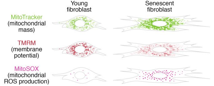
Mitochondrial dysfunction and cell senescence are hallmarks of aging and are closely interconnected. Mitochondrial dysfunction, operationally defined as a decreased respiratory capacity per mitochondrion together with a decreased mitochondrial membrane potential, typically accompanied by increased production of oxygen free radicals, is a cause and a consequence of cellular senescence and figures prominently in multiple feedback loops that induce and maintain the senescent phenotype. Here, we summarize pathways that cause mitochondrial dysfunction in senescence and aging and discuss the major consequences of mitochondrial dysfunction and how these consequences contribute to senescence and aging. We also highlight the potential of senescence-associated mitochondrial dysfunction as an antiaging and antisenescence intervention target, proposing the combination of multiple interventions converging onto mitochondrial dysfunction as novel, potent senolytics.
Overview
The claims and statistics of this paper haven’t been checked yet.
Free: it reads the open-access text and checks the first 5 claims that cite a source we can check. Create a free account to check every claim.
Integrity signals
Registry
Cites
Statistics
Figures

Verification
Claims the paper makes
The claims and statistics of this paper haven’t been checked yet.
Free: it reads the open-access text and checks the first 5 claims that cite a source we can check. Create a free account to check every claim.
Claims other papers make about this one
Each is checked against this paper’s own text: does it say what the citing paper says it does?
The source backs part of the claim, not all of it — or backs it for a different population, condition or comparison.
In the citing paper
“Both proliferation-competent glial cells (which constitute approximately 50% of brain cells) and postmitotic neurons can be positive for markers of cellular senescence in aging, obesity, traumatic injury, and neurodegeneration31,45–49, among other conditions.”
From Guidelines for minimal information on cellular senescence experimentation in vivo (2024) · cites this paper as [45]
Experimental — accuracy not yet measuredThe source backs part of the claim, not all of it — or backs it for a different population, condition or comparison.
In the citing paper
“Therefore, the dysregulation of mitochondrial fusion and fission pathways in senescent astrocytes likely contributes to mitochondrial dysfunction, oxidative stress, and the overall senescence-associated phenotype observed in these cells (Ma et al., 2020; Kleele et al., 2021; Miwa et al., 2022).”
From Aging promotes an increase in mitochondrial fragmentation in astrocytes (2024) · cites this paper as Miwa et al., 2022
Experimental — accuracy not yet measuredThe source backs part of the claim, not all of it — or backs it for a different population, condition or comparison.
In the citing paper
“Dysfunctional mitochondrial dynamics, as observed here, particularly excessive mitochondrial fission, have been associated with oxidative stress, mitochondrial DNA damage, and cellular senescence in various cell types (Twig et al., 2008; Miwa et al., 2022).”
From Aging promotes an increase in mitochondrial fragmentation in astrocytes (2024) · cites this paper as Miwa et al., 2022
Experimental — accuracy not yet measuredThe cited source says what the claim attributes to it.
In the citing paper
“while the presence of senescence markers has been proven even in post-mitotic cells”
From Guidelines for minimal information on cellular senescence experimentation in vivo (2024) · cites this paper as [45]
The check wasn’t confident either way, so it gives no verdict — never disputed.
In the citing paper
“Evidence from the literature indicates that senescent cells exhibit an increase in oxygen consumption rate (Hutter et al., 2004; Son et al., 2017; Miwa et al., 2022; Bang et al., 2023; Diniz et al., 2024a).”
From Aging promotes an increase in mitochondrial fragmentation in astrocytes (2024) · cites this paper as Miwa et al., 2022
Experimental — accuracy not yet measured
2 more claims about this paper
Create a free account to see every claim other papers make about this one.
Self-check
Percentages, sums and reported differences re-computed from the paper’s own numbers.
The self-check needs the paper’s full text.
Citations
Cites
The serial cultivation of human diploid cell strains
Hayflick, L., & Moorhead, P. S. (1961) · Experimental Cell Research · doi:10.1016/0014-4827(61)90192-6 · Cited by 8,508 · cited 1×
“Introduction What is cell senescence? Cell senescence was originally defined as the irreversible loss of replicative potential of primary cells in culture (1), initiated as a persistent DNA damage response to dysfunctional telomeres (2).”
BackgroundTelomeres shorten during ageing of human fibroblasts
Harley, C. B., Futcher, A. B., & Greider, C. W. (1990) · Nature · doi:10.1038/345458a0 · Cited by 5,750 · cited 2×
“Telomere dysfunction might be caused by telomere shortening during DNA replication (3, 4), replication-independent telomere damage (5), or a variety of other stresses (6).”
BackgroundA role for mitochondria in nlrp3 inflammasome activation
Zhou, R., Yazdi, A. S., Menu, P., & Tschopp, J. (2011) · Nature · doi:10.1038/nature09663 · Cited by 5,602
The senescence-associated secretory phenotype: the dark side of tumor suppression
Coppé, J.-P., Desprez, P.-Y., Krtolica, A., & Campisi, J. (2010) · Annual Review of Pathology: Mechanisms of Disease · doi:10.1146/annurev-pathol-121808-102144 · Cited by 5,206
Wu, Z., Puigserver, P., Andersson, U., Zhang, C., Adelmant, G., Mootha, V., Troy, A., Cinti, S., Lowell, B., Scarpulla, R. C., et al. (1999) · Cell · doi:10.1016/s0092-8674(00)80611-x · Cited by 4,139 · cited 1×
“PGC-1α activates the transcription factors Nrf1/2 and ERRα, which further control the TFAM transcription in response to increased mitochondrial biogenesis (65).”
BackgroundClearance of p16Ink4a-positive senescent cells delays ageing-associated disorders
Baker, D. J., Wijshake, T., Tchkonia, T., LeBrasseur, N. K., Childs, B. G., van de Sluis, B., Kirkland, J. L., & van Deursen, J. M. (2011) · Nature · doi:10.1038/nature10600 · Cited by 3,670
AMPK regulates energy expenditure by modulating NAD+ metabolism and SIRT1 activity
Cantó, C., Gerhart-Hines, Z., Feige, J. N., Lagouge, M., Noriega, L., Milne, J. C., Elliott, P. J., Puigserver, P., & Auwerx, J. (2009) · Nature · doi:10.1038/nature07813 · Cited by 3,300
Cellular Senescence: Defining a Path Forward
Gorgoulis, V., Adams, P. D., Alimonti, A., Bennett, D. C., Bischof, O., Bishop, C., Campisi, J., Collado, M., Evangelou, K., Ferbeyre, G., et al. (2019) · Cell · doi:10.1016/j.cell.2019.10.005 · Cited by 3,225 · cited 1×
“Telomere dysfunction might be caused by telomere shortening during DNA replication (3, 4), replication-independent telomere damage (5), or a variety of other stresses (6).”
BackgroundNakahira, K., Haspel, J. A., Rathinam, V. A. K., Lee, S.-J., Dolinay, T., Lam, H. C., Englert, J. A., Rabinovitch, M., Cernadas, M., Kim, H. P., et al. (2011) · Nature Immunology · doi:10.1038/ni.1980 · Cited by 3,049
A DNA damage checkpoint response in telomere-initiated senescence
Fagagna, F. D. D., Reaper, P. M., Clay-Farrace, L., Fiegler, H., Carr, P., von Zglinicki, T., Saretzki, G., Carter, N. P., & Jackson, S. P. (2003) · Nature · doi:10.1038/nature02118 · Cited by 2,917 · cited 1×
“Introduction What is cell senescence? Cell senescence was originally defined as the irreversible loss of replicative potential of primary cells in culture (1), initiated as a persistent DNA damage response to dysfunctional telomeres (2).”
Background
Research works printed but not linked: 2
- von Zglinicki T. Mechanisms of cell senescence in aging. In: Musi N, Hornsby P eds. Handbook of the Biology of Aging. Academic Press; 2021:53–68.
- Miwa S, Johnson TE. Stress and aging. Vijg J, et al., eds. In: The Molecular and Cellular Biology of Aging. Gerontological Society of America; 2016:216-249.
Cited by
Items 201–300 of 999
Hoseini, R., Hoseini, Z., & Kamangar, A. (2025) · Cellular and Molecular Life Sciences · doi:10.1007/s00018-025-05836-0 · Cited by 22
“Mitochondrial dysfunc - tion is a key contributor to metabolic disorders, including insulin resistance and T2DM, due to its impact on energy metabolism, oxidative stress, and cellular senescence [81, 82].”
García-Trejo, S. S., Gómez‐Sierra, T., Eugenio-Pérez, D., Medina‐Campos, O. N., & Pedraza‐Chaverrí, J. (2024) · Antioxidants · doi:10.3390/antiox13040415 · Cited by 23
“…from the ER and causes the VDAC/MCU channels to initiate the flow towards the mitochondria, the increase in calcium levels in senescent cells decreases the membrane potential, induces alterations in the calcium transport and electron transport chain and therefore increases ROS production [83,85].”
BackgroundChen, P., Wang, Y., Xie, J., Lei, J., & Zhou, B. (2024) · Neuropharmacology · doi:10.1016/j.neuropharm.2024.109950 · Cited by 23
Li, M., Yu, X., Chen, X., Jiang, Y., Zeng, Y., Ren, R., Nie, M., Zhang, Z., Bao, Y., & Kang, H. (2024) · International Immunopharmacology · doi:10.1016/j.intimp.2024.112101 · Cited by 24
Liu, S., Hu, H., Zhang, M., Zhang, Y., Geng, R., Jin, Y., Cao, Y., Guo, W., Liu, J., & Fu, S. (2024) · Journal of Agricultural and Food Chemistry · doi:10.1021/acs.jafc.3c09444 · Cited by 23
Leeson, H. C., Aguado, J., Gómez‐Inclán, C., Chaggar, H. K., Fard, A. T., Hunter, Z. L., Lavin, M. F., Mackay‐Sim, A., & Wolvetang, E. J. (2024) · Neurobiology of Disease · doi:10.1016/j.nbd.2024.106562 · Cited by 24
Shao, M., Qiu, Y., Shen, M., Liu, W. Q., Feng, D., Luo, Z., & Zhou, Y. (2024) · The FASEB Journal · doi:10.1096/fj.202302547rr · Cited by 27
“49,50 Many factors promote the expression of cyclin-dependent kinase inhibitors, particularly p16 and p21, which antagonize cyclin-dependent kinases and impede cell cycle progression, a pivotal mechanism underlying cellular aging.”
Qu, Y., Meng, B., Cai, S., Yang, B., He, Y., Fu, C., Li, X., Li, P., Cao, Z., Mao, X., et al. (2024) · Journal of Nanobiotechnology · doi:10.1186/s12951-024-02820-x · Cited by 22
“Since mitochondrial dysfunction is one hallmark of aging [26, 27], we examined mitochondrial morphological and functional changes to evaluate the impact of ESC-apoVs in aging BMMSCs. TEM and immunofluorescent staining analysis revealed alterations in mitochondrial ultrastructure, including…”
BackgroundHeadley, C. A., Gautam, S., Olmo-Fontánez, A. M., Garcia‐Vilanova, A., Dwivedi, V., Schami, A., Weintraub, S. T., Tsao, P. S., Torrelles, J. B., & Turner, J. (2024) · Advanced Science · doi:10.1002/advs.202401077 · Cited by 34
Talifu, Z., Ren, Z., Chen, C., Guo, S., Wu, Y., Li, Y., Su, B., & Zheng, X. (2025) · Aging Cell · doi:10.1111/acel.70142 · Cited by 33
“Secondly, comorbidity imposes simultaneous energy demands on multiple organ systems (Miwa et al. 2022).”
Huang, L., Shen, Y., Pan, X., Li, J., Li, C., Ruan, L., He, S., Huang, L., Liu, K., Zhao, X., et al. (2025) · Nature Communications · doi:10.1038/s41467-025-63152-4 · Cited by 31
“Excessive Ca 2+ in fl ux triggers mitochondrial dysfunction, leading to membrane depolarization, ROS generation, and cellular senescence 51,52 .”
Li, S., Fan, R., Wang, Y., He, K., Xu, J., & Li, H. (2024) · Frontiers in Pharmacology · doi:10.3389/fphar.2024.1352377 · Cited by 27
“Notably, mitochondria are the core sites of calcium overload in tumor cells (Miwa et al., 2022).”
BackgroundZhang, Q.-S., Wang, J.-N., Yang, T.-L., Li, S., Li, J., Liu, D.-N., Shang, H., & Zhang, Z. (2025) · EBioMedicine · doi:10.1016/j.ebiom.2024.105533 · Cited by 20
“57,58 In our study, we observed that interfering with SHMT2 expression resulted in increased ROS levels.”
Time Restricted Eating: A Valuable Alternative to Calorie Restriction for Addressing Obesity?
Parrotta, M. E., Colangeli, L., Scipione, V., Vitale, C., Sbraccia, P., & Guglielmi, V. (2025) · Current Obesity Reports · doi:10.1007/s13679-025-00609-z · Cited by 21
Tian, Y., Pan, P., Luo, X., Sun, Y., Yang, X., Gao, H., & Yang, Y. (2025) · Chemico-Biological Interactions · doi:10.1016/j.cbi.2025.111450 · Cited by 20
Casorati, B., Zafferri, I., Castiglioni, S., & Maier, J. A. M. (2025) · Antioxidants · doi:10.3390/antiox14040446 · Cited by 22
“…in senescent bMSCs. Contrary to previous reports suggesting an increase in mitochondrial mass during senescence both in vitro [27] and in vivo [27], our findings indicate a reduction in mitochondrial content in senescent bMSCs, as evidenced by decreased CYP F levels and MT-ND1 copy number,…”
InfluentialSynergistic targeting of cancer cells through simultaneous inhibition of key metabolic enzymes
Dreute, J., Stengel, J., Becher, J., van den Borre, D. R., Pfisterer, M., Bartkuhn, M., Beutgen, V. M., Ndreshkjana, B., Gärtner, U., Graumann, J., et al. (2025) · Cell Death and Differentiation · doi:10.1038/s41418-025-01532-5 · Cited by 23
“After 16h of transduction, medium was replaced with fresh complete medium.”
Wang, Q., Zhang, M., Jiang, W., Li, D., Qi, B., Shi, S., Shi, S., & Yi, C. (2025) · Journal of Nanobiotechnology · doi:10.1186/s12951-025-03716-0 · Cited by 24
“Notably, the loss or impairment of mitochondrial key functions is predominantly manifested in mitochondrial membrane depolarization or the loss of mitochondrial membrane potential, abnormal ROS production, and the depletion of ATP generation [36].”
Cheng, Y., Hou, B.-H., Xie, G., Shao, Y.-T., Yang, J., & Xu, C. (2023) · Free Radical Biology and Medicine · doi:10.1016/j.freeradbiomed.2023.03.264 · Cited by 22
Kristensen, N. P., Dionisio, E., Bentzen, A. K., Tamhane, T., Kemming, J. S., Nos, G., Voss, L. F., Hansen, U. K., Lauer, G. M., & Hadrup, S. R. (2024) · Science Advances · doi:10.1126/sciadv.adm8951 · Cited by 22
The senotherapeutic potential of phytochemicals for age-related intestinal disease
Costa, C. M., Pedrosa, S. S., Kirkland, J. L., Reis, F., & Madureira, A. R. (2024) · Ageing Research Reviews · doi:10.1016/j.arr.2024.102619 · Cited by 19
The impact of COVID-19 on accelerating of immunosenescence and brain aging
Müller, L., & Benedetto, S. D. (2024) · Frontiers in Cellular Neuroscience · doi:10.3389/fncel.2024.1471192 · Cited by 24
Fu, L., Wang, S., Zhang, N., Lin, Y., Zhang, S., Mao, Y., & Zhou, P. (2024) · Chemical Engineering Journal · doi:10.1016/j.cej.2024.158540 · Cited by 19
Zhang, M., Wei, J., He, C., Sui, L., Jiao, C., Zhu, X., & Pan, X. (2024) · Cellular & Molecular Biology Letters · doi:10.1186/s11658-024-00669-4 · Cited by 23
Guo, X., Qiao, X., Li, X., Zhou, W., Liu, C., Yu, F., Chen, Q., Pan, M., Niu, X., Wang, L., et al. (2025) · Phytomedicine · doi:10.1016/j.phymed.2025.156547 · Cited by 21
Addressing osteoblast senescence: Molecular pathways and the frontier of anti‐ageing treatments
Zhang, Z., Liu, P., Song, Y., Ma, L., Xu, Y., Lei, J., Tong, B., Zhu, D., Liang, H., Wang, H., et al. (2025) · Clinical and Translational Medicine · doi:10.1002/ctm2.70417 · Cited by 26
“157 The main characteristics of mitochondrial dysfunction include changes in mitochondrial DNA (mtDNA), defects in mitochondrial biogenesis and dynamics, disruptions in mitochondrial autophagy, impairments in OXPHOS and excessive accumulation of ROS.”
HIV infection and immunosenescence: challenges and intervention strategies
Jin, J., Xu, Q., Zhang, X., Zhu, A., Xia, W., Moog, C., Chan, A. S. W., Zhang, T., & Su, B. (2025) · BMC Medicine · doi:10.1186/s12916-025-04545-6 · Cited by 23
“This oxidative stress inhibits proteasomal activity and is recognized as a key driver of the detrimental effects of ageing, contributing to chronic inflammation and cellular senescence [160].”
Park, J. Y., Lee, H., Song, E. S., Lee, Y. H., Kuk, M. U., Ko, G., Kwon, H. W., Byun, Y., & Park, J. T. (2022) · Rejuvenation Research · doi:10.1089/rej.2022.0043 · Cited by 17
“40 The simultaneous reduction in mitochondrial mass was shown in SB203580-treated senescent cells, suggesting that mito-phagy, an autophagic mechanism that selectively degrades defective mitochondria, was triggered (Fig.”
Wang, J., Shen, Y., Li, M., Li, T., Shi, D., Lu, S., Qiu, F., & Wu, Z. (2023) · The Journal of Nutritional Biochemistry · doi:10.1016/j.jnutbio.2023.109361 · Cited by 19
Liu, M., He, H., Fan, F., Qiu, L.-J., Zheng, F., Guan, Y., Yang, G., & Chen, L. (2023) · Journal of Molecular and Cellular Cardiology · doi:10.1016/j.yjmcc.2023.05.005 · Cited by 17
Sonar, S. A., Watanabe, M., & Nikolich‐Žugich, J. (2023) · Seminars in Immunology · doi:10.1016/j.smim.2023.101835 · Cited by 21
Forqani, M. A., Akbarian, M., Amirahmadi, S., Soukhtanloo, M., Hosseini, M., & Forouzanfar, F. (2023) · International Journal of Neuroscience · doi:10.1080/00207454.2023.2257877 · Cited by 26
“Oxidative stress and deoxyribonucleic acid (DNa) methylation are examples of these kinds of damage [3,4]. aging is responsible for many human diseases, such as cancer, cardiovascular diseases, diabetes, and neurodegenerative diseases [5,6].”
Aryl hydrocarbon receptor: Linking environment to aging process in elderly patients with asthma
Yang, T., Wan, R., Tu, W., Avvaru, S. N., & Gao, P. (2024) · Chinese Medical Journal · doi:10.1097/cm9.0000000000002960 · Cited by 18
“[15] Of these, mtDNA shows an increased susceptibility to ROS, leading to severe oxidative damage.”
BackgroundInfluentialFlensted-Jensen, M., Oró, D., Rørbeck, E. A., Zhang, C., Madsen, M. R., Madsen, A. N., Norlin, J., Feigh, M., Larsen, S., & Hansen, H. H. (2024) · BMC Gastroenterology · doi:10.1186/s12876-024-03141-x · Cited by 17
“Given that mitochondria are critically involved in cell survival and death [18], and the main source of reactive oxygen species (ROS) [19], mitochondrial dysfunction can trigger cellular senescence programs [20–22].”
BackgroundChiariello, A., Rossetti, L. M., Valente, S., Pasquinelli, G., Sollazzo, M., Iommarini, L., Porcelli, A. M., Tognocchi, M., Conte, G., Santoro, A., et al. (2024) · Aging Cell · doi:10.1111/acel.14111 · Cited by 18
“In fact, mitochondrial dysfunction is also known to elicit a cell se - nescence phenomenon (Miwa et al., 2022 ; Wiley et al., 2016) indicated as MiDAS (mitochondrial dysfunction-associated senescence) characterized by a peculiar pattern of secretory phenotype (Wiley et al., 2016).”
BackgroundAir pollution aggravates renal ischaemia–reperfusion‐induced acute kidney injury
Sanches, T. R., Parra, A. C., Sun, P., Graner, M., Itto, L. Y. U., Butter, L. M., Claessen, N., Roelofs, J. J. T. H., Florquin, S., Veras, M. M., et al. (2024) · The Journal of Pathology · doi:10.1002/path.6302 · Cited by 23
“TECs can also be affected by AKI-induced mitochondrial damage and STING-mediated in fl ammation [50], both of which induce cellular senescence [51].”
BackgroundThe interplay of NAD and hypoxic stress and its relevance for ageing
Burtscher, J., Denti, V., Gostner, J. M., Weiss, A. K., Strasser, B., Hüfner, K., Burtscher, M., Paglia, G., Kopp, M., & Dünnwald, T. (2024) · Ageing Research Reviews · doi:10.1016/j.arr.2024.102646 · Cited by 21
Li, W., Huang, Y., Fan, L., Yangzom, D., Zhang, K., Shen, L., SuiZhong, C., Gu, C., & Yu, S. (2024) · Scientific Reports · doi:10.1038/s41598-024-82614-1 · Cited by 21
Senescence, Aging and Disease Throughout the Gastrointestinal System
Ferreira-González, S., Matsumoto, T., Hara, E., & Forbes, S. J. (2025) · Gastroenterology · doi:10.1053/j.gastro.2025.06.010 · Cited by 20
Bai, Z., Ren, J., Liu, J., Zhang, C., Huang, H., Zhao, X., Chen, X., Wei, J., Qi, J., Yang, S., et al. (2025) · Redox Biology · doi:10.1016/j.redox.2025.103818 · Cited by 18
“Mitochondrial dysfunction is a closely interconnected hallmark of T cell senescence characterized by increased mitochondrial mass, low MMP, and increased production of MtROS [21,52].”
Nunes, J. M., Kell, L., Slaghekke, A., Wüst, R. C. I., Fielding, B. C., Kell, D. B., & Pretorius, E. (2026) · Cell Death and Disease · doi:10.1038/s41419-025-08162-2 · Cited by 22
“Organelle dysfunction is also highly apparent in senescent cells, and includes disrupted mitochondrial ATP production and lysosomal hypertrophy [65].”
Senolytic and senomorphic interventions to defy senescence-associated mitochondrial dysfunction
Nehlin, J. O. (2023) · Advances in protein chemistry and structural biology · doi:10.1016/bs.apcsb.2023.02.020 · Cited by 20
Impaired mitophagosome–lysosome fusion mediates olanzapine‐induced aging
Chen, X., Wang, Z., Zheng, P., Dongol, A., Xie, Y., Ge, X., Zheng, M., Dang, X., Seyhan, Z. B., Nagaratnam, N., et al. (2023) · Aging Cell · doi:10.1111/acel.14003 · Cited by 20
“Mitochondria are responsible for supplying the necessary energy (adenosine triphosphate, ATP) for cell survival and function, while progressive mitochondrial dysfunction is a hallmark characteristic of aging (Andrews et al., 2005 ; Miwa et al., 2022 ).”
BackgroundRojas-Solé, C., Pinilla-González, V., Lillo‐Moya, J., González-Fernández, T., Saso, L., & Rodrigo, R. (2023) · Redox Report · doi:10.1080/13510002.2023.2289740 · Cited by 24
Yuan, L., Cao, Y., Zhang, Q., Pan, J.-C., Wu, C., Ye, Y., Jiao, Q., Zhu, H., & Wang, Z. (2024) · Biosensors and Bioelectronics · doi:10.1016/j.bios.2024.116123 · Cited by 17
Nie, L., He, K., Qiu, C., Li, Q., Xiong, B., Gao, C., Zhang, X., Jing, M., Wu, W., Liu, J., et al. (2024) · Biomedicine & Pharmacotherapy · doi:10.1016/j.biopha.2024.116415 · Cited by 20
Rejuvenation of young blood on aging organs: Effects, circulating factors, and mechanisms
Liu, M., Lan, Q., Wu, H., & Qiu, C.-W. (2024) · Heliyon · doi:10.1016/j.heliyon.2024.e32652 · Cited by 18
“There is currently a belief that restoring the impaired function of aging mitochondria can help alleviate cell senescence [17].”
BackgroundWang, Q., Zhu, K., & Zhang, A. (2024) · The Science of The Total Environment · doi:10.1016/j.scitotenv.2024.174502 · Cited by 19
Non-Coding RNAs of Mitochondrial Origin: Roles in Cell Division and Implications in Cancer
Piergentili, R., & Sechi, S. (2024) · International Journal of Molecular Sciences · doi:10.3390/ijms25137498 · Cited by 18
“Indeed, in many instances a direct link has been shown between aging and the dysfunction of various OXPHOS (oxidative phosphorylation) complexes (reviewed in [27,32–34]).”
BackgroundQu, X., Pan, P., Cao, S., Ma, Y., Yang, J., Gao, H., Pei, X., & Yang, Y. (2024) · International Journal of Molecular Sciences · doi:10.3390/ijms252011122 · Cited by 16
“Thus, an imbalance in MQC can accelerate cellular senescence and aging, and appropriate interventions in MQC can delay the aging process and extend the lifespan [3].”
BackgroundLee, H., Massaro, M., Abdelfattah, N., Baudo, G., Liu, H., Yun, K., & Blanco, E. (2024) · Aging Cell · doi:10.1111/acel.14446 · Cited by 19
“Cell senescence is closely associated with mitochondrial dysfunction, with senescent cells exhibiting reduced mitochondrial membrane potential ( ΔΨ m) and respiratory capacity per mitochondrion (Miwa et al., 2022).”
Zhao, L., Liang, K., Cheng, W., Zhou, X., Yang, L., Mao, X., Qi, W., Jin, H., Zhang, W., Pan, H., et al. (2025) · Nature Communications · doi:10.1038/s41467-025-63194-8 · Cited by 24
“Meanwhile, ROS accumulates, damaging DNA,proteins,andlipids,ultimatelyimpairingnormalcellfunction 21,22 .”
Wu, H., Zhang, Q., Zhu, J., Wu, L., Xiao, Y., & Yang, X. (2025) · Bioactive Materials · doi:10.1016/j.bioactmat.2025.09.002 · Cited by 35
“Directly targeting and repairing mitochondrial function offers a path to address the metabolic dysregulation of senescent cells at its source [75,214,215].”
Cardiac aging: Molecular mechanisms and therapeutic interventions
Li, X., Pang, X., Sun, H., Zhang, B., Wang, H., Wu, N., & Yang, L. (2025) · Pharmacological Research · doi:10.1016/j.phrs.2025.107954 · Cited by 21
Xie, Z., Zhang, X., Li, Y., & Zhu, R. (2025) · Experimental Gerontology · doi:10.1016/j.exger.2025.112913 · Cited by 22
Targeting mitochondrial autophagy for anti-aging
Shan, W., Liu, Y., Tang, R., Li, L., Yang, H., & Lin, L. (2025) · Cell Death Discovery · doi:10.1038/s41420-025-02913-y · Cited by 21
“During aging, mitochondrial dysfunction is usually manifested as a breakdown of energy metabolism, the dissipation of membrane potential, and the accumulation of oxidative damage [5].”
The Mitochondrial ATP Synthase/IF1 Axis in Cancer Progression: Targets for Therapeutic Intervention
Domínguez-Zorita, S., & Cuezva, J. M. (2023) · Cancers · doi:10.3390/cancers15153775 · Cited by 17
Metabolic regulation of endothelial senescence
Le, N.-T. (2023) · Frontiers in Cardiovascular Medicine · doi:10.3389/fcvm.2023.1232681 · Cited by 16
“The intricate interplay between aging and mitochondrial dysfunction establishes a detrimental cycle that contributes to the development of a senescent phenotype (133, 134).”
BackgroundNiu, S., Qiu, P., Meng, J., Tao, C., Wen, M., Yu, N., & Chen, Z. (2024) · Journal of Colloid and Interface Science · doi:10.1016/j.jcis.2024.01.002 · Cited by 16
Shafqat, A., Masters, M. C., Tripathi, U., Tchkonia, T., Kirkland, J. L., & Hashmi, S. K. (2024) · Ageing Research Reviews · doi:10.1016/j.arr.2024.102400 · Cited by 15
The mitochondrial function of peripheral blood mononuclear cells in frail older patients
Huang, T., Li, Q., Zhang, D., Tong, Q., Zhu, Q., Ding, G., & Liu, J. (2024) · Experimental Gerontology · doi:10.1016/j.exger.2024.112594 · Cited by 16
Nguyen, T. T., Mazzucco, G., Kyriacou, E., Lunardi, T., Brandl, L., Ahmed, W., Doksani, Y., & Lingner, J. (2025) · Nucleic Acids Research · doi:10.1093/nar/gkaf285 · Cited by 23
“Oxidative stress has been implicated in aging and multiple human diseases including neurodegenerative disorders and cancer [ 1 , 2 ].”
Mitochondria – the CEO of the cell
Lee-Glover, L. P., Picard, M. A., & Shutt, T. E. (2025) · Journal of Cell Science · doi:10.1242/jcs.263403 · Cited by 17
Hong, J., Jin, L., Zhang, C.-Y., Zhong, W., Yang, H., Wang, G.-M., Ma, S., Guan, C., Li, Q., & Zhou, Y. (2023) · Heliyon · doi:10.1016/j.heliyon.2023.e17361 · Cited by 14
E-cigarette-induced changes in cell stress and mitochondrial function
Ramamurthy, C., Zuniga-Hertz, J. P., Masso-Silva, J. A., Shin, J. H., Niesman, I. R., Bojanowski, C. M., Kumar, A., Hepokoski, M., Alexander, L. E. C., Patel, H. H., et al. (2025) · Free Radical Biology and Medicine · doi:10.1016/j.freeradbiomed.2025.01.004 · Cited by 14
“Dysfunctional mitochondria are associated with activating the integrated stress response (ISR) and metabolic reprogramming, representing hallmarks of mitochondrial dysfunction [18,19].”
Zhao, S., Zhang, Y., Zhao, Y. R., & Lu, X. (2025) · Progress in Biophysics and Molecular Biology · doi:10.1016/j.pbiomolbio.2025.02.002 · Cited by 15
Li, Y., Kang, K., Bao, H., Liu, S., Zhao, B., Hu, G., & Wu, J. (2025) · Biology · doi:10.3390/biology14060643 · Cited by 18
“There is also a strong mutual influence between mitochondrial function and aging; mitochondrial dysfunction and cellular senescence are markers of aging [5,6].”
Wang, X., Luo, W., Chen, Z., Li, C., Zhou, J., Huang, Z., Tang, L., Wu, J., Wu, Z., Li, Y., et al. (2025) · Journal for ImmunoTherapy of Cancer · doi:10.1136/jitc-2024-010822 · Cited by 19
“These 15×21 CAR-NK cells exhibited enhanced effector functions and were capable of recruiting T cells for cooperative interactions.”
Zhang, Y., Gu, Q., Li, Y., Li, J., Han, Y., Feng, L., Qin, X., Zhang, J., Zhou, Z., & Chang, X. (2025) · Journal of Hazardous Materials · doi:10.1016/j.jhazmat.2025.138972 · Cited by 15
Polyphenol-Based Therapeutic Strategies for Mitochondrial Dysfunction in Aging
Maksimović, T., Gădău, C., Antal, G., Čoban, M., Eșanu, O., Atyim, E., Mioc, A., & Şoica, C. (2025) · Biomolecules · doi:10.3390/biom15081116 · Cited by 22
“Mitochondrial sirtuins, SIRT 3-5, have a role in mitochondrial biogenesis, metabolism, and signaling [52,79].”
InfluentialGut Microbiota: An Ally in the Mechanisms and Interventions of Healthy Aging
Chatterjee, S. S., Hebbani, A. V., & Syed, K. (2025) · Gastrointestinal Disorders · doi:10.3390/gidisord7040068 · Cited by 17
Morciano, G., Pellielo, G., Agyapong, E. D., Pellegrino, C., Patergnani, S., Franceschini, D., Koutsikos, K., Rigon, L., Pinton, P., & Rimessi, A. (2026) · Journal of Translational Medicine · doi:10.1186/s12967-026-07783-1 · Cited by 20
“Mitochondria in senescent cells exhibit a fragmented morphology and impaired function, leading to increased mtROS production and mtDAMPs release, which further amplifies inflammatory signaling [94].”
Lee, I., Piao, S., Kim, S., Nagar, H., Choi, S., Kim, M., Vu, G.-H., Jeon, B. H., & Kim, C. (2023) · Antioxidants · doi:10.3390/antiox12030585 · Cited by 15
“Endothelial senescence is associated with the accumulation of dysfunctional mitochondria, including altered mitochondrial dynamics, decreased mitochondrial mass, and impaired mitochondrial biogenesis [5,6].”
BackgroundLiu, S. Y., Zhao, Y., Yao, H., Zhang, L., Chen, C., Zheng, Z., & Jin, S. (2023) · Experimental Cell Research · doi:10.1016/j.yexcr.2023.113688 · Cited by 18
Lessons in aging from Myc knockout mouse models
Prochownik, E. V., & Wang, H. (2023) · Frontiers in Cell and Developmental Biology · doi:10.3389/fcell.2023.1244321 · Cited by 17
“Impaired mitochondrial function, metabolism and translation have all been described in association with aging and all of these are dependent upon Myc to balance and maintain their normal function (Li et al., 2005; van Riggelen et al., 2010; Barrientos, 2012; Graves et al., 2012; Edmunds et al., 2016; D’Aquila et al.,…”
BackgroundMolecularly Targeted Fluorescent Sensors for Visualizing and Tracking Cellular Senescence
He, Z., Xu, K., Li, Y., Gao, H. J., Miao, T., Zhao, R., & Huang, Y. (2023) · Biosensors · doi:10.3390/bios13090838 · Cited by 13
“In addition to the lysosome, the mitochondrion is another organelle sensitively responding to the occurrence of senescence [62,63].”
The Emerging Role of the Mitochondrial Respiratory Chain in Skeletal Aging
Tao, H., Zhu, P., Xia, W., Chu, M., Chen, K., Wang, Q., Gu, Y., Lu, X., Bai, J., & Geng, D. (2023) · Aging and Disease · doi:10.14336/ad.2023.0924 · Cited by 24
“Mitochondrial dysfunction is believed to be the root cause of aging, inflammation, and oxidative stress imbalance [27, 43, 44].”
BackgroundXBP1 Modulates the Aging Cardiorenal System by Regulating Oxidative Stress
Zhang, J., Zhao, Y., & Gong, N. (2023) · Antioxidants · doi:10.3390/antiox12111933 · Cited by 15
“…events driven by oxidative stress contribute to increased susceptibility of the aging kidneys and hearts to acute and chronic injuries [68]. mitochondrial events driven by oxidative stress contribute to increased susceptibility of the aging kidneys and hearts to acute and chronic…”
BackgroundPiao, C., Wang, Y., Liu, T., Liu, T., Ma, Y., Li, Y., Zhang, J., & Wang, H. (2024) · Free Radical Biology and Medicine · doi:10.1016/j.freeradbiomed.2024.01.049 · Cited by 15
Imbalanced mitochondrial dynamics contributes to the pathogenesis of X-linked adrenoleukodystrophy
Launay, N., Lopez-Erauskin, J., Bianchi, P., Guha, S., Parameswaran, J., Coppa, A., Torreni, L., Schlüter, A., Fourcade, S., Paredes-Fuentes, A. J., et al. (2024) · Brain · doi:10.1093/brain/awae038 · Cited by 14
Liu, H., Liu, H., Yang, Q., & Fan, Z. (2024) · Stem Cell Research & Therapy · doi:10.1186/s13287-024-03793-2 · Cited by 16
“When the ROS concentration is too high, it will destroy the mitochondrial membrane, change the permeability of the membrane, reduce the concentration difference of ions inside and outside the membrane, and lead to a decrease in the membrane potential and ATP synthesis [62, 68].”
BackgroundHong, X., Tian, G., Dai, B., Zhou, X., Gao, Y., Zhu, L., Liu, H., Zhu, Q., Zhang, L., Zhu, Y., et al. (2024) · Advanced Science · doi:10.1002/advs.202401527 · Cited by 19
Unveiling the Neuroprotective Potential of Date Palm (Phoenix dactylifera): A Systematic Review
Asdaq, S. M. B., Almutiri, A. A., Alenzi, A., Shaikh, M. S., Shaik, M. A., Alshehri, S., & Rabbani, S. I. (2024) · Pharmaceuticals · doi:10.3390/ph17091221 · Cited by 18
“Free radicals from cell catabolism are thought to play a role in aging and other degenerative disorders [33].”
BackgroundThe role of mitochondria in cytokine and chemokine signalling during ageing
Kalykaki, M., Rubio‐Tomás, T., & Tavernarakis, N. (2024) · Mechanisms of Ageing and Development · doi:10.1016/j.mad.2024.111993 · Cited by 17
Chen, J., Dai, X., Li, X., Tang, Y.-X., Xu, X.-W., & Li, J. (2024) · Phytomedicine · doi:10.1016/j.phymed.2024.156114 · Cited by 16
Yao, J., Jiang, Y., Zhang, P., Miao, Y., Wu, X., Lei, H., Xie, Z., Tian, Y., Zhao, X., Li, J., et al. (2025) · Pharmacological Research · doi:10.1016/j.phrs.2025.107620 · Cited by 17
Munteanu, C., Galaction, A., Onose, G., Turnea, M. A., & Rotariu, M. (2025) · Pharmaceuticals · doi:10.3390/ph18030344 · Cited by 18
Rudinskiy, M., Galli, C., Raimondi, A., & Molinari, M. (2025) · Nature Cell Biology · doi:10.1038/s41556-025-01728-4 · Cited by 19
“…have been described by pathophysiologists as early hallmarks of human disorders including liver diseases 110,111 , cardiomyopathies 112 and alcohol-induced heart disease 113,114 as well as in pathophysiological conditions such as ageing 115–117 , which leads to organelle dysfunction.”
Pan, P., Cao, S., Gao, H. Y., Qu, X., Ma, Y., Yang, J., Pei, X., & Yang, Y. (2025) · International Journal of Biological Macromolecules · doi:10.1016/j.ijbiomac.2025.142368 · Cited by 21
Mechanisms of cellular senescence-induced vascular aging: evidence of senotherapeutic strategies
Mahoney, S. A., Bloom, S. I., Seals, D. R., Donato, A. J., Rossman, M. J., & Clayton, Z. S. (2025) · The Journal of Cardiovascular Aging · doi:10.20517/jca.2024.31 · Cited by 20
“Cellular senescence promotes mitochondrial dysfunction in both an autocrine and paracrine manner as a result of the proinflammatory SASP [48,49] .”
Wang, H., Gao, Y., Mingzhang, Dai, G.-C., Lu, P., Li, Y., Cao, M., Liu, X., Sheng, R., Shi, L., et al. (2025) · Journal of Advanced Research · doi:10.1016/j.jare.2025.08.013 · Cited by 19
“Organismal aging and cellular senescence induce chronic inflam-mation, which subsequently leads to mitochondrial dysfunction [73,74].”
Mitochondria in cell senescence: A Friend or Foe?
Chen, Q., Young, L. H., & Barsotti, R. (2023) · Advances in protein chemistry and structural biology · doi:10.1016/bs.apcsb.2023.02.019 · Cited by 15
Autophagy Alters the Susceptibility of Candida albicans Biofilms to Antifungal Agents
Shen, J., Ma, M., Duan, W., Huang, Y., Shi, B., Wu, Q., & Wei, X. (2023) · Microorganisms · doi:10.3390/microorganisms11082015 · Cited by 15
“MMP depends on sequential intramitochondrial biochemical reactions [50].”
BackgroundCellular Senescence in Liver Cancer: How Dying Cells Become “Zombie” Enemies
Gazzillo, A., Volponi, C., Soldani, C., Polidoro, M. A., Franceschini, B., Lleò, A., Bonavita, E., & Donadon, M. (2023) · Biomedicines · doi:10.3390/biomedicines12010026 · Cited by 15
Fernando, P. D. S. M., Piao, M. J., Herath, H. M. U. L., Kang, K. A., Hyun, C. L., Kim, E. T., Koh, Y. S., & Hyun, J. W. (2024) · Toxicology in Vitro · doi:10.1016/j.tiv.2024.105870 · Cited by 15
Identification of Cellular Isoschaftoside-Mediated Anti-Senescence Mechanism in RAC2 and LINC00294
Lee, Y. H., So, B., Lee, K. S., Kuk, M. U., Park, J. H., Yoon, J. H., Lee, Y. J., Kim, D. Y., Kim, M., Kwon, H. W., et al. (2024) · Molecules · doi:10.3390/molecules29174182 · Cited by 16
“As part of cellular senescence, defective mitochondria accumulate and undergo structural changes that significantly increase their volume and size [10].”
BackgroundLu, Q., Hu, X., Hou, Q., Yu, L., Cao, K., Ding, D., Lu, Y., & Dai, C. (2024) · Cellular Signalling · doi:10.1016/j.cellsig.2024.111451 · Cited by 17
Apparoo, Y., Phan, C., Kuppusamy, U. R., & Chan, E. W. C. (2024) · Neuroscience · doi:10.1016/j.neuroscience.2024.11.082 · Cited by 13
Fan, W.-Y., Fan, X., Xie, Y.-J., Yan, X., Tao, M.-X., Zhao, S., Yu, B., & Li, R. (2025) · Food & medicine homology. · doi:10.26599/fmh.2026.9420108 · Cited by 18
Molecular Mechanisms of Cellular Senescence in Age-Related Endometrial Dysfunction
Kobayashi, H., Umetani, M., Nishio, M., Shigetomi, H., Imanaka, S., & Hashimoto, H. (2025) · Cells · doi:10.3390/cells14120858 · Cited by 17
“Specifically, aged endometrial cells exhibit hyperactivation of glycolysis [185], reduced OXPHOS, and mitochondrial dysfunction [186].”
Metadata
- Authors
- Miwa, S., Kashyap, S., Chini, E., & von Zglinicki, T.
- Affiliations
- Newcastle University (GB)
- Mayo Clinic (US)
- Venue
- Journal of Clinical Investigation
- Published
- 2022-07-01
- Volume
- 132
- Issue
- 13
- ISSN
- 0021-9738 (ISSN-L)
- 1558-8238
- PMC
- PMC9246372
- OpenAlex
- W4283723802
- Cited by
- 1,061
- References
- 145
- Citations by year
- 2026: 294 · 2025: 348 · 2024: 263 · 2023: 136 · 2022: 20
- FWCI
- 19.97
- Type
- journal article
- Language
- en
- Open access
- gold
- doi.org
- Licence
- Locations
- Journal of Clinical Investigation (publishedVersion, cc-by)
- PubMed Central (publishedVersion, cc-by)
- PubMed (publishedVersion)
- DOAJ (DOAJ: Directory of Open Access Journals) (submittedVersion)
- Newcastle University ePrints (Newcastle Univesity) (cc-by)
- Funders
- Glenn Foundation for Medical Research
- Ted Nash Long Life Foundation
- Cancer Research UK (C12161/A24009)
- Newcastle University
- Fondation pour la Recherche Médicale
- Versus Arthritis
- National Institutes of Health (H2020, AG26094, CA233790, AG58812)
- Directorate for Biological Sciences
- Horizon 2020 Framework Programme (857524)
- Medical Research Council (MR/P020941/1)
- Biotechnology and Biological Sciences Research Council (BB/S006710/1)
- National Institute on Aging (AG-58812, R01AG058812, AG-26094)
- National Cancer Institute (R01CA233790)
- Topics
- Life Sciences › Biochemistry, Genetics and Molecular Biology › Molecular Biology › Mitochondrial Function and Pathology
- Life Sciences › Biochemistry, Genetics and Molecular Biology › Cell Biology › Telomeres, Telomerase, and Senescence
- Life Sciences › Biochemistry, Genetics and Molecular Biology › Aging › Genetics, Aging, and Longevity in Model Organisms
- Keywords
- mitochondrial dysfunction, cellular senescence, aging, mitochondrial membrane potential, respiratory capacity, reactive oxygen species, senolytics
- MeSH terms
- Mitochondria, Phenotype, Cellular Senescence
About these checks
Registry, retraction, Cites and statistics results are computed, not judged, with one exception: a printed reference that no index lists is read once by a model before it counts as a citation error. Claims are extracted by a model and kept only when their quote appears verbatim in the text; each cited source is then checked, with a model’s help, for whether it says what the claim relies on. Those verdicts can be wrong — read the claim’s quote and the cited source before relying on one.
Statistics and arithmetic are recomputed, not judged.
Confidence bands are shown only for fields calibrated on our annotated test set. None is calibrated yet, so no band is shown.
What counts as a claim ›How we measure confidence ›How we check this ›
Are you an author, or do you see a problem with this page? Request a review
Data sources
Bibliographic data: Semantic Scholar, OpenAlex, Crossref.
Contains information from the Semantic Scholar Academic Graph Datasets, made available under the ODC Attribution License (ODC-BY 1.0).
Semantic Scholar’s data is described in: Kinney, R., et al. (2023). The Semantic Scholar Open Data Platform. arXiv:2301.10140.
