

Mitochondrial dysfunction in cell senescence and aging
Open accessThe DOI matches its Crossref record; 145 of the 145 references it cites were found in a registry or an index.
Abstract
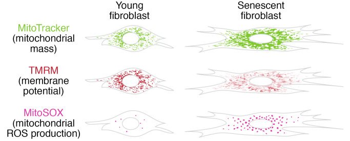
Mitochondrial dysfunction and cell senescence are hallmarks of aging and are closely interconnected. Mitochondrial dysfunction, operationally defined as a decreased respiratory capacity per mitochondrion together with a decreased mitochondrial membrane potential, typically accompanied by increased production of oxygen free radicals, is a cause and a consequence of cellular senescence and figures prominently in multiple feedback loops that induce and maintain the senescent phenotype. Here, we summarize pathways that cause mitochondrial dysfunction in senescence and aging and discuss the major consequences of mitochondrial dysfunction and how these consequences contribute to senescence and aging. We also highlight the potential of senescence-associated mitochondrial dysfunction as an antiaging and antisenescence intervention target, proposing the combination of multiple interventions converging onto mitochondrial dysfunction as novel, potent senolytics.
Overview
The claims and statistics of this paper haven’t been checked yet.
Free: it reads the open-access text and checks the first 5 claims that cite a source we can check. Create a free account to check every claim.
Integrity signals
Registry
Cites
Statistics
Figures

Verification
Claims the paper makes
The claims and statistics of this paper haven’t been checked yet.
Free: it reads the open-access text and checks the first 5 claims that cite a source we can check. Create a free account to check every claim.
Claims other papers make about this one
Each is checked against this paper’s own text: does it say what the citing paper says it does?
The source backs part of the claim, not all of it — or backs it for a different population, condition or comparison.
In the citing paper
“Both proliferation-competent glial cells (which constitute approximately 50% of brain cells) and postmitotic neurons can be positive for markers of cellular senescence in aging, obesity, traumatic injury, and neurodegeneration31,45–49, among other conditions.”
From Guidelines for minimal information on cellular senescence experimentation in vivo (2024) · cites this paper as [45]
Experimental — accuracy not yet measuredThe source backs part of the claim, not all of it — or backs it for a different population, condition or comparison.
In the citing paper
“Therefore, the dysregulation of mitochondrial fusion and fission pathways in senescent astrocytes likely contributes to mitochondrial dysfunction, oxidative stress, and the overall senescence-associated phenotype observed in these cells (Ma et al., 2020; Kleele et al., 2021; Miwa et al., 2022).”
From Aging promotes an increase in mitochondrial fragmentation in astrocytes (2024) · cites this paper as Miwa et al., 2022
Experimental — accuracy not yet measuredThe source backs part of the claim, not all of it — or backs it for a different population, condition or comparison.
In the citing paper
“Dysfunctional mitochondrial dynamics, as observed here, particularly excessive mitochondrial fission, have been associated with oxidative stress, mitochondrial DNA damage, and cellular senescence in various cell types (Twig et al., 2008; Miwa et al., 2022).”
From Aging promotes an increase in mitochondrial fragmentation in astrocytes (2024) · cites this paper as Miwa et al., 2022
Experimental — accuracy not yet measuredThe cited source says what the claim attributes to it.
In the citing paper
“while the presence of senescence markers has been proven even in post-mitotic cells”
From Guidelines for minimal information on cellular senescence experimentation in vivo (2024) · cites this paper as [45]
The check wasn’t confident either way, so it gives no verdict — never disputed.
In the citing paper
“Evidence from the literature indicates that senescent cells exhibit an increase in oxygen consumption rate (Hutter et al., 2004; Son et al., 2017; Miwa et al., 2022; Bang et al., 2023; Diniz et al., 2024a).”
From Aging promotes an increase in mitochondrial fragmentation in astrocytes (2024) · cites this paper as Miwa et al., 2022
Experimental — accuracy not yet measured
2 more claims about this paper
Create a free account to see every claim other papers make about this one.
Self-check
Percentages, sums and reported differences re-computed from the paper’s own numbers.
The self-check needs the paper’s full text.
Citations
Cites
The serial cultivation of human diploid cell strains
Hayflick, L., & Moorhead, P. S. (1961) · Experimental Cell Research · doi:10.1016/0014-4827(61)90192-6 · Cited by 8,508 · cited 1×
“Introduction What is cell senescence? Cell senescence was originally defined as the irreversible loss of replicative potential of primary cells in culture (1), initiated as a persistent DNA damage response to dysfunctional telomeres (2).”
BackgroundTelomeres shorten during ageing of human fibroblasts
Harley, C. B., Futcher, A. B., & Greider, C. W. (1990) · Nature · doi:10.1038/345458a0 · Cited by 5,750 · cited 2×
“Telomere dysfunction might be caused by telomere shortening during DNA replication (3, 4), replication-independent telomere damage (5), or a variety of other stresses (6).”
BackgroundA role for mitochondria in nlrp3 inflammasome activation
Zhou, R., Yazdi, A. S., Menu, P., & Tschopp, J. (2011) · Nature · doi:10.1038/nature09663 · Cited by 5,602
The senescence-associated secretory phenotype: the dark side of tumor suppression
Coppé, J.-P., Desprez, P.-Y., Krtolica, A., & Campisi, J. (2010) · Annual Review of Pathology: Mechanisms of Disease · doi:10.1146/annurev-pathol-121808-102144 · Cited by 5,206
Wu, Z., Puigserver, P., Andersson, U., Zhang, C., Adelmant, G., Mootha, V., Troy, A., Cinti, S., Lowell, B., Scarpulla, R. C., et al. (1999) · Cell · doi:10.1016/s0092-8674(00)80611-x · Cited by 4,139 · cited 1×
“PGC-1α activates the transcription factors Nrf1/2 and ERRα, which further control the TFAM transcription in response to increased mitochondrial biogenesis (65).”
BackgroundClearance of p16Ink4a-positive senescent cells delays ageing-associated disorders
Baker, D. J., Wijshake, T., Tchkonia, T., LeBrasseur, N. K., Childs, B. G., van de Sluis, B., Kirkland, J. L., & van Deursen, J. M. (2011) · Nature · doi:10.1038/nature10600 · Cited by 3,670
AMPK regulates energy expenditure by modulating NAD+ metabolism and SIRT1 activity
Cantó, C., Gerhart-Hines, Z., Feige, J. N., Lagouge, M., Noriega, L., Milne, J. C., Elliott, P. J., Puigserver, P., & Auwerx, J. (2009) · Nature · doi:10.1038/nature07813 · Cited by 3,300
Cellular Senescence: Defining a Path Forward
Gorgoulis, V., Adams, P. D., Alimonti, A., Bennett, D. C., Bischof, O., Bishop, C., Campisi, J., Collado, M., Evangelou, K., Ferbeyre, G., et al. (2019) · Cell · doi:10.1016/j.cell.2019.10.005 · Cited by 3,225 · cited 1×
“Telomere dysfunction might be caused by telomere shortening during DNA replication (3, 4), replication-independent telomere damage (5), or a variety of other stresses (6).”
BackgroundNakahira, K., Haspel, J. A., Rathinam, V. A. K., Lee, S.-J., Dolinay, T., Lam, H. C., Englert, J. A., Rabinovitch, M., Cernadas, M., Kim, H. P., et al. (2011) · Nature Immunology · doi:10.1038/ni.1980 · Cited by 3,049
A DNA damage checkpoint response in telomere-initiated senescence
Fagagna, F. D. D., Reaper, P. M., Clay-Farrace, L., Fiegler, H., Carr, P., von Zglinicki, T., Saretzki, G., Carter, N. P., & Jackson, S. P. (2003) · Nature · doi:10.1038/nature02118 · Cited by 2,917 · cited 1×
“Introduction What is cell senescence? Cell senescence was originally defined as the irreversible loss of replicative potential of primary cells in culture (1), initiated as a persistent DNA damage response to dysfunctional telomeres (2).”
Background
Research works printed but not linked: 2
- von Zglinicki T. Mechanisms of cell senescence in aging. In: Musi N, Hornsby P eds. Handbook of the Biology of Aging. Academic Press; 2021:53–68.
- Miwa S, Johnson TE. Stress and aging. Vijg J, et al., eds. In: The Molecular and Cellular Biology of Aging. Gerontological Society of America; 2016:216-249.
Cited by
Items 601–700 of 999
Senescence Programs Shape the Chronic Wound Microenvironment
Shvedova, M., Cho, M., Eyckmans, J., & Roh, D. S. (2026) · Advances in Wound Care · doi:10.1177/21621918261441046 · Cited by 3
Vrouvaki, A., Christodoulou, M., Hillas, G., Loukides, S., & Fouka, E. (2026) · International Journal of Molecular Sciences · doi:10.3390/ijms27083542 · Cited by 3
“Mitochondrial dysfunction is consistently found across multiple senescence contexts, including replicative, stress-induced, oncogene-induced and telomere-associated senescence [55,56].”
Pîrlog, L.-M., Catana, A. M., Pitforodeschi, A.-D., Pilatec, A.-N., Băilă, R.-M., Rusu, I., Militaru, M.-S., Iordănescu, I. I., & Belbe, A.-A. (2026) · Genes · doi:10.3390/genes17060681 · Cited by 4
“Other strategies attempt to restore apoptotic competence by disrupting FOXO4–TP53 interactions, inhibiting HSP90-dependent senescence signalling, or targeting vulnerabilities linked to mitochondrial dysfunction, redox imbalance, proteostasis stress, and altered glycolytic metabolism [187–193].”
Aging of mesenchymal stem cells in bone aging: Mechanisms, impact, and therapeutic perspectives
Zhou, Z., Wang, H. W., Jin, Z., Wen, B., Wu, T., Zhu, T., & Liu, D. (2026) · Ageing Research Reviews · doi:10.1016/j.arr.2026.103240 · Cited by 6
Oyama, T., Yamamoto, T., Kameda, T., Kamiya, T., Abe, H., Abe, T., & Tanuma, S. (2024) · Life · doi:10.3390/life14030413 · Cited by 5
“NAD + is directly involved in this system as a substrate for SIRT3 [15], making mitochondria-specific proteins like SIRT3 and SOD2 valuable indicators of mitochondrial health [15–17].”
BackgroundArioka, M., Miura, K., Han, R., Igawa, K., Takahashi‐Yanaga, F., & Sasaguri, T. (2024) · Life Sciences · doi:10.1016/j.lfs.2024.122807 · Cited by 4
The Mitochondrial Transplantation: A New Frontier in Plastic Surgery
Li, H., & Mu, D. (2024) · Journal of Craniofacial Surgery · doi:10.1097/scs.0000000000010706 · Cited by 3
“62 In 2022, Miwa et al 63 investigated this complex relationship be-tween mitochondrial dysfunction and the aging process.”
Samiminemati, A., Shahzaib, M., Moriello, C., Alessio, N., Aprile, D., Squillaro, T., Bernardo, G. D., & Galderisi, U. (2024) · Methods in molecular biology · doi:10.1007/7651_2024_581 · Cited by 4
Targeting mitochondrial function as a potential therapeutic approach for allergic asthma
Chen, D., Wu, W., Li, J., Huang, X., Chen, S., Zheng, T., Huang, G., & Ouyang, S. (2025) · Inflammation Research · doi:10.1007/s00011-024-01972-8 · Cited by 6
“These changes raise AMP/ADP and NAD + /NADH ratios, activating AMP-activated protein kinase (AMPK) and p53-dependent cell cycle arrest [24].”
Yu, X., Song, X., Chen, Y., Lv, Y., & Tan, M. (2025) · Food Bioscience · doi:10.1016/j.fbio.2025.106537 · Cited by 3
Lee, Y. H., Lim, H., Kim, G., Jang, G., Kuk, M. U., Park, J. H., Yoon, J. H., Lee, Y. J., Kim, D., So, B., et al. (2025) · Cell Proliferation · doi:10.1111/cpr.70049 · Cited by 5
Metallic Nanoparticles and Cosmetics: The Role of Mitochondria and Premature Aging
Montesinos‐Cruz, V., & Olmanson, J. (2025) · Wiley Interdisciplinary Reviews Nanomedicine and Nanobiotechnology · doi:10.1002/wnan.70014 · Cited by 5
Mavioğlu, R. N., Gumpp, A. M., Hummel, E. M., Moser, D., Ammerpohl, O., Behnke, A., Mack, M., & Kolassa, I. (2025) · Brain Behavior and Immunity · doi:10.1016/j.bbi.2025.05.022 · Cited by 3
Liu, B., Wang, M., Pu, N., Wang, J., Fan, W., Qu, C., Wu, H., Song, Z., & Tao, Y. (2025) · Ageing Research Reviews · doi:10.1016/j.arr.2025.102788 · Cited by 3
Fu, W., Kadeer, G., He, Y., & Feng, Y. J. (2022) · Frontiers in Genetics · doi:10.3389/fgene.2022.975886 · Cited by 3
“, 2022), insulin resistance (Hayden, 2022), as well as aging (Miwa et al., 2022).”
BackgroundAging, Cellular Senescence, and Glaucoma
Zhang, Y., Huang, S., Xie, B., & Zhong, Y. (2023) · Aging and Disease · doi:10.14336/ad.2023.0631 · Cited by 87
Yang, X., Wang, J., Dai, X., Ma, N., Hu, C., Guo, H., Chen, S., Huang, Y., & Wu, J. (2023) · Free Radical Biology and Medicine · doi:10.1016/j.freeradbiomed.2023.08.030 · Cited by 3
miR-195b is required for proper cellular homeostasis in the elderly
Muñoz-Gallardo, M. D. M., García‐Padilla, C., Vicente‐García, C., Carvajal, J. J., Arenega, A., & Franco, D. (2024) · Scientific Reports · doi:10.1038/s41598-024-51256-8 · Cited by 4
“…are previous evidence of the functional role of miR-195 modulating cell proliferation and metabolism, there is scarce information on its plausible role in other essential cellular processes such as redox homeostasis and cellular senescence 91,92 , even if they are intimately co-regulated 50–53 .”
mtDNA extramitochondrial replication mediates mitochondrial defect effects
Shan, Z., Li, S., Gao, Y., Jian, C., Ti, X., Zuo, H., Wang, Y., Zhao, G., Wang, Y., & Zhang, Q. (2024) · iScience · doi:10.1016/j.isci.2024.108970 · Cited by 3
“46,54 However, how mitochondrial dysfunction is related to aging is elusive.”
BackgroundWang, D., Lin, D., Yang, X., Wu, D., Li, P., Zhang, Z., Zhang, W., Guo, Y., Fu, S., & Zhang, N. (2024) · Molecular Biology Reports · doi:10.1007/s11033-024-09238-6 · Cited by 3
“Several molecular mechanisms underlie the aging process, including interactions between mitochondria and telomeres, suggesting common molecular mechanisms at work [42].”
Miwa, S., Fielder, E., Ishaq, A., Low, E., Laws, J., Calista, A., Castle, J., & von Zglinicki, T. (2024) · doi:10.59368/agingbio.20240022 · Cited by 5
Self-citationMaternal Ezh1/2 deficiency impairs the function of mitochondria in mouse oocytes and early embryos
Zhang, D., Deng, W., Jiang, T., Zhao, Y., Bai, D., Tian, Y., Kong, S., Zhang, L., Wang, H., Gao, S., et al. (2024) · Journal of Cellular Physiology · doi:10.1002/jcp.31244 · Cited by 3
“The membrane potential plays a critical role in mitochondrial activity and function (Miwa et al., 2022).”
BackgroundZhong, W., Chen, J., He, Y., Li, X., & Yuan, C. (2024) · Neuroreport · doi:10.1097/wnr.0000000000002024 · Cited by 3
“…this signaling pathway in regulating mitochondrial quality control mechanisms, the inhibition of the PI3K/AKT/ mTOR signaling pathway has become a hopeful treat-ment strategy for neurodegeneration due to its significant involvement in maintaining mitochondrial quality con-trol in recent years [30].”
BackgroundCombined Endurance and Resistance Exercise Mitigates Age‐Associated Cardiac Dysfunction
Han, X., Ashraf, M. Z., Shi, H., Nkembo, A. T., Tipparaju, S. M., & Xuan, W. (2024) · Advanced Biology · doi:10.1002/adbi.202400137 · Cited by 5
Velmurugan, S., Pauline, R., Chandrashekar, G., Kulanthaivel, L., & Subbaraj, G. K. (2024) · Nigerian Postgraduate Medical Journal · doi:10.4103/npmj.npmj_9_24 · Cited by 4
Wang, H., Stevens, T., Lu, J. L., Roberts, A., Land, C. V., Muzumdar, R. H., Gong, Z., Vockley, J., & Prochownik, E. V. (2024) · Advanced Science · doi:10.1002/advs.202401593 · Cited by 3
Combating Cellular Aging: Frontiers in Biomaterials and Therapies
Liu, S., Mou, J., Zhao, C., Du, C., Liu, J., Bao, W., Xiao, H., Huang, W., Liao, J., & Lei, Y. (2024) · ACS Materials Letters · doi:10.1021/acsmaterialslett.4c02108 · Cited by 6
Kurmaev, D. P., Bulgakova, S. V., Treneva, E. V., Kosareva, O. V., Merzlova, P. Y., Sharonova, L. A., & Dolgikh, Y. A. (2025) · Экспериментальная и клиническая гастроэнтерология · doi:10.31146/1682-8658-ecg-228-8-226-241 · Cited by 5
Glytsou, C. (2025) · Methods in molecular biology · doi:10.1007/978-1-0716-4426-3_13 · Cited by 5
Uric Acid Promotes Human Umbilical Vein Endothelial Cell Senescence In Vitro
Lewandowska, K., Mikuła‐Pietrasik, J., Książek, K., Tykarski, A., & Uruski, P. (2025) · Metabolites · doi:10.3390/metabo15060402 · Cited by 4
“The present study reveals that JC-1 decreased with mitochondrial damage [26], and as a compensatory response, the cell produces more healthy mitochondria, reflected by an increase in NAO levels, a marker of mitochondrial mass [27].”
Yao, Y., Lu, X., Zhao, L., Liao, H.-Y., Liu, Z., Zhao, H., Wang, H., Xu, D., & Tan, Z.-X. (2025) · Toxicology and Applied Pharmacology · doi:10.1016/j.taap.2025.117524 · Cited by 3
EHE cell cultures are a platform for mechanistic and therapeutic investigation
Scalora, N., DeWane, G., Drebot, Y., Khan, A. A., Sinha, S., Ghosh, K., Robinson, D., Cogswell, P. C., Bellizzi, A. M., Snow, A. N., et al. (2025) · Scientific Reports · doi:10.1038/s41598-025-19453-1 · Cited by 4
“5E) shows an enrichment for processes consistent with oxidative phosphorylation consistent with upregulation/dysregulation of mitochondrial genes (Table S5), often seen in senescent cells 39,51 .”
Pelliccia, A., Capradossi, F., Corsi, F., Tarquini, G. D., Bruni, E., Reichle, A., Torino, F., & Ghibelli, L. (2023) · International Journal of Molecular Sciences · doi:10.3390/ijms24032040 · Cited by 5
“Indeed, mitochondrial dysfunctions are often considered hallmarks of senescence [27,28].”
BackgroundWoronzow, V., Möhner, J., Remane, D., & Zischler, H. (2023) · Frontiers in Cell and Developmental Biology · doi:10.3389/fcell.2023.1274807 · Cited by 2
The role of senescence in cancer therapy-associated cardiovascular toxicity
Alshoubaki, Y. K., Grüterich, V., Zollet, V., & Kuster, G. M. (2024) · The Journal of Cardiovascular Aging · doi:10.20517/jca.2024.14 · Cited by 2
Ling, L., Li, M., Lu, J.-W., Zhang, F., Pan, F., Yao, S., Myatt, L., Wang, W., Sun, K., & Ying, H. (2025) · Mechanisms of Ageing and Development · doi:10.1016/j.mad.2025.112053 · Cited by 3
Pearngam, P., Promthep, K., Prasertporn, T., Songsomboon, K., Polvat, T., Charoenkul, J., Mukda, S., Govitrapong, P., Nopparat, C., & Panmanee, J. (2025) · Pesticide Biochemistry and Physiology · doi:10.1016/j.pestbp.2025.106520 · Cited by 2
Dehkordi, S. K., Sajedi, S., Heshmat, A., Orr, M. E., & Zare, H. (2025) · npj Aging · doi:10.1038/s41514-025-00235-y · Cited by 3
“It is a hallmark of both cellular senescence 60 and AD 61 .”
Chen, D., Yao, L., Mo, K., Li, L., Liu, Z., Xia, Z., & Ding, H. (2025) · Scientific Reports · doi:10.1038/s41598-025-05849-6 · Cited by 4
“Miwa et al. revealed the central role of mitochondrial dysfunction in cellular senescence 24 , while Camacho-Encina et al. further demonstrated the synergistic effect of mitochondrial dysfunction and cellular senescence in cardiovascular disease 37 .”
Murayama, R., Horisawa, K., Miura, S., Taniguchi, S., Shu, J., Takahashi, M., Izumi, Y., Bamba, T., Ishigami, K., & Suzuki, A. (2025) · Aging Cell · doi:10.1111/acel.70197 · Cited by 2
“Another possibility is an increase in mitochondrial stress caused by ROS owing to reduced mitochondrial function in the hepatocytes of old mice (Miwa et al. 2022).”
Jiang, B., Guan, J., Lv, Y., Yang, C., Yang, M., Shen, X., Xu, B., Wang, Q., Qin, Y., & Wu, L. J. (2025) · Sensors and Actuators B Chemical · doi:10.1016/j.snb.2025.138511 · Cited by 3
The toxicity of insecticide broflanilide on human SH-SY5Y cells
Xu, Y., Wang, X., Yang, X., Liu, X., Wang, M., An, S., Na, R., & Hao, Y. (2025) · Environmental Toxicology and Chemistry · doi:10.1093/etojnl/vgaf243 · Cited by 2
Wang, X., Tu, Q., Mao, A., Meng, K., Jun, L., Li, B., Zhang, T., Zhang, H., Ding, Y., & Cao, Z. (2025) · Phytomedicine · doi:10.1016/j.phymed.2025.157385 · Cited by 2
Hao, X., Li, Y., & Gao, X. (2025) · Biogerontology · doi:10.1007/s10522-025-10337-5 · Cited by 2
“This process ultimately contributes to the decline of macroscopic cardiovascular structure and function (Miwa et al. 2022).”
Brown adipose tissue and skeletal muscle coordinately contribute to thermogenesis in mice
Izumi-Mishima, Y., Tsutsumi, R., Shiuchi, T., Fujimoto, S., Taniguchi, M., Sugiuchi, M., Tsutsumi, M., Okamatsu‐Ogura, Y., Yoneshiro, T., Kuroda, M., et al. (2025) · eLife · doi:10.7554/elife.99982.3 · Cited by 7
Shang, S., Li, Z., Liu, H.-D., Li, S., Zheng, K., & Ruan, L. (2025) · Current Medical Science · doi:10.1007/s11596-025-00129-w · Cited by 2
“In addition, the inflammatory environment can affect mitochondrial function, reduce ATP generation efficiency, exacerbate cellular metabolic disorders, and further promote the aging process [46, 47].”
An, J., Su, H., Wang, X., Zhang, G., Xiao, C.-D., Wollenberga, B., Chen, Y.-X., Yang, H., Luo, H., Yang, L., et al. (2025) · Chinese Medicine · doi:10.1186/s13020-025-01254-3 · Cited by 2
“Mitochondria undergo significant remodeling during cardiomyocyte senescence [8].”
Li, Y., Xia, Y., Han, Z., Liu, J., & Hou, L. (2025) · QJM · doi:10.1093/qjmed/hcaf290 · Cited by 2
cAMP-MFN2 signaling suppresses cochlear cell senescence and age-related hearing loss
Liu, Y., Cao, J., Cui, J., Chen, H., Qin, S., Li, Q., & Su, Z. (2025) · Frontiers in Immunology · doi:10.3389/fimmu.2025.1715738 · Cited by 4
“This fi nding aligns with the emerging paradigm that positions mitochondria not just as metabolic hubs, but as central platforms for integrating signals that determine cell fate, including senescence and in fl ammation (37, 38).”
Hong, E., Yang, G., Oh, S., & Kim, E. (2025) · BioFactors · doi:10.1002/biof.70069 · Cited by 2
“…linked to cellular senescence, a state of irreversible cell cycle arrest, during which senescent cells increase the secretion of proinflammatory cytokines, chemokines, growth factors, and matrix-degrading factors, collectively termed the senescence-associated secretory phenotype (SASP) [5, 6].”
Yang, X., Hua, L., Gao, D., Wu, Y., Yang, Y., Jin, X., Jiang, X., Jin, C., Feng, B., Che, L., et al. (2025) · Molecular Metabolism · doi:10.1016/j.molmet.2025.102306 · Cited by 3
“Mitochondrial reactive oxygen species (ROS) accumulation impairs mitochondrial function while simultaneously acting as signaling molecules to trigger cellular senescence [45,46].”
Cortactin Suppresses mTOR-Dependent Senescence in Circulating Tumor Cells
Hu, J., Zhang, B., Chen, J., Huang, G., Zhao, M., Zhou, H., Yang, F., Liu, K., Zheng, S., Tan, H., et al. (2025) · Cancer Research · doi:10.1158/0008-5472.can-25-1175 · Cited by 5
Mitochondrial DNA Variation in the Aging Human Cerebral Cortex and Cerebellum
Omidsalar, A. A., Tyrpak, D. R., MacKay, J. A., Yen, K., Cohen, P., Serrano, G. E., Beach, T. G., Nalls, M. A., Hernandez, D. G., Cookson, M., et al. (2025) · Aging Cell · doi:10.1111/acel.70340 · Cited by 4
Redox Regulation of Complement Pathway Activation in Aging and Related Diseases
Ferdowsi, S., Arjuna, S., Sudharshan, S. J., & Zennadi, R. (2025) · Antioxidants · doi:10.3390/antiox15010029 · Cited by 3
“Dysreg-ulated complement activation contributes to mitochondrial dysfunction [75,76], cellular senescence [77], blood–brain barrier breakdown [78], and chronic low-grade inflammation (“inflammaging”) [76,78].”
Ginsenoside Rg1 delays chronological aging in a yeast model via SSE1-Mediated mitophagy
Yao, Z., Lu, M., Li, C., Li, X., Shang, H., & Bie, S. (2025) · Gene · doi:10.1016/j.gene.2025.149986 · Cited by 2
Wang, H., Fan, Y., Liang, Q., Tao, W., Chen, X. G., Wang, J., Cao, S., Ye, J., Zuo, S., Zhang, C., et al. (2025) · Journal for ImmunoTherapy of Cancer · doi:10.1136/jitc-2025-011477 · Cited by 5
Zhang, J., Liu, C., Liao, Y., He, W., Zheng, Y., Shi, H., Liu, Q., Lin, J., Gao, C., Wang, X., et al. (2025) · Journal of Hazardous Materials · doi:10.1016/j.jhazmat.2025.140998 · Cited by 3
Non-Canonical Senescence Phenotype in Resistance to CDK4/6 Inhibitors in ER-Positive Breast Cancer
Mammadova, A., Gu, Y., Ruan, L., Badve, S., & Gökmen‐Polar, Y. (2026) · Biomolecules · doi:10.3390/biom16010093 · Cited by 3
“This metabolic state defined by lower NAD + /NADH ra-tios lacks the IL-1-dependent inflammatory arm of the SASP [17,18].”
Duan, R., Li, Z. Y., Li, X., Ren, W., Li, J., Bai, Y., Lü, A., Yan, Y., Zhao, A., & Liu, S. (2026) · European journal of medical research · doi:10.1186/s40001-025-03788-6 · Cited by 3
“This finding aligns with biological models where chronic inflammation, mitochondrial dysfunction, and neurohormonal activa-tion—exacerbated in advanced CKM stages—accelerate multi-system decline [43, 44].”
Blaauw, M. J. T., Navas, A., Rios-Vazquez, V., Vadaq, N., Berrevoets, M. A. H., Otten, T., Vos, W. A. J. W., van Eekeren, L. E., Groenendijk, A. L., Weijers, G., et al. (2026) · The Journal of Infectious Diseases · doi:10.1093/infdis/jiag018 · Cited by 2
“Another hallmark of T cell senescence is mitochondrial dysfunction, characterized by impaired electron transport chain activity and reduced complex IV assembly [27, 42, 43].”
Chen, S., Yu, L., Tian, N., Zhao, Q., Qin, M., Shao, L., Sun, P., Ma, P., Wang, J., Fang, P., et al. (2026) · Phytomedicine · doi:10.1016/j.phymed.2026.157827 · Cited by 2
Mitochondrial Dysfunction in Renal Aging: A Vicious Cycle Driving Kidney Disease Progression
Yu, W., Ouyang, Q., Liu, W., Jia, L., & Wang, B. (2026) · Antioxidants and Redox Signaling · doi:10.1177/15230864251410936 · Cited by 4
“More importantly, NAD + levels play a signi fi cant role in regulating cell cycle arrest, MiDAS, and SASP during senescence, as noted in a recent review (Miwa et al., 2022).”
SIRT3 attenuates AGEs-induced senescence in human granulosa cells through enhancing mitophagy
Li, S., Tang, M., Zhu, S., Zhang, Z., Cao, Y., & Xue, R. (2026) · Cell Biology and Toxicology · doi:10.1007/s10565-026-10138-7 · Cited by 2
“Mitochondrial dysfunction increases during aging, which in turn contributes to aggravated cellular senescence and age-related diseases (Miwa et al. 2022).”
Lee, Y. H., Jeong, E. Y., Kim, Y. H., Oh, S., Yoon, J. H., Park, J. H., Lee, Y. J., Kim, D., So, B., Kim, M., et al. (2026) · Cosmetics · doi:10.3390/cosmetics13010022 · Cited by 2
Chan, K., Zheng, B., Zheng, Y., Zheng, W.-D., Zeng, Y.-X., Long, W. S., Zhang, S., Hou, N., Yuan, W., & Wong, W. (2026) · ACS Chemical Biology · doi:10.1021/acschembio.5c00562 · Cited by 2
MTFR1L is a cardiac antiaging factor for maintenance of mitochondrial homeostasis
Shi, L. P., Sun, Z., Cao, Y., Li, Y., Qing, W., Zhou, L., Xu, M., Mai, X., Ou, L., 杨小平, et al. (2026) · Proceedings of the National Academy of Sciences · doi:10.1073/pnas.2527247123 · Cited by 3
“Finally, circulating IL - 6, a marker of the senescence - associated secretory phenotype (32), was significantly increased in aged KO mice but remained unchanged in young animals (Fig.”
Single-cell aging clocks: A precision tool for dissecting and targeting the aging process
Hao, Y., Xie, S., Wei, Y., Sun, X., Xiao, Y., Xie, X., Han, K., Wang, T., Wang, Z., Zhang, L., et al. (2026) · Ageing Research Reviews · doi:10.1016/j.arr.2026.103040 · Cited by 2
Immune Aging as a Failure of Programmed Cell Death Coordination
Lee, H.-M., Kim, E.-J., Hasan, A., & Kang, T. (2026) · International Journal of Molecular Sciences · doi:10.3390/ijms27031554 · Cited by 4
“In aged immune cells, mitochondrial damage leads to impaired apoptotic signaling, excessive production of reactive oxygen species, and release of mitochondrial DNA into the cytosol [75,176,177].”
Wu, H., Jiang, X., Cai, J., Li, C., Yuan, X., Huang, Y., Xiong, Y., Qi, X., & Li, C. (2026) · Biochimica et Biophysica Acta (BBA) - Molecular Basis of Disease · doi:10.1016/j.bbadis.2026.168187 · Cited by 3
Llop‐Hernández, À., Verdura, S., López, J., Martín-Castillo, B., Menéndez, J. A., & Cuyàs, E. (2026) · Cell Death Discovery · doi:10.1038/s41420-026-02967-6 · Cited by 4
“Dissecting these components, including potential non-canonical mitochondrial DAMP outputs [3, 106], remains an open task.”
Xu, L., Li, X., Fan, X., Yan, W., Wu, W., Li, J., Deng, R., Zhu, H., Mao, A., Sun, P., et al. (2026) · Acta Biochimica et Biophysica Sinica · doi:10.3724/abbs.2026031 · Cited by 2
Aging-Related Changes in the Injury Response of the Peripheral Nervous System
Cen, Y.-Y., Wang, M.-Y., Song, Q., Gao, X.-L., Zhou, C., Li, C.-J., Liu, F., Zhang, Y.-Y., & Shen, J. (2026) · Neuroscience Bulletin · doi:10.1007/s12264-025-01564-4 · Cited by 2
“…elevated rate of mitochondrial DNA (mtDNA) mutations [245], mitochondrial Ca 2+ over-load [77], an imbalanced nicotinamide adenine dinucleotide (NAD+)/nicotinamide adenine dinucleotide, reduced form (NADH) ratio [77, 244], and disruption of other mitochondrial homeostasis mechanisms [78] (Fig 4).”
InfluentialHuang, J., Hu, J., Shi, C., Chu, C., & Hu, H. (2026) · Cancer Cell International · doi:10.1186/s12935-026-04235-0 · Cited by 2
“This interplay is particularly relevant in the context of cancer, where both ageing and mitochondrial dysfunction can contribute to tumor initiation, progression, and resistance to therapy [13].”
Tian, K., Du, Q., Guo, J. F., Liao, W., Yang, X., Lai, S., Liao, J., Mu, Y., & Xiao, L. (2026) · Communications Biology · doi:10.1038/s42003-026-09773-x · Cited by 2
Saka, T., Hayashi, R., Yoshino, Y., Mitsui, T., Maruyama, H., Kono, H., Kawaguchi, M., Nakagawa, H., Ikari, A., & Endo, S. (2026) · Chemico-Biological Interactions · doi:10.1016/j.cbi.2026.112012 · Cited by 2
Liu, Y., Li, W., Yang, Z., Long, H., Cai, S., Sun, C., Xiong, Y., Zhang, Y., Liu, Y., Luo, G., et al. (2026) · Marine Drugs · doi:10.3390/md24030112 · Cited by 3
“Therapeutic modulation of mitochondrial dysfunction may ease photoaging via attenuation of DNA damage and restraint of SASP-associated secretory activity [9,10].”
Mao, S., Tian, Y., Li, D., Tian, L., Yu, H., Liu, Z., Gao, X., Wen, Y., Wan, F., Wang, Z., et al. (2026) · Animal Research and One Health · doi:10.1002/aro2.70068 · Cited by 3
“Peroxisome proliferator‐ activated receptor gamma coactivator‐1α (PGC‐1α), nuclear respiratory factor 1 (Nrf1), transcription factor A (TFAM), and MFN2 are crucial proteins in mitochondrial homeostasis [54].”
Intercompartmental communication in senescence
Mazan-Mamczarz, K., Wind, E. J., Leng, J., & Gorospe, M. (2026) · FEBS Open Bio · doi:10.1002/2211-5463.70236 · Cited by 3
“As such, senescent cells exhibit impaired oxidative phosphorylation, reduced energy production, and increased oxidative stress; together, these changes lead to metabolic dysfunction and the secretion of pro-inflammatory factors implicated in age-related decline and disease [146,148 – 150].”
FTMT-mediated suppression of mitophagy links iron accumulation to osteoporosis
Zhang, R., Wang, Y., Li, L., Li, J., Feng, G., Hu, Y., Liu, G., Wang, X., Zhang, J., Wei, P., et al. (2026) · Redox Biology · doi:10.1016/j.redox.2026.104157 · Cited by 3
“This defect, characterized by reduced PINK1/PARKIN signaling and P62 accumulation, provides a mechanistic explanation for the accrual of damaged mitochondria and the subsequent progression of BMSC senescence, a phenomenon previously observed in aged BMSCs [47].”
Mitochondria at the heart of aging: structure, function, and failure
Marei, H. E. S. (2026) · Journal of Translational Medicine · doi:10.1186/s12967-026-08047-8 · Cited by 8
“A separate state of senescence has been identified as Mitochondrial Dysfunction-Associated Senescence (MiDAS) that occurs mainly due to mitochondrial dysfunction and metabolic problems as opposed to classic genotoxins [100].”
Wang, Z., Feng, W., Zhou, J., Huang, L., Fang, C., Yuan, J., Chen, H., & Xue, L. (2026) · Journal of Stroke and Cerebrovascular Diseases · doi:10.1016/j.jstrokecerebrovasdis.2026.108645 · Cited by 2
Renal IGFBP6 Interacts With THBS1 to Drive Renal Cellular Senescence and Fibrosis
Yu, J.-T., Xu, C., Hu, X., Shan, R.-R., Wang, J., Li, X. Z., Wang, Y., Dong, Y., Huang, S., Sun, S. X., et al. (2026) · Advanced Science · doi:10.1002/advs.75802 · Cited by 2
“Cell senescence is closely linked to renal fibrosis, characterized by cell cycle arrest, impaired DNA repair, and increased inflammatory factors [ 22–24 ].”
Physical exercise therapy as an anti-aging strategy for osteosarcopenia: a narrative review
Liu, T. T., Zhang, P., Dong, W. T., Gong, Y. L., Fang, Z. J., Li, N., & Ma, X. (2026) · Frontiers in Aging · doi:10.3389/fragi.2026.1791384 · Cited by 3
“It was first described as a consequence of telomere shortening during repeated cell division, a process known as replicative senescence (Miwa et al., 2022).”
TIA-1 promotes FUNDC1-mediated mitophagy to protect against stress-induced cellular senescence
Cha, S., Jung, M., Tak, H., Ryu, S., Han, S., Chae, D., Kim, J., Jeong, S. M., Kim, W., & Lee, E. K. (2026) · Experimental & Molecular Medicine · doi:10.1038/s12276-026-01752-w · Cited by 2
Molecular mechanisms and recent advances in cellular senescence
Hussain, A., Tatar, M., & Gong, R. (2026) · Cell & Bioscience · doi:10.1186/s13578-026-01596-x · Cited by 4
“Senescent cells communicate with the neighboring cells through protein bridges, extracellular vesicle, cytoplasmic bridges and also by NOTCH/AG1 signaling [4, 440, 441].”
Herskind, C., O’Neill, P., & Horsman, M. R. (2026) · Radiation Oncology · doi:10.1186/s13014-026-02907-9 · Cited by 2
Yu, Z., Zhang, Z., Zhang, X., Bao, J., Li, H., Yu, J., Shi, N., Nan, F., Cao, L., Li, C., et al. (2023) · Journal of Functional Foods · doi:10.1016/j.jff.2023.105450 · Cited by 2
Sun, J.-Y., Zhao, J., Qiu, Y., & Fan, T. (2024) · Chemico-Biological Interactions · doi:10.1016/j.cbi.2024.110898 · Cited by 2
Shen, L., Zeng, X., & Zhang, H. (2024) · Aging · doi:10.18632/aging.205884 · Cited by 4
“Cellular senescence is an irreversible cell cycle arrest that includes DNA damage, loss of mitochondrial function, activation of proto-oncogenes or tumor suppressor genes, and oxidative stress [18].”
BackgroundYan, Q., Zhang, H., Ma, Y., Sun, L., Chen, Z., Zhang, Y., & Guo, W. (2024) · Free Radical Biology and Medicine · doi:10.1016/j.freeradbiomed.2024.11.029 · Cited by 2
Lee, Y. H., Jeong, E. Y., Kim, Y. H., Park, J. H., Yoon, J. H., Lee, Y. J., Lee, S. H., Nam, Y. K., Cha, S. Y., Park, J., et al. (2025) · Aging · doi:10.18632/aging.206207 · Cited by 8
Zhu, L., Huang, X., Zhang, F., Yang, J., & Xu, Z. (2025) · Cancer Medicine · doi:10.1002/cam4.71012 · Cited by 2
Air Pollution Aggravates Renal Ischemia-Reperfusion-Induced Acute Kidney InjuryPreprint
Sanches, T. R., Parra, A. C., Sun, P., Graner, M., Itto, L. Y. U., Butter, L. M., Claessen, N., Roelofs, J. J. T. H., Florquin, S., Veras, M. M., et al. (2023) · bioRxiv (Cold Spring Harbor Laboratory) · doi:10.1101/2023.08.17.553734 · Cited by 2
Fielder, E., Ishaq, A., Low, E., Laws, J., Calista, A., Castle, J., von Zglinicki, T., & Miwa, S. (2023) · bioRxiv (Cold Spring Harbor Laboratory) · doi:10.1101/2023.08.23.554476 · Cited by 2
Self-citationSenolytic Treatment Prevents Endothelial Dysfunction in Arterial AgingPreprint
Lin, J. M., Tan, D., Guo, W., Yang, X., Cang, P., Yang, Z., Zha, J., Lin, H., Zhang, Q., Yang, J. H., et al. (2025) · SSRN Electronic Journal · doi:10.2139/ssrn.5129394 · Cited by 1
Calcium-Magnesium Balance—Clinical Implications for Global Human HealthPreprint
Chambers, P. W., & Wimalawansa, S. J. (2025) · Qeios · doi:10.32388/swbgwr.2 · Cited by 2
AQP5 deficiency promotes the senescence of lens epithelial cells through mitochondrial dysfunction
Zhang, K., Di, G., Li, B., Ge, H., Bai, Y., Bian, W., Wang, D., & Chen, P. (2023) · Biochemical and Biophysical Research Communications · doi:10.1016/j.bbrc.2023.09.051 · Cited by 1
Accelerated Aging process in Patients Living with HIV: Role of Mitochondrial Dysfunction.
Gnoni, M. L. (2024) · Norton Healthcare Medical Journal · doi:10.59541/001c.117234 · Cited by 2
Gnoni, M. L. (2024) · Norton Healthcare Medical Journal · doi:10.59541/001c.118562 · Cited by 1
From Reactive to Proactive – The Future Life Design to Promote Health and Extend the Human Lifespan
Yao, L., Yang, C., Graff, J. C., Wang, G., Wang, G., & Gu, W. (2024) · Advanced Biology · doi:10.1002/adbi.202400148 · Cited by 3
Metadata
- Authors
- Miwa, S., Kashyap, S., Chini, E., & von Zglinicki, T.
- Affiliations
- Newcastle University (GB)
- Mayo Clinic (US)
- Venue
- Journal of Clinical Investigation
- Published
- 2022-07-01
- Volume
- 132
- Issue
- 13
- ISSN
- 0021-9738 (ISSN-L)
- 1558-8238
- PMC
- PMC9246372
- OpenAlex
- W4283723802
- Cited by
- 1,061
- References
- 145
- Citations by year
- 2026: 294 · 2025: 348 · 2024: 263 · 2023: 136 · 2022: 20
- FWCI
- 19.97
- Type
- journal article
- Language
- en
- Open access
- gold
- doi.org
- Licence
- Locations
- Journal of Clinical Investigation (publishedVersion, cc-by)
- PubMed Central (publishedVersion, cc-by)
- PubMed (publishedVersion)
- DOAJ (DOAJ: Directory of Open Access Journals) (submittedVersion)
- Newcastle University ePrints (Newcastle Univesity) (cc-by)
- Funders
- Glenn Foundation for Medical Research
- Ted Nash Long Life Foundation
- Cancer Research UK (C12161/A24009)
- Newcastle University
- Fondation pour la Recherche Médicale
- Versus Arthritis
- National Institutes of Health (H2020, AG26094, CA233790, AG58812)
- Directorate for Biological Sciences
- Horizon 2020 Framework Programme (857524)
- Medical Research Council (MR/P020941/1)
- Biotechnology and Biological Sciences Research Council (BB/S006710/1)
- National Institute on Aging (AG-58812, R01AG058812, AG-26094)
- National Cancer Institute (R01CA233790)
- Topics
- Life Sciences › Biochemistry, Genetics and Molecular Biology › Molecular Biology › Mitochondrial Function and Pathology
- Life Sciences › Biochemistry, Genetics and Molecular Biology › Cell Biology › Telomeres, Telomerase, and Senescence
- Life Sciences › Biochemistry, Genetics and Molecular Biology › Aging › Genetics, Aging, and Longevity in Model Organisms
- Keywords
- mitochondrial dysfunction, cellular senescence, aging, mitochondrial membrane potential, respiratory capacity, reactive oxygen species, senolytics
- MeSH terms
- Mitochondria, Phenotype, Cellular Senescence
About these checks
Registry, retraction, Cites and statistics results are computed, not judged, with one exception: a printed reference that no index lists is read once by a model before it counts as a citation error. Claims are extracted by a model and kept only when their quote appears verbatim in the text; each cited source is then checked, with a model’s help, for whether it says what the claim relies on. Those verdicts can be wrong — read the claim’s quote and the cited source before relying on one.
Statistics and arithmetic are recomputed, not judged.
Confidence bands are shown only for fields calibrated on our annotated test set. None is calibrated yet, so no band is shown.
What counts as a claim ›How we measure confidence ›How we check this ›
Are you an author, or do you see a problem with this page? Request a review
Data sources
Bibliographic data: Semantic Scholar, OpenAlex, Crossref.
Contains information from the Semantic Scholar Academic Graph Datasets, made available under the ODC Attribution License (ODC-BY 1.0).
Semantic Scholar’s data is described in: Kinney, R., et al. (2023). The Semantic Scholar Open Data Platform. arXiv:2301.10140.
