

Mitochondrial dysfunction in cell senescence and aging
Open accessThe DOI matches its Crossref record; 145 of the 145 references it cites were found in a registry or an index.
Abstract
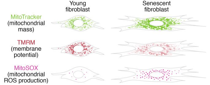
Mitochondrial dysfunction and cell senescence are hallmarks of aging and are closely interconnected. Mitochondrial dysfunction, operationally defined as a decreased respiratory capacity per mitochondrion together with a decreased mitochondrial membrane potential, typically accompanied by increased production of oxygen free radicals, is a cause and a consequence of cellular senescence and figures prominently in multiple feedback loops that induce and maintain the senescent phenotype. Here, we summarize pathways that cause mitochondrial dysfunction in senescence and aging and discuss the major consequences of mitochondrial dysfunction and how these consequences contribute to senescence and aging. We also highlight the potential of senescence-associated mitochondrial dysfunction as an antiaging and antisenescence intervention target, proposing the combination of multiple interventions converging onto mitochondrial dysfunction as novel, potent senolytics.
Overview
The claims and statistics of this paper haven’t been checked yet.
Free: it reads the open-access text and checks the first 5 claims that cite a source we can check. Create a free account to check every claim.
Integrity signals
Registry
Cites
Statistics
Figures

Verification
Claims the paper makes
The claims and statistics of this paper haven’t been checked yet.
Free: it reads the open-access text and checks the first 5 claims that cite a source we can check. Create a free account to check every claim.
Claims other papers make about this one
Each is checked against this paper’s own text: does it say what the citing paper says it does?
The source backs part of the claim, not all of it — or backs it for a different population, condition or comparison.
In the citing paper
“Both proliferation-competent glial cells (which constitute approximately 50% of brain cells) and postmitotic neurons can be positive for markers of cellular senescence in aging, obesity, traumatic injury, and neurodegeneration31,45–49, among other conditions.”
From Guidelines for minimal information on cellular senescence experimentation in vivo (2024) · cites this paper as [45]
Experimental — accuracy not yet measuredThe source backs part of the claim, not all of it — or backs it for a different population, condition or comparison.
In the citing paper
“Therefore, the dysregulation of mitochondrial fusion and fission pathways in senescent astrocytes likely contributes to mitochondrial dysfunction, oxidative stress, and the overall senescence-associated phenotype observed in these cells (Ma et al., 2020; Kleele et al., 2021; Miwa et al., 2022).”
From Aging promotes an increase in mitochondrial fragmentation in astrocytes (2024) · cites this paper as Miwa et al., 2022
Experimental — accuracy not yet measuredThe source backs part of the claim, not all of it — or backs it for a different population, condition or comparison.
In the citing paper
“Dysfunctional mitochondrial dynamics, as observed here, particularly excessive mitochondrial fission, have been associated with oxidative stress, mitochondrial DNA damage, and cellular senescence in various cell types (Twig et al., 2008; Miwa et al., 2022).”
From Aging promotes an increase in mitochondrial fragmentation in astrocytes (2024) · cites this paper as Miwa et al., 2022
Experimental — accuracy not yet measuredThe cited source says what the claim attributes to it.
In the citing paper
“while the presence of senescence markers has been proven even in post-mitotic cells”
From Guidelines for minimal information on cellular senescence experimentation in vivo (2024) · cites this paper as [45]
The check wasn’t confident either way, so it gives no verdict — never disputed.
In the citing paper
“Evidence from the literature indicates that senescent cells exhibit an increase in oxygen consumption rate (Hutter et al., 2004; Son et al., 2017; Miwa et al., 2022; Bang et al., 2023; Diniz et al., 2024a).”
From Aging promotes an increase in mitochondrial fragmentation in astrocytes (2024) · cites this paper as Miwa et al., 2022
Experimental — accuracy not yet measured
2 more claims about this paper
Create a free account to see every claim other papers make about this one.
Self-check
Percentages, sums and reported differences re-computed from the paper’s own numbers.
The self-check needs the paper’s full text.
Citations
Cites
The serial cultivation of human diploid cell strains
Hayflick, L., & Moorhead, P. S. (1961) · Experimental Cell Research · doi:10.1016/0014-4827(61)90192-6 · Cited by 8,508 · cited 1×
“Introduction What is cell senescence? Cell senescence was originally defined as the irreversible loss of replicative potential of primary cells in culture (1), initiated as a persistent DNA damage response to dysfunctional telomeres (2).”
BackgroundTelomeres shorten during ageing of human fibroblasts
Harley, C. B., Futcher, A. B., & Greider, C. W. (1990) · Nature · doi:10.1038/345458a0 · Cited by 5,750 · cited 2×
“Telomere dysfunction might be caused by telomere shortening during DNA replication (3, 4), replication-independent telomere damage (5), or a variety of other stresses (6).”
BackgroundA role for mitochondria in nlrp3 inflammasome activation
Zhou, R., Yazdi, A. S., Menu, P., & Tschopp, J. (2011) · Nature · doi:10.1038/nature09663 · Cited by 5,602
The senescence-associated secretory phenotype: the dark side of tumor suppression
Coppé, J.-P., Desprez, P.-Y., Krtolica, A., & Campisi, J. (2010) · Annual Review of Pathology: Mechanisms of Disease · doi:10.1146/annurev-pathol-121808-102144 · Cited by 5,206
Wu, Z., Puigserver, P., Andersson, U., Zhang, C., Adelmant, G., Mootha, V., Troy, A., Cinti, S., Lowell, B., Scarpulla, R. C., et al. (1999) · Cell · doi:10.1016/s0092-8674(00)80611-x · Cited by 4,139 · cited 1×
“PGC-1α activates the transcription factors Nrf1/2 and ERRα, which further control the TFAM transcription in response to increased mitochondrial biogenesis (65).”
BackgroundClearance of p16Ink4a-positive senescent cells delays ageing-associated disorders
Baker, D. J., Wijshake, T., Tchkonia, T., LeBrasseur, N. K., Childs, B. G., van de Sluis, B., Kirkland, J. L., & van Deursen, J. M. (2011) · Nature · doi:10.1038/nature10600 · Cited by 3,670
AMPK regulates energy expenditure by modulating NAD+ metabolism and SIRT1 activity
Cantó, C., Gerhart-Hines, Z., Feige, J. N., Lagouge, M., Noriega, L., Milne, J. C., Elliott, P. J., Puigserver, P., & Auwerx, J. (2009) · Nature · doi:10.1038/nature07813 · Cited by 3,300
Cellular Senescence: Defining a Path Forward
Gorgoulis, V., Adams, P. D., Alimonti, A., Bennett, D. C., Bischof, O., Bishop, C., Campisi, J., Collado, M., Evangelou, K., Ferbeyre, G., et al. (2019) · Cell · doi:10.1016/j.cell.2019.10.005 · Cited by 3,225 · cited 1×
“Telomere dysfunction might be caused by telomere shortening during DNA replication (3, 4), replication-independent telomere damage (5), or a variety of other stresses (6).”
BackgroundNakahira, K., Haspel, J. A., Rathinam, V. A. K., Lee, S.-J., Dolinay, T., Lam, H. C., Englert, J. A., Rabinovitch, M., Cernadas, M., Kim, H. P., et al. (2011) · Nature Immunology · doi:10.1038/ni.1980 · Cited by 3,049
A DNA damage checkpoint response in telomere-initiated senescence
Fagagna, F. D. D., Reaper, P. M., Clay-Farrace, L., Fiegler, H., Carr, P., von Zglinicki, T., Saretzki, G., Carter, N. P., & Jackson, S. P. (2003) · Nature · doi:10.1038/nature02118 · Cited by 2,917 · cited 1×
“Introduction What is cell senescence? Cell senescence was originally defined as the irreversible loss of replicative potential of primary cells in culture (1), initiated as a persistent DNA damage response to dysfunctional telomeres (2).”
Background
Research works printed but not linked: 2
- von Zglinicki T. Mechanisms of cell senescence in aging. In: Musi N, Hornsby P eds. Handbook of the Biology of Aging. Academic Press; 2021:53–68.
- Miwa S, Johnson TE. Stress and aging. Vijg J, et al., eds. In: The Molecular and Cellular Biology of Aging. Gerontological Society of America; 2016:216-249.
Cited by
Items 701–800 of 999
Oxidative Stress and Cardiovascular Disease: Implication of Gender and Age
Karam, M., Itani, M. M., Faraj, M., Jaffa, A. A., Jelwan, J., Aldeen, K. S., & Jaffa, A. A. (2024) · Oxidative stress in applied basic research and clinical practice · doi:10.1007/978-3-031-62131-4_1 · Cited by 3
Li, X., Ma, Q., Huang, Y., Cheng, L., Liu, Y., Li, H., Zhan, H., Zhang, F., Liu, Y., & Li, Y. (2024) · Immunological Investigations · doi:10.1080/08820139.2024.2402824 · Cited by 1
“An increase in mitochondrial mass indicates mitochondrial dysfunction (Miwa et al., 2022).”
InfluentialImpacts of Senolytic Phytochemicals on Gut Microbiota: A Comprehensive Review
Kim, H. S., & Jung, C. H. (2024) · Journal of Microbiology and Biotechnology · doi:10.4014/jmb.2408.08032 · Cited by 1
Wang, H., Wang, L., Xiao, J., Cong, Y., Zong, S., Zhang, Y.-X., Liu, J., & Zhu, M. (2024) · Biological and Pharmaceutical Bulletin · doi:10.1248/bpb.b24-00537 · Cited by 1
Ruiz‐Velasco, A., Jouve, C., Deshayes, L., Kohlhaas, M., Maack, C., & Hulot, J. (2026) · Pediatric Research · doi:10.1038/s41390-026-05208-9 · Cited by 2
Zhang, Z., Luo, Y., Zhang, H., Zeng, Z., Zheng, W., Zhao, Y., Huang, Y., & Shen, L. (2025) · Journal of Ethnopharmacology · doi:10.1016/j.jep.2025.119593 · Cited by 1
Kalyoncu, M., Demirci, D., Eris, S., Dayanc, B., Çakıroğlu, E., Basol, M., Uysal, M., Cakan‐Akdogan, G., Liu, F., Öztürk, M., et al. (2025) · Molecular Oncology · doi:10.1002/1878-0261.70021 · Cited by 2
“S11F), suggesting potential metabolic alterations associated with the senescent state [43].”
Qiu, Y., Li, Y., Li, M., Wang, Y., Shen, M., Shao, J., Zhang, F., Xu, X., Wang, F., Zhang, Z., et al. (2025) · Chinese Journal of Natural Medicines · doi:10.1016/s1875-5364(25)60836-3 · Cited by 4
Mampay, M., Alhity, G., Rolle, S. O., Alzboon, W., Stewart, N. A., Flint, M. S., & Sheridan, G. K. (2025) · Journal of Neurochemistry · doi:10.1111/jnc.70052 · Cited by 1
Panico, F., Mirra, D., Petito, G., Spaziano, G., Vecchio, V. D., Esposito, R., Senese, R., Desiderio, V., Lanni, A., & D’Agostino, B. (2025) · Biomedicines · doi:10.3390/biomedicines13051014 · Cited by 1
“In this context, the mitochondrial mass is considered a well-established hallmark of aging [64].”
Zhang, Y., Shen, M., Zhang, B., Li, X., Cheng, H., Feng, D., Han, Y., Luo, Z., & Zhou, Y. (2025) · The FASEB Journal · doi:10.1096/fj.202403361r · Cited by 3
“Given that mitochondrial dysfunction plays an important role in the induction of senescence [28], and it has been demonstrated that mtROS levels increase during renal fibrosis [29].”
Impacts of aging on brain metabolism
Rodrigues, B. A., Santiago, T. C., & Silva, J. D. N. (2025) · Progress in brain research · doi:10.1016/bs.pbr.2025.05.009 · Cited by 4
Dehydroacteoside rejuvenates senescence via TVP23C-CDRT4 regulation
Lee, Y. J., Song, E. S., Lee, Y. H., Lee, K. S., So, B., Park, J. H., Yoon, J. H., Kim, D., Kim, M., Kwon, H. W., et al. (2025) · Experimental Gerontology · doi:10.1016/j.exger.2025.112800 · Cited by 1
“Next, we investigated whether mitochondrial MMP was impacted by TVP23C-CDRT4 knockdown.”
InfluentialChen, X., Wang, D., Zhou, Y., Li, L., Zhu, T., Zhao, R., Lu, M., & Mu, B. (2025) · Molecular Biology Reports · doi:10.1007/s11033-025-10665-2 · Cited by 3
Pan, Y., Liu, H., Chen, J., Deng, H., Yang, C., Huang, Y., Cai, Q., Huang, W., Huang, M., Xiong, S., et al. (2025) · Biogerontology · doi:10.1007/s10522-025-10271-6 · Cited by 1
“To view a copy of this licence, visit http://creativecom-mons.org/licenses/by-nc-nd/4.0/.”
InfluentialXu, Y., Zhang, W., Xu, W., Li, S., Zhang, D., Lin, X., Liu, J., Fu, Q., Pei-jie, H., & Si, H. (2025) · Journal of Pharmaceutical Analysis · doi:10.1016/j.jpha.2025.101432 · Cited by 1
“As essential organelles responsible for energy production, mitochondrial dysfunction not only leads to diminished ATP generation but also contributes to impaired cellular signaling [41].”
Understanding Cellular Senescence and Senotherapeutics for Nutri-geriatric Solutions
Chawla, S., Bhattacharya, S., & Gupta, A. (2025) · Biomedical & Pharmacology Journal · doi:10.13005/bpj/3214 · Cited by 3
Suwitono, M. R., Wahyuniari, I. A. I., Luo, H., Suwitono, M. C., & Sulastri, T. (2025) · Ankara Universitesi Eczacilik Fakultesi Dergisi · doi:10.33483/jfpau.1588979 · Cited by 3
Ehara, Y., Mifune, Y., Inui, A., Yamaura, K., Shinohara, I., Kusunose, M., Tanaka, S., Nakabayashi, D., Matsumoto, T., & Kuroda, R. (2025) · Journal of Orthopaedic Research® · doi:10.1002/jor.70090 · Cited by 1
“Improvement in mitochondrial function is defined as an increase in MMP and respiratory capacity [31].”
Iacoban, A. C., Bacalum, M., Răileanu, M., Stoica, R., Culita, D. C., Radu, D., Alexandru-Dinu, A., Neațu, F., Rostas, A. M., & Vlaicu, I. D. (2025) · Applied Surface Science · doi:10.1016/j.apsusc.2025.165176 · Cited by 1
Chatzikalil, E., Bistas, K., Kymioni, V. M., Diamantopoulos, P. T., & Solomou, E. E. (2025) · Cells · doi:10.3390/cells14221788 · Cited by 1
“Dysregulation of mitochondrial dysfunction and cell senescence are considered key features of aging and are interconnected [190].”
InfluentialZhong, S., Wang, X., Guo, Y., Chen, J., Lin, Q., Wen, S., Chen, Y., & He, S. (2025) · Acupuncture & Electro-Therapeutics Research · doi:10.1177/03601293251356851 · Cited by 1
“Research on the causes and mechanisms of aging in recent years has shown that oxygen-free radicals in cells and tissues may be the main factor in accelerating aging (Hu et al., 2022; Miwa et al., 2022).”
Balkrishna, A., Gohel, V., Singh, A., Pathak, N., Tomer, M., Dev, R., & Varshney, A. (2025) · Fitoterapia · doi:10.1016/j.fitote.2025.107000 · Cited by 2
Wang, X., Wen, K., Li, Y., He, Y., Shen, W., & Wang, H. (2025) · Neuroepidemiology · doi:10.1159/000549912 · Cited by 1
Polystyrene nanoplastics induce ovarian granulosa cell senescence via autophagy suppression
Hu, H., Ouyang, H., Ma, L., Xu, Q., Peng, Y., Meng, M., & Zhang, T. (2025) · NanoImpact · doi:10.1016/j.impact.2025.100607 · Cited by 1
Gerashchenko, G. V., & Tukalo, M. A. (2025) · Biopolymers and Cell · doi:10.7124/bc.000b25 · Cited by 1
Xiao, H., Chen, Y., Liu, X., Chen, R., Yang, C., Yang, F., Chen, C., Geng, B., & Xia, Y. (2025) · Biology · doi:10.3390/biology14121801 · Cited by 2
“…hypoxic-osteogenic microenvironment; nevertheless, some hypotheses, such as the senescence state through the p21 pathway, mitochondrial dysfunction, and ROS metabolism, are not well-developed at the level of the molecular mechanisms that can explain the involvement of hypoxia in osteoporosis [15].”
T-cell apoptosis is more susceptible to overactivated neutrophils delivered-NETs-MPO/NE complex
Lu, C., Shao, L., Li, H., Gao, X., Mei, Y., & Sun, B. (2025) · International Immunopharmacology · doi:10.1016/j.intimp.2025.116100 · Cited by 1
The biology of PKM2 in the metabolism and senescence in diabetic kidney disease
Kayes, M. I., & Kanasaki, K. (2025) · Journal of Diabetes Investigation · doi:10.1111/jdi.70231 · Cited by 1
“Mitochondrial dysfunction under diabetic conditions further increases reactive oxygen species (ROS), reinforcing senescence 14 , while epigenetic alterations sustain the senescent phenotype and SASP output 15 .”
Li, L., Huang, X., & Li, P. (2026) · Basic & Clinical Pharmacology & Toxicology · doi:10.1111/bcpt.70188 · Cited by 1
Zhang, E., Wan, Z., Ma, Y., Li, Y., Zhou, Q., Cai, B., Li, Y., Li, F., Ying, B., & Yan, L. (2026) · Frontiers in Immunology · doi:10.3389/fimmu.2025.1721097 · Cited by 1
“To establish the physiological relevance of mitochondrial indicators, we fi rst examined their association with age in the entire cohort of HC (N=44), given the known impact of immunosenescence on immune cell metabolism and mitochondrial dynamics (24 – 26).”
Oligodendrocytes at the crossroads: Central players and interactive partners in white matter aging
Zhang, X., Yu, P., Chen, Y., Xu, Y., Xu, R., Yang, X., Lv, S., Shang, H., & Hu, Y. (2026) · Ageing Research Reviews · doi:10.1016/j.arr.2026.103034 · Cited by 3
Wolf, S. T., Bangle, J. F., Jennings, W. E., Albino, G. R., Gorejena, M., Clary, B. L., Heo, J., & Call, J. A. (2026) · American Journal of Physiology-Regulatory, Integrative and Comparative Physiology · doi:10.1152/ajpregu.00262.2025 · Cited by 1
Jiang, P., Jia, Y., Zhang, J., Wang, Z., Ma, H., Guo, Y., Wang, M., Yan, W., & Xi, X. (2026) · Medicine · doi:10.1097/md.0000000000047376 · Cited by 1
“[3,4] Cell senescence is a state of permanent proliferative arrest that cells enter after experiencing various stress factors such as DNA damage, oxidative stress, and telomere shortening.”
Sobolewski, P., Koper, M., Wasążnik-Jędras, A., Kołos, M., & Walecka, I. (2026) · International Journal of Molecular Sciences · doi:10.3390/ijms27031520 · Cited by 1
“Several molecular markers have been proposed as indicators of mitochondrial dysfunction–associated senescence (MiDAS) in keratinocytes [32].”
Supplementation with Lycium barbarum glycopeptide (LbGP) rescues the quality of aged oocytes
Guo, A., Shi, R., Liu, S., Zhang, Y., Mao, Y., Yan, G., & Cao, G. (2026) · Journal of Ethnopharmacology · doi:10.1016/j.jep.2026.121318 · Cited by 2
Rodriguez, M. R., Woessner, A. E., & Quinn, K. P. (2026) · Advances in Wound Care · doi:10.1177/21621918261421628 · Cited by 2
Zhong, H., Bao, S., Cao, W., He, X., & Ban, Z. (2026) · Tobacco Induced Diseases · doi:10.18332/tid/216136 · Cited by 1
“It also impairs mitochondrial function, increasing ROS generation, mitochondrial dysfunction is a central driver of aging biology 30 .”
Li, Y., Hu, X., & Zhou, X. (2026) · Blood Cancer Discovery · doi:10.1158/2643-3230.bcd-25-0338 · Cited by 1
Li, X., Chen, M., Zhang, Y., Li, J., Xiang, L., Xiao, Y., Xiang, Y., Chen, L., Ran, Q., & Li, Z. (2026) · Current Issues in Molecular Biology · doi:10.3390/cimb48020196 · Cited by 3
“Therefore, mitochondrial dysfunction is both a hallmark and a driver of cellular senescence and aging [70].”
Zheng, X., Huang, R., Liu, Z., Liu, T., Lin, H., Yin, L., Wu, Q., & Zhao, M. (2026) · Bioengineering · doi:10.3390/bioengineering13030256 · Cited by 2
“…control such as insufficient mitochondrial biogenesis and defective mitophagy/mitochondrial clearance can drive sustained ROS generation, forming a feed-forward “ROS–mitochondrial damage–ROS” loop that shifts senescence from a transient stress response to a long-term steady-state alteration [6].”
Targeted therapies in lung cancer: personalizing treatment across the age spectrum
Xu, Y., Chen, F., Zhang, H., & Wang, X. (2026) · Frontiers in Oncology · doi:10.3389/fonc.2026.1743620 · Cited by 1
“…intracellular changes are ampli fi ed by tumor-derived factors that manipulate immune signaling through cAMP/PKA-CREB, NF-k B, and cGAS-STING pathways, further suppressing T-cell activation and skewing immune cell metabolism toward glycolysis, reducing the durability of antitumor immunity (66, 67).”
Microglial Senescence in Neurodegenerative Diseases: Mechanisms and Therapeutic Perspectives
Liu, Y., Hu, Z., Xiao, Y., Han, X., Cheng, X., & Ye, T. (2026) · ACS Chemical Neuroscience · doi:10.1021/acschemneuro.6c00016 · Cited by 1
Escitalopram accelerated aging via impaired IIS/autophagy signaling pathway
Chen, Z.-Y., Qi, C.-Y., Ji, F., Xue, K., Quan, H.-B., Zhou, J., Liu, W., & Lu, G.-D. (2026) · GeroScience · doi:10.1007/s11357-026-02190-2 · Cited by 1
Sun, Y., Fu, X., Li, Y., Peng, M., & Yang, L. (2026) · Molecular Biology of the Cell · doi:10.1091/mbc.e25-11-0560 · Cited by 1
Ou, L., Cui, B., Zhou, L., Liu, K., Shi, L., Zhou, T. G., Zeng, L., Zhou, H., Xu, H., Zheng, Y., et al. (2026) · Investigative Ophthalmology & Visual Science · doi:10.1167/iovs.67.3.11 · Cited by 1
“15,22 In dysfunctional mitochondria, impaired electron transport chain activity leads to NADH accumulation and a collapsed NAD + /NADH ratio.”
Zheng, L., Huang, X., Peng, Z., Qian, S., Lu, Y., Lin, L., & Su, X. (2026) · Experimental Cell Research · doi:10.1016/j.yexcr.2026.114973 · Cited by 1
Cheng, J., Xia, Y., Ji, Z., Xu, L., Gao, R., Xiong, Z. L., Huang, L., Zhang, X., Ding, W., Sun, Y., et al. (2026) · Journal of Neurochemistry · doi:10.1111/jnc.70402 · Cited by 1
Hiki, M. (2026) · Medical Hypotheses · doi:10.1016/j.mehy.2026.111951 · Cited by 1
Wang, C., Lu, J., Fu, H., Feng, X., Xu, Z., Luo, P., Yang, B., He, Q., & Yang, X. (2026) · Phytotherapy Research · doi:10.1002/ptr.70310 · Cited by 1
“Emerging evidence strongly suggests a significant correlation between inflammation and apoptosis (Miwa et al. 2022; Pei et al. 2024).”
Lee, C. H., Nyakuma, B. B., Zhou, T., Xue, J., Cheong, T. H., Li, Y., Wong, S. L., Gao, L., & Wu, X. (2026) · Food Research International · doi:10.1016/j.foodres.2026.119189 · Cited by 1
Redox rhythms promote fitness by modulating ageing-dependent reprogramming
Wang, X., Cui, S.-S., Li, X.-K., Qu, S., Chang, W., Tang, A., Jin, Y., Peng, S.-J., Wang, H.-P., Fang, X.-Q., et al. (2026) · Nature Metabolism · doi:10.1038/s42255-026-01515-x · Cited by 2
“Mitochondrial dysfunction is a hallmark of skeletal muscle ageing, characterized by reductions in both quantity and quality that accelerate sarcopenia 35,36 .”
Wang, Z., Jiang, X., Lin, J., An, R., He, Y., & Li, S. (2026) · Antioxidants · doi:10.3390/antiox15040515 · Cited by 2
“Elevated ROS levels significantly diminish the MMP, although it can recover upon the removal of ROS [56,57].”
Li, J., Chen, G., & Bao, X. (2026) · Inflammation · doi:10.1007/s10753-026-02514-6 · Cited by 1
Gao, X., Pan, Y., Liu, Q., & Wu, H. (2026) · Frontiers in Cardiovascular Medicine · doi:10.3389/fcvm.2026.1748411 · Cited by 3
“Aging is associated with an increased prevalence of mitochondrial dysfunction, as evidenced by studies on aging-related mitochondrial dysfunction and arrhythmia following MI (78, 79).”
New insights into Rett syndrome pathogenesis: defining the role of MEPC2 in DNA damage
Mansooralavi, N., & Lowry, W. E. (2026) · Frontiers in Neurology · doi:10.3389/fneur.2026.1711771 · Cited by 1
Contact-electro-catalysis in biological applications
Hua, L., Irwin, M. G., & Wang, S. (2026) · Nano Energy · doi:10.1016/j.nanoen.2026.112052 · Cited by 2
Ma, Y., Chen, J., Song, R., & Yu, X. (2026) · Molecular Medicine · doi:10.1186/s10020-026-01503-7 · Cited by 2
“Aging is fundamentally a process of progressive metabolic decline (Sun et al. 2025), characterized by mitochondrial dysfunction (Miwa et al. 2022), disrupted proteostasis (Ding et al. 2025), and chronic low-grade inflammation (Fu et al. 2025).”
Xin, Y., He, Y., & Liu, X. (2026) · Burns & Trauma · doi:10.1093/burnst/tkag037 · Cited by 1
Nguyen, H. V. M., Hu, F., Shakya, E., Fender, C. L., Lee, K., Schindler, J., García‐Jaramillo, M., Ahsan, N., Siebert, J., Borengasser, S. J., et al. (2026) · The FASEB Journal · doi:10.1096/fj.202502189rrr · Cited by 1
Cell senescence in chronic pain: An integrative perspective on cellular and neural mechanisms
Ma, L., Wang, Y., Wang, J., Li, Y., Hu, X., Zou, X., Zhang, D., Zhu, T., & Zhou, C. (2026) · Neuroscience & Biobehavioral Reviews · doi:10.1016/j.neubiorev.2026.106789 · Cited by 3
Zhao, M., 吴巧芳, Ding, Y., Lu, C., Du, Y., Lin, X., Bi, L., Wang, C., Ji, J., Sun, W., et al. (2026) · BMC Medicine · doi:10.1186/s12916-026-04967-w · Cited by 2
“Furthermore, previous studies have shown that impaired mitochondrial OXPHOS is a common characteristic of senescent cells [53].”
Huang, J., Wang, A., Li, X., Huang, P., Zeng, K., Sun, L., Li, M., Chen, Y., & Wang, J. (2026) · Cell Proliferation · doi:10.1111/cpr.70237 · Cited by 1
Targeting Mitochondria in Aging‐Related Diseases: Therapeutic Potential and Obstacles
Xiang, Z., Chen, Y., Liu, X., Lu, H., Yang, Y., Xing, L., Zhang, Y., Lang, C., Zhang, S., Zhao, S., et al. (2026) · MedComm · doi:10.1002/mco2.70790 · Cited by 4
“Organismal aging is a multifaceted, gradual process marked by the step-by-step decline of cellular and systemic functions, as well as the deterioration of various tissues, resulting in compromised function and heightened mortality risk [ 3 ].”
Chen, Y., Li, W., Zilong, M. E. N. G., Li, H., Chen, Y., Fan, J., & Zhu, M. (2026) · Reproductive Biology · doi:10.1016/j.repbio.2026.101238 · Cited by 1
Senotherapeutics: Emerging pharmacological strategies to target cellular senescence
Nagy, D., Brunner, E., Ekambaram, S., Nagaraja, R. Y., Kordestan, K. V., Patai, R., Kristóf, R., Nagykaldi, M., Chandragiri, S. S., Shanmugarama, S., et al. (2026) · Advances in Translational Research · doi:10.1556/1661.2026.00015 · Cited by 3
Wang, Y., Zhang, C., Li, X. Y., Li, S. X., Jiang, W., Duan, B., Liu, X., Zhou, S., & Zhang, Y. (2026) · Journal of Gastroenterology and Hepatology · doi:10.1111/jgh.70480 · Cited by 1
“…upregulation, consistent with known crosstalk: Mitochondrial dysfunction induces senescence via ROS, and senescence worsens mitochondrial damage [36]; for example, iron chelation-induced senescence models show that reduced hepatic mitochondrial complex II activity precedes p27kip1-mediated…”
Quercetin Mitigates Oxidative Stress-Induced Premature Senescence in SH-SY5Y Neuronal-like Cells
Salamone, F. L., Molonia, M. S., Trischitta, S., Saija, A., Cimino, F., & Speciale, A. (2026) · International Journal of Molecular Sciences · doi:10.3390/ijms27135759 · Cited by 4
Han, Q., Han, Q., Deng, H., Alifu, A., Li, J., Chen, Z., Deng, L., Cai, X., Chen, Y., & Lin, Y. (2026) · Biomaterials · doi:10.1016/j.biomaterials.2026.124425 · Cited by 2
Cellular Senescence and Aging: Mechanisms, Disease Convergence, and Therapeutic Frontiers
Cai, G., Ren, J., Wang, L., Wu, Y., Wang, H., Cao, X., Shang, H., Hou, X., Wang, Y., Xue, H., et al. (2026) · MedComm · doi:10.1002/mco2.70827 · Cited by 2
“Mitochondrial dysfunction, a canonical aging hallmark, manifests primarily as impaired oxidative phosphorylation and bioenergetic failure [ 53 ].”
Nuclear‐Mitochondria Crosstalk in Senescent Adipose‐Derived Stem Cells
Zhang, Y., Du, Y., Ling, S., Wang, H., He, J., Li, J., Huo, K., Liu, B., Li, Q., Wei, J., et al. (2026) · Smart Medicine · doi:10.1002/smmd.70043 · Cited by 1
“Oxidative damage, replication errors, and insufficient repair mechanisms are major factors in the accumulation of mtDNA mutations [94–96]. mtDNA mutations can lead to electron transport chain (ETC) dysfunction, decrease ATP production and increase ROS production, which further exacerbates oxidative…”
Dong, J. I. A. N. Z. E., Yan, B., Zhao, X., Bai, J., Shan, H., Gao, X., Sheng, L., Dai, J., Jiang, F., Zhang, M., et al. (2026) · Journal of Orthopaedic Translation · doi:10.1016/j.jot.2026.101171 · Cited by 1
“Previous studies have shown that mitochondrial dysfunction drives cell senescence and that cell senescence has a negative feedback effect on mitochondrial function through the secretion of SASP components and cell cycle arrest [59 – 62].”
Extracellular vesicles in senescence-associated chronic lung diseases
Wang, R., Chen, Y., Liu, X., & Barnes, P. J. (2026) · Chinese Medical Journal · doi:10.1097/cm9.0000000000004208 · Cited by 1
Li, J., Zhao, X., Shen, Y., Huang, L., Guo, J., & Wang, J. (2026) · Biochimica et Biophysica Acta (BBA) - Molecular Basis of Disease · doi:10.1016/j.bbadis.2026.168388 · Cited by 2
Tong, A., Zhou, Y., Ding, Y., Zhao, Y., Han, C., Wei, L., & Zhao, J. (2026) · Materials Today Bio · doi:10.1016/j.mtbio.2026.103514 · Cited by 1
The Physical Senotype in cellular senescence: Mechanical and physical strategies for senotherapy
Wang, B., Qi, X., Huard, J., & Mu, X. (2026) · Ageing Research Reviews · doi:10.1016/j.arr.2026.103283 · Cited by 1
Müller, L., Benedetto, S. D., & Müller, V. (2026) · Frontiers in Cellular and Infection Microbiology · doi:10.3389/fcimb.2026.1896480 · Cited by 1
“Mitochondria regulate not only cellular energy production but also redox balance, innate immune signaling, apoptosis, and in fl ammatory responses (Miwa et al., 2022; Lopez-Otin et al., 2023).”
Lee, Y. J., Yoon, J. H., Lim, H., Song, E. S., Park, J. H., Kim, D., Yang, J.-E., So, M. K., Kim, M., Song, J., et al. (2026) · Antioxidants · doi:10.3390/antiox15081006 · Cited by 1
“Mitochondrial mass was significantly higher in senescent than in young fibroblasts, consistent with the accumulation of damaged or dysfunctional mitochondria during cellular senescence [53] (Figures 4B and 9B).”
Tang, R., Yin, J., Li, S., Wang, S., Fei, J., & Zhou, Z. (2026) · Experimental Gerontology · doi:10.1016/j.exger.2026.113276 · Cited by 2
Zhang, X., Yin, T., Cheng, Y., 常英新, Guo, Z., Liu, J., Shi, J., Tan, K., Yang, J., Guo, L., et al. (2026) · Ageing Research Reviews · doi:10.1016/j.arr.2026.103318 · Cited by 1
Zheng, Y., Chen, C. F., Li, X., Li, Z., Li, H., Tan, M., Zhai, D., Zhang, F., Lü, W., 陈志华, et al. (2026) · 中国科学通报:英文版 · doi:10.1016/j.scib.2026.08.050 · Cited by 1
Mitochondrial homeodynamics in ageing: mechanisms, resilience, and interventions
Chmielewski, P. P. (2026) · Biogerontology · doi:10.1007/s10522-026-10506-0 · Cited by 2
“…cardiomyocytes, and skeletal muscle, impaired proteostasis and organelle turnover can progressively increase the fraction of mitochondria that are structurally intact but functionally constrained (Affourtit and Carré 2024; Miwa et al. 2022; Moehle et al. 2019; Ravindran and Gustafsson 2025).”
Targeting cellular senescence as a therapeutic approach in non-alcoholic steatohepatitis
Nakano, Y., & Johmura, Y. (2023) · Annals of Hepatology · doi:10.1016/j.aohep.2023.100900 · Cited by 2
Protective Effect of SGLT2 Inhibitor on D-Galactose-Induced Senescence in Mice and Its Mechanism
Ma, Z., Xu, M., Jin, K., Zhu, G., & Chen, X. (2023) · Journal of Biosciences and Medicines · doi:10.4236/jbm.2023.115021 · Cited by 2
The role of cellular lipid metabolism in aging
Chen, X., & Liu, J. (2023) · Elsevier eBooks · doi:10.1016/b978-0-323-95582-9.00013-9 · Cited by 3
Garrod-Ketchley, C., d’Algue, L. M., Littlewood, K., Hood, G., Worthington, A., Pattrick, M., Sutcliffe, C., Valla, Z., Joti, N., Zalak, U., et al. (2023) · Discovery Immunology · doi:10.1093/discim/kyad026 · Cited by 2
Luo, M., Yuan, Q., Xie, Y., Mai, M., Song, W., Wang, Y., Shi, H., Xia, E.-Q., & Guo, H. (2024) · The Journals of Gerontology Series A · doi:10.1093/gerona/glae014 · Cited by 3
Mitochondria in aging: A review of structure, function, and interorganelle relationships
Chen, M. (2024) · Theoretical and Natural Science · doi:10.54254/2753-8818/29/20240749 · Cited by 1
Effect of Ficus Carica Leaves Extract on the Hippocampus of the Aged Rats
Zeair, E. A., Zolfakar, A. S., El-Kholy, W. B., & Faried, M. A. (2024) · Menoufia Medical Journal · doi:10.59204/2314-6788.1110 · Cited by 1
Lu, Y., Han, S., Hu, J. R., Lv, K., Ruan, Y., Cheng, G., Zhang, J., Wu, X., Weng, Z., & Zhou, X. (2025) · IUBMB Life · doi:10.1002/iub.70001 · Cited by 1
“34 Senescent cells contribute to the tumor heterogeneity of pancreatic cancer.”
Rana, K. S., Marwah, M. K., Raja, F. N. S., Dias, I. H. K., Hindalekar, Y. S., Tahan, M. A. A., Brown, J. E. P., & Bellary, S. (2025) · Journal of Receptors and Signal Transduction · doi:10.1080/10799893.2025.2475441 · Cited by 1
“Dysregulated mitochondria decreases respiratory capacity per mitochon-drion and membrane potential, typically accompanied by increased production of oxygen free radicals [40].”
Tanaka, S., Mifune, Y., Inui, A., Yamaura, K., Furukawa, T., Kato, T., Kusunose, M., Matsumoto, T., Matsushita, T., & Kuroda, R. (2025) · Journal of Orthopaedic Research® · doi:10.1002/jor.26064 · Cited by 1
Hwang, H. J., Amrit, F. R. G., Berry, B. J., Croix, C. M. S., Wojtovich, A. P., Shiva, S., & Ghazi, A. (2025) · Journal of Biosciences · doi:10.1007/s12038-025-00539-x · Cited by 1
“Mitochondria play crucial roles in mediating the link between reproduction and longevity as they regulate energy production, metabolic homeostasis and signaling, which are essential for reproductive fitness, stress response and healthy longevity (Miwa et al .”
T Cell Aging: An Important Target for Perioperative Immunomodulation
Lan, H., Xu, S., Li, H., Guo, R., Feng, Z., & Wang, Y. (2025) · Clinical Interventions in Aging · doi:10.2147/cia.s519438
“24 Besides inducing progressive changes in mitochondrial morphology, activity, and signaling function, these factors could also cause telomere wear and reduced enzyme repair activity, leading to decreased telomere length in T cells.”
Song, D., Hong, T., Song, J., Bazer, F. W., Song, G., Jeong, W., & Lim, W. (2025) · Food and Chemical Toxicology · doi:10.1016/j.fct.2025.115622 · Cited by 1
Metabolic Effects of Healing Touch During Cervical Cancer Treatment: An Exploratory Analysis
van Wietmarschen, H., Área-Gómez, E., Picard, M. A., Goodheart, M. J., Sood, A. K., & Lutgendorf, S. K. (2025) · Integrative Cancer Therapies · doi:10.1177/15347354251367793 · Cited by 1
“…(NAD + , of which kynurenine is a precursor), which decreases with aging and whose supplementation improves resilience to age-related deterioration and mitochondrial respiratory capacity in animal studies (reviewed in Castro-Portuguez and Sutphin, 48 Mitchell et al., 49 and Miwa et al. 50 ).”
Grapentine, S., Agarwal, P., Dolinsky, V. W., & Bakovic, M. (2022) · Research Square · doi:10.21203/rs.3.rs-2143064/v1 · Cited by 1
Fielder, E., Ishaq, A., Low, E., Laws, J., Calista, A., Castle, J., von Zglinicki, T., & Miwa, S. (2023) · SSRN Electronic Journal · doi:10.2139/ssrn.4631509 · Cited by 1
Self-citationShi, J., Shringarpure, S., Hinds, D. A., Auton, A., & Holmes, M. V. (2024) · medRxiv · doi:10.1101/2024.07.24.24310950 · Cited by 2
Metadata
- Authors
- Miwa, S., Kashyap, S., Chini, E., & von Zglinicki, T.
- Affiliations
- Newcastle University (GB)
- Mayo Clinic (US)
- Venue
- Journal of Clinical Investigation
- Published
- 2022-07-01
- Volume
- 132
- Issue
- 13
- ISSN
- 0021-9738 (ISSN-L)
- 1558-8238
- PMC
- PMC9246372
- OpenAlex
- W4283723802
- Cited by
- 1,061
- References
- 145
- Citations by year
- 2026: 294 · 2025: 348 · 2024: 263 · 2023: 136 · 2022: 20
- FWCI
- 19.97
- Type
- journal article
- Language
- en
- Open access
- gold
- doi.org
- Licence
- Locations
- Journal of Clinical Investigation (publishedVersion, cc-by)
- PubMed Central (publishedVersion, cc-by)
- PubMed (publishedVersion)
- DOAJ (DOAJ: Directory of Open Access Journals) (submittedVersion)
- Newcastle University ePrints (Newcastle Univesity) (cc-by)
- Funders
- Glenn Foundation for Medical Research
- Ted Nash Long Life Foundation
- Cancer Research UK (C12161/A24009)
- Newcastle University
- Fondation pour la Recherche Médicale
- Versus Arthritis
- National Institutes of Health (H2020, AG26094, CA233790, AG58812)
- Directorate for Biological Sciences
- Horizon 2020 Framework Programme (857524)
- Medical Research Council (MR/P020941/1)
- Biotechnology and Biological Sciences Research Council (BB/S006710/1)
- National Institute on Aging (AG-58812, R01AG058812, AG-26094)
- National Cancer Institute (R01CA233790)
- Topics
- Life Sciences › Biochemistry, Genetics and Molecular Biology › Molecular Biology › Mitochondrial Function and Pathology
- Life Sciences › Biochemistry, Genetics and Molecular Biology › Cell Biology › Telomeres, Telomerase, and Senescence
- Life Sciences › Biochemistry, Genetics and Molecular Biology › Aging › Genetics, Aging, and Longevity in Model Organisms
- Keywords
- mitochondrial dysfunction, cellular senescence, aging, mitochondrial membrane potential, respiratory capacity, reactive oxygen species, senolytics
- MeSH terms
- Mitochondria, Phenotype, Cellular Senescence
About these checks
Registry, retraction, Cites and statistics results are computed, not judged, with one exception: a printed reference that no index lists is read once by a model before it counts as a citation error. Claims are extracted by a model and kept only when their quote appears verbatim in the text; each cited source is then checked, with a model’s help, for whether it says what the claim relies on. Those verdicts can be wrong — read the claim’s quote and the cited source before relying on one.
Statistics and arithmetic are recomputed, not judged.
Confidence bands are shown only for fields calibrated on our annotated test set. None is calibrated yet, so no band is shown.
What counts as a claim ›How we measure confidence ›How we check this ›
Are you an author, or do you see a problem with this page? Request a review
Data sources
Bibliographic data: Semantic Scholar, OpenAlex, Crossref.
Contains information from the Semantic Scholar Academic Graph Datasets, made available under the ODC Attribution License (ODC-BY 1.0).
Semantic Scholar’s data is described in: Kinney, R., et al. (2023). The Semantic Scholar Open Data Platform. arXiv:2301.10140.
