

The DOI matches its Crossref record; 78 of the 79 references it cites were found in a registry or an index.
Microglial Interactions with Synapses Are Modulated by Visual Experience
Open accessAbstract
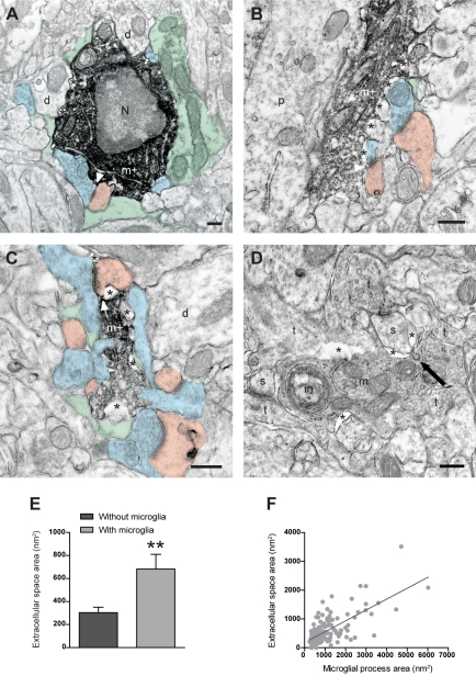
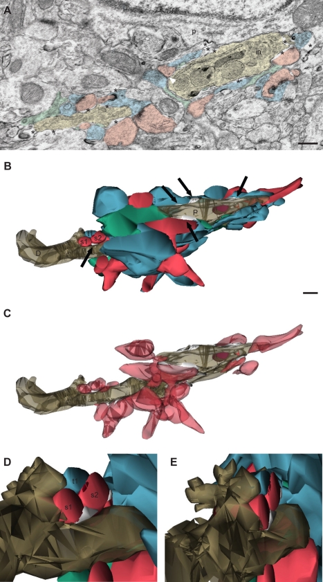
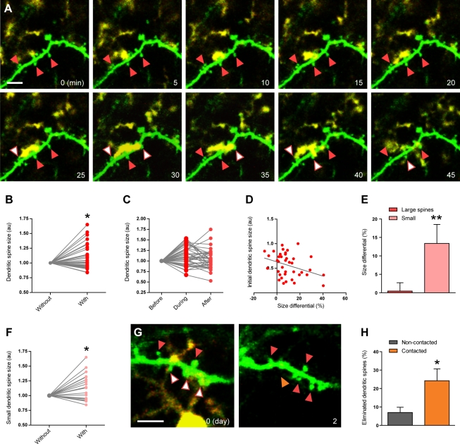
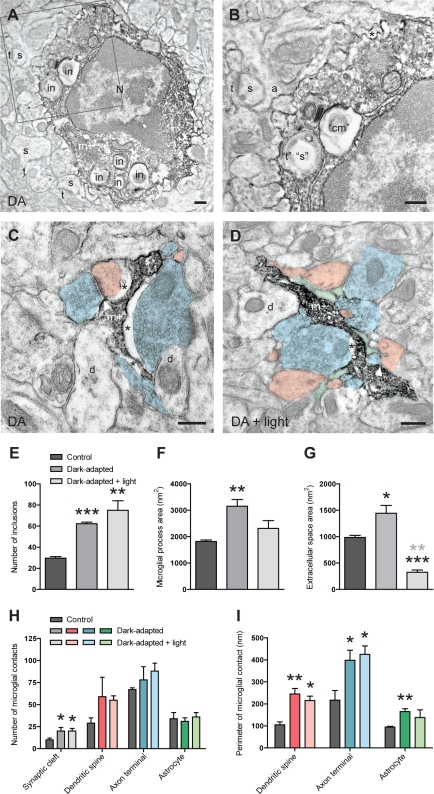
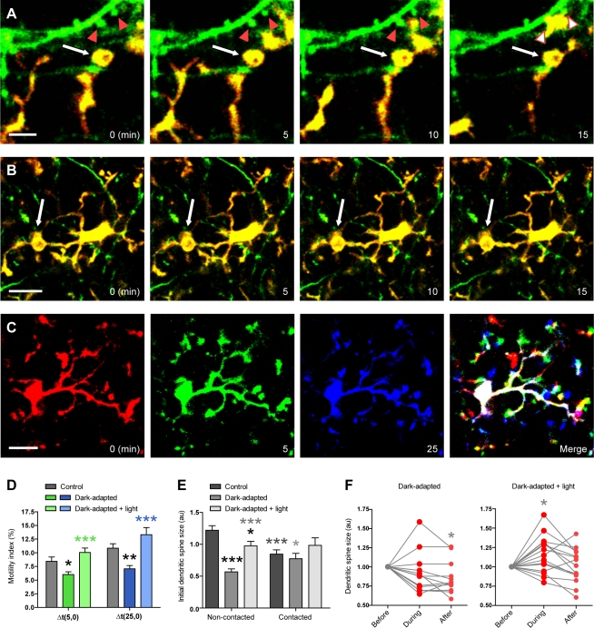
Microglia are the immune cells of the brain. In the absence of pathological insult, their highly motile processes continually survey the brain parenchyma and transiently contact synaptic elements. Aside from monitoring, their physiological roles at synapses are not known. To gain insight into possible roles of microglia in the modification of synaptic structures, we used immunocytochemical electron microscopy, serial section electron microscopy with three-dimensional reconstructions, and two-photon in vivo imaging to characterize microglial interactions with synapses during normal and altered sensory experience, in the visual cortex of juvenile mice. During normal visual experience, most microglial processes displayed direct apposition with multiple synapse-associated elements, including synaptic clefts. Microglial processes were also distinctively surrounded by pockets of extracellular space. In terms of dynamics, microglial processes localized to the vicinity of small and transiently growing dendritic spines, which were typically lost over 2 d. When experience was manipulated through light deprivation and reexposure, microglial processes changed their morphology, showed altered distributions of extracellular space, displayed phagocytic structures, apposed synaptic clefts more frequently, and enveloped synapse-associated elements more extensively. While light deprivation induced microglia to become less motile and changed their preference of localization to the vicinity of a subset of larger dendritic spines that persistently shrank, light reexposure reversed these behaviors. Taken together, these findings reveal different modalities of microglial interactions with synapses that are subtly altered by sensory experience. These findings suggest that microglia may actively contribute to the experience-dependent modification or elimination of a specific subset of synapses in the healthy brain.
Overview
The claims and statistics of this paper haven’t been checked yet.
Run the check — free, no account needed. It reads the open-access text and checks the sources it cites.
Integrity signals
Registry
Cites
Statistics
Figures
Verification
Claims the paper makes
The claims and statistics of this paper haven’t been checked yet.
Run the check — free, no account needed. It reads the open-access text and checks the sources it cites.
Claims other papers make about this one
Each is checked against this paper’s own text: does it say what the citing paper says it does?
In the citing paper
“Microglia have recently been implicated as active participants in brain plasticity per se (42–45).”
From Prolonged Period of Cortical Plasticity upon Redox Dysregulation in Fast-Spiking Interneurons (2015) · cites this paper as [42]
In the citing paper
“microglia participate in experience-dependent remodeling of neural circuits in the normal mature brain by removing extraneous synaptic elements [6, 74–77]”
From Microglial dysfunction in brain aging and Alzheimer's disease (2014) · cites this paper as [74]
In the citing paper
“Microglia also closely monitor neural activity and regulate synaptic plasticity.”
From Adult neurogenesis, glia, and the extracellular matrix (2019) · cites this paper as Tremblay et al., 2010
In the citing paper
“Microglia dynamically interact with neuronal circuits, making direct contacts with synaptic elements1819 in a way that is modulated by experience-dependent changes in neuronal activity19.”
From Microglial P2Y12 is necessary for synaptic plasticity in mouse visual cortex (2016) · cites this paper as [19]
In the citing paper
“The motility of microglial processes and spine contact are actively regulated by sensory experience and are involved in spine elimination (Tremblay et al., 2010).”
From Spatiotemporal dynamics of dendritic spines in the living brain (2014) · cites this paper as Tremblay et al., 2010
In the citing paper
“Another type of glial cells, microglia, have also been found to be in close contact with dendritic spines.”
From Spatiotemporal dynamics of dendritic spines in the living brain (2014) · cites this paper as Tremblay et al., 2010
In the citing paper
“The motility of microglial processes and spine contact are actively regulated by sensory experience and are involved in spine elimination (Tremblay et al., 2010).”
From Spatiotemporal dynamics of dendritic spines in the living brain (2014) · cites this paper as Tremblay et al., 2010
Self-check
Percentages, sums and reported differences re-computed from the paper’s own numbers.
The self-check needs the paper’s full text.
Citations
Cites
The mouse brain in stereotaxic coordinates
Konsman, J. (2003) · Psychoneuroendocrinology · doi:10.1016/s0306-4530(03)00088-x · Cited by 16,796
The Mouse Brain in Stereotaxic Coordinates
Paxinos, G. (2001) · Cited by 12,859
Resting microglial cells are highly dynamic surveillants of brain parenchyma in vivo
Nimmerjahn, A., Kirchhoff, F., & Helmchen, F. (2005) · Science · doi:10.1126/science.1110647 · Cited by 5,827
ATP mediates rapid microglial response to local brain injury in vivo
Davalos, D., Grutzendler, J., Yang, G., Kim, J. V., Zuo, Y., Jung, S., Littman, D. R., Dustin, M. L., & Gan, W.-B. (2005) · Nature Neuroscience · doi:10.1038/nn1472 · Cited by 3,913
Microglia: Active sensor and versatile effector cells in the normal and pathologic brain
Hanisch, U.-K., & Kettenmann, H. (2007) · Nature Neuroscience · doi:10.1038/nn1997 · Cited by 3,676
The Classical Complement Cascade Mediates CNS Synapse Elimination
Stevens, B., Allen, N. J., Vazquez, L. E., Howell, G. R., Christopherson, K. S., Nouri, N., Micheva, K. D., Mehalow, A. K., Huberman, A. D., Stafford, B., et al. (2007) · Cell · doi:10.1016/j.cell.2007.10.036 · Cited by 3,313
mouse brain in stereotaxic coordinates
Franklin, K. B. J., & Paxinos, G. (1997) · Cited by 3,258
Imaging Neuronal Subsets in Transgenic Mice Expressing Multiple Spectral Variants of GFP
Feng, G., Mellor, R. H., Bernstein, M., Keller-Peck, C., Nguyen, Q. T., Wallace, M., Nerbonne, J. M., Lichtman, J. W., & Sanes, J. R. (2000) · Neuron · doi:10.1016/s0896-6273(00)00084-2 · Cited by 3,042
Jung, S., Aliberti, J., Graemmel, P., Sunshine, M. J., Kreutzberg, G. W., Sher, A., & Littman, D. R. (2000) · Molecular and Cellular Biology · doi:10.1128/mcb.20.11.4106-4114.2000 · Cited by 2,632
Structural basis of long-term potentiation in single dendritic spines
Matsuzaki, M., Honkura, N., Ellis-Davies, G. C. R., & Kasai, H. (2004) · Nature · doi:10.1038/nature02617 · Cited by 2,483
Cited by
Sorted by citations
Items 301–400 of 1,003
Ultrastructural evidence of microglial heterogeneity in Alzheimer’s disease amyloid pathology
El Hajj, H., Savage, J. C., Bisht, K., Parent, M., Vallières, L., Rivest, S., & Tremblay, M.-È. (2019) · Journal of Neuroinflammation · doi:10.1186/s12974-019-1473-9 · Cited by 119
“In the healthy adolescent brain, pockets of extracellular space are tightly associated with microglial cell bodies and processes, without noticeable accumulation of materials [45].”
BackgroundSelf-citationMicroglia: A Central Player in Depression
Deng, S.-L., Chen, J.-G., & Wang, F. (2020) · Current Medical Science · doi:10.1007/s11596-020-2193-1 · Cited by 119
Maternal immune activation induces sustained changes in fetal microglia motility
Ozaki, K., Kato, D., Ikegami, A., Hashimoto, A., Sugio, S., Guo, Z., Shibushita, M., Tatematsu, T., Haruwaka, K., Moorhouse, A. J., et al. (2020) · Scientific Reports · doi:10.1038/s41598-020-78294-2 · Cited by 123
Microglial morphometric analysis: so many options, so little consistency
Reddaway, J., Richardson, P., Bevan, R. J., Stoneman, J., & Palombo, M. (2023) · Frontiers in Neuroinformatics · doi:10.3389/fninf.2023.1211188 · Cited by 131
Rangarajan, P., Jiang, B., Baby, N., Manivannan, J., Jayapal, M., Lu, J., Tay, S. S. W., Ling, E., & Dheen, S. T. (2012) · BMC Neuroscience · doi:10.1186/1471-2202-13-64 · Cited by 121
Skaper, S. D., & Facci, L. (2012) · Philosophical Transactions of the Royal Society B Biological Sciences · doi:10.1098/rstb.2011.0391 · Cited by 121
Microglia in neurodegenerative diseases
Xu, Y., Jin, M.-Z., Yang, Z., & Jin, M.-Z. (2020) · Neural Regeneration Research · doi:10.4103/1673-5374.290881 · Cited by 121
Tuning neural circuits and behaviors by microglia in the adult brain
Zhao, S., Umpierre, A. D., & Wu, L. (2024) · Trends in Neurosciences · doi:10.1016/j.tins.2023.12.003 · Cited by 122
Kongsui, R., Beynon, S. B., Johnson, S. J., & Walker, F. R. (2014) · Journal of Neuroinflammation · doi:10.1186/s12974-014-0182-7 · Cited by 115
Rapid and specific gray matter changes in M1 induced by balance training
Taubert, M., Mehnert, J., Pleger, B., & Villringer, A. (2016) · NeuroImage · doi:10.1016/j.neuroimage.2016.03.017 · Cited by 120
Ronzano, R., Roux, T., Thétiot, M., Aigrot, M., Richard, L., Lejeune, F., Mazuir, E., Vallat, J., Lubetzki, C., & Desmazières, A. (2021) · Nature Communications · doi:10.1038/s41467-021-25486-7 · Cited by 122
Glia: A Neglected Player in Non-invasive Direct Current Brain Stimulation
Gellner, A., Reis, J., & Fritsch, B. (2016) · Frontiers in Cellular Neuroscience · doi:10.3389/fncel.2016.00188 · Cited by 114
Eyo, U. B., Peng, J., Murugan, M., Mo, M., Lalani, A., Xie, P., Xu, P., Margolis, D. J., & Wu, L. (2016) · eNeuro · doi:10.1523/eneuro.0209-16.2016 · Cited by 117
Microglia, Cytokines, and Neural Activity: Unexpected Interactions in Brain Development and Function
Ferro, A., Auguste, Y. S. S., & Cheadle, L. (2021) · Frontiers in Immunology · doi:10.3389/fimmu.2021.703527 · Cited by 116
Synaptic Elimination in Neurological Disorders
Cardozo, P. L., de Lima, I. B. Q., Maciel, E. M. A., Silva, N. C., Dobránsky, T., & Ribeiro, F. M. (2019) · Current Neuropharmacology · doi:10.2174/1570159x17666190603170511 · Cited by 115
Kolodziejczak, M., Béchade, C., Gervasi, N., Irinopoulou, T., Banas, S. M., Cordier, C., Rebsam, A., Roumier, A., & Maroteaux, L. (2015) · ACS Chemical Neuroscience · doi:10.1021/cn5003489 · Cited by 113
Innate Inflammation in Parkinson's DiseasePreprint
Perry, V. H. (2012) · Cold Spring Harbor Perspectives in Medicine · doi:10.1101/cshperspect.a009373 · Cited by 118
The microglia in healthy and diseased retina
Li, L., Eter, N., & Heiduschka, P. (2015) · Experimental Eye Research · doi:10.1016/j.exer.2015.04.020 · Cited by 112
Masuda, T., Croom, D., Hida, H., & Kirov, S. A. (2011) · Glia · doi:10.1002/glia.21220 · Cited by 117
Neuroinflammation responses after subarachnoid hemorrhage: A review
Zheng, V. Z. Y., & Wong, G. K. C. (2017) · Journal of Clinical Neuroscience · doi:10.1016/j.jocn.2017.02.001 · Cited by 112
New tricks for an ancient system: Physiological and pathological roles of complement in the CNS
Tenner, A. J., Stevens, B., & Woodruff, T. M. (2018) · Molecular Immunology · doi:10.1016/j.molimm.2018.06.264 · Cited by 115
Microglia: Lifelong patrolling immune cells of the brain
Eyo, U. B., & Wu, L. (2019) · Progress in Neurobiology · doi:10.1016/j.pneurobio.2019.04.003 · Cited by 118
Microglial trogocytosis and the complement system regulate axonal pruning in vivo
Lim, T. K. Y., & Ruthazer, E. S. (2021) · eLife · doi:10.7554/elife.62167 · Cited by 114
The glial perspective of autism spectrum disorders
Zeidán‐Chuliá, F., Salmina, A. B., Malinovskaya, N. A., Noda, M., Verkhratsky, A., & Moreira, J. C. F. (2013) · Neuroscience & Biobehavioral Reviews · doi:10.1016/j.neubiorev.2013.11.008 · Cited by 116
Central nervous system myeloid cells as drug targets: current status and translational challenges
Biber, K. P. H., Möller, T., Boddeke, E., & Prinz, M. (2015) · Nature Reviews Drug Discovery · doi:10.1038/nrd.2015.14 · Cited by 111
Brain Ultrastructure: Putting the Pieces Together
Nahirney, P. C., & Tremblay, M. (2021) · Frontiers in Cell and Developmental Biology · doi:10.3389/fcell.2021.629503 · Cited by 111
Self-citationOligodendrocytes and Microglia: Key Players in Myelin Development, Damage and Repair
Kalafatakis, I., & Karagogeos, D. (2021) · Biomolecules · doi:10.3390/biom11071058 · Cited by 126
Rosin, J. M., Vora, S. R., & Kurrasch, D. M. (2018) · Brain Behavior and Immunity · doi:10.1016/j.bbi.2018.07.023 · Cited by 113
Microglia: The constant gardeners
HUGHES, V. A. (2012) · Nature · doi:10.1038/485570a · Cited by 114
Receptors, Ion Channels, and Signaling Mechanisms Underlying Microglial Dynamics
Madry, C., & Attwell, D. (2015) · Journal of Biological Chemistry · doi:10.1074/jbc.r115.637157 · Cited by 109
“microglial processes, and light re-exposure increased their velocity back to control levels (7).”
BackgroundIn vivo dynamics of innate immune sentinels in the CNS
Nayak, D., Zinselmeyer, B. H., Corps, K. N., & McGavern, D. B. (2012) · IntraVital · doi:10.4161/intv.22823 · Cited by 109
Physiological roles of microglia during development
Pont‐Lezica, L., Béchade, C., Cantaut-Belarif, Y., Pascual, O., & Bessis, A. (2011) · Journal of Neurochemistry · doi:10.1111/j.1471-4159.2011.07504.x · Cited by 121
Microglial interactions with the neurovascular system in physiology and pathology
Zhao, X., Eyo, U. B., Murugan, M., & Wu, L. (2018) · Developmental Neurobiology · doi:10.1002/dneu.22576 · Cited by 112
Nerve injury and neuropathic pain — A question of age
Fitzgerald, M., & McKelvey, R. (2015) · Experimental Neurology · doi:10.1016/j.expneurol.2015.07.013 · Cited by 109
The role of microglia at synapses in the healthy CNS: novel insights from recent imaging studies
Tremblay, M. (2011) · Neuron Glia Biology · doi:10.1017/s1740925x12000038 · Cited by 117
Self-citationThe role of microglia and their CX3CR1 signaling in adult neurogenesis in the olfactory bulb
Reshef, R., Kudryavitskaya, E., Shani-Narkiss, H., Isaacson, B., Rimmerman, N., Mizrahi, A., & Yirmiya, R. (2017) · eLife · doi:10.7554/elife.30809 · Cited by 112
From Maternal Diet to Neurodevelopmental Disorders: A Story of Neuroinflammation
Bordeleau, M., de Cossío, L. F., Chakravarty, M. M., & Tremblay, M. (2021) · Frontiers in Cellular Neuroscience · doi:10.3389/fncel.2020.612705 · Cited by 110
Self-citationSexual dimorphism of microglia and synapses during mouse postnatal development
Weinhard, L., Neniskyte, U., Vadisiute, A., di Bartolomei, G., Aygün, N., Riviere, L., Zonfrillo, F., Dymecki, S., & Gross, C. (2018) · Developmental Neurobiology · doi:10.1002/dneu.22568 · Cited by 107
Guadagno, A., Wong, T. P., & Walker, C. (2017) · Progress in Neuro-Psychopharmacology and Biological Psychiatry · doi:10.1016/j.pnpbp.2017.09.025 · Cited by 112
Complement: The Emerging Architect of the Developing Brain
Coulthard, L. G., Hawksworth, O. A., & Woodruff, T. M. (2018) · Trends in Neurosciences · doi:10.1016/j.tins.2018.03.009 · Cited by 107
Communication, Cross Talk, and Signal Integration in the Adult Hippocampal Neurogenic Niche
Vicidomini, C., Guo, N., & Sahay, A. (2020) · Neuron · doi:10.1016/j.neuron.2019.11.029 · Cited by 108
Augusto‐Oliveira, M., Arrifano, G. D. P., Delage, C. I., Tremblay, M., Lopez, M. E. C., & Verkhratsky, A. (2021) · Biological reviews/Biological reviews of the Cambridge Philosophical Society · doi:10.1111/brv.12797 · Cited by 114
Self-citationMicroglia as Central Protagonists in the Chronic Stress Response
Schramm, E., & Waisman, A. (2022) · Neurology Neuroimmunology & Neuroinflammation · doi:10.1212/nxi.0000000000200023 · Cited by 113
TAM Receptors Affect Adult Brain Neurogenesis by Negative Regulation of Microglial Cell Activation
Ji, R., Tian, S., Lu, H. J., Lu, Q., Zheng, Y., Wang, X., Ding, J., Lu, Q., & Lu, Q. (2013) · The Journal of Immunology · doi:10.4049/jimmunol.1302229 · Cited by 110
Dexamethasone retrodialysis attenuates microglial response to implanted probes in vivo
Kozai, T. D. Y., Jaquins‐Gerstl, A., Vazquez, A. L., Michael, A. C., & Cui, X. T. (2016) · Biomaterials · doi:10.1016/j.biomaterials.2016.02.013 · Cited by 108
Microglia-mediated synaptic pruning is impaired in sleep-deprived adolescent mice
Tuan, L., & Lee, L. (2019) · Neurobiology of Disease · doi:10.1016/j.nbd.2019.104517 · Cited by 107
Parellada, E., & Gassó, P. (2021) · Translational Psychiatry · doi:10.1038/s41398-021-01385-9 · Cited by 108
Wyatt‐Johnson, S. K., Witt, T. C., Barbaro, N. M., Cohen‐Gadol, A., & Brewster, A. L. (2017) · Experimental Neurology · doi:10.1016/j.expneurol.2017.06.009 · Cited by 109
Picard, K., Bisht, K., Poggini, S., Garofalo, S., Golia, M. T., Basilico, B., Abdallah, F., Albanese, N. C., Amrein, I., Vernoux, N., et al. (2021) · Brain Behavior and Immunity · doi:10.1016/j.bbi.2021.07.022 · Cited by 107
Self-citationImpact on the brain of the inflammatory response to surgery
Saxena, S., & Maze, M. (2018) · La Presse Médicale · doi:10.1016/j.lpm.2018.03.011 · Cited by 104
Bar, E., & Barak, B. (2019) · Glia · doi:10.1002/glia.23637 · Cited by 108
Microglia control glutamatergic synapses in the adult mouse hippocampus
Basilico, B., Ferrucci, L., Ratano, P., Golia, M. T., Grimaldi, A., Rosito, M., Ferretti, V., Reverte, I., Sanchini, C., Marrone, M. C., et al. (2021) · Glia · doi:10.1002/glia.24101 · Cited by 104
Self-citationUnderstanding the physical basis of memory: Molecular mechanisms of the engram
Luis, C. O. S., & Ryan, T. J. (2022) · Journal of Biological Chemistry · doi:10.1016/j.jbc.2022.101866 · Cited by 114
Dendritic Spine Elimination: Molecular Mechanisms and Implications
Stein, I. S., & Zito, K. (2018) · The Neuroscientist · doi:10.1177/1073858418769644 · Cited by 110
Neuroinflammation, Pain and Depression: An Overview of the Main Findings
Campos, A. C. P., Antunes, G. F., Matsumoto, M., Pagano, R. L., & Martinez, R. C. R. (2020) · Frontiers in Psychology · doi:10.3389/fpsyg.2020.01825 · Cited by 107
Nakayama, H., Abe, M., Morimoto, C., Iida, T., Okabe, S., Sakimura, K., & Hashimoto, K. (2018) · Nature Communications · doi:10.1038/s41467-018-05100-z · Cited by 103
Morphological and Phagocytic Profile of Microglia in the Developing Rat Cerebellum
Perez‐Pouchoulen, M., VanRyzin, J. W., & McCarthy, M. M. (2015) · eNeuro · doi:10.1523/eneuro.0036-15.2015 · Cited by 100
A limited capacity for microglial repopulation in the adult brain
Najafi, A. R., Crapser, J., Jiang, S., Ng, W., Mortazavi, A., West, B. L., & Green, K. N. (2018) · Glia · doi:10.1002/glia.23477 · Cited by 99
The role of microglia in brain maintenance: implications for Rett syndrome
Derecki, N. C., Cronk, J. C., & Kipnis, J. (2012) · Trends in Immunology · doi:10.1016/j.it.2012.10.002 · Cited by 106
Savage, J. C., St‐Pierre, M., Carrier, M., Hajj, H. E., Novak, S. W., Sánchez, M. G., Cicchetti, F., & Tremblay, M. (2020) · Journal of Neuroinflammation · doi:10.1186/s12974-020-01782-9 · Cited by 103
Self-citationWhole Brain Radiation-Induced Vascular Cognitive Impairment: Mechanisms and Implications
Warrington, J. P., Ashpole, N. M., Csiszár, A., Lee, Y. W., Ungvari, Z. I., & Sonntag, W. E. (2013) · Journal of Vascular Research · doi:10.1159/000354227 · Cited by 99
Microglial modulation of neuronal activity in the healthy brain
York, E. M., Bernier, L., & MacVicar, B. A. (2018) · Developmental Neurobiology · doi:10.1002/dneu.22571 · Cited by 98
New Insights into Microglia–Neuron Interactions: A Neuron’s Perspective
Pósfai, B., Cserép, C., Orsolits, B., & Dénes, Á. (2019) · Neuroscience · doi:10.1016/j.neuroscience.2018.04.046 · Cited by 98
Müller, L., Benedetto, S. D., & Müller, V. (2025) · Cells · doi:10.3390/cells14010054 · Cited by 102
Oligodendrocyte progenitor cells in Alzheimer's disease: from physiology to pathology
Zou, P., Wu, C., Liu, T. C.-Y., Duan, R., & Yang, L. (2023) · Translational Neurodegeneration · doi:10.1186/s40035-023-00385-7 · Cited by 97
Microglia responses to interleukin‐6 and type I interferons in neuroinflammatory disease
West, P. K., Viengkhou, B., Campbell, I. L., & Hofer, M. J. (2019) · Glia · doi:10.1002/glia.23634 · Cited by 101
Neuroinflammation in Sepsis: Molecular Pathways of Microglia Activation
Moraes, C. A., Zaverucha-do-Valle, C., Fleurance, R., Sharshar, T., Bozza, F. A., & d’Avila, J. C. (2021) · Pharmaceuticals · doi:10.3390/ph14050416 · Cited by 105
Microglia: Phagocytosing to Clear, Sculpt, and Eliminate
Hong, S. Y., & Stevens, B. (2016) · Developmental Cell · doi:10.1016/j.devcel.2016.07.006 · Cited by 102
Stratoulias, V., Ruiz, R., Kanatani, S., Osman, A. M., Keane, L., Armengol, J. A., Rodríguez-Moreno, A., Murgoci, A.-N., García-Domínguez, I., Alonso-Bellido, I., et al. (2023) · Nature Neuroscience · doi:10.1038/s41593-023-01326-3 · Cited by 96
Self-citationFrom the Cajal alumni Achúcarro and Río-Hortega to the rediscovery of never-resting microglia
Tremblay, M., Lecours, C., Samson, L., Sánchez-Zafra, V., & Sierra, A. (2015) · Frontiers in Neuroanatomy · doi:10.3389/fnana.2015.00045 · Cited by 96
Self-citationSantos, S. E. D., Medeiros, M., Porfírio, J., Tavares, W. C., Pessôa, L. M., Grinberg, L. T., Leite, R. E. P., Ferretti‐Rebustini, R. E. D. L., Suemoto, C. K., Filho, W. J., et al. (2020) · Journal of Neuroscience · doi:10.1523/jneurosci.2339-19.2020 · Cited by 104
Arnoux, I., Hoshiko, M., Mandavy, L., Avignone, E., Yamamoto, N., & Audinat, É. (2013) · Glia · doi:10.1002/glia.22503 · Cited by 96
Microglia shape presynaptic properties at developing glutamatergic synapses
Basilico, B., Pagani, F., Grimaldi, A., Cortese, B., Angelantonio, S. D., Weinhard, L., Gross, C. T., Limatola, C., Maggi, L., & Ragozzino, D. (2018) · Glia · doi:10.1002/glia.23508 · Cited by 100
Activity Shapes Neural Circuit Form and Function: A Historical Perspective
Pan, Y., & Monje, M. (2020) · Journal of Neuroscience · doi:10.1523/jneurosci.0740-19.2019 · Cited by 98
From development to dysfunction: Microglia and the complement cascade in CNS homeostasis
Zabel, M. K., & Kirsch, W. M. (2013) · Ageing Research Reviews · doi:10.1016/j.arr.2013.02.001 · Cited by 100
Defective microglial development in the hippocampus of Cx3cr1 deficient mice
Pagani, F., Paolicelli, R. C., Murana, E., Cortese, B., Angelantonio, S. D., Zurolo, E., Guiducci, E., Ferreira, T., Garofalo, S., Catalano, M., et al. (2015) · Frontiers in Cellular Neuroscience · doi:10.3389/fncel.2015.00111 · Cited by 107
Neonatal microglia: The cornerstone of brain fate
Pierre, W. C., Smith, P. L. P., Londoño, I., Chemtob, S., Mallard, C. E., & Lodygensky, G. A. (2016) · Brain Behavior and Immunity · doi:10.1016/j.bbi.2016.08.018 · Cited by 102
Sandvig, I., Augestad, I. L., Håberg, A. K., & Sandvig, A. (2018) · European Journal of Neuroscience · doi:10.1111/ejn.13959 · Cited by 94
Hemichannels: New pathways for gliotransmitter release
Montero, T. D., & Orellana, J. A. (2014) · Neuroscience · doi:10.1016/j.neuroscience.2014.11.048 · Cited by 100
New insights into mononuclear phagocyte biology from the visual system
Reyes, N. S., O’Koren, E. G., & Saban, D. R. (2017) · Nature reviews. Immunology · doi:10.1038/nri.2017.13 · Cited by 95
Labrousse, V. F., Leyrolle, Q., Amadieu, C., Aubert, A., Séré, A., Coutureau, É., Gregoire, S., Brétillon, L., Pallet, V., Gressèns, P., et al. (2018) · Brain Behavior and Immunity · doi:10.1016/j.bbi.2018.06.004 · Cited by 99
Microglia‐dependent remodeling of neuronal circuits
Guedes, J. R., Ferreira, P. A., Costa, J. M., Cardoso, A. L., & Peça, J. (2022) · Journal of Neurochemistry · doi:10.1111/jnc.15689 · Cited by 93
Fate of microglia during HIV‐1 infection: From activation to senescence?
Chen, N. C., Partridge, A. T., Sell, C., Torres, C. A., & Martín‐García, J. (2016) · Glia · doi:10.1002/glia.23081 · Cited by 94
Microglia: Driving critical periods and sexual differentiation of the brain
VanRyzin, J. W., Pickett, L. A., & McCarthy, M. M. (2017) · Developmental Neurobiology · doi:10.1002/dneu.22569 · Cited by 97
Savage, J. C., Picard, K., Ibáñez, F. G., & Tremblay, M. (2018) · Frontiers in Immunology · doi:10.3389/fimmu.2018.00803 · Cited by 96
Self-citationDevelopment of ramified microglia from early macrophages in the zebrafish optic tectum
Svahn, A. J., Graeber, M. B., Ellett, F. E., Lieschke, G. J., Rinkwitz, S., Bennett, M. R., & Becker, T. S. (2012) · Developmental Neurobiology · doi:10.1002/dneu.22039 · Cited by 93
Prolonged Period of Cortical Plasticity upon Redox Dysregulation in Fast-Spiking Interneurons
Morishita, H., Cabungcal, J.-H., Chen, Y., Do, K. Q., & Hensch, T. K. (2015) · Biological Psychiatry · doi:10.1016/j.biopsych.2014.12.026 · Cited by 92
Deciphering Resting Microglial Morphology and Process Motility from a Synaptic Prospect
Hristovska, I., & Pascual, O. (2016) · Frontiers in Integrative Neuroscience · doi:10.3389/fnint.2015.00073 · Cited by 92
Krukowski, K., Feng, X., Paladini, M. S., Chou, A., Sacramento, K., Grue, K., Riparip, L.-K., Jones, T., Campbell-Beachler, M., Nelson, G., et al. (2018) · Scientific Reports · doi:10.1038/s41598-018-26039-7 · Cited by 92
Alterations in CA1 hippocampal synapses in a mouse model of fragile X syndrome
Jawaid, S., Kidd, G. J., Wang, J., Swetlik, C., Dutta, R., & Trapp, B. D. (2017) · Glia · doi:10.1002/glia.23284 · Cited by 93
Kim, I., Mlsna, L. M., Yoon, S., Le, B., Yu, S., Xu, D., & Koh, S. (2015) · Brain and Behavior · doi:10.1002/brb3.403 · Cited by 91
“Additionally, emerging evidence for the integral role played by microglia in developmental synapse maturation and elimination suggests that they not only are present, but are functionally active by this time (Stevens et al. 2007; Tremblay et al. 2010; Paolicelli et al. 2011; Schafer et al. 2012).”
BackgroundThe emerging tale of microglia in psychiatric disorders
Rahimian, R., Wakid, M., O’Leary, L. A., & Mechawar, N. (2021) · Neuroscience & Biobehavioral Reviews · doi:10.1016/j.neubiorev.2021.09.023 · Cited by 96
Transformation of cortical and hippocampal neural circuit by environmental enrichment
Hirase, H., & Shinohara, Y. (2014) · Neuroscience · doi:10.1016/j.neuroscience.2014.09.031 · Cited by 96
Flaviviruses and the Central Nervous System: Revisiting Neuropathological Concepts
Maximova, O. A., & Pletnev, A. G. (2018) · Annual Review of Virology · doi:10.1146/annurev-virology-092917-043439 · Cited by 94
The role of glial cells in synapse elimination
Chung, W.-S., & Barres, B. A. (2012) · Current Opinion in Neurobiology · doi:10.1016/j.conb.2011.10.003 · Cited by 89
Microglia in action: how aging and injury can change the brain’s guardians
Lourbopoulos, A. I., Ertürk, A., & Hellal, F. (2015) · Frontiers in Cellular Neuroscience · doi:10.3389/fncel.2015.00054 · Cited by 97
Defining the Microglia Response during the Time Course of Chronic Neurodegeneration
Vincenti, J. E., Murphy, L., Grabert, K., McColl, B. W., Cancellotti, E., Freeman, T. C., & Manson, J. C. (2016) · Journal of Virology · doi:10.1128/jvi.02613-15 · Cited by 88
Jin, L.-W., Horiuchi, M., Wulff, H., Liu, X.-B., Cortopassi, G. A., Erickson, J. D., & Maezawa, I. (2015) · The Journal of Neuroscience · doi:10.1523/jneurosci.2778-14.2015 · Cited by 88
“Microglia regulate neurodevelopment by actively surveilling and responding to brain microenvironments (Davalos et al., 2005; Nimmerjahn et al., 2005; Tremblay et al., 2010; Paolicelli et al., 2011; Schafer et al., 2012) and could be strongly influenced by epigenetic mechanisms.”
BackgroundSpatiotemporal dynamics of dendritic spines in the living brain
Chen, C.-C., Lu, J., & Zuo, Y. (2014) · Frontiers in Neuroanatomy · doi:10.3389/fnana.2014.00028 · Cited by 88
Phagocytic astrocytes: Emerging from the shadows of microglia
Konishi, H., Koizumi, S., & Kiyama, H. (2022) · Glia · doi:10.1002/glia.24145 · Cited by 88
Metadata
- Authors
- Tremblay, M.-È., Lowery, R. L., & Majewska, A. K.
- Affiliations
- University of Rochester Medicine (US)
- Corresponding author
- Tremblay, Marie-Ève
- Majewska, Ania K.
- Venue
- PLoS Biology
- Publisher
- Public Library of Science (PLoS)
- Published
- 2010-11-02
- Volume
- 8
- Issue
- 11
- Pages
- e1000527
- ISSN
- 1544-9173 (ISSN-L)
- 1545-7885
- PMC
- PMC2970556
- OpenAlex
- W2066943555
- Cited by
- 1,542
- References
- 79
- Citations by year
- 2026: 62 · 2025: 74 · 2024: 127 · 2023: 90 · 2022: 122 · 2021: 145 · 2020: 127 · 2019: 149 · 2018: 107 · 2017: 97 · 2016: 101 · 2015: 95 · 2014: 103 · 2013: 66 · 2012: 48
- FWCI
- 27.48
- Type
- journal article
- Language
- en
- Open access
- gold
- doi.org
- Locations
- PLoS Biology (publishedVersion, cc-by)
- Europe PMC (PubMed Central) (publishedVersion, cc-by)
- PubMed Central (publishedVersion, cc-by)
- PubMed (publishedVersion)
- DOAJ (DOAJ: Directory of Open Access Journals) (submittedVersion, cc-by-sa)
- Figshare (submittedVersion, cc-by)
- Funders
- Burroughs Wellcome Fund
- Alfred P. Sloan Foundation
- University of Rochester
- Stiftung für die Erforschung der Muskelkrankheiten
- National Institutes of Health
- Medical Center, University of Rochester
- National Eye Institute (R01EY019277, EY019277)
- Topics
- Life Sciences › Neuroscience › Neurology › Neuroinflammation and Neurodegeneration Mechanisms
- Life Sciences › Neuroscience › Cellular and Molecular Neuroscience › Neuroscience and Neuropharmacology Research
- Life Sciences › Neuroscience › Developmental Neuroscience › Neurogenesis and neuroplasticity mechanisms
- Keywords
- microglia, synapses, visual cortex, sensory experience, light deprivation, dendritic spines, synaptic cleft, extracellular space, microglial motility, phagocytosis, experience-dependent synaptic plasticity, synaptic pruning, juvenile mice
- MeSH terms
- Animals, Extracellular Space, Microscopy, Electron, Synapses, Vision, Ocular, Microglia, Mice
About these checks
Registry, retraction, Cites and statistics results are computed, not judged, with one exception: a printed reference that no index lists is read once by a model before it counts as a citation error. Claims are extracted by a model and kept only when their quote appears verbatim in the text; each cited source is then checked, with a model’s help, for whether it says what the claim relies on. Those verdicts can be wrong — read the claim’s quote and the cited source before relying on one.
Statistics and arithmetic are recomputed, not judged.
Confidence bands are shown only for fields calibrated on our annotated test set. None is calibrated yet, so no band is shown.
What counts as a claim ›How we measure confidence ›How we check this ›
Are you an author, or do you see a problem with this page? Request a review
