

The DOI matches its Crossref record; 49 of the 49 references it cites were found in a registry or an index.
Homocysteine-Lowering by B Vitamins Slows the Rate of Accelerated Brain Atrophy in Mild Cognitive Impairment: A Randomized Controlled Trial
Open accessAbstract
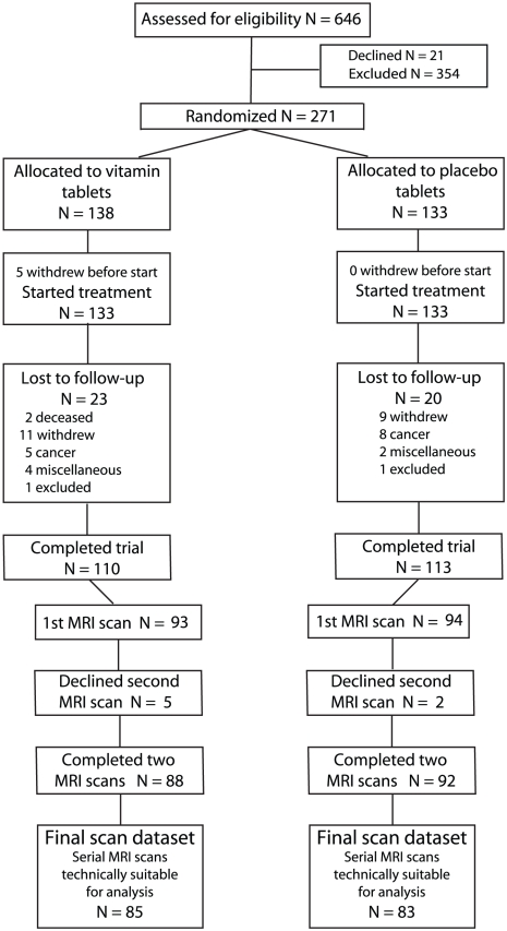
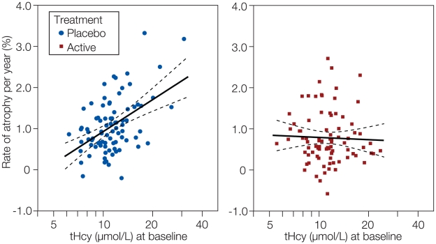
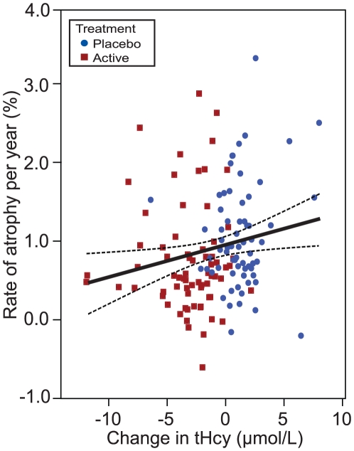
BACKGROUND: An increased rate of brain atrophy is often observed in older subjects, in particular those who suffer from cognitive decline. Homocysteine is a risk factor for brain atrophy, cognitive impairment and dementia. Plasma concentrations of homocysteine can be lowered by dietary administration of B vitamins. OBJECTIVE: To determine whether supplementation with B vitamins that lower levels of plasma total homocysteine can slow the rate of brain atrophy in subjects with mild cognitive impairment in a randomised controlled trial (VITACOG, ISRCTN 94410159). METHODS AND FINDINGS: Single-center, randomized, double-blind controlled trial of high-dose folic acid, vitamins B(6) and B(12) in 271 individuals (of 646 screened) over 70 y old with mild cognitive impairment. A subset (187) volunteered to have cranial MRI scans at the start and finish of the study. Participants were randomly assigned to two groups of equal size, one treated with folic acid (0.8 mg/d), vitamin B(12) (0.5 mg/d) and vitamin B(6) (20 mg/d), the other with placebo; treatment was for 24 months. The main outcome measure was the change in the rate of atrophy of the whole brain assessed by serial volumetric MRI scans. RESULTS: A total of 168 participants (85 in active treatment group; 83 receiving placebo) completed the MRI section of the trial. The mean rate of brain atrophy per year was 0.76% [95% CI, 0.63-0.90] in the active treatment group and 1.08% [0.94-1.22] in the placebo group (P = 0.001). The treatment response was related to baseline homocysteine levels: the rate of atrophy in participants with homocysteine >13 µmol/L was 53% lower in the active treatment group (P = 0.001). A greater rate of atrophy was associated with a lower final cognitive test scores. There was no difference in serious adverse events according to treatment category. CONCLUSIONS AND SIGNIFICANCE: The accelerated rate of brain atrophy in elderly with mild cognitive impairment can be slowed by treatment with homocysteine-lowering B vitamins. Sixteen percent of those over 70 y old have mild cognitive impairment and half of these develop Alzheimer's disease. Since accelerated brain atrophy is a characteristic of subjects with mild cognitive impairment who convert to Alzheimer's disease, trials are needed to see if the same treatment will delay the development of Alzheimer's disease. TRIAL REGISTRATION: Controlled-Trials.com ISRCTN94410159.
Overview
The claims and statistics of this paper haven’t been checked yet.
Run the check — free, no account needed. It reads the open-access text and checks the sources it cites.
Integrity signals
Registry
Cites
Statistics
Figures
Verification
Claims the paper makes
The claims and statistics of this paper haven’t been checked yet.
Run the check — free, no account needed. It reads the open-access text and checks the sources it cites.
Claims other papers make about this one
Each is checked against this paper’s own text: does it say what the citing paper says it does?
The source backs part of the claim, not all of it — or backs it for a different population, condition or comparison.
In the citing paper
“Finally, dietary approaches in terms of nutrition supplementation, such as combined formulation of specific active ingredients or probiotics, or nutritional guidance, such as fasting intermitting or Mediterranean diet, ameliorate both cognition and function of prodromal and mild AD patients [200,201,202].”
From Treatment of Alzheimer’s Disease: Beyond Symptomatic Therapies (2023) · cites this paper as [200]
Experimental — accuracy not yet measuredThe cited source says what the claim attributes to it.
In the citing paper
“While some degree of brain atrophy is common in older age, even in people who are cognitively healthy, atrophy is accelerated in people with Alzheimer's dementia.”
From 2024 Alzheimer's disease facts and figures (2024) · cites this paper as [20]
Self-check
Percentages, sums and reported differences re-computed from the paper’s own numbers.
The self-check needs the paper’s full text.
Citations
Cites
Folstein, M. F., Folstein, S. E., & McHugh, P. R. (1975) · Journal of Psychiatric Research · doi:10.1016/0022-3956(75)90026-6 · Cited by 88,282 · cited 5×
“First, in studies over longer periods (up to 5 years) it has been found that the rate of whole brain atrophy in MCI is correlated with cognitive decline in several tests, including the MMSE [5].”
InfluentialMoms, J. C., Heyman, A., Mohs, R. C., Hughes, J. P., van Belle, G., Fillenbaum, G., Mellits, E. D., & Clark, C. (1989) · Neurology · doi:10.1212/wnl.39.9.1159 · Cited by 4,130 · cited 1×
“…daily living based on the Cambridge Behavioural Inventory [26], an objective memory problem assessed with the ‘Telephone interview of cognitive status, modified’ (TICS-M) [27], a test without a ceiling effect [28], and category fluency [29] based on previously defined cut-off scores for MCI [28].”
Consortium to Establish a Registry for Alzheimer's Disease (CERAD)
Fillenbaum, G., & Heyman, A. (2002) · doi:10.1002/0470846410.ch42(iii · Cited by 2,859
Accurate, Robust, and Automated Longitudinal and Cross-Sectional Brain Change Analysis
Smith, S. M., Zhang, Y., Jenkinson, M., Chen, J. M., Matthews, P. M., Federico, A., & Stefano, N. D. (2002) · NeuroImage · doi:10.1006/nimg.2002.1040 · Cited by 2,013 · cited 3×
“A cross-sectional method (SIENAX) was used to estimate normalized brain volume from a single image, using the skull to normalize spatially, with respect to a standard image [31].”
InfluentialSelf-citationMild Cognitive Impairment: Ten Years Later
Petersen, R. C., Roberts, R. O., Knopman, D. S., Boeve, B. F., Geda, Y. E., Ivnik, R. J., Smith, G. E., & Jack, C. R. (2009) · Archives of Neurology · doi:10.1001/archneurol.2009.266 · Cited by 1,447
Longitudinal Magnetic Resonance Imaging Studies of Older Adults: A Shrinking Brain
Resnick, S. M., Pham, D. L., Kraut, M. A., Zonderman, A. B., & Davatzikos, C. (2003) · The Journal of Neuroscience · doi:10.1523/jneurosci.23-08-03295.2003 · Cited by 1,425 · cited 1×
“The atrophy occurs even in cognitively healthy subjects [1] but is much accelerated in patients suffering from Alzheimer’s disease [2,3,4,5].”
Folate, Vitamin B12, and Serum Total Homocysteine Levels in Confirmed Alzheimer Disease
Clarke, R., Smith, A. D., Jobst, K. A., Refsum, H., Sutton, L., & Ueland, P. M. (1998) · Archives of Neurology · doi:10.1001/archneur.55.11.1449 · Cited by 1,408 · cited 3×
“The results also fit with our earlier finding that raised tHcy predicts the rate of shrinkage of the medial temporal lobe in patients with Alzheimer’s disease [12].”
Self-citationMorris, J. C., Mohs, R. C., Rogers, H., Fillenbaum, G. G., & Heyman, A. (1988) · PubMed · Cited by 1,131
Facts and recommendations about total homocysteine determinations: an expert opinion
Refsum, H., Smith, A. D., Ueland, P. M., Nexo, E., Clarke, R., McPartlin, J., Johnston, C., Engbaek, F., Schneede, J., McPartlin, C., et al. (2004) · Clinical Chemistry · doi:10.1373/clinchem.2003.021634 · Cited by 1,056 · cited 1×
“The tissue and plasma concentrations of homocysteine are largely determined by the body’s status of certain B vitamins (folate, B6 and B12), which are cofactors or substrates for enzymes involved in homocysteine metabolism [22].”
Self-citationPrevalence of Cognitive Impairment without Dementia in the United States
Plassman, B. L., Langa, K. M., Fisher, G. G., Heeringa, S. G., Weir, D. R., Ofstedal, M. B., Burke, J. R., Hurd, M. D., Potter, G. G., Rodgers, W. L., et al. (2008) · Annals of Internal Medicine · doi:10.7326/0003-4819-148-6-200803180-00005 · Cited by 946 · cited 1×
“The prevalence of MCI is between 14% and 18% in those over 70 years-old [24,41], which means that about 5 million people in the USA and 14 million in greater Europe suffer from this condition.”
Cited by
Sorted by citations
Items 701–800 of 813
Hughes, J. C., Poole, M., Louw, S., Greener, H., & Emmett, C. (2020) · Cambridge University Press eBooks · doi:10.1017/9781108594714.026
Lilford, P. (2020) · Cambridge University Press eBooks · doi:10.1017/9781108594714.028
Series, H. G., & Esiri, M. M. (2020) · Cambridge University Press eBooks · doi:10.1017/9781108594714.005
Hughes, J. C. (2020) · Cambridge University Press eBooks · doi:10.1017/9781108594714.003
Supporting Self-Management in Early Dementia
Cheffey, J., Hill, L., & Roberts, G. A. (2020) · Cambridge University Press eBooks · doi:10.1017/9781108594714.017
Deardorff, W. J., & Grossberg, G. T. (2018) · Springer eBooks · doi:10.1007/978-3-319-74604-3_9
Jakubowski, H. (2013) · Springer eBooks · doi:10.1007/978-3-7091-1410-0_3
Brindle, N., & Walsh, C. (2020) · Cambridge University Press eBooks · doi:10.1017/9781108594714.025
Colgate, R., Turner, A., & Rafferty, D. (2020) · Cambridge University Press eBooks · doi:10.1017/9781108594714.023
The Home Assessment in Old Age Psychiatry
Ng, B., & Atkins, M. (2020) · Cambridge University Press eBooks · doi:10.1017/9781108594714.012
Cognitive decline and Alzheimer’s disease
Leray, C. (2017) · CRC Press eBooks · doi:10.1201/9781315268828-5
Jefferies, K., & Agrawal, N. (2020) · Cambridge University Press eBooks · doi:10.1017/9781108594714.006
Richards, F., & Curtice, M. J. R. (2020) · Cambridge University Press eBooks · doi:10.1017/9781108594714.008
Hughes, J. C., Jolley, D. J., Jordan, A., & Sampson, E. L. (2020) · Cambridge University Press eBooks · doi:10.1017/9781108594714.020
Alcohol Misuse in Older People
Rao, R., & CROME, I. B. (2020) · Cambridge University Press eBooks · doi:10.1017/9781108594714.009 · Cited by 1
Wagle, A., & Badrakalimuthu, V. R. (2020) · Cambridge University Press eBooks · doi:10.1017/9781108594714.010
Reducing the Healthcare Burden of Delirium
Meagher, D. J., O'Connell, H. P., Cullen, W., & Adamis, D. (2020) · Cambridge University Press eBooks · doi:10.1017/9781108594714.022
Gupta, S., Fiertag, O., Thanulingam, T., Strange, B. A., & Warner, J. P. (2020) · Cambridge University Press eBooks · doi:10.1017/9781108594714.007
Wilson, S., & Pinner, G. (2020) · Cambridge University Press eBooks · doi:10.1017/9781108594714.013
Hughes, J. C., & Lilford, P. (2020) · Cambridge University Press eBooks · doi:10.1017/9781108594714.001
Biomarkers and the Diagnosis of Preclinical Alzheimer’s Disease
Lilford, P., & Hughes, J. C. (2020) · Cambridge University Press eBooks · doi:10.1017/9781108594714.015
Understanding the Person with Dementia
Hughes, J. C., & Beatty, A. (2020) · Cambridge University Press eBooks · doi:10.1017/9781108594714.027
Hughes, J. C., & Lilford, P. (2020) · Cambridge University Press eBooks · doi:10.1017/9781108594714.002
Sullivan, V., Majumdar, B., Richman, A. V., & Vinjamuri, S. (2020) · Cambridge University Press eBooks · doi:10.1017/9781108594714.016
Sultana, A., & Alauddin, M. (2022) · IntechOpen eBooks · doi:10.5772/intechopen.101611
Inflammatory potential of diet and aging
Orchard, T., Lohman, M. C., & Kopec, R. E. (2022) · Elsevier eBooks · doi:10.1016/b978-0-12-822130-3.00001-6
Brain Health and B Vitamins in Ageing
HAMAMCIOĞLU, A. C., & KARABAŞ, E. (2021) · DergiPark (Istanbul University)
Yaşlanmada Beyin Sağlığı ve B Vitamini
HAMAMCIOĞLU, A. C., & KARABAŞ, E. (2021) · Medical Journal of Western Black Sea · doi:10.29058/mjwbs.871573
ALSUntangled No. 54: “LEAP2BFIT”
THE ALSUNTANGLED GROUP (2020) · Amyotrophic Lateral Sclerosis and Frontotemporal Degeneration · doi:10.1080/21678421.2020.1743470
“B vitamins can decrease homocysteine levels (45), which are elevated in PALS and may contribute to oxidative stress and excitotoxicity (46).”
Background(2016) · Elsevier eBooks · doi:10.1016/b978-0-12-804538-1.00036-5
Tu, M.-C., Hsu, Y., Lo, C., & Huang, C.-F. (2017) · Springer eBooks · doi:10.1007/978-3-319-40007-5_35-1
Yaghoubi, A. (2021) · Research Square · doi:10.21203/rs.3.rs-427829/v2
Hughes, J. C., & Lilford, P. (2020) · Cambridge University Press eBooks · doi:10.1017/9781108594714.029
(2006) · WORLD SCIENTIFIC eBooks · doi:10.1142/9789812707321_0001
Redox regulation of cobalamin status in human neuronal cells and cerebral cortex
Zhang, Y. (2016) · Digital Repository Service (Northeastern University) · doi:10.17760/d20213496
Nechiporuk, V. M., Pentyuk, L. О., & Korda, M. M. (2022) · Bulletin of Problems Biology and Medicine · doi:10.29254/2077-4214-2022-2-2-165-109-123
Nieraad, H. (2022) · Publication Server of Goethe University Frankfurt am Main (Goethe University Frankfurt) · doi:10.21248/gups.68191
B 12 -tilskudd uten B 12 -mangel?
Matre, Å. O., & Lysne, V. (2019) · Norsk tidsskrift for ernæring · doi:10.18261/ntfe.17.3.2
Resolving Geroplasticity to the Balance of Rejuvenins and Geriatrins
TABIBZADEH, S. S. (2022) · Aging and Disease · doi:10.14336/ad.2022.0414
“Even in old but healthy individuals, hyperhomocysteinemia can contribute to the regional or widespread cortical atrophy associated with cognitive decline [578].”
Backgroundde Aquino, A. M. I., Alexandrino, K. A. L. G., de Brito, L. L. M., de Lima, L. D., Gomes, E. R. D. M., & Andrade, S. M. (2023) · doi:10.22541/au.167303809.98273320/v1
Who benefits more from the cognitive effects of B vitamins?
Kwok, T. C. Y. (2023) · Open Access Government · doi:10.56367/oag-037-9490
Student and Trainee Column: The experiences of a doctoral researcher in cognitive neuroscience
Denniss, R. (2018) · The Neuropsychologist · doi:10.53841/bpsneur.2018.1.5.78
Park, K., Masato, U. E. T. A., Nose, A., Yamashita, F., & Nasu, S. (2017) · JOURNAL OF THE BREWING SOCIETY OF JAPAN · doi:10.6013/jbrewsocjapan.112.849
Gou, J., Zhang, X., He, K., Wang, F., & Zou, L. (2023) · Research Square · doi:10.21203/rs.3.rs-2995217/v1
Malnutrition and early-stage Alzheimer's disease
Chaudhary, S., Chaudhary, S. S., Rawat, S., Prasanan, J., & Ashraf, G. M. (2023) · Elsevier eBooks · doi:10.1016/b978-0-323-89834-8.00024-6
Impact of Multidomain Lifestyle Intervention on Brain MRI Markers Related to Cognitive Reserve
Moon, S. Y. (2023) · Journal of Cognitive Intervention and Digital Health · doi:10.58558/jcd.2023.2.1.10
Dasgupta, M., & Brymer, C. (2023) · Health Science Reports · doi:10.1002/hsr2.1500
Sieben auf einen Streich: Neuroprotektive (Mikro-)Nährstoffe als kognitive Enhancer
Gröber, U., & Friedrichsen, H.-P. (2023) · Zeitschrift für Orthomolekulare Medizin · doi:10.1055/a-2153-9332
Reichenberg, L. W., & Seligman, L. (2016) · Wiley eBooks · doi:10.1002/9781394259465.ch18
In symphony: nutrient combinations for healthy brain ageing
van Soest, A. P. M. (2024) · doi:10.18174/652954
Elias, H. A., Mohib, K., Saleem, M. A., Rajput, L. S., & Ellahi, A. (2024) · Cureus · doi:10.7759/cureus.63792
“Furthermore, elevated Hcy levels have been associated with the aggravation of various neurological conditions including epilepsy, Parkinson's disease, and brain atrophy [7].”
BackgroundWang, J. (2024) · Baltic Journal of Sport and Health Sciences · doi:10.33607/bjshs.v1i132.1498
Age-related cognitive decline and omega-3
Lembke, P. (2024) · Elsevier eBooks · doi:10.1016/b978-0-443-29114-2.00025-8
Nutritional Supplements for Brain Health
Hasbal‐Celikok, G. (2024) · doi:10.69860/nobel.9786053359241.10
Shirai, T., Inaba, S., Hayashi, Y., Marui, Y., Wakana, N., Homma, K., & Fukuyama, N. (2024) · The Japanese Journal of Nutrition and Dietetics · doi:10.5264/eiyogakuzashi.82.146
Li, S., Qi, M., Wang, Y., Xin, L., & Li, X. (2025) · Frontiers in Public Health · doi:10.3389/fpubh.2025.1494916
Sex-specific modulation of FOLR1 and its cycle enzyme genes in Alzheimer’s disease brain regions
Sanfilippo, C., Castrogiovanni, P., Imbesi, R., Vecchio, M. M., Vinciguerra, M., Blennow, K., Zetterberg, H., & Rosa, M. D. (2025) · Metabolic Brain Disease · doi:10.1007/s11011-025-01578-x
“For instance, a randomized controlled trial by Smith et al. ( 2010) demonstrated that folic acid and vitamin B12 supplementation effectively lowered plasma homocysteine levels and slowed brain atrophy in elderly individuals with mild cognitive impairment.”
Mourya, V. K., Tiwari, S., Nisha, N., & Paramanik, V. (2025) · IBRO Neuroscience Reports · doi:10.1016/j.ibneur.2025.03.007
“FA deficiency during pregnancy has been associated with cognitive impairments, disruptions in neurotransmitter synthesis, and neurotox-icity, which can impair brain function and behavior in rodents (Smith et al., 2010).”
Alzheimer's disease: Interventions and preventive approaches
ZEIDAN, H. M., & ZEIDAN, I. H. (2025) · Elsevier eBooks · doi:10.1016/b978-0-443-21632-9.00007-8
Nutritional Deficiencies in Perimenopausal and Postmenopausal Women: A Public Health Concern
Vohra, A. (2025) · International Journal of Leading Research Publication. · doi:10.70528/ijlrp.v6.i7.1630
Epigenetic effects of Nosustrophine in Alzheimer’s disease
Martínez-Iglesias, O. A., Naidoo, V., Carrera, I., Corzo, L., & Cacabelos, R. (2025) · Elsevier eBooks · doi:10.1016/b978-0-443-18722-3.00024-4
Huchegowda, R., Harbishettar, V., Sinha, P., Das, S., Sivakumar, P., & John, J. P. (2025) · medRxiv · doi:10.1101/2025.08.02.25332580
“It is possible that low Vitamin B12 and high HCY contribute to the initial development of cognitive impairment as risk factors, but once impairment is present, their association with cognitive test scores can be weak, as observed in this study (22) (Smith et al., 2010).”
Zhang, G., Hu, W., Yang, S., Hou, Y., Li, M., Yang, Y., & Zhao, R. (2025) · Nutrición Hospitalaria · doi:10.20960/nh.05723
Kačerová, T., Miller, J. J. J. J., Radford‐Smith, D. E., Smith, S. M., Jeans, A. F., Jernerén, F., Shepherd, D., Pires, E., de Jel, S., Gong, Q., et al. (2025) · medRxiv · doi:10.1101/2025.11.12.25339932
Self-citationJoshi, S. M., McKee, A. M., Ille, S., Buss, K., Beach, T. G., Serrano, G. E., & Jadavji, N. M. (2025) · bioRxiv (Cold Spring Harbor Laboratory) · doi:10.1101/2025.11.21.689865
“Intervention trials with B vitamins in elderly individuals with cognitive impairment demonstrate a significant slowing of whole and regional brain atrophy, along with a deceleration of cognitive decline [24].”
Yablonsky, E., Higginbotham, E., Chrzanowski, D. T., & Holroyd, S. (2025) · Case Reports in Clinical Medicine · doi:10.4236/crcm.2025.1412086
Jakubowski, H. (2013) · Springer eBooks · doi:10.1007/978-3-7091-1410-0_5
Li, S., Guo, Y., Men, J., Fu, H., & Xu, T. (2021) · Figshare · doi:10.6084/m9.figshare.14795483
Li, S., Guo, Y., Men, J., Fu, H., & Xu, T. (2021) · Figshare · doi:10.6084/m9.figshare.14795477
Li, S., Guo, Y., Men, J., Fu, H., & Xu, T. (2021) · Figshare · doi:10.6084/m9.figshare.14795480
Li, S., Guo, Y., Men, J., Fu, H., & Xu, T. (2021) · Figshare · doi:10.6084/m9.figshare.14795474
Heffernan, M., Doherty, L. C., Hack-Mendes, R., Clarke, M., Hodge, S. J., Clements, M., McAnena, L. B., Rivelsrud, M., Ward, M., Strain, J. J., et al. (2021) · Figshare · doi:10.6084/m9.figshare.17140909
Yu, J. H., Kim, R. E. Y., Jung, J.-M., Park, S. Y., Lee, D. Y., Cho, H. J., Kim, N. H., Yoo, H. J., Seo, J. A., Kim, S. G., et al. (2021) · Figshare · doi:10.6084/m9.figshare.16921778
Li, S., Guo, Y., Men, J., Fu, H., & Xu, T. (2021) · Figshare · doi:10.6084/m9.figshare.14795486
Neuro-Nutrition: Fortifying Foods for Cognitive Well-Being
Patel, P., Bishoyi, A. K., & Sanghvi, G. (2025) · Springer eBooks · doi:10.1007/978-981-95-1217-1_13
Parkinson’s Disease and Diet: Nutritional Interventions for Motor and Cognitive Functions
Manhas, M. W., Javid, S., Bushra, Choudhary, D., & Rahman, A. (2026) · Springer eBooks · doi:10.1007/978-981-95-4169-0_9
Nutritional Interventions for Dementia Prevention: A Brief Perspective
Kamohara, S., & Sakatani, K. (2025) · Personalized Medicine Universe · doi:10.46459/pmu.2025002
Dietary interventions and brain health
Joshi, S. M., McCabe, R., & Jadavji, N. M. (2026) · Elsevier eBooks · doi:10.1016/b978-0-443-33841-0.00020-8
Patten, R. V., Hallowell, E. S., Kamper, J. E., Jennette, K. J., & Kanser, R. J. (2026) · Springer eBooks · doi:10.1007/978-3-032-05880-5_7
Kanouchi, H., Yamamoto, A., Kuwabara, A., Takenaka, S., Nishikubo, E., Nomura, Y., Naruto, T., Watanabe, K., Mizuno, K., & Watanabe, Y. (2026) · Nutrients · doi:10.3390/nu18060941
“For example, Hcy accumulation can reduce the availability of S-adenosylmethionine (SAM), a universal methyl donor required for the synthesis of dopamine and serotonin, thereby impairing neuromodulation and neuroplasticity [15–17].”
Ponce‐Fuentes, F., Velásquez, C. Á., Mercado, M. Á., López, R. N., Neira-Maldonado, C., Salazar‐Méndez, J., Coronado, J.-C., & Cuyúl-Vásquez, I. (2026) · Nutrición Hospitalaria · doi:10.20960/nh.06434
Choline status and related health outcomes in humans: A scoping review
Yoo, S., Aoun, A., Montazeri, A., Collins, É., & Little, J. (2026) · Nutrition and Health · doi:10.1177/02601060261430655
“…as suggesting that choline intake/status has bene fi cial effects on cognitive function among older adults (Aguree et al., 2023; Liu et al., 2021a; Smith et al., 2010; Velazquez et al., 2020; Ylilauri et al., 2019) and among infants (Irvine et al., 2022; Jiang et al., 2014; Obeid et al., 2022).”
An Integrated Approach on Vascular Cognitive Impairment
Jianu, D. C., Jianu, S. N., Bora, B. I., Munteanu, G., Bertici, R.-A., Dan, T. F., & Petrică, L. (2026) · IntechOpen eBooks · doi:10.5772/intechopen.1014841
Lefèvre‐Arbogast, S. (2019) · theses.fr (ABES) · doi:10.70675/6915e632z663fz4caaz9c7az455ad25755da
La place des biomarqueurs dans les essais de prévention de la maladie d'Alzheimer
Delrieu, J. (2021) · theses.fr (ABES) · doi:10.70675/2b5f4719z01f9z4376z9dabzd5a185dc4d79
Koshy, R. R., Bahl, D., Muhammed, M., Kumar, A. A., Preman, P., Raziya, F., Lewis, C. V., Gopalan, G. L., & Gunasekaran, J. (2026) · Karnataka Pediatric Journal · doi:10.25259/kpj_7_2026
B-Group Vitamins in Internal Medicine, Gynaecology and Neurology: A Comprehensive Clinical Review
Pršo, A.-M. L., Pejić, M. K., Milotić, D. M., Matijaca, A., Matijaca, H., Marton, U., Sruk, A., & Gojo-Tomic, N. (2026) · Biochemistry · doi:10.5772/intechopen.1014859
Vitamin B12 Supplementation: Is More Always Better?
Yepes-Calderón, M., Doorenbos, C. S. E., Stegmann, M., Touw, D. J., Harmsen, H. J. M., Heiner-Fokkema, M. R., van Spronsen, F. J., Corpeleijn, E., & Bakker, S. J. L. (2026) · Nutrients · doi:10.3390/nu18101597
“…at baseline (mean plasma B 12 ~330 pmol/L) showed that combined supplementation with vitamin B 12 , folic acid, and vitamin B 6 for 24 months slowed brain atrophy and cognitive decline, particularly among participants with elevated homocysteine or sufficient long-chain omega-3 status [48,49].”
Д.С., К. (2026) · Український медичний часопис · doi:10.32471/umj.1680-3051.278467
Castagna, A., Cotroneo, A. M., Mosele, M., Sapone, P., Martinelli, E., Rahmati, M., Smith, L., & Veronese, N. (2026) · Aging Clinical and Experimental Research · doi:10.1007/s40520-026-03433-0
“[25] Moreover, neuroimaging work also supports the concept that B vitamins can slow atrophy in brain regions relevant to Alzheimer’s pathology.”
Karmakar, V., Ghosh, A., Majie, A., Chatterjee, K., & Gorain, B. (2026) · Ageing Research Reviews · doi:10.1016/j.arr.2026.103262
Zhang, X., Li, J., An, Y., Zheng, J., Xu, G., Wang, J., Chen, L., & Lu, Y. (2026) · Progress in Neuro-Psychopharmacology and Biological Psychiatry · doi:10.1016/j.pnpbp.2026.111861
Alzheimer's disease and nutrition: Focus on the impact of hyperhomocysteinemia
Clément, G., Hansmannel, F., Hopes, L., Bossenmeyer‐Pourié, C., & Renaud, M. (2026) · Journal of Alzheimer s Disease · doi:10.1177/13872877261471813
“126 In another study, the FACIT trial was based on a sample of older patients without cognitive disorders but with HHCY.”
Huchegowda, R., Harbishettar, V., Sinha, P., Das, S., Sivakumar, P. T., & John, J. P. (2026) · Journal of Nutrition in Gerontology and Geriatrics · doi:10.1080/21551197.2026.2713421
Clinical Applications of Blood Tests
Yang, C. S.-Y., & Sabbagh, M. N. (2026) · Springer eBooks · doi:10.1007/978-3-032-23863-4_5
Draht, F. (2026) · Zeitschrift für Komplementärmedizin · doi:10.1055/a-2896-4441
Metadata
- Authors
- Smith, A. D., Smith, S. M., de Jager, C. A., Whitbread, P., Johnston, C., Agacinski, G., Oulhaj, A., Bradley, K. M., Jacoby, R., & Refsum, H.
- Affiliations
- University of Oxford (GB)
- University of Oslo (NO)
- Corresponding author
- Smith, A. David
- Venue
- PLoS ONE
- Publisher
- Public Library of Science (PLoS)
- Published
- 2010-09-08
- Volume
- 5
- Issue
- 9
- Pages
- e12244
- ISSN
- 1932-6203 (ISSN-L)
- PMC
- PMC2935890
- OpenAlex
- W2156635660
- Cited by
- 813
- References
- 49
- Citations by year
- 2026: 50 · 2025: 49 · 2024: 34 · 2023: 44 · 2022: 58 · 2021: 58 · 2020: 66 · 2019: 39 · 2018: 43 · 2017: 55 · 2016: 59 · 2015: 49 · 2014: 44 · 2013: 58 · 2012: 59
- FWCI
- 36.35
- Type
- journal article
- Language
- en
- Open access
- gold
- doi.org
- Locations
- PLoS ONE (publishedVersion, cc-by)
- PubMed Central (publishedVersion, cc-by)
- PubMed (publishedVersion)
- ORCA Online Research @Cardiff (Cardiff University) (acceptedVersion)
- DOAJ (DOAJ: Directory of Open Access Journals) (submittedVersion, cc-by-sa)
- Figshare (submittedVersion, cc-by)
- Funders
- Alzheimer's Research Trust
- National Institute for Health and Care Research
- University of Oxford
- Medical Research Council
- Topics
- Health Sciences › Nursing › Nutrition and Dietetics › Folate and B Vitamins Research
- Health Sciences › Medicine › Psychiatry and Mental health › Dementia and Cognitive Impairment Research
- Health Sciences › Medicine › Clinical Neurology › Alcoholism and Thiamine Deficiency
- Keywords
- homocysteine, B vitamins, folic acid, vitamin B6, vitamin B12, brain atrophy, mild cognitive impairment, cognitive decline, dementia prevention, older adults, plasma homocysteine, volumetric MRI, cognitive performance, adverse events
- MeSH terms
- Aged, Aged, 80 and over, Atrophy, Brain, Cognition Disorders, Female, Homocysteine, Humans, Magnetic Resonance Imaging, Male, Radiography, Vitamin B Complex, Treatment Outcome
About these checks
Registry, retraction, Cites and statistics results are computed, not judged, with one exception: a printed reference that no index lists is read once by a model before it counts as a citation error. Claims are extracted by a model and kept only when their quote appears verbatim in the text; each cited source is then checked, with a model’s help, for whether it says what the claim relies on. Those verdicts can be wrong — read the claim’s quote and the cited source before relying on one.
Statistics and arithmetic are recomputed, not judged.
Confidence bands are shown only for fields calibrated on our annotated test set. None is calibrated yet, so no band is shown.
What counts as a claim ›How we measure confidence ›How we check this ›
Are you an author, or do you see a problem with this page? Request a review
