

The DOI matches its Crossref record; 14 of the 14 references it cites were found in a registry or an index.
The Extracellular Matrix in Skin Inflammation and Infection
Abstract
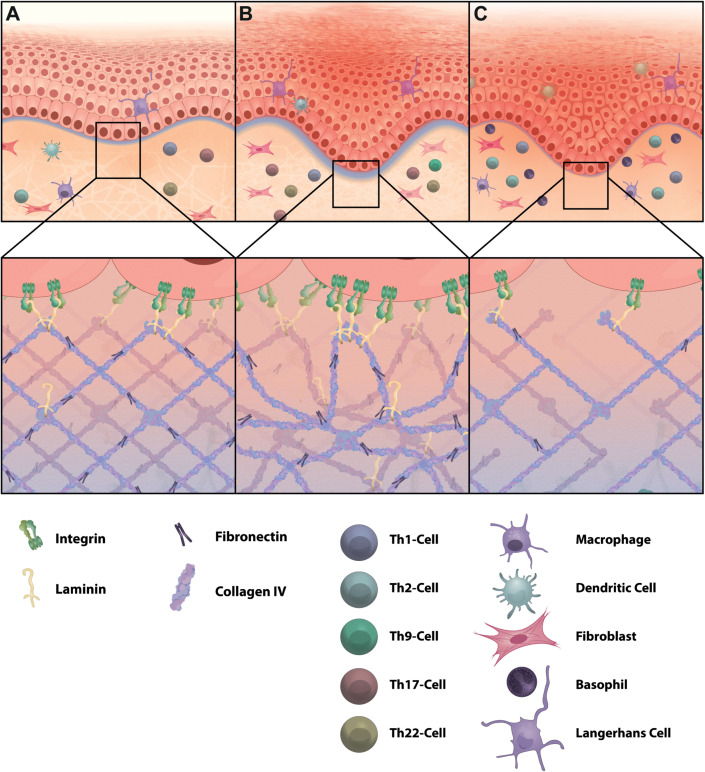
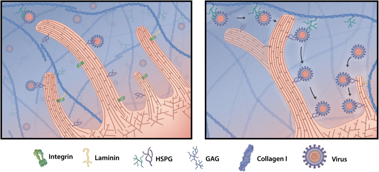
The extracellular matrix (ECM) is an integral component of all organs and plays a pivotal role in tissue homeostasis and repair. While the ECM was long thought to mostly have passive functions by providing physical stability to tissues, detailed characterization of its physical structure and biochemical properties have uncovered an unprecedented broad spectrum of functions. It is now clear that the ECM not only comprises the essential building block of tissues but also actively supports and maintains the dynamic interplay between tissue compartments as well as embedded resident and recruited inflammatory cells in response to pathologic stimuli. On the other hand, certain pathogens such as bacteria and viruses have evolved strategies that exploit ECM structures for infection of cells and tissues, and mutations in ECM proteins can give rise to a variety of genetic conditions. Here, we review the composition, structure and function of the ECM in cutaneous homeostasis, inflammatory skin diseases such as psoriasis and atopic dermatitis as well as infections as a paradigm for understanding its wider role in human health.
Overview
The claims and statistics of this paper haven’t been checked yet.
Run the check — free, no account needed. It reads the open-access text and checks the sources it cites.
Integrity signals
Registry
Cites
Statistics
Figures
Verification
Claims the paper makes
The claims and statistics of this paper haven’t been checked yet.
Run the check — free, no account needed. It reads the open-access text and checks the sources it cites.
Claims other papers make about this one
Each is checked against this paper’s own text: does it say what the citing paper says it does?
In the citing paper
“Chronic wound-edge SEM images are complex and consist of diverse features.53-56”
From SEMTWIST Quantification of Biofilm Infection in Human Chronic Wound Using Scanning Electron Microscopy and Machine Learning (2025) · cites this paper as [55]
In the citing paper
“The ECM architecture is highly intricate and can sometimes be misinterpreted as biofilm due to protuberances or abnormal topographical modifications on its membrane that occur during ECM remodeling.55,56”
From SEMTWIST Quantification of Biofilm Infection in Human Chronic Wound Using Scanning Electron Microscopy and Machine Learning (2025) · cites this paper as [55]
In the citing paper
“For example, membrane projections or ECM features can be mistaken for biofilm, especially in samples with low biofilm abundance.55,56”
From SEMTWIST Quantification of Biofilm Infection in Human Chronic Wound Using Scanning Electron Microscopy and Machine Learning (2025) · cites this paper as [55]
Self-check
Percentages, sums and reported differences re-computed from the paper’s own numbers.
The self-check needs the paper’s full text.
Citations
Cites
Tensional homeostasis and the malignant phenotype
Paszek, M. J., Zahir, N., Johnson, K. R., Lakins, J. N., Rozenberg, G. I., Gefen, A., Reinhart-King, C. A., Margulies, S. S., Dembo, M., Boettiger, D., et al. (2005) · Cancer Cell · doi:10.1016/j.ccr.2005.08.010 · Cited by 3,941
Local force and geometry sensing regulate cell functions
Vogel, V., & Sheetz, M. (2006) · Nature Reviews Molecular Cell Biology · doi:10.1038/nrm1890 · Cited by 2,328 · cited 2×
“…expose otherwise inaccessible cryptic peptide sequences on the ECM, open mechanosensitive ion channels, change receptor or ligand conformation to strengthen their interaction, and thereby alter cell-matrix interactions and biochemical signaling responses (Vogel and Sheetz, 2006; Wang et al., 2019).”
BackgroundExtracellular Matrix Degradation and Remodeling in Development and Disease
Lu, P., Takai, K., Weaver, V. M., & Werb, Z. (2011) · Cold Spring Harbor Perspectives in Biology · doi:10.1101/cshperspect.a005058 · Cited by 2,265
Extracellular Matrix Assembly: A Multiscale Deconstruction
Mouw, J. K., Ou, G., & Weaver, V. M. (2014) · Nature Reviews Molecular Cell Biology · doi:10.1038/nrm3902 · Cited by 1,534
Nuclear envelope rupture and repair during cancer cell migration
Denais, C. M., Gilbert, R. M., Isermann, P., McGregor, A. L., te Lindert, M., Weigelin, B., Davidson, P. M., Friedl, P., Wolf, K., & Lammerding, J. (2016) · Science · doi:10.1126/science.aad7297 · Cited by 1,318
Elosegui-Artola, A., Oria, R., Chen, Y., Kosmalska, A., Pérez-González, C., Castro, N., Zhu, C., Trepat, X., & Roca-Cusachs, P. (2016) · Nature Cell Biology · doi:10.1038/ncb3336 · Cited by 858
Sowinski, S., Jolly, C., Berninghausen, O., Purbhoo, M. A., Chauveau, A., Köhler, K., Oddos, S., Eissmann, P., Brodsky, F. M., Hopkins, C., et al. (2008) · Nature Cell Biology · doi:10.1038/ncb1682 · Cited by 754
Martino, M. M., Tortelli, F., Mochizuki, M., Traub, S., Ben-David, D., Kuhn, G. A., Müller, R., Livne, E., Eming, S. A., & Hubbell, J. A. (2011) · Science Translational Medicine · doi:10.1126/scitranslmed.3002614 · Cited by 459
Hair follicle aging is driven by transepidermal elimination of stem cells via COL17A1 proteolysis
Matsumura, H., Mohri, Y., Binh, N. T., Morinaga, H., Fukuda, M., Ito, M., Kurata, S., Hoeijmakers, J., & Nishimura, E. K. (2016) · Science · doi:10.1126/science.aad4395 · Cited by 416
Actin- and myosin-driven movement of viruses along filopodia precedes their entry into cells
Lehmann, M. J., Sherer, N. M., Marks, C. B., Pypaert, M., & Mothes, W. (2005) · The Journal of Cell Biology · doi:10.1083/jcb.200503059 · Cited by 403
Research works printed but not linked: 111
- Abercrombie, 1980, The Croonian Lecture, 1978-The crawling movement of metazoan cells., Proc. Roy. Soc. Lon., 207, 129, 10.1098/rspb.1980.0017 · DOI
- Adair-Kirk, 2008, Fragments of extracellular matrix as mediators of inflammation., Int. J. Biochem. Cell Biol., 40, 1101, 10.1016/j.biocel.2007.12.005 · DOI
- Adams, 2000, Stimulation of fascin spikes by thrombospondin-1 is mediated by the GTPases Rac and Cdc42., J. Cell Biol., 150, 807, 10.1083/jcb.150.4.807 · DOI
- Adams, 1999, Cell-matrix adhesions differentially regulate fascin phosphorylation., Mol. Biol. Cell, 10, 4177, 10.1091/mbc.10.12.4177 · DOI
- Aguiar, 2020, Molecular alterations in the extracellular matrix in the brains of newborns with congenital Zika syndrome., Sci. Signal., 13, 10.1126/scisignal.aay6736 · DOI
- Alfano, 2016, The interplay of extracellular matrix and microbiome in urothelial bladder cancer., Nat. Rev. Urol., 13, 77, 10.1038/nrurol.2015.292 · DOI
- Andriessen, 1997, Basal membrane heparan sulphate proteoglycan expression during wound healing in human skin., J. Pathol., 183, 264, 10.1002/(sici)1096-9896(199711)183:3<264::aid-path940>3.0.co;2-3 · DOI
- Bailey, 2001, Molecular mechanisms of ageing in connective tissues., Mech. Age. Devel., 122, 735, 10.1016/s0047-6374(01)00225-1 · DOI
- Bansode, 2020, Glycation changes molecular organization and charge distribution in type I collagen fibrils., Scient. Rep., 10
- Becker, 2019, Infectious Entry of Merkel Cell Polyomavirus., J. Virol., 93, e2004, 10.1128/JVI.02004-18 · DOI
- Behren, 2005, Papillomavirus E2 protein induces expression of the matrix metalloproteinase-9 via the extracellular signal-regulated kinase/activator protein-1 signaling pathway., Cancer Res., 65, 11613, 10.1158/0008-5472.can-05-2672 · DOI
- Bhattacharjee, 2019, Unraveling the ECM-Immune Cell Crosstalk in Skin Diseases., Front. Cell Devel. Biol., 7
- Bienkowska-Haba, 2011, The cytoskeleton in papillomavirus infection., Viruses, 3, 260, 10.3390/v3030260 · DOI
- Bodor, 2020, Of Cell Shapes and Motion: The Physical Basis of Animal Cell Migration., Devel. Cell, 52, 550, 10.1016/j.devcel.2020.02.013 · DOI
- Bonnans, 2014, Remodelling the extracellular matrix in development and disease., Nat. Rev. Mol. Cell Biol., 15, 786, 10.1038/nrm3904 · DOI · Paper page
- Boothby, 2020, Regulatory T cells in skin injury: At the crossroads of tolerance and tissue repair., Sci. Immunol., 5, 10.1126/sciimmunol.aaz9631 · DOI
- Bosman, 2003, Functional structure and composition of the extracellular matrix., J. Pathol., 200, 423, 10.1002/path.1437 · DOI
- Bouhaddou, 2020, The Global Phosphorylation Landscape of SARS-CoV-2 Infection., Cell, 182, 685, 10.1016/j.cell.2020.06.034 · DOI
- Cai, 2017, The Role of the Lysyl Oxidases in Tissue Repair and Remodeling: A Concise Review., Tissue Eng. Regen. Med., 14, 15, 10.1007/s13770-016-0007-0 · DOI
- Chang, 2020, Circadian control of the secretory pathway maintains collagen homeostasis., Nat. Cell Biol., 22, 74, 10.1038/s41556-019-0441-z · DOI
- Chang, 2017, Force-dependent breaching of the basement membrane., Matrix Biol., 178, 10.1016/j.matbio.2016.12.005 · DOI
- Chaudhuri, 2020, Effects of extracellular matrix viscoelasticity on cellular behaviour., Nature, 584, 535, 10.1038/s41586-020-2612-2 · DOI · Paper page
- Cho, 2001, Fibronectin and fibrinogen contribute to the enhanced binding of Staphylococcus aureus to atopic skin., J. Allerg. Clin. Immunol., 108, 269, 10.1067/mai.2001.117455 · DOI
- Culp, 2006, Keratinocyte-secreted laminin 5 can function as a transient receptor for human papillomaviruses by binding virions and transferring them to adjacent cells., J. Virol., 80, 8940, 10.1128/jvi.00724-06 · DOI
- Dandia, 2019, Glycated collagen - a 3D matrix system to study pathological cell behavior., Biomater. Sci., 7, 3480, 10.1039/c9bm00184k · DOI
- Das, 2019, Differential Influence of IL-9 and IL-17 on Actin Cytoskeleton Regulates the Migration Potential of Human Keratinocytes., J. Immunol., 202, 1949, 10.4049/jimmunol.1800823 · DOI
- Deng, 2016, Hypoxia-inducible factor 1 in autoimmune diseases., Cell. Immunol., 303, 7, 10.1016/j.cellimm.2016.04.001 · DOI
- DuFort, 2011, Balancing forces: architectural control of mechanotransduction., Nat. Rev. Mol. Cell Biol., 12, 308, 10.1038/nrm3112 · DOI
- Dyer, 2020, Understanding the mechanisms that facilitate specificity, not redundancy, of chemokine-mediated leukocyte recruitment., Immunology, 160, 336, 10.1111/imm.13200 · DOI
- Engel, 1989, EGF-like domains in extracellular matrix proteins: Localized signals for growth and differentiation?, FEBS Lette., 251, 1, 10.1016/0014-5793(89)81417-6 · DOI
- Fidler, 2018, The triple helix of collagens – an ancient protein structure that enabled animal multicellularity and tissue evolution., J. Cell Sci., 131, 10.1242/jcs.203950 · DOI
- Foxall, 2019, PAK4 Kinase Activity Plays a Crucial Role in the Podosome Ring of Myeloid Cells., Cell Rep., 29, 3385, 10.1016/j.celrep.2019.11.016 · DOI
- Ghetti, 2018, Subpopulations of dermal skin fibroblasts secrete distinct extracellular matrix: implications for using skin substitutes in the clinic., Br. J. Dermatol., 179, 381
- Gopal, 2020, Calcium in Cell-Extracellular Matrix Interactions., Adv. Exp. Med. Biol., 1131, 1079, 10.1007/978-3-030-12457-1_43 · DOI
- Gralinski, 2013, Mechanisms of Severe Acute Respiratory Syndrome Coronavirus-Induced Acute Lung Injury., mBio, 4, e271, 10.1128/mBio.00271-13 · DOI
- Grau-Bové, 2015, Origin and evolution of lysyl oxidases., Scient. Rep., 5, 10568, 10.1038/srep10568 · DOI
- Hadigal, 2020, Heparanase-Regulated Syndecan-1 Shedding Facilitates Herpes Simplex Virus 1 Egress., J. Virol., 94, e1672, 10.1128/JVI.01672-19 · DOI
- Han, 2001, TNF-alpha stimulates activation of pro-MMP2 in human skin through NF-(kappa)B mediated induction of MT1-MMP., J. Cell Sci., 114, 131, 10.1242/jcs.114.1.131 · DOI
- Hetmanski, 2019, Membrane Tension Orchestrates Rear Retraction in Matrix-Directed Cell Migration., Dev. Cell, 51, 460, 10.1016/j.devcel.2019.09.006 · DOI
- Huang, 2019, Interleukin-17 Drives Interstitial Entrapment of Tissue Lipoproteins in Experimental Psoriasis., Cell Metabol., 29, 475, 10.1016/j.cmet.2018.10.006 · DOI
- Humphrey, 2014, Mechanotransduction and extracellular matrix homeostasis., Nat. Rev. Mol. Cell Biol., 15, 802, 10.1038/nrm3896 · DOI · Paper page
- Hynes, 2009, The extracellular matrix: not just pretty fibrils., Science, 326, 1216, 10.1126/science.1176009 · DOI · Paper page
- Jayo, 2016, Fascin Regulates Nuclear Movement and Deformation in Migrating Cells., Dev. Cell, 38, 371, 10.1016/j.devcel.2016.07.021 · DOI
- Kakavandi, 2018, Anoikis resistance and oncoviruses., J. Cell. Biochem., 119, 2484, 10.1002/jcb.26363 · DOI
- Kelsh, 2014, Regulation of the innate immune response by fibronectin: synergism between the III-1 and EDA domains., PLoS One, 9, 10.1371/journal.pone.0102974 · DOI
- Kisling, 2019, What is the role of peptide fragments of collagen I and IV in health and disease?, Life Sci., 228, 30, 10.1016/j.lfs.2019.04.042 · DOI
- Knappe, 2007, Surface-exposed amino acid residues of HPV16 L1 protein mediating interaction with cell surface heparan sulfate., J. Biol. Chem., 282, 27913, 10.1074/jbc.m705127200 · DOI
- Knipper, Johanna, 2015, Interleukin-4 Receptor α Signaling in Myeloid Cells Controls Collagen Fibril Assembly in Skin Repair., Immunity, 43, 803, 10.1016/j.immuni.2015.09.005 · DOI
- Kobayashi, 2010, Sensing substrate rigidity by mechanosensitive ion channels with stress fibers and focal adhesions., Curr. Opin. Cell Biol., 22, 669, 10.1016/j.ceb.2010.08.023 · DOI
- Kuo, 2011, Rhinovirus infection induces extracellular matrix protein deposition in asthmatic and nonasthmatic airway smooth muscle cells., Am. J. Physiol., 300, L951
- Kurbet, 2016, Sterile Inflammation Enhances ECM Degradation in Integrin beta1 KO Embryonic Skin., Cell Rep., 16, 3334, 10.1016/j.celrep.2016.08.062 · DOI
- Lang, 2011, Inhibition of SARS pseudovirus cell entry by lactoferrin binding to heparan sulfate proteoglycans., PLOS ONE, 6, 10.1371/journal.pone.0023710 · DOI
- Lawson, 2018, Rho GTPase signaling complexes in cell migration and invasion., J. Cell Biol., 217, 447, 10.1083/jcb.201612069 · DOI · Paper page
- Leithner, 2016, Diversified actin protrusions promote environmental exploration but are dispensable for locomotion of leukocytes., Nat. Cell Biol., 18, 1253, 10.1038/ncb3426 · DOI · Paper page
- Levental, 2009, Matrix crosslinking forces tumor progression by enhancing integrin signaling., Cell, 139, 891, 10.1016/j.cell.2009.10.027 · DOI · Paper page
- Liguori, 2011, Tumor-associated macrophages as incessant builders and destroyers of the cancer stroma., Cancers, 3, 3740, 10.3390/cancers3043740 · DOI
- Martín-Villar, 2015, Podoplanin mediates ECM degradation by squamous carcinoma cells through control of invadopodia stability., Oncogene, 34, 4531, 10.1038/onc.2014.388 · DOI
- Mason, 2013, Tuning three-dimensional collagen matrix stiffness independently of collagen concentration modulates endothelial cell behavior., Acta Biomater., 9, 4635, 10.1016/j.actbio.2012.08.007 · DOI
- Matrosovich, 2013, SialoGlyco Chemistry and Biology II 1-28.
- Matsubayashi, 2017, A Moving Source of Matrix Components Is Essential for De Novo Basement Membrane Formation., Curr. Biol., 27, 3526, 10.1016/j.cub.2017.10.001 · DOI
- Michael, 2020, New perspectives on integrin-dependent adhesions., Curr. Opin. Cell Biol., 63, 31, 10.1016/j.ceb.2019.12.008 · DOI
- Michopoulou, 2020, A novel mechanism in wound healing: Laminin 332 drives MMP9/14 activity by recruiting syndecan-1 and CD44., Matrix Biol., 94, 1, 10.1016/j.matbio.2020.06.004 · DOI
- Milewska, 2014, Human coronavirus NL63 utilizes heparan sulfate proteoglycans for attachment to target cells., Journal of virology, 88, 13221, 10.1128/jvi.02078-14 · DOI
- Mishra, 2020, Periostin Activation of Integrin Receptors on Sensory Neurons Induces Allergic Itch., Cell Rep., 31, 10.1016/j.celrep.2020.03.036 · DOI
- Nikolic, 2011, HIV-1 activates Cdc42 and induces membrane extensions in immature dendritic cells to facilitate cell-to-cell virus propagation., Blood, 118, 4841, 10.1182/blood-2010-09-305417 · DOI
- Norris, 2007, Periostin regulates collagen fibrillogenesis and the biomechanical properties of connective tissues., J. Cell. Biochem., 101, 695, 10.1002/jcb.21224 · DOI
- Norsgaard, 2014, Calcipotriol counteracts betamethasone-induced decrease in extracellular matrix components related to skin atrophy., Arch. Dermatol. Res., 306, 719, 10.1007/s00403-014-1485-3 · DOI
- Nwogu, 2018, Cellular sheddases are induced by Merkel Cell Polyomavirus small tumour antigen to mediate cell dissociation and invasiveness., PLoS Pathog, 14, e1007276, 10.1371/journal.ppat.1007276 · DOI
- Nystrom, 2019, Matrix molecules and skin biology., Semin. Cell Dev. Biol., 89, 136, 10.1016/j.semcdb.2018.07.025 · DOI · Paper page
- Orban, 2004, Crosslinking of collagen gels by transglutaminase., J. Biomed. Mater. Res. Part A, 756, 10.1002/jbm.a.20110 · DOI
- Owczarek, 2018, Early events during human coronavirus OC43 entry to the cell., Sci. Rep., 8, 10.1038/s41598-018-25640-0 · DOI
- Oyoshi, 2012, Eosinophil-derived leukotriene C4 signals via type 2 cysteinyl leukotriene receptor to promote skin fibrosis in a mouse model of atopic dermatitis., Proc. Natl. Acad. Sci., 109, 4992, 10.1073/pnas.1203127109 · DOI
- Pfisterer, 2020, FMNL2 regulates dynamics of fascin in filopodia., J. Cell Biol., 219, 10.1083/jcb.201906111 · DOI
- Quan, 2015, Role of Age-Associated Alterations of the Dermal Extracellular Matrix Microenvironment in Human Skin Aging: A Mini-Review., Gerontology, 61, 427, 10.1159/000371708 · DOI · Paper page
- Rafiq, 2019, A mechano-signalling network linking microtubules, myosin IIA filaments and integrin-based adhesions., Nat. Mater., 18, 638, 10.1038/s41563-019-0371-y · DOI
- Reversat, 2020, Cellular locomotion using environmental topography., Nature, 582, 582, 10.1038/s41586-020-2283-z · DOI
- Ridley, 2011, Life at the leading edge., Cell, 145, 1012, 10.1016/j.cell.2011.06.010 · DOI
- Ringer, 2017, Sensing the mechano-chemical properties of the extracellular matrix., Matrix Biol., 64, 6, 10.1016/j.matbio.2017.03.004 · DOI
- Rodriguez-Pascual, 2018, Lysyl Oxidases: Functions and Disorders., J. Glaucoma, 27, S15, 10.1097/IJG.0000000000000910 · DOI
- Roy, 2010, Processing of type I collagen gels using nonenzymatic glycation., J. Biomed. Mater. Res. A., 93, 843, 10.1002/jbm.a.32231 · DOI
- Sapp, 2009, Viral entry mechanisms: human papillomavirus and a long journey from extracellular matrix to the nucleus., FEBS J., 276, 7206, 10.1111/j.1742-4658.2009.07400.x · DOI
- Sarkar, 2017, Endogenous Glucocorticoid Deficiency in Psoriasis Promotes Inflammation and Abnormal Differentiation., J. Investig. Dermatol., 137, 1474, 10.1016/j.jid.2017.02.972 · DOI
- Schneider, 2019, Orf virus infection of human keratinocytes and dermal fibroblasts: Limited virus detection and interference with intercellular adhesion molecule-1 up-regulation., Exp. Dermatol., 28, 142, 10.1111/exd.13861 · DOI
- Scott, 2010, LIM kinases are required for invasive path generation by tumor and tumor-associated stromal cells., J. Cell Biol., 191, 169, 10.1083/jcb.201002041 · DOI
- Shao, 2019, Physical properties of the photodamaged human skin dermis: Rougher collagen surface and stiffer/harder mechanical properties., Exp. Dermatol., 28, 914, 10.1111/exd.13728 · DOI
- Shen, 2020, Reduction of Liver Metastasis Stiffness Improves Response to Bevacizumab in Metastatic Colorectal Cancer., Cancer Cell, 37, 800, 10.1016/j.ccell.2020.05.005 · DOI
- Simon, 2017, Regulation of the Immune System by Laminins., Trends Immunol., 38, 858, 10.1016/j.it.2017.06.002 · DOI
- Singh, 2012, Human pathogens utilize host extracellular matrix proteins laminin and collagen for adhesion and invasion of the host., FEMS Microbiol. Rev., 36, 1122, 10.1111/j.1574-6976.2012.00340.x · DOI · Paper page
- Smith, 2008, Virus activated filopodia promote human papillomavirus type 31 uptake from the extracellular matrix., Virology, 381, 16, 10.1016/j.virol.2008.08.040 · DOI
- Sunyer, 2020, Durotaxis., Curr. Biol., 30, R383, 10.1016/j.cub.2020.03.051 · DOI
- Tajpara, 2019, A Preclinical Model for Studying Herpes Simplex Virus Infection., J. Investig. Dermatol., 139, 673, 10.1016/j.jid.2018.08.034 · DOI
- Talmi-Frank, 2016, Extracellular Matrix Proteolysis by MT1-MMP Contributes to Influenza-Related Tissue Damage and Mortality., Cell Host Microbe, 20, 458, 10.1016/j.chom.2016.09.005 · DOI
- Tan, 2020, Regulation and dynamics of force transmission at individual cell-matrix adhesion bonds., Sci. Adv., 6, 10.1126/sciadv.aax0317 · DOI
- Taylor, 2011, Subversion of the actin cytoskeleton during viral infection., Nat. Rev. Microbiol., 9, 427, 10.1038/nrmicro2574 · DOI
- Tominaga, 2011, Matrix Metalloproteinase-8 Is Involved in Dermal Nerve Growth: Implications for Possible Application to Pruritus from In Vitro Models., J. Investig. Dermatol., 131, 2105, 10.1038/jid.2011.173 · DOI
- Tortorici, 2019, Structural basis for human coronavirus attachment to sialic acid receptors., Nat. Struct. Mol. Biol., 26, 481, 10.1038/s41594-019-0233-y · DOI
- Tse, 2018, Hepatitis transactivator protein X promotes extracellular matrix modification through HIF/LOX pathway in liver cancer., Oncogenesis, 7, 10.1038/s41389-018-0052-8 · DOI
- Uitto, 1989, Extracellular Matrix of the Skin: 50 Years of Progress., J. Investig. Dermatol., 92, S61, 10.1038/jid.1989.34 · DOI
- Vallet, 2019, Lysyl oxidases: from enzyme activity to extracellular matrix cross-links., Essays Biochem., 63, 349, 10.1042/ebc20180050 · DOI
- van den Dries, 2019, Probing the mechanical landscape - new insights into podosome architecture and mechanics., J. Cell Sci., 132, 10.1242/jcs.236828 · DOI
- van Helvert, 2018, Mechanoreciprocity in cell migration., Nat. Cell Biol., 20, 8, 10.1038/s41556-017-0012-0 · DOI · Paper page
- Vorstandlechner, 2020, Deciphering the functional heterogeneity of skin fibroblasts using single-cell RNA sequencing., FASEB J., 34, 3677, 10.1096/fj.201902001rr · DOI
- Wang, 2019, Force-Dependent Binding Constants., Biochemistry, 58, 4696, 10.1021/acs.biochem.9b00453 · DOI
- Weaver, 2006, Invadopodia: specialized cell structures for cancer invasion., Clin. Exp. Metast., 23, 97, 10.1007/s10585-006-9014-1 · DOI
- Weninger, 2014, Leukocyte migration in the interstitial space of non-lymphoid organs., Nat. Rev. Immunol., 14, 232, 10.1038/nri3641 · DOI
- Wolf, 2009, Collagen-based cell migration models in vitro and in vivo., Semin. Cell Dev. Biol., 20, 931, 10.1016/j.semcdb.2009.08.005 · DOI · Paper page
- Yamada, 2019, Mechanisms of 3D cell migration., Nat. Rev. Mol. Cell Biol., 20, 738, 10.1038/s41580-019-0172-9 · DOI
- Yang, 2014, Histamine Contributes to Tissue Remodeling via Periostin Expression., J. Investig. Dermatol., 134, 2105, 10.1038/jid.2014.120 · DOI
- Ye, 2015, Adeno-Associated Virus Vector Mediated Delivery of the HBV Genome Induces Chronic Hepatitis B Virus Infection and Liver Fibrosis in Mice., PLoS One, 10, 10.1371/journal.pone.0130052 · DOI
- Zhang, 2020, Targeting Tissue Stiffness in Metastasis: Mechanomedicine Improves Cancer Therapy., Cancer Cell, 37, 754, 10.1016/j.ccell.2020.05.011 · DOI
- Zhou, 2010, Spatiotemporal expression of periostin during skin development and incisional wound healing: lessons for human fibrotic scar formation., J. Cell Commun. Signal., 4, 99, 10.1007/s12079-010-0090-2 · DOI
Cited by
Sorted by citations
Effects of Air Pollution on Cellular Senescence and Skin Aging
Martic, I., Jansen-Dürr, P., & Cavinato, M. (2022) · Cells · doi:10.3390/cells11142220 · Cited by 95
Towards a comprehensive definition of the human fascial system
Stecco, C., Pratt, R., Nemetz, L. D., Schleip, R., Stecco, A., & Theise, N. D. (2025) · Journal of Anatomy · doi:10.1111/joa.14212 · Cited by 53
Zhao, G., Lyu, J., Veniaminova, N. A., Zarnowski, R., Mattos, E., Johnson, C. J., Quintanilla, D., Hautau, H., Hold, L. A., Xu, B., et al. (2025) · Nature Communications · doi:10.1038/s41467-025-60876-1 · Cited by 32
Singh, S., Muniz De Oliveira, F., Wang, C., Kumar, M., Xuan, Y., DeMazumder, D., Sen, C. K., & Roy, S. (2025) · Advances in Wound Care · doi:10.1089/wound.2024.0291 · Cited by 6
Zhao, G., Lyu, J., Veniaminova, N. A., Zarnowski, R., Mattos, E., Johnson, C. J., Quintanilla, D., Hautau, H., Hold, L. A., Xu, B., et al. (2025) · doi:10.1101/2025.02.13.637978 · Cited by 2
Metadata
- Authors
- Pfisterer, K., Shaw, L. E., Symmank, D., & Weninger, W.
- Venue
- Frontiers in Cell and Developmental Biology
- Publisher
- Frontiers Media SA
- Published
- 2021-07-06
- Volume
- 9
- ISSN
- 2296-634X
- PMC
- PMC8290172
- Cited by
- 244
- References
- 14
- Funders
- Vienna Science and Technology Fund
- Amt der NÖ Landesregierung
About these checks
Registry, retraction, Cites and statistics results are computed, not judged, with one exception: a printed reference that no index lists is read once by a model before it counts as a citation error. Claims are extracted by a model and kept only when their quote appears verbatim in the text; each cited source is then checked, with a model’s help, for whether it says what the claim relies on. Those verdicts can be wrong — read the claim’s quote and the cited source before relying on one.
Statistics and arithmetic are recomputed, not judged.
Confidence bands are shown only for fields calibrated on our annotated test set. None is calibrated yet, so no band is shown.
What counts as a claim ›How we measure confidence ›How we check this ›
Are you an author, or do you see a problem with this page? Request a review
How to cite
Cite CoChat for the checks on this page, and the paper for its own findings.
- This page
- CoChat. (2026). Citation check: The Extracellular Matrix in Skin Inflammation and Infection. https://app.cochat.ai/papers/10.3389/fcell.2021.682414
- The paper
- Pfisterer, K., Shaw, L. E., Symmank, D., & Weninger, W. (2021). The Extracellular Matrix in Skin Inflammation and Infection. Frontiers in Cell and Developmental Biology. https://doi.org/10.3389/fcell.2021.682414
