

The DOI matches its Crossref record; 115 of the 115 references it cites were found in a registry or an index.
Gut-Lung Crosstalk in Sepsis-Induced Acute Lung Injury
Open accessAbstract
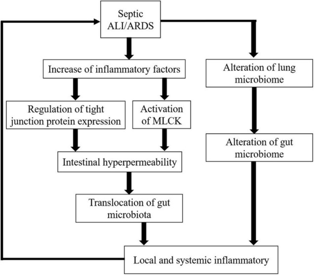
Acute lung injury (ALI) and acute respiratory distress syndrome (ARDS) are common acute and severe cases of the respiratory system with complicated pathogenesis and high mortality. Sepsis is the leading indirect cause of ALI/ARDS in the intensive care unit (ICU). The pathogenesis of septic ALI/ARDS is complex and multifactorial. In the development of sepsis, the disruption of the intestinal barrier function, the alteration of gut microbiota, and the translocation of the intestinal microbiome can lead to systemic and local inflammatory responses, which further alter the immune homeostasis in the systemic environment. Disruption of homeostasis may promote and propagate septic ALI/ARDS. In turn, when ALI occurs, elevated levels of inflammatory cytokines and the shift of the lung microbiome may lead to the dysregulation of the intestinal microbiome and the disruption of the intestinal mucosal barrier. Thus, the interaction between the lung and the gut can initiate and potentiate sepsis-induced ALI/ARDS. The gut–lung crosstalk may be a promising potential target for intervention. This article reviews the underlying mechanism of gut-lung crosstalk in septic ALI/ARDS.
Overview
The claims and statistics of this paper haven’t been checked yet.
Run the check — free, no account needed. It reads the open-access text and checks the sources it cites.
Integrity signals
Registry
Cites
Statistics
Figures
Verification
Claims the paper makes
The claims and statistics of this paper haven’t been checked yet.
Run the check — free, no account needed. It reads the open-access text and checks the sources it cites.
Claims other papers make about this one
Each is checked against this paper’s own text: does it say what the citing paper says it does?
The cited source says what the claim attributes to it.
In the citing paper
“The inflammatory cascade stemming from GI-released bacteria and endotoxins can exacerbate pulmonary dysfunction, leading to acute respiratory distress syndrome (ARDS) [27], worsening pulmonary outcomes [28].”
From Gastrointestinal function and nutritional interventions in septic shock (2025) · cites this paper as [27]
Self-check
Percentages, sums and reported differences re-computed from the paper’s own numbers.
The self-check needs the paper’s full text.
Citations
Cites
Bellani, G., Laffey, J. G., Pham, T., Fan, E., Brochard, L., Esteban, A., Gattinoni, L., van Haren, F., Larsson, A., McAuley, D. F., et al. (2016) · JAMA · doi:10.1001/jama.2016.0291 · Cited by 5,766 · cited 9×
“Sepsis is the leading indirect cause of ALI/ARDS in the intensive care unit (ICU).”
BackgroundInfluentialMacrophage plasticity, polarization, and function in health and disease
Shapouri‐Moghaddam, A., Mohammadian, S., Vazini, H., Taghadosi, M., Esmaeili, S., Mardani, F., Seifi, B., Mohammadi, A., Afshari, J. T., & Sahebkar, A. (2018) · Journal of Cellular Physiology · doi:10.1002/jcp.26429 · Cited by 4,930 · cited 1×
“Macrophages, commonly polarized into M1 (proinflammatory) and M2 (anti-inflammatory) types according to responses to environmental stimuli, are key orchestrators in the pathogenesis of ALI/ARDS (Shapouri-Moghaddam et al., 2018).”
BackgroundHost-gut microbiota metabolic interactions
Nicholson, J. K., Holmes, E., Kinross, J., Burcelin, R., Gibson, G., Jia, W., & Pettersson, S. (2012) · Science · doi:10.1126/science.1223813 · Cited by 4,760
Parada Venegas, D., De la Fuente, M. K., Landskron, G., González, M. J., Quera, R., Dijkstra, G., Harmsen, H. J. M., Faber, K. N., & Hermoso, M. A. (2019) · Frontiers in Immunology · doi:10.3389/fimmu.2019.00277 · Cited by 3,820
The human microbiome: at the interface of health and disease
Cho, I., & Blaser, M. J. (2012) · Nature Reviews Genetics · doi:10.1038/nrg3182 · Cited by 3,449 · cited 1×
“Notably, the differential immune responses to sepsis are determined by the microbiome (Cho and Blaser, 2012).”
BackgroundAcute respiratory distress syndrome
Matthay, M. A., Zemans, R. L., Zimmerman, G. A., Arabi, Y. M., Beitler, J. R., Mercat, A., Herridge, M. S., Randolph, A. G., & Calfee, C. S. (2019) · Nature Reviews Disease Primers · doi:10.1038/s41572-019-0069-0 · Cited by 2,514
A microbial symbiosis factor prevents intestinal inflammatory disease
Mazmanian, S. K., Round, J. L., & Kasper, D. L. (2008) · Nature · doi:10.1038/nature07008 · Cited by 2,340
The gut microbiota–brain axis in behaviour and brain disorders
Morais, L. H., Schreiber, H. L., & Mazmanian, S. K. (2021) · Nature Reviews Microbiology · doi:10.1038/s41579-020-00460-0 · Cited by 2,149
The microbiota in adaptive immune homeostasis and disease
Honda, K., & Littman, D. R. (2016) · Nature · doi:10.1038/nature18848 · Cited by 1,846
Bacteria as vitamin suppliers to their host: A gut microbiota perspective
LeBlanc, J. G., Milani, C., de Giori, G. S., Sesma, F., van Sinderen, D., & Ventura, M. (2013) · Current Opinion in Biotechnology · doi:10.1016/j.copbio.2012.08.005 · Cited by 1,613
Acute Respiratory Distress Syndrome
Fan, E., Brodie, D., & Slutsky, A. S. (2018) · JAMA · doi:10.1001/jama.2017.21907 · Cited by 1,482
Claudins and the Modulation of Tight Junction Permeability
Günzel, D., & Yu, A. S. L. (2013) · Physiological Reviews · doi:10.1152/physrev.00019.2012 · Cited by 1,456 · cited 1×
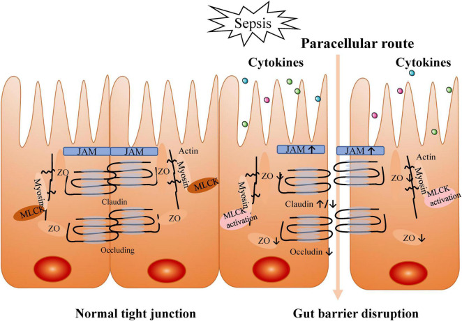
“Members of the claudin family are major driving forces in the formation of paracellular barriers (Günzel and Yu, 2013).”
BackgroundImpact of the Gut Microbiota on Intestinal Immunity Mediated by Tryptophan Metabolism
Gao, J., Xu, K., Liu, H., Liu, G., Bai, M., Peng, C., Li, T., & Yin, Y. (2018) · Frontiers in Cellular and Infection Microbiology · doi:10.3389/fcimb.2018.00013 · Cited by 1,357
Hydrocortisone, Vitamin C, and Thiamine for the Treatment of Severe Sepsis and Septic Shock
Marik, P. E., Khangoora, V., Rivera, R., Hooper, M. H., & Catravas, J. (2017) · Chest · doi:10.1016/j.chest.2016.11.036 · Cited by 887
Mechanisms and treatment of organ failure in sepsis
Lelubre, C., & Vincent, J. (2018) · Nature Reviews Nephrology · doi:10.1038/s41581-018-0005-7 · Cited by 877
Dickson, R. P., Singer, B. H., Newstead, M. W., Falkowski, N. R., Erb-Downward, J. R., Standiford, T. J., & Huffnagle, G. B. (2016) · Nature Microbiology · doi:10.1038/nmicrobiol.2016.113 · Cited by 724
The microbial metabolite desaminotyrosine protects from influenza through type I interferon
Steed, A. L., Christophi, G. P., Kaiko, G. E., Sun, L., Goodwin, V. M., Jain, U., Esaulova, E., Artyomov, M. N., Morales, D. J., Holtzman, M. J., et al. (2017) · Science · doi:10.1126/science.aam5336 · Cited by 569 · cited 1×
“Phenolic One of the degradation products of flavonoids and amino acids by commensal bacteria is desaminotyrosine (DAT; Steed et al., 2017).”
BackgroundZhao, Y., Chen, F., Wu, W. H., Sun, M., Bilotta, A. J., Yao, S., Xiao, Y., Huang, X., Eaves-Pyles, T. D., Golovko, G., et al. (2018) · Mucosal Immunology · doi:10.1038/mi.2017.118 · Cited by 536
Guidelines on the management of acute respiratory distress syndrome
Griffiths, M., McAuley, D. F., Perkins, G. D., Barrett, N. A., Blackwood, B., Boyle, A. J., Chee, N., Connolly, B. A., Dark, P. M., Finney, S. J., et al. (2019) · BMJ Open Respiratory Research · doi:10.1136/bmjresp-2019-000420 · Cited by 519
Tang, J., Xu, L., Zeng, Y., & Gong, F. (2020) · International Immunopharmacology · doi:10.1016/j.intimp.2020.107272 · Cited by 517 · cited 2×
“This inhibits the activation of the TLR4/NF-κB signaling pathway in the lung, inflammation, and the release of oxidative stress factors in ALI animals (Tang et al., 2021).”
BackgroundWang, J., Li, F., Wei, H., Lian, Z.-X., Sun, R., & Tian, Z. (2014) · Journal of Experimental Medicine · doi:10.1084/jem.20140625 · Cited by 490 · cited 1×
“In the context of experimental influenza infection, it has been reported that the IFN-γ produced by lung-derived CCR9 + CD4 + T cells changed the composition of the gut microbiota, and caused intestinal immune injury (Wang et al., 2014).”
BackgroundPathogenesis of Acute Respiratory Distress Syndrome
Huppert, L. A., Matthay, M. A., & Ware, L. B. (2019) · Seminars in Respiratory and Critical Care Medicine · doi:10.1055/s-0039-1683996 · Cited by 475
The microbiome and critical illness
Dickson, R. P. (2016) · The Lancet Respiratory Medicine · doi:10.1016/s2213-2600(15)00427-0 · Cited by 460 · cited 1×
“, 2016), which allows intact microbes and microbiota products to translocate, resulting in amplification of the systemic inflammatory response and contribution to multiple organ failure (Dickson, 2016).”
BackgroundChen, X., Tang, J., Shuai, W., Meng, J., Feng, J., & Han, Z. (2020) · Inflammation Research · doi:10.1007/s00011-020-01378-2 · Cited by 438 · cited 1×
“Thus, the regulation of macrophage polarization from M1 to M2 may be a novel therapeutic strategy of ALI/ARDS (Chen et al., 2020).”
BackgroundThe Gut as the Motor of Multiple Organ Dysfunction in Critical Illness
Klingensmith, N. J., & Coopersmith, C. M. (2016) · Critical Care Clinics · doi:10.1016/j.ccc.2015.11.004 · Cited by 429
The role of the gut microbiota in sepsis
Haak, B. W., & Wiersinga, W. J. (2017) · The Lancet. Gastroenterology & hepatology · doi:10.1016/s2468-1253(16)30119-4 · Cited by 392
Dickson, R. P., Erb-Downward, J. R., & Huffnagle, G. B. (2014) · The Lancet Respiratory Medicine · doi:10.1016/s2213-2600(14)70028-1 · Cited by 371 · cited 1×
“, 2015) may alter the lung microbiome that in turn perpetuates a positive-feedback loop of inflammation, injury, and further dysbiosis (Dickson et al., 2014).”
BackgroundLung Microbiota Predict Clinical Outcomes in Critically Ill Patients
Dickson, R. P., Schultz, M. J., van der Poll, T., Schouten, L. R. A., Falkowski, N. R., Luth, J. E., Sjoding, M. W., Brown, C. A., Chanderraj, R., Huffnagle, G. B., et al. (2020) · American Journal of Respiratory and Critical Care Medicine · doi:10.1164/rccm.201907-1487oc · Cited by 369
Intestinal Permeability Defects: Is It Time to Treat?
Odenwald, M. A., & Turner, J. R. (2013) · Clinical Gastroenterology and Hepatology · doi:10.1016/j.cgh.2013.07.001 · Cited by 366
Gut-origin sepsis: evolution of a concept
Deitch, E. A. (2012) · The Surgeon · doi:10.1016/j.surge.2012.03.003 · Cited by 359 · cited 1×
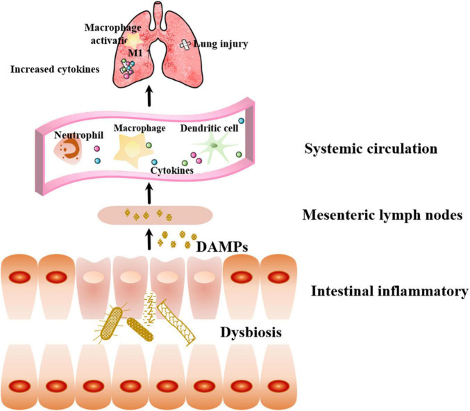
“A massive release of DAMPs accelerates the progression of organ damage and multiple organ dysfunction syndrome (MODS), further aggravating gut injury, forming a vicious cycle (Deitch, 2012; Assimakopoulos et al., 2018; Haussner et al., 2019).”
BackgroundClinical Application and Potential of Fecal Microbiota Transplantation
Ooijevaar, R. E., Terveer, E. M., Verspaget, H. W., Kuijper, E. J., & Keller, J. J. (2018) · Annual Review of Medicine · doi:10.1146/annurev-med-111717-122956 · Cited by 348
The gut microbiome’s role in the development, maintenance, and outcomes of sepsis
Adelman, M. W., Woodworth, M. H., Langelier, C., Busch, L. M., Kempker, J. A., Kraft, C. S., & Martin, G. S. (2020) · Critical Care · doi:10.1186/s13054-020-02989-1 · Cited by 342 · cited 2×
“An increasing body of evidence has illustrated the role of the gut microbiome in the occurrence, development, and outcomes of sepsis (Adelman et al., 2020).”
BackgroundRegulation of alveolar macrophage death in acute lung inflammation
Fan, E., & Fan, J. (2018) · Respiratory Research · doi:10.1186/s12931-018-0756-5 · Cited by 320
Lankelma, J. M., van Vught, L. A., Belzer, C., Schultz, M. J., van der Poll, T., de Vos, W. M., & Wiersinga, W. J. (2016) · Intensive Care Medicine · doi:10.1007/s00134-016-4613-z · Cited by 282
Deriu, E., Boxx, G. M., He, X., Pan, C., Benavidez, S. D., Cen, L., Rozengurt, N., Shi, W., & Cheng, G. (2016) · PLoS Pathogens · doi:10.1371/journal.ppat.1005572 · Cited by 280 · cited 1×
“Moreover, the pulmonary production of IFN-Is promotes the consumption of obligate anaerobic bacteria and the enrichment of proteobacteria in the gut, leading to significant intestinal dysregulation (Deriu et al., 2016).”
BackgroundRole of gut microbiota on intestinal barrier function in acute pancreatitis
Li, X.-Y., He, C., Zhu, Y., & Lü, N. (2020) · World Journal of Gastroenterology · doi:10.3748/wjg.v26.i18.2187 · Cited by 258
The Immature Gut Barrier and Its Importance in Establishing Immunity in Newborn Mammals
Weström, B. R., Sureda, E. A., Pierzynowska, K. G., Pierzynowski, S. G., & Pérez-Cano, F. J. (2020) · Frontiers in Immunology · doi:10.3389/fimmu.2020.01153 · Cited by 258 · cited 1×
“When the selective barrier functions normally, it allows the cell-side movement of water, solutes, and immunomodulatory factors but prevents the movement of macromolecules and microorganisms (Weström et al., 2020).”
BackgroundMechanisms of Intestinal Barrier Dysfunction in Sepsis
Yoseph, B. P., Klingensmith, N. J., Liang, Z., Breed, E. R., Burd, E. M., Mittal, R., Dominguez, J. A., Petrie, B., Ford, M. L., & Coopersmith, C. M. (2016) · Shock · doi:10.1097/shk.0000000000000565 · Cited by 256 · cited 3×
“Sepsis induces significantly compromised gut barrier integrity (Yoseph et al., 2016), which allows intact microbes and microbiota products to translocate, resulting in amplification of the systemic inflammatory response and contribution to multiple organ failure (Dickson, 2016).”
BackgroundInfluentialThiamine deficiency in critically ill patients with sepsis
Donnino, M. W., Carney, E., Cocchi, M. N., Barbash, I. J., Chase, M., Joyce, N. R., Chou, P. P., & Ngo, L. H. (2010) · Journal of Critical Care · doi:10.1016/j.jcrc.2010.03.003 · Cited by 253
Gut-origin sepsis in the critically ill patient: pathophysiology and treatment
Assimakopoulos, S. F., Triantos, C., Thomopoulos, K., Fligou, F., Maroulis, I., Marangos, M., & Gogos, C. A. (2018) · Infection · doi:10.1007/s15010-018-1178-5 · Cited by 244 · cited 2×
“A massive release of DAMPs accelerates the progression of organ damage and multiple organ dysfunction syndrome (MODS), further aggravating gut injury, forming a vicious cycle (Deitch, 2012; Assimakopoulos et al., 2018; Haussner et al., 2019).”
BackgroundWang, J., Li, F., Sun, R., Gao, X., Wei, H., Li, L.-J., & Tian, Z. (2013) · Nature Communications · doi:10.1038/ncomms3106 · Cited by 239 · cited 1×
“In clinical cases, the most common source of sepsis is the lung (Wang et al., 2013).”
BackgroundGut microbiota dependent trimethylamine N-oxide aggravates angiotensin II–induced hypertension
Jiang, S., Shui, Y., Cui, Y., Tang, C., Wang, X., Qiu, X., Hu, W., Fei, L., Li, Y., Zhang, S., et al. (2021) · Redox Biology · doi:10.1016/j.redox.2021.102115 · Cited by 237
Shimizu, K., Yamada, T., Ogura, H., Mohri, T., Kiguchi, T., Fujimi, S., Asahara, T., Yamada, T., Ojima, M., Ikeda, M., et al. (2018) · Critical Care · doi:10.1186/s13054-018-2167-x · Cited by 237
Commensal Microbes Induce Serum IgA Responses that Protect against Polymicrobial Sepsis
Wilmore, J. R., Gaudette, B. T., Gómez, D., Hashemi, T., Jones, D. D., Gardner, C. A., Cole, S. D., Misic, A. M., Beiting, D. P., & Allman, D. (2018) · Cell Host & Microbe · doi:10.1016/j.chom.2018.01.005 · Cited by 235 · cited 1×
“Commensal bacteria directly produce IgA, which has a protective activity against mucosal barrier disruption and the resulting sepsis (Wilmore et al., 2018).”
BackgroundTrimethylamine N-oxide (TMAO) in human health
Gątarek, P., & Kałużna‐Czaplińska, J. (2020) · EXCLI journal · doi:10.17179/excli2020-3239 · Cited by 235
Challenge to the Intestinal Mucosa During Sepsis
Haußner, F., Chakraborty, S., Halbgebauer, R., & Huber-Lang, M. S. (2019) · Frontiers in Immunology · doi:10.3389/fimmu.2019.00891 · Cited by 233
Li, X., Jamal, M., Guo, P., Jin, Z. Y., Zheng, F., Song, X., Zhan, J., & Wu, H. (2019) · Biomedicine & Pharmacotherapy · doi:10.1016/j.biopha.2019.109363 · Cited by 196
Interplay between gut microbiota and antimicrobial peptides
Zong, X., Fu, J., Xu, B., Wang, Y., & Jin, M. (2020) · Animal nutrition · doi:10.1016/j.aninu.2020.09.002 · Cited by 193
Sze, M. A., Tsuruta, M., Yang, S.-W. J., Oh, Y., Man, S. F. P., Hogg, J. C., & Sin, D. D. (2014) · PLoS ONE · doi:10.1371/journal.pone.0111228 · Cited by 187
for the LUNG-SAFE Investigators and the ESICM Trials Group, Abe, T., Madotto, F., Pham, T. O., Nagata, I., Uchida, M., Tamiya, N., Kurahashi, K., Bellani, G., & Laffey, J. G. (2018) · Critical Care · doi:10.1186/s13054-018-2126-6 · Cited by 186 · cited 8×
“Sepsis is the leading indirect cause of ALI/ARDS in the intensive care unit (ICU).”
BackgroundInfluentialCollapse of the Microbiome, Emergence of the Pathobiome, and the Immunopathology of Sepsis
Alverdy, J. C., & Krezalek, M. A. (2017) · Critical Care Medicine · doi:10.1097/ccm.0000000000002172 · Cited by 185 · cited 3×
“DAMPS are recognized by cells expressing intrinsic pattern receptors in the intrinsic immune system, including macrophages, white blood cells, and dendritic cells, then secreting pro-inflammatory factors that promote lung injury and the development of MODS, which further aggravate intestinal barrier injury and lead to…”
BackgroundMicrobial Contribution to the Human Metabolome: Implications for Health and Disease
Treuren, W. V., & Dodd, D. (2019) · Annual Review of Pathology Mechanisms of Disease · doi:10.1146/annurev-pathol-020117-043559 · Cited by 180
Pulmonary infections complicating ARDS
Luyt, C. E., Bouadma, L., Morris, A. C., Dhanani, J., Kollef, M. H., Lipman, J., Martín‐Loeches, I., Nseir, S., Ranzani, O., Roquilly, A., et al. (2020) · Intensive Care Medicine · doi:10.1007/s00134-020-06292-z · Cited by 177
Flavonoids and Immune Function in Human: A Systematic Review
Peluso, I., Miglio, C., Morabito, G., Ioannone, F., & Serafini, M. (2013) · Critical Reviews in Food Science and Nutrition · doi:10.1080/10408398.2012.656770 · Cited by 172
Homeostasis and its disruption in the lung microbiome
Dickson, R. P., Erb‐Downward, J. R., & Huffnagle, G. B. (2015) · American Journal of Physiology-Lung Cellular and Molecular Physiology · doi:10.1152/ajplung.00279.2015 · Cited by 166 · cited 1×
“, 2019), the flow of protein-rich alveolar exudate, the establishment of oxygen gradients, the surge of inflammatory mediators, and the impairment of local immune defenses (Dickson et al., 2015) may alter the lung microbiome that in turn perpetuates a positive-feedback loop of inflammation, injury, and further dysbios…”
BackgroundAgudelo-Ochoa, G. M., Valdés-Duque, B. E., Giraldo-Giraldo, N. A., Jaillier-Ramírez, A. M., Giraldo-Villa, A., Acevedo-Castaño, I., Yepes-Molina, M. A., Barbosa-Barbosa, J., & Benítez-Paéz, A. (2020) · Gut Microbes · doi:10.1080/19490976.2019.1707610 · Cited by 166 · cited 1×
“Concomitantly, researchers have detected an increased abundance of Enterococcus and other pathogenic bacteria in sepsis patients who died, suggesting that these bacteria may be potential biomarkers for ICU care (Agudelo-Ochoa et al., 2020).”
BackgroundGut integrity in critical illness
Otani, S., & Coopersmith, C. M. (2019) · Journal of Intensive Care · doi:10.1186/s40560-019-0372-6 · Cited by 165 · cited 1×
“Three factors contribute to intestinal barrier damage: visceral hypoperfusion or ischemia, restoration of intestinal blood flow during resuscitation leading to ischemiareperfusion injury, and loss of the intestinal barrier function, which allows bacteria, endotoxins, or both to cross the mucosal barrier (Otani and Coo…”
BackgroundTherapeutic Potential of the Gut Microbiota in the Prevention and Treatment of Sepsis
Haak, B. W., Prescott, H. C., & Wiersinga, W. J. (2018) · Frontiers in Immunology · doi:10.3389/fimmu.2018.02042 · Cited by 163
Panzer, A. R., Lynch, S. V., Langelier, C., Christie, J. D., McCauley, K. E., Nelson, M. F., Cheung, C. K., Benowitz, N. L., Cohen, M. J. M., & Calfee, C. S. (2017) · American Journal of Respiratory and Critical Care Medicine · doi:10.1164/rccm.201702-0441oc · Cited by 157
Overexpression of Bcl-2 in the intestinal epithelium improves survival in septic mice
Coopersmith, C. M., Chang, K. C., Swanson, P. E., Tinsley, K. W., Stromberg, P. E., Buchman, T. G., Karl, I. E., & Hotchkiss, R. S. (2002) · Critical Care Medicine · doi:10.1097/00003246-200201000-00028 · Cited by 152 · cited 1×
“, 2003), with increased intestinal epithelial cells (IECs; Coopersmith et al., 2002) and crypt cell apoptosis (Perrone et al.”
BackgroundAdministration of nicotinamide riboside prevents oxidative stress and organ injury in sepsis
Hong, G., Dong, Z., Zhang, L., Ni, R., Wang, G., Fan, G., Lu, Z., & Peng, T. (2018) · Free Radical Biology and Medicine · doi:10.1016/j.freeradbiomed.2018.05.073 · Cited by 149
Neal, M. D., Sodhi, C. P., Dyer, M., Craig, B. T., Good, M. L., Jia, H., Yazji, I., Afrazi, A., Richardson, W. M., Beer–Stolz, D., et al. (2013) · The Journal of Immunology · doi:10.4049/jimmunol.1202264 · Cited by 147
Kim, S. M., DeFazio, J. R., Hyoju, S. K., Sangani, K. A., Keskey, R. C., Krezalek, M. A., Khodarev, N. N., Sangwan, N., Christley, S., Harris, K. G., et al. (2020) · Nature Communications · doi:10.1038/s41467-020-15545-w · Cited by 144
Kim, W., Jo, E., Eom, J. S., Mok, J., Kim, M., Kim, K. U., Park, H.-K., Lee, M. K., & Lee, K. (2018) · Journal of Critical Care · doi:10.1016/j.jcrc.2018.07.004 · Cited by 132
Increased gut permeability and bacterial translocation in Pseudomonas pneumonia-induced sepsis
Yu, P. W., & Martin, C. M. (2000) · Critical Care Medicine · doi:10.1097/00003246-200007000-00065 · Cited by 119
Gut Microbiota in Intestinal and Liver Disease
Jones, R. M., & Neish, A. S. (2020) · Annual Review of Pathology Mechanisms of Disease · doi:10.1146/annurev-pathol-030320-095722 · Cited by 117
Coopersmith, C. M., Stromberg, P. E., Davis, C. G., Dunne, W. M., Amiot, D. M., Karl, I. E., Hotchkiss, R. S., & Buchman, T. G. (2003) · Critical Care Medicine · doi:10.1097/01.ccm.0000055385.29232.11 · Cited by 110 · cited 1×
“At the cellular level, crypt cell proliferation is markedly decreased (Coopersmith et al., 2003), with increased intestinal epithelial cells (IECs; Coopersmith et al.”
BackgroundDisruption of tight junctions during polymicrobial sepsis in vivo
Li, Q., Zhang, Q., Wang, C., Liu, X., Li, N., & Li, J. (2009) · The Journal of Pathology · doi:10.1002/path.2525 · Cited by 106
Breakdown of Mucin as Barrier to Digestive Enzymes in the Ischemic Rat Small Intestine
Chang, M., Alsaigh, T., Kistler, E. B., & Schmid‐Schönbein, G. W. (2012) · PLoS ONE · doi:10.1371/journal.pone.0040087 · Cited by 103 · cited 1×
“Moreover, cytokines cause an abnormal intestinal and barrier function of the mucous layer, characterized by reduced thickness, diminished lumen coverage, and poor adhesion (Chang et al., 2012).”
BackgroundAlterations of lung microbiota in a mouse model of LPS-induced lung injury
Poroyko, V., Meng, F., Meliton, A., Afonyushkin, T., Ulanov, A., Semenyuk, E., Latif, O., Tesic, V., Birukova, A. A., & Birukov, K. G. (2015) · American Journal of Physiology-Lung Cellular and Molecular Physiology · doi:10.1152/ajplung.00061.2014 · Cited by 102
The gut microbiome alters immunophenotype and survival from sepsis
Fay, K. T., Klingensmith, N. J., Chen, C., Zhang, W., Sun, Y., Morrow, K. N., Liang, Z., Burd, E. M., Ford, M. L., & Coopersmith, C. M. (2019) · The FASEB Journal · doi:10.1096/fj.201802188r · Cited by 101
Liu, J., Chang, G., Huang, J., Wang, Y., Ma, N., Roy, A.-C., & Shen, X. (2019) · Journal of Agricultural and Food Chemistry · doi:10.1021/acs.jafc.8b06359 · Cited by 100
Pinheiro, N. M., Santana, F. P. R., Almeida, R. R., Guerreiro, M., Martins, M. A., Caperuto, L. C., Câmara, N. O. S., Wensing, L. A., Prado, V. F., Tibério, I. D. F. L. C., et al. (2016) · The FASEB Journal · doi:10.1096/fj.201600431r · Cited by 95
Shimizu, K., Ogura, H., Asahara, T., Nomoto, K., Morotomi, M., Nakahori, Y., Osuka, A., Yamano, S., Goto, M., Matsushima, A., et al. (2010) · Neurogastroenterology & Motility · doi:10.1111/j.1365-2982.2010.01653.x · Cited by 94
Yamada, T., Shimizu, K., Ogura, H., Asahara, T., Nomoto, K., Yamakawa, K., Hamasaki, T., Nakahori, Y., Ohnishi, M., Kuwagata, Y., et al. (2014) · Journal of Parenteral and Enteral Nutrition · doi:10.1177/0148607114529596 · Cited by 93 · cited 1×
“Several studies have confirmed significantly decreased SCFA concentrations in sepsis patients (Shimizu et al., 2011, 2018; Yamada et al., 2015; Valdes-Duque et al., 2020).”
BackgroundEnteral nutrient deprivation in patients leads to a loss of intestinal epithelial barrier function
Ralls, M. W., Demehri, F. R., Feng, Y., Ignatoski, K. M. W., & Teitelbaum, D. H. (2015) · Surgery · doi:10.1016/j.surg.2014.12.004 · Cited by 84
Kapetanaki, S., Kumawat, A. K., Persson, K., & Demirel, I. (2021) · International Journal of Molecular Sciences · doi:10.3390/ijms222111864 · Cited by 80
Wang, Y., Xu, Y., Zhang, P., Ruan, W., Zhang, L., Yuan, S., Pang, T., & Jia, A. (2018) · Biochemical Pharmacology · doi:10.1016/j.bcp.2018.09.002 · Cited by 77
Claudins: vital partners in transcellular and paracellular transport coupling
Günzel, D. (2016) · Pflügers Archiv - European Journal of Physiology · doi:10.1007/s00424-016-1909-3 · Cited by 75
Sodium butyrate alleviates LPS-induced acute lung injury in mice via inhibiting HMGB1 release
Li, N., Liu, X., Hong, M., Huang, X., Chen, H., Xu, J., Wang, C., Zhang, Y.-X., Zhong, J., Nie, H., et al. (2018) · International Immunopharmacology · doi:10.1016/j.intimp.2018.01.017 · Cited by 75
Vitamin C and Thiamine for Sepsis and Septic Shock
Mitchell, A. B., Ryan, T. E., Gillion, A. R., Wells, L., & Muthiah, M. P. (2019) · The American Journal of Medicine · doi:10.1016/j.amjmed.2019.07.054 · Cited by 69
Senthil, M., Watkins, A. C., Barlos, D., Xu, D.-Z., Lu, Q., Abungu, B., Caputo, F. J., Feinman, R., & Deitch, E. A. (2007) · Annals of Surgery · doi:10.1097/sla.0b013e3180caa3af · Cited by 67 · cited 1×
“Moreover, mesenteric lymphatic vessels collected from critically ill mice induces lung injury upon intravenous injection in healthy mice (Senthil et al., 2007).”
BackgroundAlterations of the murine gut microbiome with age and allergic airway disease
Vital, M., Harkema, J. R., Rizzo, M., Tiedje, J., & Brandenberger, C. (2015) · Journal of Immunology Research · doi:10.1155/2015/892568 · Cited by 65 · cited 2×
“Thus, lung inflammation directly changes the intestinal bacterial community structure and further worsens lung inflammation (Vital et al., 2015).”
BackgroundPerrone, E. E., Jung, E., Breed, E. R., Dominguez, J. A., Liang, Z., Clark, A. T., Dunne, W. M., Burd, E. M., & Coopersmith, C. M. (2012) · Shock · doi:10.1097/shk.0b013e318259abdb · Cited by 65
Cathelicidin preserves intestinal barrier function in polymicrobial sepsis
Ho, J., Chan, H., Liang, Y., Liu, X., Zhang, L., Li, Q., Zhang, Y., Zeng, J., Ugwu, F. N., Ho, I. H. T., et al. (2020) · Critical Care · doi:10.1186/s13054-020-2754-5 · Cited by 62
Wang, J., Li, R., Peng, Z., Zhou, W.-H., Hu, B., Rao, X., Yang, X., & Li, J. (2018) · Shock · doi:10.1097/shk.0000000000001144 · Cited by 60
MESENTERIC LYMPH DUCT LIGATION IMPROVES SURVIVAL IN A LETHAL SHOCK MODEL
Badami, C. D., Senthil, M., Caputo, F. J., Rupani, B., Doucet, D. R., Pisarenko, V., Xu, D.-Z., Lu, Q., Feinman, R., & Deitch, E. A. (2008) · Shock · doi:10.1097/shk.0b013e318173edd1 · Cited by 59 · cited 1×
“Studies have suggested that ligation of mesenteric lymphatic vessels attenuates lung injury and neutrophil activation and improves survival in mice with endotoxemia (Badami et al., 2008).”
BackgroundLiu, M.-J., Bao, S., Napolitano, J. R., Burris, D. L., Yu, L., Tridandapani, S., & Knoell, D. L. (2014) · PLoS ONE · doi:10.1371/journal.pone.0094934 · Cited by 58
Zhuo, Y., Li, D., Cui, L., Li, C., Zhang, S., Zhang, Q., Zhang, L., Wang, X., & Yang, L. (2019) · Biomedicine & Pharmacotherapy · doi:10.1016/j.biopha.2019.109012 · Cited by 57
Inhibition of myeloid differentiation factor 2 by baicalein protects against acute lung injury
Chen, H., Zhang, Y., Zhang, W., Liu, H., Sun, C., Zhang, B., Bai, B., Wu, D., Xiao, Z., Lum, H., et al. (2019) · Phytomedicine · doi:10.1016/j.phymed.2019.152997 · Cited by 57 · cited 1×
“It is worth noting that the mortality rate of ARDS caused by sepsis is higher than that of ARDS caused by other factors (Chen et al., 2019).”
BackgroundDiagnosis and management of acute respiratory distress syndrome
Fernando, S. M., Ferreyro, B. L., Urner, M., Munshi, L., & Fan, E. (2021) · Canadian Medical Association Journal · doi:10.1503/cmaj.202661 · Cited by 56
Bittencourt-Mernak, M. I., Pinheiro, N. M., Santana, F. P. R., Guerreiro, M., Saraiva-Romanholo, B. M., Grecco, S. S., Caperuto, L. C., Felizardo, R. J. F., Câmara, N. O. S., Tibério, I. D. F. L. C., et al. (2016) · American Journal of Physiology-Lung Cellular and Molecular Physiology · doi:10.1152/ajplung.00444.2015 · Cited by 54 · cited 1×
“Accumulating evidence indicates that some natural or synthetic materials can ameliorate the prognosis of sepsis-induced ALI in animal models by inhibiting pulmonary M1 polarization and altering the macrophage function (Bittencourt-Mernak et al., 2017; Pinheiro et al., 2017; Wang et al., 2018, 2019; Zhuo et al., 2019).”
BackgroundLorentz, A., Liang, Z., Meng, M., Chen, C., Yoseph, B. P., Breed, E. R., Mittal, R., Klingensmith, N. J., Farris, A. B., Burd, E. M., et al. (2017) · Molecular Medicine · doi:10.2119/molmed.2016.00256 · Cited by 54
Wei, Y., Gao, J., Kou, Y., Liu, M., Meng, L., Zheng, X., Xu, S., Ming, L., Sun, H., Liu, Z., et al. (2020) · The FASEB Journal · doi:10.1096/fj.201902900rr · Cited by 51
Xu, Y., Zhu, J., Feng, B., Lin, F., Zhou, J., Liu, J., Shi, X., Lu, X., Pan, Q., Yu, J., et al. (2021) · Cell Proliferation · doi:10.1111/cpr.13028 · Cited by 49 · cited 1×
“Depletion of intestinal microbiota with broad-spectrum antibiotics could exacerbate lung and intestinal damage and increase the mortality in sepsis (Xu et al., 2021).”
BackgroundThe lung lesion in four different types of shock in rabbits
Cuevas, P. (1972) · Archives of Surgery · doi:10.1001/archsurg.1972.04180030067015 · Cited by 48 · cited 1×
“When the bacterial burden of the gut is minimized, the inflammation and injury sustained with distal organs are lessened during sepsis (Cuevas et al., 1972).”
BackgroundStool Short-Chain Fatty Acids in Critically Ill Patients with Sepsis
Valdés-Duque, B. E., Giraldo, N. A. G., Jaillier-Ramírez, A. M., Villa, A. G., Acevedo-Castaño, I., Yepes-Molina, M. A., Barbosa-Barbosa, J., Barrera-Causil, C., & Agudelo, G. M. (2020) · Journal of the American College of Nutrition · doi:10.1080/07315724.2020.1727379 · Cited by 48 · cited 1×
“Several studies have confirmed significantly decreased SCFA concentrations in sepsis patients (Shimizu et al., 2011, 2018; Yamada et al., 2015; Valdes-Duque et al., 2020).”
BackgroundIndole signaling decreases biofilm formation and related virulence of Listeria monocytogenes
Rattanaphan, P., Mittraparp‐arthorn, P., Srinoun, K., Vuddhakul, V., & Tansila, N. (2020) · FEMS Microbiology Letters · doi:10.1093/femsle/fnaa116 · Cited by 47
Liu, J., Wang, M., Chen, W., Ma, J., Peng, Y., Zhang, M., Wang, C., Yan, G., & Lü, G. (2021) · Frontiers in Pediatrics · doi:10.3389/fped.2021.645060 · Cited by 46
Dominguez, J. A., Vithayathil, P. J., Khailova, L. S., Lawrance, C. P., Samocha, A. J., Jung, E., Leathersich, A. M., Dunne, W. M., & Coopersmith, C. M. (2011) · Shock · doi:10.1097/shk.0b013e31822793c4 · Cited by 45
New markers for sepsis caused by Pseudomonas aeruginosa during burn infection
Elmassry, M. M., Mudaliar, N. S., Colmer‐Hamood, J. A., Francisco, M. J. S., Griswold, J., Dissanaike, S., & Hamood, A. N. (2020) · Metabolomics · doi:10.1007/s11306-020-01658-2 · Cited by 38
Vitamin B6 reduces oxidative stress in lungs and liver in experimental sepsis
Giustina, A. D., Danielski, L. G., NOVOCHADLO, M. M., Goldim, M. P. D. S., Joaquim, L. D. S., Metzker, K. L. L., Carli, R. J. D., Denicol, T. L., Cidreira, T., Vieira, T. C., et al. (2019) · Anais da Academia Brasileira de Ciências · doi:10.1590/0001-3765201920190434 · Cited by 36
Special Issue on “The Tight Junction and Its Proteins: More than Just a Barrier”
Krug, S. M., & Fromm, M. (2020) · International Journal of Molecular Sciences · doi:10.3390/ijms21134612 · Cited by 36
5-HT Drives Mortality in Sepsis Induced by Cecal Ligation and Puncture in Mice
Zhang, J., Bi, J., Liu, S., Pang, Q., Zhang, R., Wang, S., & Liu, C. (2017) · Mediators of Inflammation · doi:10.1155/2017/6374283 · Cited by 34
Schmitt, F. C. F., Lipinski, A. L., Hofer, S., Uhle, F., Nußhag, C., Hackert, T., Dalpke, A. H., Weigand, M. A., Brenner, T., & Boutin, S. (2020) · Journal of Hospital Infection · doi:10.1016/j.jhin.2020.04.028 · Cited by 31 · cited 1×
“A recent clinical study (Schmitt et al., 2020) compared the pulmonary microbiota in 15 patients with sepsisinduced ARDS undergoing abdominal surgery and 15 patients post esophagectomy.”
BackgroundLevesque, C. L., Turner, J. M., Li, J., Wizzard, P. R., Pierre, B. S., Lim, D. W., & Wales, P. W. (2016) · Journal of Parenteral and Enteral Nutrition · doi:10.1177/0148607115626903 · Cited by 27
[ The intestine-liver-lung axis in septic syndrome].
Pugin, J., & Chevrolet, J.-C. (1991) · PubMed · Cited by 24
Vancomycin-induced gut dysbiosis duringPseudomonas aeruginosapulmonary infection in a mice model
Rosa, C. P., Pereira, J. A., Santos, N. C. D. M., Brancaglion, G. A., Silva, E. N., Tagliati, C. A., Novaes, R. D., Corsetti, P. P., & de Almeida, L. A. (2019) · Journal of Leukocyte Biology · doi:10.1002/jlb.4ab0919-432r · Cited by 23
Su, H., Zhao, W., Zhang, F., Song, M., Liu, F., Zheng, J., Ling, M., Yang, X., Yang, Q., He, H., et al. (2020) · Food & Function · doi:10.1039/d0fo00376j · Cited by 21
Xu, M., Wang, C., Li, N., Wang, J., Zhang, Y., & Deng, X. (2019) · Medical Science Monitor · doi:10.12659/msm.911444 · Cited by 19 · cited 1×
“In addition, acetic acid alleviates septic ALI by regulating the mitogen-activated protein kinase (MAPK) pathway, improving the alveolar permeability, reducing inflammatory factors, and inhibiting oxygen free radical production (Xu et al., 2019).”
BackgroundChou, R., Wu, P.-S., Wang, S., Wu, C., Lu, S., Lien, R., Tsai, Y., Lu, Y., Kuo, M.-R., Guo, J., et al. (2021) · Journal of Intensive Care · doi:10.1186/s40560-021-00581-5 · Cited by 17 · cited 1×
“However, a recent single-center prospective study reveals that TMAO may play different roles in CV and infectious diseases (Chou et al., 2021).”
BackgroundChen, Y., Yang, X.-Q., Tseng, B.-Y., Tsai, Y., Tseng, S., Lee, C.-H., & Yao, C. (2018) · Journal of Pediatric Surgery · doi:10.1016/j.jpedsurg.2018.01.023 · Cited by 12 · cited 1×
“Notably, toll-like receptor 4 (TLR4) is widely expressed in intestinal stem cells and regulates their proliferation or apoptosis (Chen et al., 2018).”
BackgroundALTERATIONS IN MUCOSAL IMMUNE CELL DISTRIBUTION IN SEPTIC RATS
Österberg, J., Johnsson, C., Gannedahl, G., Westlund, A., & Haglund, U. (1997) · Shock · doi:10.1097/00024382-199703000-00005 · Cited by 11
Yoo, J., Kim, R. B., Ju, S., Lee, S. J., Cho, Y. J., Jeong, Y. Y., Lee, J. D., & Kim, H. C. (2020) · Tuberculosis & respiratory diseases · doi:10.4046/trd.2020.0008 · Cited by 8 · cited 1×
“However, an recent single center, retrospective study didn’t find that vitamin B1 and C supplementation improve clinical outcomes (mortality rates, ventilator and ICU-free days) in septic ARDS patients requiring invasive mechanical ventilation (Yoo et al., 2020).”
BackgroundYin, G., Li, B., & Fan, X. M. (2019) · PubMed · doi:10.3760/cma.j.issn.0376-2491.2019.20.013 · Cited by 6 · cited 1×
“FMT intervention could correct the changes in the intestinal flora and improve lung injury by inhibiting the activation of the PI3K/AKT/NF-κB signaling pathway and decreasing the expression of intercellular cell adhesion molecule-1 (ICAM-1; Yin et al., 2019).”
Background
Cited by
Sorted by citations
Lung microbiome: new insights into the pathogenesis of respiratory diseases
Li, R., Li, J., & Zhou, X. (2024) · Signal Transduction and Targeted Therapy · doi:10.1038/s41392-023-01722-y · Cited by 483
“16,137,138 The gut-lung axis is bidirectional, which means that the lung can affect gut homeostasis.”
BackgroundSelf-citationAcute lung injury caused by sepsis: how does it happen?
Sun, B., Lei, M., Zhang, J., Kang, H., Liu, H., & Zhou, F. (2023) · Frontiers in Medicine · doi:10.3389/fmed.2023.1289194 · Cited by 154
“It is also one of the most important causes of death of critically ill patients (4, 5).”
BackgroundQiu, F.-S., Wang, J., Guo, M.-Y., Li, X.-J., Shi, C.-Y., Wu, F., Zhang, H., Ying, H., & Yu, C.-H. (2023) · Biomedicine & Pharmacotherapy · doi:10.1016/j.biopha.2023.115007 · Cited by 110
Exploring immune-related pathogenesis in lung injury: Providing new insights Into ALI/ARDS
Zheng, J., Li, Y., Kong, X., & Guo, J. (2024) · Biomedicine & Pharmacotherapy · doi:10.1016/j.biopha.2024.116773 · Cited by 115
The leaky gut and the gut microbiome in sepsis – targets in research and treatment
Chancharoenthana, W., Kamolratanakul, S., Schultz, M. J., & Leelahavanichkul, A. (2023) · Clinical Science · doi:10.1042/cs20220777 · Cited by 86
Metadata
- Authors
- Zhou, X., & Liao, Y.
- Affiliations
- Wuhan University (CN)
- Corresponding author
- Zhou, Xin
- ORCID
- Zhou, Xin
- Venue
- Frontiers in Microbiology
- Publisher
- Frontiers Media SA
- Published
- 2021-12-23
- Volume
- 12
- Pages
- 779620-779620
- ISSN
- 1664-302X (ISSN-L)
- PMC
- PMC8733643
- OpenAlex
- W4200159901
- Cited by
- 153
- References
- 115
- Citations by year
- 2026: 36 · 2025: 48 · 2024: 38 · 2023: 25 · 2022: 6
- FWCI
- 2.64
- Type
- journal article
- Language
- en
- Open access
- gold
- doi.org
- Licence
- Locations
- Frontiers in Microbiology (publishedVersion, cc-by)
- PubMed Central (publishedVersion, cc-by)
- PubMed (publishedVersion)
- DOAJ (DOAJ: Directory of Open Access Journals) (submittedVersion, cc-by-sa)
- Topics
- Health Sciences › Medicine › Critical Care and Intensive Care Medicine › Sepsis Diagnosis and Treatment
- Health Sciences › Medicine › Pulmonary and Respiratory Medicine › Respiratory Support and Mechanisms
- Life Sciences › Biochemistry, Genetics and Molecular Biology › Molecular Biology › Gut microbiota and health
- Keywords
- sepsis, acute lung injury, acute respiratory distress syndrome, intestinal barrier dysfunction, gut microbiota, lung microbiome, inflammatory cytokines, immune dysregulation, sepsis pathogenesis
About these checks
Registry, retraction, Cites and statistics results are computed, not judged, with one exception: a printed reference that no index lists is read once by a model before it counts as a citation error. Claims are extracted by a model and kept only when their quote appears verbatim in the text; each cited source is then checked, with a model’s help, for whether it says what the claim relies on. Those verdicts can be wrong — read the claim’s quote and the cited source before relying on one.
Statistics and arithmetic are recomputed, not judged.
Confidence bands are shown only for fields calibrated on our annotated test set. None is calibrated yet, so no band is shown.
What counts as a claim ›How we measure confidence ›How we check this ›
Are you an author, or do you see a problem with this page? Request a review
