

The DOI matches its Crossref record; 47 of the 48 references it cites were found in a registry or an index, and 1 are not checked yet.
Effects of Meditation Experience on Functional Connectivity of Distributed Brain Networks
Open accessPublished version of a preprint
Abstract
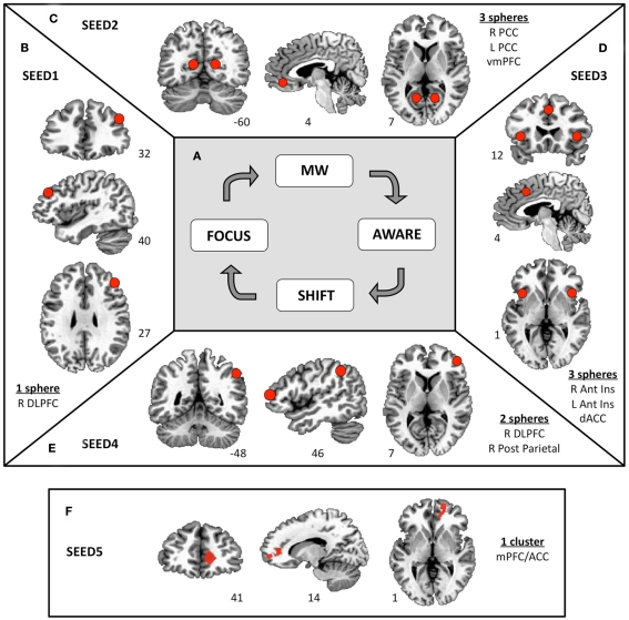
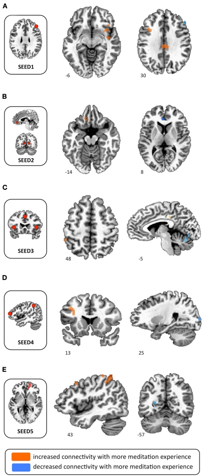
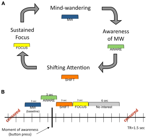
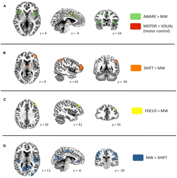
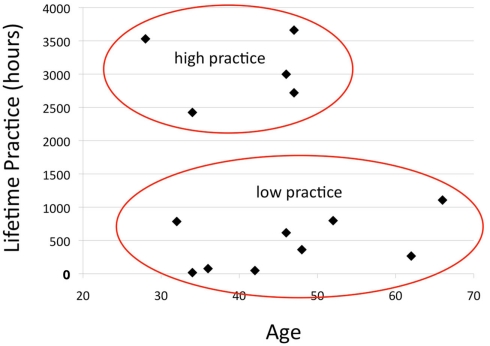
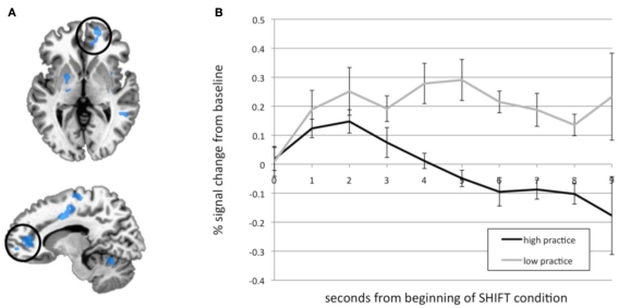
This study sought to examine the effect of meditation experience on brain networks underlying cognitive actions employed during contemplative practice. In a previous study, we proposed a basic model of naturalistic cognitive fluctuations that occur during the practice of focused attention meditation. This model specifies four intervals in a cognitive cycle: mind wandering (MW), awareness of MW, shifting of attention, and sustained attention. Using subjective input from experienced practitioners during meditation, we identified activity in salience network regions during awareness of MW and executive network regions during shifting and sustained attention. Brain regions associated with the default mode were active during MW. In the present study, we reasoned that repeated activation of attentional brain networks over years of practice may induce lasting functional connectivity changes within relevant circuits. To investigate this possibility, we created seeds representing the networks that were active during the four phases of the earlier study, and examined functional connectivity during the resting state in the same participants. Connectivity maps were then contrasted between participants with high vs. low meditation experience. Participants with more meditation experience exhibited increased connectivity within attentional networks, as well as between attentional regions and medial frontal regions. These neural relationships may be involved in the development of cognitive skills, such as maintaining attention and disengaging from distraction, that are often reported with meditation practice. Furthermore, because altered connectivity of brain regions in experienced meditators was observed in a non-meditative (resting) state, this may represent a transference of cognitive abilities "off the cushion" into daily life.
Overview
The claims and statistics of this paper haven’t been checked yet.
Run the check — free, no account needed. It reads the open-access text and checks the sources it cites.
Integrity signals
Registry
Cites
Statistics
Figures
Verification
Claims the paper makes
The claims and statistics of this paper haven’t been checked yet.
Run the check — free, no account needed. It reads the open-access text and checks the sources it cites.
Claims other papers make about this one
Each is checked against this paper’s own text: does it say what the citing paper says it does?
The cited source says what the claim attributes to it.
In the citing paper
“Generally, mindfulness often begins with FA meditation (Hasenkamp and Barsalou, 2012), which is restricted to a specific object (normally the sensory experience of the breath).”
From Enhanced Resting-State Functional Connectivity With Decreased Amplitude of Low-Frequency Fluctuations of the Salience Network in Mindfulness Novices (2022) · cites this paper as Hasenkamp and Barsalou, 2012
In the citing paper
“Functional connectivity studies of meditation have indicated increased connectivity between anterior and posterior nodes of the DMN at rest123,124 and during meditation125,126 in experienced meditators with 10,000 h of practice but not following 8 weeks of training,127”
From Awakening is not a metaphor: the effects of Buddhist meditation practices on basic wakefulness (2014) · cites this paper as [125]
Self-check
Percentages, sums and reported differences re-computed from the paper’s own numbers.
The self-check needs the paper’s full text.
Citations
Cites
Advances in functional and structural MR image analysis and implementation as FSL
Smith, S. M., Jenkinson, M., Woolrich, M. W., Beckmann, C. F., Behrens, T. E. J., Johansen-Berg, H., Bannister, P. R., De Luca, M., Drobnjak, I., Flitney, D. E., et al. (2004) · NeuroImage · doi:10.1016/j.neuroimage.2004.07.051 · Cited by 13,630
AFNI: Software for analysis and visualization of functional magnetic resonance neuroimages
Cox, R. W. (1996) · Computers and Biomedical Research · doi:10.1006/cbmr.1996.0014 · Cited by 10,913
The Brain's Default Network: Anatomy, Function, and Relevance to Disease
Buckner, R. L., Andrews‐Hanna, J. R., & Schacter, D. L. (2008) · Annals of the New York Academy of Sciences · doi:10.1196/annals.1440.011 · Cited by 9,761
The human brain is intrinsically organized into dynamic, anticorrelated functional networks
Fox, M. D., Snyder, A. Z., Vincent, J. L., Corbetta, M., Van Essen, D. C., & Raichle, M. E. (2005) · Proceedings of the National Academy of Sciences · doi:10.1073/pnas.0504136102 · Cited by 8,553
Dissociable intrinsic connectivity networks for salience processing and executive control
Seeley, W. W., Menon, V., Schatzberg, A. F., Keller, J., Glover, G. H., Kenna, H., Reiss, A. L., & Greicius, M. D. (2007) · The Journal of Neuroscience · doi:10.1523/jneurosci.5587-06.2007 · Cited by 7,426
How do you feel — now? The anterior insula and human awareness
(Bud) Craig, A. D. (2009) · Nature Reviews Neuroscience · doi:10.1038/nrn2555 · Cited by 6,657 · cited 2×
“The insula has been implicated in a vast array Frontiers in Human Neuroscience www.frontiersin.org March 2012 | Volume 6 | Article 38 | 5 of tasks, including present moment awareness and sensation of internal states (Craig, 2002, 2009).”
BackgroundHow do you feel? Interoception: The sense of the physiological condition of the body
Craig, A. D. (2002) · Nature Reviews Neuroscience · doi:10.1038/nrn894 · Cited by 6,006
The reorienting system of the human brain: from environment to theory of mind
Corbetta, M., Patel, G., & Shulman, G. L. (2008) · Neuron · doi:10.1016/j.neuron.2008.04.017 · Cited by 3,889
The PANAS-X: Manual for the Positive and Negative Affect Schedule - Expanded Form
Watson, D., & Clark, L. A. (1994) · doi:10.17077/48vt-m4t2 · Cited by 3,790
Nolen-Hoeksema, S., & Morrow, J. (1991) · Journal of Personality and Social Psychology · doi:10.1037/0022-3514.61.1.115 · Cited by 3,276
Research works printed but not linked: 1
- Taylor V. Daneault V. Grant J. A. Scavone G. Breton E. Roffe-Vidal S. Courtemanche J. Lavarenne A. S. Marrelec G. Benali H. Beauregard M. (in press). Impact of mindfulness training on the default mode network during a restful state: a functional connectivity study. Soc. Cogn. Affect. Neurosci. [Manuscript ID: SCAN-11–106]. PMC3541485 22446298
Cited by
Sorted by citations
The Neuroscience of Mindfulness Meditation
Tang, Y.-Y., Hölzel, B. K., & Posner, M. I. (2015) · Nature reviews. Neuroscience · doi:10.1038/nrn3916 · Cited by 2,889
Van Dam, N. T., van Vugt, M. K., Vago, D. R., Schmalzl, L., Saron, C. D., Olendzki, A., Meissner, T., Lazar, S. W., Kerr, C. E., Gorchov, J., et al. (2018) · Perspectives on Psychological Science · doi:10.1177/1745691617709589 · Cited by 1,506
Fox, K. C. R., Spreng, R. N., Ellamil, M., Andrews-Hanna, J. R., & Christoff, K. (2015) · NeuroImage · doi:10.1016/j.neuroimage.2015.02.039 · Cited by 690
Lutz, A., Jha, A. P., Dunne, J. D., & Saron, C. D. (2015) · American Psychologist · doi:10.1037/a0039585 · Cited by 697
“The potentially reciprocal nature of activation of these large-scale networks and their functional roles have started to be explored in the context of meditation sessions or meditation expertise, and is a promising avenue of research (Hasenkamp & Barsalou, 2012).”
BackgroundRusch, H. L., Rosario, M., Levison, L. M., Olivera, A., Livingston, W. S., Wu, T., & Gill, J. M. (2019) · Annals of the New York Academy of Sciences · doi:10.1111/nyas.13996 · Cited by 500
Desbordes, G., Negi, L. T., Pace, T. W. W., Wallace, B. A., Raison, C. L., & Schwartz, E. L. (2012) · Frontiers in Human Neuroscience · doi:10.3389/fnhum.2012.00292 · Cited by 458
Palhano-Fontes, F., Andrade, K. C., Tofoli, L. F., Santos, A. C., Crippa, J. A. S., Hallak, J. E. C., Ribeiro, S., & de Araujo, D. B. (2015) · PLOS ONE · doi:10.1371/journal.pone.0118143 · Cited by 454
Boyd, J. E., Lanius, R. A., & McKinnon, M. C. (2018) · Journal of Psychiatry and Neuroscience · doi:10.1503/jpn.170021 · Cited by 407
Psychedelics, meditation, and self-consciousness
Millière, R., Carhart-Harris, R. L., Roseman, L., Trautwein, F.-M., & Berkovich-Ohana, A. (2018) · Frontiers in Psychology · doi:10.3389/fpsyg.2018.01475 · Cited by 390
Breath of Life: The Respiratory Vagal Stimulation Model of Contemplative Activity
Gerritsen, R. J. S., & Band, G. P. H. (2018) · Frontiers in Human Neuroscience · doi:10.3389/fnhum.2018.00397 · Cited by 342
Functional Connectivity Measures After Psilocybin Inform a Novel Hypothesis of Early Psychosis
Carhart-Harris, R. L., Leech, R., Erritzoe, D., Williams, T. M., Stone, J. M., Evans, J., Sharp, D. J., Feilding, A., Wise, R. G., & Nutt, D. J. (2013) · Schizophrenia Bulletin · doi:10.1093/schbul/sbs117 · Cited by 256
Taren, A. A., Gianaros, P. J., Greco, C. M., Lindsay, E. K., Fairgrieve, A., Brown, K. W., Rosen, R. K., Ferris, J. L., Julson, E., Marsland, A. L., et al. (2015) · Social Cognitive and Affective Neuroscience · doi:10.1093/scan/nsv066 · Cited by 236
Conversano, C., Ciacchini, R., Orrù, G., Di Giuseppe, M., Gemignani, A., & Poli, A. (2020) · Frontiers in Psychology · doi:10.3389/fpsyg.2020.01683 · Cited by 243
“, 2020a,b), and the effects were modulated by lifetime meditation experience (Brewer et al., 2011; Hasenkamp and Barsalou, 2012).”
BackgroundLippelt, D. P., Hommel, B., & Colzato, L. S. (2014) · Frontiers in Psychology · doi:10.3389/fpsyg.2014.01083 · Cited by 278
King, A. P., Block, S. R., Sripada, R. K., Rauch, S., Giardino, N., Favorite, T., Angstadt, M., Kessler, D., Welsh, R., & Liberzon, I. (2016) · Depression and Anxiety · doi:10.1002/da.22481 · Cited by 225
Lutz, A., McFarlin, D. R., Perlman, D. M., Salomons, T. V., & Davidson, R. J. (2013) · NeuroImage · doi:10.1016/j.neuroimage.2012.09.030 · Cited by 233
Mindful attention to breath regulates emotions via increased amygdala–prefrontal cortex connectivity
Doll, A., Hölzel, B. K., Bratec, S. M., Boucard, C. C., Xie, X., Wohlschläger, A. M., & Sorg, C. (2016) · NeuroImage · doi:10.1016/j.neuroimage.2016.03.041 · Cited by 245
Grant, S., Colaiaco, B., Motala, A., Shanman, R., Booth, M., Sorbero, M., & Hempel, S. (2017) · Journal of Addiction Medicine · doi:10.1097/adm.0000000000000338 · Cited by 220
Mindfulness-induced selflessness: a MEG neurophenomenological study
Dor-Ziderman, Y., Berkovich-Ohana, A., Glicksohn, J., & Goldstein, A. (2013) · Frontiers in Human Neuroscience · doi:10.3389/fnhum.2013.00582 · Cited by 205
Witkiewitz, K., Lustyk, M. K. B., & Bowen, S. (2013) · Psychology of Addictive Behaviors · doi:10.1037/a0029258 · Cited by 207
Neural mechanisms of mindfulness and meditation: Evidence from neuroimaging studies
Marchand, W. R. (2014) · World Journal of Radiology · doi:10.4329/wjr.v6.i7.471 · Cited by 207
Pelletier, R., Higgins, J., & Bourbonnais, D. (2015) · BMC Musculoskeletal Disorders · doi:10.1186/s12891-015-0480-y · Cited by 215
Wells, R. E., Yeh, G. Y., Kerr, C. E., Wolkin, J. R., Davis, R. B., Tan, Y. H. V., Spaeth, R. B., Wall, R. B., Walsh, J. N., Kaptchuk, T. J., et al. (2013) · Neuroscience Letters · doi:10.1016/j.neulet.2013.10.001 · Cited by 179
Doll, A., Hölzel, B. K., Boucard, C. C., Wohlschläger, A. M., & Sorg, C. (2015) · Frontiers in Human Neuroscience · doi:10.3389/fnhum.2015.00461 · Cited by 165
Dynamic network interactions supporting internally-oriented cognition
Zabelina, D. L., & Andrews‐Hanna, J. R. (2016) · Current Opinion in Neurobiology · doi:10.1016/j.conb.2016.06.014 · Cited by 183
Experts bodies, experts minds: How physical and mental training shape the brain
Debarnot, U., Sperduti, M., Rienzo, F. D., & Guillot, A. (2014) · Frontiers in Human Neuroscience · doi:10.3389/fnhum.2014.00280 · Cited by 187
The Neuroscience of Mindfulness: How Mindfulness Alters the Brain and Facilitates Emotion Regulation
Wheeler, M. S., Arnkoff, D. B., & Glass, C. R. (2017) · Mindfulness · doi:10.1007/s12671-017-0742-x · Cited by 168
Cognitive control in the self-regulation of physical activity and sedentary behavior
Buckley, J., Cohen, J. D., Kramer, A. F., McAuley, E., & Mullen, S. P. (2014) · Frontiers in Human Neuroscience · doi:10.3389/fnhum.2014.00747 · Cited by 180
Tomasino, B., Fregona, S., Skrap, M., & Fabbro, F. (2013) · Frontiers in Human Neuroscience · doi:10.3389/fnhum.2012.00346 · Cited by 145
Awakening is not a metaphor: the effects of Buddhist meditation practices on basic wakefulness
Britton, W. B., Lindahl, J. R., Cahn, B. R., Davis, J. H., & Goldman, R. E. (2014) · Annals of the New York Academy of Sciences · doi:10.1111/nyas.12279 · Cited by 134
Ellamil, M., Fox, K. C. R., Dixon, M. L., Pritchard, S., Todd, R. M., Thompson, E. T., & Kalina Christoff, A. (2016) · NeuroImage · doi:10.1016/j.neuroimage.2016.04.034 · Cited by 149
Reduced mind wandering in experienced meditators and associated EEG correlates
Brandmeyer, T., & Arnaud Delorme, A. (2016) · Experimental Brain Research · doi:10.1007/s00221-016-4811-5 · Cited by 153
Domain-specific enhancement of metacognitive ability following meditation training.
Baird, B., Mrazek, M. D., Phillips, D. T., & Schooler, J. W. (2014) · Journal of Experimental Psychology General · doi:10.1037/a0036882 · Cited by 144
Cahn, B. R., Arnaud Delorme, A., & Polich, J. M. (2012) · Social Cognitive and Affective Neuroscience · doi:10.1093/scan/nss060 · Cited by 142
Bhayee, S., Tomaszewski, P., Lee, D. H., Moffat, G., Pino, L., Moreno, S., & Farb, N. A. S. (2016) · BMC Psychology · doi:10.1186/s40359-016-0168-6 · Cited by 118
Britton, W. B., Davis, J. H., Loucks, E. B., Peterson, B., Cullen, B. H., Reuter, L., Rando, A., Rahrig, H., Lipsky, J., & Lindahl, J. R. (2018) · Behaviour Research and Therapy · doi:10.1016/j.brat.2017.09.010 · Cited by 100
“…outcomes (Brefczynski-Lewis, Lutz, Schaefer, Levinson,& Davidson, 2007; Dickenson, Berkman, Arch, & Lieberman, 2013; Hasenkamp & Barsalou, 2012; Hasenkamp, Wilson-Mendenhall, Duncan, & Barsalou, 2012; Lutz et al., 2008; Manna et al., 2010; Marzetti et al., 2014; Rapgay & Bystrisky, 2009).”
Using suggestion to modulate automatic processes: From Stroop to McGurk and beyond
Lifshitz, M., Bonn, N. A., Fischer, A., Kashem, I. F., & Raz, A. (2012) · Cortex · doi:10.1016/j.cortex.2012.08.007 · Cited by 105
Brain Network Reconfiguration and Perceptual Decoupling During an Absorptive State of Consciousness
Hove, M. J., Stelzer, J., Nierhaus, T., Thiel, S. D., Gundlach, C., Margulies, D. S., Van Dijk, K. R. A., Turner, R., Keller, P. E., & Merker, B. (2016) · Cerebral Cortex · doi:10.1093/cercor/bhv137 · Cited by 97
Executive control and felt concentrative engagement following intensive meditation training
Zanesco, A. P., King, B. G., MacLean, K. A., & Saron, C. D. (2013) · Frontiers in Human Neuroscience · doi:10.3389/fnhum.2013.00566 · Cited by 99
The default mode network as a biomarker for monitoring the therapeutic effects of meditation
Simon, R., & Engström, M. (2015) · Frontiers in Psychology · doi:10.3389/fpsyg.2015.00776 · Cited by 95
Jazaieri, H., Lee, I. A., McGonigal, K. M., Jinpa, T., Doty, J. R., Gross, J. J., & Goldin, P. R. (2015) · The Journal of Positive Psychology · doi:10.1080/17439760.2015.1025418 · Cited by 99
Panda, R., Bharath, R. D., Upadhyay, N., Mangalore, S., Chennu, S., & Rao, S. L. (2016) · Frontiers in Human Neuroscience · doi:10.3389/fnhum.2016.00372 · Cited by 91
Mindful movement and skilled attention
Clark, D., Schumann, F., & Mostofsky, S. H. (2015) · Frontiers in Human Neuroscience · doi:10.3389/fnhum.2015.00297 · Cited by 102
Mooneyham, B. W., Mrazek, M. D., Mrazek, A. J., Mrazek, K. L., Phillips, D. T., & Schooler, J. W. (2017) · Journal of Cognitive Neuroscience · doi:10.1162/jocn_a_01066 · Cited by 86
Rahrig, H., Vago, D. R., Passarelli, M., Auten, A., Lynn, N. A., & Brown, K. W. (2022) · Scientific Reports · doi:10.1038/s41598-022-15195-6 · Cited by 101
Tops, M., Boksem, M. A. S., Quirin, M., IJzerman, H., & Koole, S. L. (2014) · Frontiers in Psychology · doi:10.3389/fpsyg.2014.00429 · Cited by 93
Meditation Interventions for Chronic Disease Populations
Chan, R. R., & Larson, J. L. (2015) · Journal of Holistic Nursing · doi:10.1177/0898010115570363 · Cited by 83
Mindfulness‐of‐breathing exercise modulates EEG alpha activity during cognitive performance
Bing‐Canar, H., Pizzuto, J., & Compton, R. J. (2016) · Psychophysiology · doi:10.1111/psyp.12678 · Cited by 89
Bauer, C. C. C., Okano, K., Ghosh, S., Lee, Y. J., Melero, H., Angeles, C. D. L., Nestor, P. G., del Re, E. C., Northoff, G., Niznikiewicz, M. A., et al. (2020) · Psychiatry Research · doi:10.1016/j.psychres.2020.112770 · Cited by 78
Yang, C.-C., Barrós-Loscertales, A., Li, M., Pinazo, D., Borchardt, V., Ávila, C., & Walter, M. (2019) · Scientific Reports · doi:10.1038/s41598-019-47470-4 · Cited by 81
From State-to-Trait Meditation: Reconfiguration of Central Executive and Default Mode Networks
Bauer, C. C. C., Whitfield-Gabrieli, S., Díaz, J. L., Pasaye, E. H., & Barrios, F. A. (2019) · eneuro · doi:10.1523/eneuro.0335-18.2019 · Cited by 81
Issues and Perspectives in Meditation Research: In Search for a Definition
Awasthi, B. (2013) · Frontiers in Psychology · doi:10.3389/fpsyg.2012.00613 · Cited by 90
Berkovich‐Ohana, A., Glicksohn, J., Ben‐Soussan, T. D., & Goldstein, A. (2016) · Mindfulness · doi:10.1007/s12671-016-0649-y · Cited by 86
Tao, J., Chen, X., Liu, J., Egorova, N., Xue, X., Liu, W., Zheng, G., Li, M., Wu, J., Hu, K., et al. (2017) · Frontiers in Human Neuroscience · doi:10.3389/fnhum.2017.00514 · Cited by 78
A review on research and evaluation methods for investigating self-transcendence
Kitson, A., Chirico, A., Gaggioli, A., & Riecke, B. E. (2020) · Frontiers in Psychology · doi:10.3389/fpsyg.2020.547687 · Cited by 76
Roland, L. T., Lenze, E. J., Hardin, F. M., Kallogjeri, D., Nicklaus, J., Wineland, A. M., Fendell, G., Peelle, J. E., & Piccirillo, J. F. (2015) · Otolaryngology–Head and Neck Surgery · doi:10.1177/0194599815571556 · Cited by 77
Do Lifestyle Activities Protect Against Cognitive Decline in Aging? A Review
Christie, G. J., Hamilton, T., Manor, B., Farb, N. A. S., Farzan, F., Sixsmith, A. J., Temprado, J.-J., & Moreno, S. (2017) · Frontiers in Aging Neuroscience · doi:10.3389/fnagi.2017.00381 · Cited by 80
The Effects of Mindfulness on Older Adults with Mild Cognitive Impairment
Wong, W. P., Coles, J. Y., Chambers, R., Wu, D. B., & Hassed, C. S. (2017) · Journal of Alzheimer s Disease Reports · doi:10.3233/adr-170031 · Cited by 78
Berkovich‐Ohana, A., Harel, M., Hahamy, A., Arieli, A., & Malach, R. (2016) · NeuroImage · doi:10.1016/j.neuroimage.2016.04.024 · Cited by 73
Studying the default mode and its mindfulness-induced changes using EEG functional connectivity
Berkovich-Ohana, A., Glicksohn, J., & Goldstein, A. (2014) · Social Cognitive and Affective Neuroscience · doi:10.1093/scan/nst153 · Cited by 87
Rosenkranz, M. A., Dunne, J. D., & Davidson, R. J. (2019) · Current Opinion in Psychology · doi:10.1016/j.copsyc.2018.12.022 · Cited by 81
Meditation and the Wandering Mind: A Theoretical Framework of Underlying Neurocognitive Mechanisms
Brandmeyer, T., & Arnaud Delorme, A. (2020) · Perspectives on Psychological Science · doi:10.1177/1745691620917340 · Cited by 86
The (In)flexible self: Psychopathology, mindfulness, and neuroscience
Giommi, F., Bauer, P. R., Berkovich‐Ohana, A., Barendregt, H. P., Brown, K. W., Gallagher, S., Nyklíček, I., Ostafin, B. D., Raffone, A., Slagter, H. A., et al. (2023) · International Journal of Clinical and Health Psychology · doi:10.1016/j.ijchp.2023.100381 · Cited by 84
Dynamic correlations between heart and brain rhythm during Autogenic meditation
Kim, D.-K., Lee, K.-M., Kim, J., Whang, M., & Kang, S. W. (2013) · Frontiers in Human Neuroscience · doi:10.3389/fnhum.2013.00414 · Cited by 71
Bettinardi, R. G., Tort-Colet, N., Ruiz-Mejias, M., Sanchez-Vives, M. V., & Deco, G. (2015) · NeuroImage · doi:10.1016/j.neuroimage.2015.03.037 · Cited by 68
Sevinc, G., Hölzel, B. K., Greenberg, J., Gard, T., Brunsch, V., Hashmi, J. A., Vangel, M. G., Orr, S. P., Milad, M. R., & Lazar, S. W. (2019) · Biological Psychiatry · doi:10.1016/j.biopsych.2019.05.017 · Cited by 66
King, A. P., Block, S. R., Sripada, R. K., Rauch, S. A. M., Porter, K. E., Favorite, T. K., Giardino, N., & Liberzon, I. (2016) · Frontiers in Psychiatry · doi:10.3389/fpsyt.2016.00154 · Cited by 64
Trait Mindfulness and Functional Connectivity in Cognitive and Attentional Resting State Networks
Parkinson, T. D., Kornelsen, J., & Smith, S. D. (2019) · Frontiers in Human Neuroscience · doi:10.3389/fnhum.2019.00112 · Cited by 62
Unifying turbulent dynamics framework distinguishes different brain states
Escrichs, A., Perl, Y. S., Uribe, C., Càmara, E., Türker, B., Pyatigorskaya, N., López‐González, A., Pallavicini, C., Panda, R., Annen, J., et al. (2022) · Communications Biology · doi:10.1038/s42003-022-03576-6 · Cited by 66
Mapping complex mind states: EEG neural substrates of meditative unified compassionate awareness
Schoenberg, P. L. A., Ruf, A., Churchill, J., Brown, D. P., & Brewer, J. A. (2017) · Consciousness and Cognition · doi:10.1016/j.concog.2017.11.003 · Cited by 68
Re-examining the Mysterious Role of the Cerebellum in Pain
Li, C. N., Keay, K. A., Henderson, L. A., & Mychasiuk, R. (2024) · Journal of Neuroscience · doi:10.1523/jneurosci.1538-23.2024 · Cited by 65
Resting state functional connectivity associated with trait emotional intelligence
Takeuchi, H., Taki, Y., Nouchi, R., Sekiguchi, A., Hashizume, H., Sassa, Y., Kotozaki, Y., Miyauchi, C. M., Yokoyama, R., Iizuka, K., et al. (2013) · NeuroImage · doi:10.1016/j.neuroimage.2013.06.044 · Cited by 60
Hauswald, A., Übelacker, T., Leske, S., & Weisz, N. (2015) · NeuroImage · doi:10.1016/j.neuroimage.2014.12.065 · Cited by 70
Brandmeyer, T., & Arnaud Delorme, A. (2020) · Frontiers in Human Neuroscience · doi:10.3389/fnhum.2020.00246 · Cited by 61
Bilevicius, E., Smith, S. D., & Kornelsen, J. (2017) · Brain Connectivity · doi:10.1089/brain.2017.0520 · Cited by 66
Neuroenhancement of Memory for Children with Autism by a Mind–Body Exercise
Chan, A. S.-Y., Han, Y. M. Y., Sze, S. L., & Lau, E. M. (2015) · Frontiers in Psychology · doi:10.3389/fpsyg.2015.01893 · Cited by 60
Altered resting state connectivity of the default mode network in alexithymia
Liemburg, E. J., Swart, M., Bruggeman, R., Kortekaas, R., Knegtering, H., Ćurčić-Blake, B., & Aleman, A. (2012) · Social Cognitive and Affective Neuroscience · doi:10.1093/scan/nss048 · Cited by 56
Mooneyham, B. W., Mrazek, M. D., Mrazek, A. J., & Schooler, J. W. (2016) · Annals of the New York Academy of Sciences · doi:10.1111/nyas.13044 · Cited by 56
The contemplative exercise through the lenses of predictive processing: A promising approach
Pagnoni, G. (2019) · Progress in brain research · doi:10.1016/bs.pbr.2018.10.022 · Cited by 63
Beyond the veil of duality—topographic reorganization model of meditation
Cooper, A. C., Ventura, B., & Northoff, G. (2022) · Neuroscience of Consciousness · doi:10.1093/nc/niac013 · Cited by 60
Kim, H., Tegethoff, M., Meinlschmidt, G., Stalujanis, E., Belardi, A., Jo, S., Lee, J., Kim, D., Yoo, S., & Lee, J. (2019) · NeuroImage · doi:10.1016/j.neuroimage.2019.03.066 · Cited by 54
Cognitive Behavioral Therapy and Mindfulness-Based Cognitive Therapy for Depressive Disorders
Lee, S., & Cho, S. (2021) · Advances in experimental medicine and biology · doi:10.1007/978-981-33-6044-0_16 · Cited by 61
Meditation training modulates brain electric microstates and felt states of awareness
Zanesco, A. P., Skwara, A. C., King, B. G., Powers, C., Wineberg, K., & Saron, C. D. (2021) · Human Brain Mapping · doi:10.1002/hbm.25430 · Cited by 55
Ictal alterations of consciousness during ecstatic seizures
Picard, F., & Kurth, F. (2013) · Epilepsy & Behavior · doi:10.1016/j.yebeh.2013.09.036 · Cited by 52
Mindfulness:A Transtherapeutic Approach for Transdiagnostic Mental Processes
Greeson, J., Garland, E. L., & Black, D. (2014) · The Wiley Blackwell Handbook of Mindfulness · doi:10.1002/9781118294895.ch28 · Cited by 50
Meditation and Mindfulness in Pregnancy and Postpartum: A Review of the Evidence
Babbar, S., Oyarzabal, A. J., & Oyarzabal, E. A. (2021) · Clinical Obstetrics & Gynecology · doi:10.1097/grf.0000000000000640 · Cited by 50
Sevinc, G., & Lazar, S. W. (2019) · Current Opinion in Psychology · doi:10.1016/j.copsyc.2019.02.006 · Cited by 49
Network-based characterization of brain functional connectivity in Zen practitioners
Kemmer, P. B., Guo, Y., Wang, Y., & Pagnoni, G. (2015) · Frontiers in Psychology · doi:10.3389/fpsyg.2015.00603 · Cited by 48
Ethical Leadership as a Balance Between Opposing Neural Networks
Rochford, K. C., Jack, A. I., Boyatzis, R. E., & French, S. E. (2016) · Journal of Business Ethics · doi:10.1007/s10551-016-3264-x · Cited by 51
Andreu, C. I., Cosmelli, D., Slagter, H. A., & Franken, I. H. A. (2018) · PLOS ONE · doi:10.1371/journal.pone.0191661 · Cited by 47
Paoletti, P., & Ben‐Soussan, T. D. (2020) · Frontiers in Psychology · doi:10.3389/fpsyg.2020.01807 · Cited by 57
Cotier, F. A., Zhang, R., & Lee, T. M. C. (2017) · Scientific Reports · doi:10.1038/s41598-017-00678-8 · Cited by 50
Sevinc, G., Rusche, J., Wong, B., Datta, T., Kaufman, R., Gutz, S. E., Schneider, M. L., Todorova, N., Gaser, C., Thomalla, G., et al. (2021) · Frontiers in Aging Neuroscience · doi:10.3389/fnagi.2021.702796 · Cited by 55
Rajyoga meditation induces grey matter volume changes in regions that process reward and happiness
G, R. B. M., Kadavigere, R., Prakashini, K., Sathian, B., & Rai, K. S. (2020) · Scientific Reports · doi:10.1038/s41598-020-73221-x · Cited by 46
Zhou, D., Kang, Y., Cosme, D., Jovanova, M., He, X., Mahadevan, A. S., Ahn, J., Stanoi, O. A., Brynildsen, J. K., Cooper, N., et al. (2023) · Proceedings of the National Academy of Sciences · doi:10.1073/pnas.2201074119 · Cited by 44
Broca’s area network in language function: a pooling-data connectivity study
Bernal, B., Ardila, A., & Rosselli, M. (2015) · Frontiers in Psychology · doi:10.3389/fpsyg.2015.00687 · Cited by 43
Weder, B. (2022) · Frontiers in Behavioral Neuroscience · doi:10.3389/fnbeh.2022.928522 · Cited by 52
Meditation and Hypnosis: Two Sides of the Same Coin?
Facco, E. (2017) · International Journal of Clinical and Experimental Hypnosis · doi:10.1080/00207144.2017.1276361 · Cited by 39
Berkovich‐Ohana, A., Harel, M., Hahamy, A., Arieli, A., & Malach, R. (2016) · Data in Brief · doi:10.1016/j.dib.2016.07.015 · Cited by 39
Metadata
- Authors
- Hasenkamp, W., & Barsalou, L. W.
- Affiliations
- Emory University (US)
- Corresponding author
- Hasenkamp, Wendy
- Venue
- Frontiers in Human Neuroscience
- Publisher
- Frontiers Media SA
- Published
- 2012
- Volume
- 6
- Pages
- 38-38
- ISSN
- 1662-5161 (ISSN-L)
- PMC
- PMC3290768
- OpenAlex
- W2139765609
- Cited by
- 367
- References
- 48
- Citations by year
- 2026: 17 · 2025: 25 · 2024: 33 · 2023: 27 · 2022: 29 · 2021: 28 · 2020: 38 · 2019: 29 · 2018: 16 · 2017: 30 · 2016: 24 · 2015: 31 · 2014: 16 · 2013: 16 · 2012: 8
- FWCI
- 11.48
- Type
- journal article
- Language
- en
- Open access
- gold
- doi.org
- Licence
- cc-by-nc
- Locations
- Frontiers in Human Neuroscience (publishedVersion, cc-by-nc)
- PubMed Central (publishedVersion)
- PubMed (publishedVersion)
- ENLIGHTEN (Jurnal Bimbingan dan Konseling Islam) (acceptedVersion)
- DOAJ (DOAJ: Directory of Open Access Journals) (submittedVersion, cc-by-sa)
- Funders
- U.S. Department of Veterans Affairs
- Emory University
- Mind and Life Institute
- Topics
- Life Sciences › Neuroscience › Cognitive Neuroscience › Functional Brain Connectivity Studies
- Social Sciences › Psychology › Clinical Psychology › Mindfulness and Compassion Interventions
- Life Sciences › Neuroscience › Cognitive Neuroscience › Mind wandering and attention
- Keywords
- meditation experience, functional connectivity, distributed brain networks, focused attention meditation, mind wandering, salience network, executive network, default mode network, sustained attention, attentional shift, resting state, meditation practitioners
About these checks
Registry, retraction, Cites and statistics results are computed, not judged, with one exception: a printed reference that no index lists is read once by a model before it counts as a citation error. Claims are extracted by a model and kept only when their quote appears verbatim in the text; each cited source is then checked, with a model’s help, for whether it says what the claim relies on. Those verdicts can be wrong — read the claim’s quote and the cited source before relying on one.
Statistics and arithmetic are recomputed, not judged.
Confidence bands are shown only for fields calibrated on our annotated test set. None is calibrated yet, so no band is shown.
What counts as a claim ›How we measure confidence ›How we check this ›
Are you an author, or do you see a problem with this page? Request a review
