

The DOI matches its Crossref record; 9 of the 9 references it cites were found in a registry or an index.
Biogenesis, Isolation, and Detection of Exosomes and Their Potential in Therapeutics and Diagnostics
Abstract
The increasing research and rapid developments in the field of exosomes provide insights into their role and significance in human health. Exosomes derived from various sources, such as mesenchymal stem cells, cardiac cells, and tumor cells, to name a few, can be potential therapeutic agents for the treatment of diseases and could also serve as biomarkers for the early detection of diseases. Cellular components of exosomes, several proteins, lipids, and miRNAs hold promise as novel biomarkers for the detection of various diseases. The structure of exosomes enables them as drug delivery vehicles. Since exosomes exhibit potential therapeutic applications, their efficient isolation from complex biological/clinical samples and precise real-time analysis becomes significant. With the advent of microfluidics, nano-biosensors are being designed to capture exosomes efficiently and rapidly. Herein, we have summarized the history, biogenesis, characteristics, functions, and applications of exosomes, along with the isolation, detection, and quantification techniques. The implications of surface modifications to enhance specificity have been outlined. The review also sheds light on the engineered nanoplatforms being developed for exosome detection and capture.
Overview
The claims and statistics of this paper haven’t been checked yet.
Run the check — free, no account needed. It reads the open-access text and checks the sources it cites.
Integrity signals
Registry
Cites
Statistics
Figures
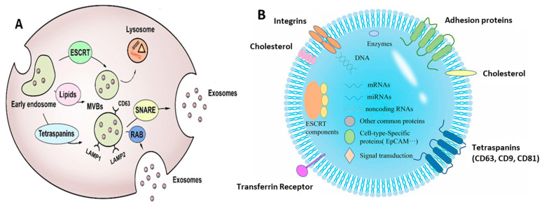
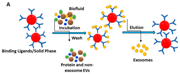
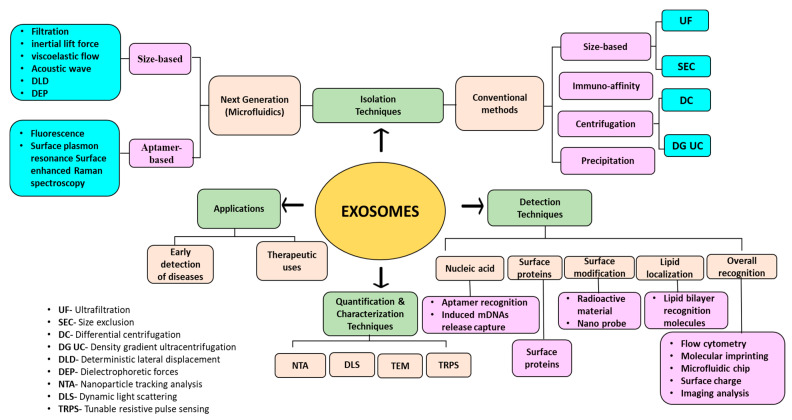
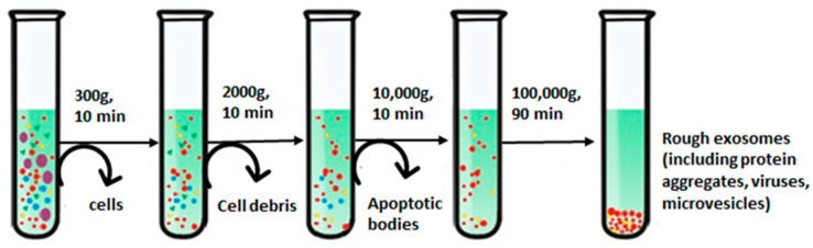
Verification
Claims the paper makes
The claims and statistics of this paper haven’t been checked yet.
Run the check — free, no account needed. It reads the open-access text and checks the sources it cites.
Claims other papers make about this one
Each is checked against this paper’s own text: does it say what the citing paper says it does?
In the citing paper
“The small size and capacity to fuse with cellular membranes enable efficient drug delivery across physiological barriers like the blood-brain barrier, a major obstacle in treating brain cancers.”
From Emerging role of exosomes in cancer therapy: progress and challenges (2025) · cites this paper as [5]
Self-check
Percentages, sums and reported differences re-computed from the paper’s own numbers.
The self-check needs the paper’s full text.
Citations
Cites
The biology , function , and biomedical applications of exosomes
Kalluri, R., & LeBleu, V. S. (2020) · Science · doi:10.1126/science.aau6977 · Cited by 10,770
B lymphocytes secrete antigen-presenting vesicles.
Raposo, G., Nijman, H. W., Stoorvogel, W., Liejendekker, R., Harding, C. V., Melief, C. J., & Geuze, H. J. (1996) · The Journal of experimental medicine · doi:10.1084/jem.183.3.1161 · Cited by 3,546
Kowal, J., Arras, G., Colombo, M., Jouve, M., Morath, J. P., Primdal-Bengtson, B., Dingli, F., Loew, D., Tkach, M., & Théry, C. (2016) · Proceedings of the National Academy of Sciences · doi:10.1073/pnas.1521230113 · Cited by 3,426
Johnstone, R. M., Adam, M., Hammond, J. R., Orr, L., & Turbide, C. (1987) · Journal of Biological Chemistry · doi:10.1016/s0021-9258(18)48095-7 · Cited by 2,931 · cited 2×
“In the beginning, when the extracellular vesicles (EVs) were first discovered, these microvesicles along with the exosomes were seen as cellular wastes or the by-products/debris of cells [21,22].”
BackgroundGlypican-1 identifies cancer exosomes and detects early pancreatic cancer
Melo, S. A., Luecke, L. B., Kahlert, C., Fernandez, A. F., Gammon, S. T., Kaye, J., LeBleu, V. S., Mittendorf, E. A., Weitz, J., Rahbari, N., et al. (2015) · Nature · doi:10.1038/nature14581 · Cited by 2,880
Syndecan–syntenin–ALIX regulates the biogenesis of exosomes
Baietti, M. F., Zhang, Z., Mortier, E., Melchior, A., Degeest, G., Geeraerts, A., Ivarsson, Y., Depoortere, F., Coomans, C., Vermeiren, E., et al. (2012) · Nature Cell Biology · doi:10.1038/ncb2502 · Cited by 1,899
Akers, J. C., Gonda, D., Kim, R., Carter, B. S., & Chen, C. C. (2013) · Journal of Neuro-Oncology · doi:10.1007/s11060-013-1084-8 · Cited by 1,423 · cited 2×
“Exosomes also carry genes from their parental cells like microRNAs, long non-coding RNAs, and circular RNAs [59,60].”
BackgroundLiquid biopsy enters the clinic — implementation issues and future challenges
Ignatiadis, M., Sledge, G. W., & Jeffrey, S. S. (2021) · Nature Reviews Clinical Oncology · doi:10.1038/s41571-020-00457-x · Cited by 1,191
Jawed, A., Saxena, V., & Pandey, L. M. (2020) · Journal of Water Process Engineering · doi:10.1016/j.jwpe.2019.101009 · Cited by 297
Self-citation
Research works printed but not linked: 203
- Sun, 2021, Progress in the research of nanomaterial-based exosome bioanalysis and exosome-based nanomaterials tumor therapy, Biomaterials, 274, 120873, 10.1016/j.biomaterials.2021.120873 · DOI
- Royo, 2021, Extracellular vesicles in hepatology: Physiological role, involvement in pathogenesis, and therapeutic opportunities, Pharmacol. Ther., 218, 107683, 10.1016/j.pharmthera.2020.107683 · DOI
- Jiang, 2020, Recent Progress on the Isolation and Detection Methods of Exosomes, Chem. Asian J., 15, 3973, 10.1002/asia.202000873 · DOI
- Fang, 2022, Nanomaterials assisted exosomes isolation and analysis towards liquid biopsy, Mater. Today Bio, 16, 100371, 10.1016/j.mtbio.2022.100371 · DOI
- Gardiner, 2013, Extracellular vesicle sizing and enumeration by nanoparticle tracking analysis, J. Extracell. Vesicles, 2, 19671, 10.3402/jev.v2i0.19671 · DOI
- Brown, W. (1993). Dynamic Light Scattering: The Method and Some Applications, Clarendon Press. · DOI
- Kurian, 2021, Elucidating methods for isolation and quantification of exosomes: A review, Mol. Biotechnol., 63, 249, 10.1007/s12033-021-00300-3 · DOI
- Cayota, 2016, Electrochemical sandwich immunosensor for determination of exosomes based on surface marker-mediated signal amplification, Anal. Chem., 88, 10466, 10.1021/acs.analchem.6b02421 · DOI
- Ashcroft, 2012, Determination of the size distribution of blood microparticles directly in plasma using atomic force microscopy and microfluidics, Biomed. Microdevices, 14, 641, 10.1007/s10544-012-9642-y · DOI
- Jung, 2018, Sample preparation and imaging of exosomes by transmission electron microscopy, JoVE (J. Vis. Exp.), 4, e56482
- Jiang, 2019, Exosomes: Diagnostic Biomarkers and Therapeutic Delivery Vehicles for Cancer, Mol. Pharm., 16, 3333, 10.1021/acs.molpharmaceut.9b00409 · DOI
- Wang, 2023, Mesoporous magnetic nanoparticles conjugated aptamers for exosomes capture and detection of Alzheimer’s disease, Eng. Regen., 4, 349
- Xin, 2012, Exosome-mediated transfer of miR-133b from multipotent mesenchymal stromal cells to neural cells contributes to neurite outgrowth, Stem Cells, 30, 1556, 10.1002/stem.1129 · DOI
- Zhang, 2015, HucMSC-Exosome Mediated-Wnt4 Signaling Is Required for Cutaneous Wound Healing, Stem Cells, 33, 2158, 10.1002/stem.1771 · DOI
- Yang, 2018, The serum exosome derived MicroRNA-135a, -193b, and -384 were potential Alzheimer’s disease biomarkers, Biomed. Environ. Sci., 31, 87
- Brennan, 2017, Protective Effect of let-7 miRNA Family in Regulating Inflammation in Diabetes-Associated Atherosclerosis, Diabetes, 66, 2266, 10.2337/db16-1405 · DOI
- An, 2016, Emergence of exosomal miRNAs as a diagnostic biomarker for Alzheimer’s disease, J. Neurol. Sci., 360, 141, 10.1016/j.jns.2015.12.005 · DOI
- Mosquera-Heredia, M.I., Morales, L.C., Vidal, O.M., Barcelo, E., Silvera-Redondo, C., Velez, J.I., and Garavito-Galofre, P. (2021). Exosomes: Potential Disease Biomarkers and New Therapeutic Targets. Biomedicines, 9. · DOI
- Lin, 2020, Exosomes in disease and regeneration: Biological functions, diagnostics, and beneficial effects, Am. J. Physiol. Heart Circ. Physiol., 319, H1162, 10.1152/ajpheart.00075.2020 · DOI
- Cui, 2018, Exosomes as a liquid biopsy for lung cancer, Lung Cancer, 116, 46, 10.1016/j.lungcan.2017.12.012 · DOI
- Pan, 1983, Fate of the transferrin receptor during maturation of sheep reticulocytes in vitro: Selective externalization of the receptor, Cell, 33, 967, 10.1016/0092-8674(83)90040-5 · DOI
- Enderle, D., Spiel, A., Coticchia, C.M., Berghoff, E., Mueller, R., Schlumpberger, M., Sprenger-Haussels, M., Shaffer, J.M., Lader, E., and Skog, J. (2015). Characterization of RNA from exosomes and other extracellular vesicles isolated by a novel spin column-based method. PLoS ONE, 10. · DOI
- Stoorvogel, 2002, The biogenesis and functions of exosomes, Traffic, 3, 321, 10.1034/j.1600-0854.2002.30502.x · DOI
- Urbanelli, 2013, Signaling pathways in exosomes biogenesis, secretion and fate, Genes, 4, 152, 10.3390/genes4020152 · DOI
- Raposo, 2013, Extracellular vesicles: Exosomes, microvesicles, and friends, J. Cell Biol., 200, 373, 10.1083/jcb.201211138 · DOI · Paper page
- Donselaar, 2002, Immunoelectron microscopic localization of cholesterol using biotinylated and non-cytolytic perfringolysin O, J. Histochem. Cytochem., 50, 43, 10.1177/002215540205000105 · DOI
- Kucharzewska, 2013, Exosomes reflect the hypoxic status of glioma cells and mediate hypoxia-dependent activation of vascular cells during tumor development, Proc. Natl. Acad. Sci. USA, 110, 7312, 10.1073/pnas.1220998110 · DOI · Paper page
- Farooqi, 2018, Exosome biogenesis, bioactivities and functions as new delivery systems of natural compounds, Biotechnol. Adv., 36, 328, 10.1016/j.biotechadv.2017.12.010 · DOI · Paper page
- Willms, 2016, Cells release subpopulations of exosomes with distinct molecular and biological properties, Sci. Rep., 6, 22519, 10.1038/srep22519 · DOI
- Zeringer, 2015, Strategies for isolation of exosomes, Cold Spring Harb. Protoc., 2015, 319, 10.1101/pdb.top074476 · DOI
- Hornick, 2015, Serum exosome microRNA as a minimally-invasive early biomarker of AML, Sci. Rep., 5, 11295, 10.1038/srep11295 · DOI
- Goto, 2018, An elevated expression of serum exosomal microRNA-191, -21,-451a of pancreatic neoplasm is considered to be efficient diagnostic marker, BMC Cancer, 18, 116, 10.1186/s12885-018-4006-5 · DOI
- Kowal, 2014, Biogenesis and secretion of exosomes, Curr. Opin. Cell Biol., 29, 116, 10.1016/j.ceb.2014.05.004 · DOI · Paper page
- Lu, 2017, Exosomal tetraspanins mediate cancer metastasis by altering host microenvironment, Oncotarget, 8, 62803, 10.18632/oncotarget.19119 · DOI
- Ronquist, 2016, Energy-requiring uptake of prostasomes and PC3 cell-derived exosomes into non-malignant and malignant cells, J. Extracell. Vesicles, 5, 29877, 10.3402/jev.v5.29877 · DOI
- Doyle, L.M., and Wang, M.Z. (2019). Overview of extracellular vesicles, their origin, composition, purpose, and methods for exosome isolation and analysis. Cells, 8. · DOI
- Andaloussi, 2013, Exosomes for targeted siRNA delivery across biological barriers, Adv. Drug Deliv. Rev., 65, 391, 10.1016/j.addr.2012.08.008 · DOI
- Hergenreder, J.R. (2011). Serum Exosome Profile as Related to Early Pregnancy Status in the Mare. [Ph.D. Thesis, Colorado State University].
- Mateos, 2017, Recruitment of RNA molecules by connexin RNA-binding motifs: Implication in RNA and DNA transport through microvesicles and exosomes, Biochim. Biophys. Acta (BBA)-Mol. Cell Res., 1864, 728, 10.1016/j.bbamcr.2017.02.001 · DOI
- Cho, 2012, Exosomes from breast cancer cells can convert adipose tissue-derived mesenchymal stem cells into myofibroblast-like cells, Int. J. Oncol., 40, 130
- Kojima, 2018, Designer exosomes produced by implanted cells intracerebrally deliver therapeutic cargo for Parkinson’s disease treatment, Nat. Commun., 9, 1305, 10.1038/s41467-018-03733-8 · DOI · Paper page
- Liu, 2021, The biology, function, and applications of exosomes in cancer, Acta Pharm. Sin. B, 11, 2783, 10.1016/j.apsb.2021.01.001 · DOI
- Chen, 2022, Review on strategies and technologies for exosome isolation and purification, Front. Bioeng. Biotechnol., 9, 811971, 10.3389/fbioe.2021.811971 · DOI · Paper page
- Li, 2017, Progress in exosome isolation techniques, Theranostics, 7, 789, 10.7150/thno.18133 · DOI
- Gurunathan, S., Kang, M.-H., Jeyaraj, M., Qasim, M., and Kim, J.-H. (2019). Review of the isolation, characterization, biological function, and multifarious therapeutic approaches of exosomes. Cells, 8. · DOI
- Bebelman, 2018, Biogenesis and function of extracellular vesicles in cancer, Pharmacol. Ther., 188, 1, 10.1016/j.pharmthera.2018.02.013 · DOI
- Castro, 2014, Ceramide: A simple sphingolipid with unique biophysical properties, Prog. Lipid Res., 54, 53, 10.1016/j.plipres.2014.01.004 · DOI
- Lobasso, 2021, A lipidomic approach to identify potential biomarkers in exosomes from melanoma cells with different metastatic potential, Front. Physiol., 12, 748895, 10.3389/fphys.2021.748895 · DOI
- Admyre, 2007, Exosomes with immune modulatory features are present in human breast milk, J. Immunol., 179, 1969, 10.4049/jimmunol.179.3.1969 · DOI
- Kalra, 2013, Comparative proteomics evaluation of plasma exosome isolation techniques and assessment of the stability of exosomes in normal human blood plasma, Proteomics, 13, 3354, 10.1002/pmic.201300282 · DOI
- Gonzales, 2009, Large-scale proteomics and phosphoproteomics of urinary exosomes, J. Am. Soc. Nephrol., 20, 363, 10.1681/ASN.2008040406 · DOI · Paper page
- Yokoi, 2021, Exosomes and extracellular vesicles: Rethinking the essential values in cancer biology, Semin. Cancer Biol., 74, 79, 10.1016/j.semcancer.2021.03.032 · DOI
- Stuffers, 2009, Multivesicular endosome biogenesis in the absence of ESCRTs, Traffic, 10, 925, 10.1111/j.1600-0854.2009.00920.x · DOI
- Valadi, 2007, Exosome-mediated transfer of mRNAs and microRNAs is a novel mechanism of genetic exchange between cells, Nat. Cell Biol., 9, 654, 10.1038/ncb1596 · DOI · Paper page
- Shu, 2020, Purity and yield of melanoma exosomes are dependent on isolation method, J. Extracell. Vesicles, 9, 1692401, 10.1080/20013078.2019.1692401 · DOI · Paper page
- Balaj, 2013, Current methods for the isolation of extracellular vesicles, Biol. Chem., 394, 1253, 10.1515/hsz-2013-0141 · DOI
- Konoshenko, 2018, Isolation of extracellular vesicles: General methodologies and latest trends, BioMed Res. Int., 2018, 8545347, 10.1155/2018/8545347 · DOI
- Yang, 2020, Progress, opportunity, and perspective on exosome isolation-efforts for efficient exosome-based theranostics, Theranostics, 10, 3684, 10.7150/thno.41580 · DOI
- Amigorena, 2006, Isolation and characterization of exosomes from cell culture supernatants and biological fluids, Curr. Protoc. Cell Biol., 30, 3
- Gupta, 2018, An improvised one-step sucrose cushion ultracentrifugation method for exosome isolation from culture supernatants of mesenchymal stem cells, Stem Cell Res. Ther., 9, 180, 10.1186/s13287-018-0923-0 · DOI
- Neves, 2009, Subcellular fractionation of human eosinophils: Isolation of functional specific granules on isoosmotic density gradients, J. Immunol. Methods, 344, 64, 10.1016/j.jim.2009.03.006 · DOI
- Dettenhofer, 1999, Highly purified human immunodeficiency virus type 1 reveals a virtual absence of Vif in virions, J. Virol., 73, 1460, 10.1128/JVI.73.2.1460-1467.1999 · DOI
- Ford, 1994, Iodixanol: A nonionic iso-osmotic centrifugation medium for the formation of self-generated gradients, Anal. Biochem., 220, 360, 10.1006/abio.1994.1350 · DOI
- Skubal, L.R. (2001). The Essential Guide to Environmental Chemistry by George Schwedt, John Wiley & Sons Inc.. Book Review.
- Ritchie, C. (2012). Protein Purification, Iowa State University. · DOI
- Rider, 2016, ExtraPEG: A polyethylene glycol-based method for enrichment of extracellular vesicles, Sci. Rep., 6, 23978, 10.1038/srep23978 · DOI
- Brownlee, 2014, A novel “salting-out” procedure for the isolation of tumor-derived exosomes, J. Immunol. Methods, 407, 120, 10.1016/j.jim.2014.04.003 · DOI
- Alvarez, 2012, Comparison of protein, microRNA, and mRNA yields using different methods of urinary exosome isolation for the discovery of kidney disease biomarkers, Kidney Int., 82, 1024, 10.1038/ki.2012.256 · DOI
- Bertokova, 2022, Exosomes from prostate cancer cell lines: Isolation optimisation and characterisation, Biomed. Pharmacother., 151, 113093, 10.1016/j.biopha.2022.113093 · DOI
- Cho, 2020, Development and comparative analysis of human urine exosome isolation strategies, Process Biochem., 88, 197, 10.1016/j.procbio.2019.09.017 · DOI
- Weng, 2016, Effective isolation of exosomes with polyethylene glycol from cell culture supernatant for in-depth proteome profiling, Analyst, 141, 4640, 10.1039/C6AN00892E · DOI
- Serra, 2016, Enrichment of extracellular vesicles from tissues of the central nervous system by PROSPR, Mol. Neurodegener., 11, 1
- Zarovni, 2015, Integrated isolation and quantitative analysis of exosome shuttled proteins and nucleic acids using immunocapture approaches, Methods, 87, 46, 10.1016/j.ymeth.2015.05.028 · DOI
- Lane, 2017, Purification protocols for extracellular vesicles, Extracell. Vesicles Methods Protoc., 1660, 111, 10.1007/978-1-4939-7253-1_10 · DOI
- Sharma, 2018, Immunoaffinity-based isolation of melanoma cell-derived exosomes from plasma of patients with melanoma, J. Extracell. Vesicles, 7, 1435138, 10.1080/20013078.2018.1435138 · DOI
- Saad, M.H., Badierah, R., Redwan, E.M., and El-Fakharany, E.M. (2021). A comprehensive insight into the role of exosomes in viral infection: Dual faces bearing different functions. Pharmaceutics, 13. · DOI
- Feng, Y., Huang, W., Wani, M., Yu, X., and Ashraf, M. (2014). Ischemic preconditioning potentiates the protective effect of stem cells through secretion of exosomes by targeting Mecp2 via miR-22. PLoS ONE, 9. · DOI
- Chen, 2019, Advances in exosomes technology, Clin. Chim. Acta, 493, 14, 10.1016/j.cca.2019.02.021 · DOI
- Boussac, 2001, Proteomic analysis of dendritic cell-derived exosomes: A secreted subcellular compartment distinct from apoptotic vesicles, J. Immunol., 166, 7309, 10.4049/jimmunol.166.12.7309 · DOI
- Chahar, 2015, Exosomes and their role in the life cycle and pathogenesis of RNA viruses, Viruses, 7, 3204, 10.3390/v7062770 · DOI
- Witwer, 2013, Standardization of sample collection, isolation and analysis methods in extracellular vesicle research, J. Extracell. Vesicles, 2, 20360, 10.3402/jev.v2i0.20360 · DOI
- 2017, Ion-exchange chromatography purification of extracellular vesicles, Biotechniques, 63, 65, 10.2144/000114575 · DOI
- Busatto, S., Vilanilam, G., Ticer, T., Lin, W.-L., Dickson, D.W., Shapiro, S., Bergese, P., and Wolfram, J. (2018). Tangential flow filtration for highly efficient concentration of extracellular vesicles from large volumes of fluid. Cells, 7. · DOI
- Kim, 2021, Defined MSC exosome with high yield and purity to improve regenerative activity, J. Tissue Eng., 12, 20417314211008626, 10.1177/20417314211008626 · DOI
- Mulder, M. (1996). Basic Principles of Membrane Technology, Springer Science & Business Media. · DOI
- Parimon, 2018, Isolation of extracellular vesicles from murine bronchoalveolar lavage fluid using an ultrafiltration centrifugation technique, J. Vis. Exp. JoVE, 9, e58310
- Xu, 2017, A protocol for isolation and proteomic characterization of distinct extracellular vesicle subtypes by sequential centrifugal ultrafiltration, Exosomes Microvesicles Methods Protoc., 1545, 91, 10.1007/978-1-4939-6728-5_7 · DOI
- Guerreiro, E.M., Vestad, B., Steffensen, L.A., Aass, H.C.D., Saeed, M., Øvstebø, R., Costea, D.E., Galtung, H.K., and Søland, T.M. (2018). Efficient extracellular vesicle isolation by combining cell media modifications, ultrafiltration, and size-exclusion chromatography. PLoS ONE, 13. · DOI
- Klymiuk, 2019, Exosomes isolation and identification from equine mesenchymal stem cells, BMC Vet. Res., 15, 42, 10.1186/s12917-019-1789-9 · DOI
- Hong, 2016, Isolation of biologically active and morphologically intact exosomes from plasma of patients with cancer, J. Extracell. Vesicles, 5, 29289, 10.3402/jev.v5.29289 · DOI
- Grootemaat, 2014, Single-step isolation of extracellular vesicles by size-exclusion chromatography, J. Extracell. Vesicles, 3, 23430, 10.3402/jev.v3.23430 · DOI
- Koh, 2018, Exosome enrichment by ultracentrifugation and size exclusion chromatography, Front. Biosci., 23, 865, 10.2741/4621 · DOI
- Wu, 2017, Microfluidics for exosome isolation and analysis: Enabling liquid biopsy for personalized medicine, Lab. A Chip, 17, 3558, 10.1039/C7LC00592J · DOI
- Fernandes, 2018, Connecting worlds–a view on microfluidics for a wider application, Biotechnol. Adv., 36, 1341, 10.1016/j.biotechadv.2018.05.001 · DOI
- Bai, 2022, Microfluidic strategies for the isolation and profiling of exosomes, TrAC Trends Anal. Chem., 158, 116834, 10.1016/j.trac.2022.116834 · DOI
- Shirejini, 2022, The Yin and Yang of exosome isolation methods: Conventional practice, microfluidics, and commercial kits, Biotechnol. Adv., 54, 107814, 10.1016/j.biotechadv.2021.107814 · DOI
- Wu, 2017, Isolation of exosomes from whole blood by integrating acoustics and microfluidics, Proc. Natl. Acad. Sci. USA, 114, 10584, 10.1073/pnas.1709210114 · DOI
- Kang, 2019, Isolation and profiling of circulating tumor-associated exosomes using extracellular vesicular lipid–protein binding affinity based microfluidic device, Small, 15, 1903600, 10.1002/smll.201903600 · DOI
- Xu, 2018, Magnetic-based microfluidic device for on-chip isolation and detection of tumor-derived exosomes, Anal. Chem., 90, 13451, 10.1021/acs.analchem.8b03272 · DOI
- Liu, 2019, Lipid nanovesicles by microfluidics: Manipulation, synthesis, and drug delivery, Adv. Mater., 31, 1804788, 10.1002/adma.201804788 · DOI
- Seo, 2022, Pervaporation-assisted in situ formation of nanoporous microchannels with various material and structural properties, Lab. A Chip, 22, 1474, 10.1039/D1LC01184G · DOI
- Ding, 2023, Magnetic-nanowaxberry-based microfluidic ExoSIC for affinity and continuous separation of circulating exosomes towards cancer diagnosis, R. Soc. Chem., 23, 1694
- Yang, 2017, Exosome separation using microfluidic systems: Size-based, immunoaffinity-based and dynamic methodologies, Biotechnol. J., 12, 1600699, 10.1002/biot.201600699 · DOI
- Morani, 2020, Electrokinetic characterization of extracellular vesicles with capillary electrophoresis: A new tool for their identification and quantification, Anal. Chim. Acta, 1128, 42, 10.1016/j.aca.2020.06.073 · DOI
- Deregibus, 2016, Charge-based precipitation of extracellular vesicles, Int. J. Mol. Med., 38, 1359, 10.3892/ijmm.2016.2759 · DOI
- Kim, 2016, Chromatographically isolated CD63+ CD81+ extracellular vesicles from mesenchymal stromal cells rescue cognitive impairments after TBI, Proc. Natl. Acad. Sci. USA, 113, 170, 10.1073/pnas.1522297113 · DOI
- Heath, 2018, Rapid isolation and enrichment of extracellular vesicle preparations using anion exchange chromatography, Sci. Rep., 8, 5730, 10.1038/s41598-018-24163-y · DOI
- Martins, 2023, A review on comparative studies addressing exosome isolation methods from body fluids, Anal. Bioanal. Chem., 415, 1239, 10.1007/s00216-022-04174-5 · DOI
- Hasan, 2018, Effect of Functional Groups of Self-Assembled Monolayers on Protein Adsorption and Initial Cell Adhesion, ACS Biomater. Sci. Eng., 4, 3224, 10.1021/acsbiomaterials.8b00795 · DOI
- Saad, M.G., Beyenal, H., and Dong, W.-J. (2021). Exosomes as powerful engines in cancer: Isolation, characterization and detection techniques. Biosensors, 11. · DOI
- Zhou, 2021, Multiplexed profiling of biomarkers in extracellular vesicles for cancer diagnosis and therapy monitoring, Anal. Chim. Acta, 1175, 338633, 10.1016/j.aca.2021.338633 · DOI
- Hessvik, 2018, Current knowledge on exosome biogenesis and release, Cell. Mol. Life Sci., 75, 193, 10.1007/s00018-017-2595-9 · DOI · Paper page
- Rajput, A., Varshney, A., Bajaj, R., and Pokharkar, V. (2022). Exosomes as New Generation Vehicles for Drug Delivery: Biomedical Applications and Future Perspectives. Molecules, 27. · DOI
- Huang, 2019, A sensitive aptasensor based on a hemin/G-Quadruplex-assisted signal amplification strategy for electrochemical detection of gastric cancer exosomes, Small, 15, 1900735, 10.1002/smll.201900735 · DOI
- Yu, 2019, Electrical and label-free quantification of exosomes with a reduced graphene oxide field effect transistor biosensor, Anal. Chem., 91, 10679, 10.1021/acs.analchem.9b01950 · DOI
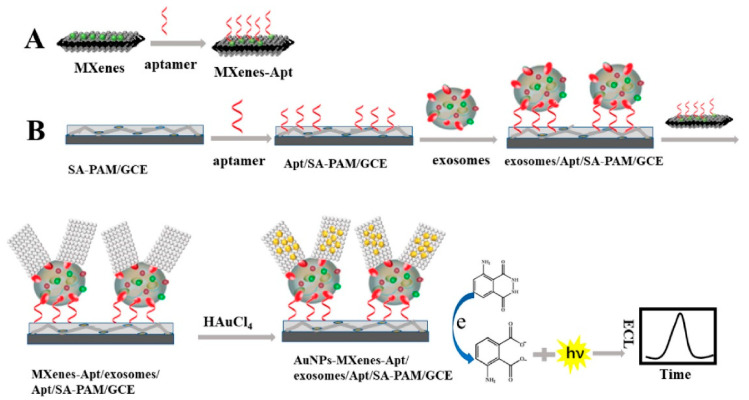
- Zhang, 2020, In situ formation of gold nanoparticles decorated Ti3C2 MXenes nanoprobe for highly sensitive electrogenerated chemiluminescence detection of exosomes and their surface proteins, Anal. Chem., 92, 5546, 10.1021/acs.analchem.0c00469 · DOI
- Xu, 2020, Optical, electrochemical and electrical (nano)biosensors for detection of exosomes: A comprehensive overview, Biosens. Bioelectron., 161, 112222, 10.1016/j.bios.2020.112222 · DOI
- Zong, 2016, Facile detection of tumor-derived exosomes using magnetic nanobeads and SERS nanoprobes, Anal. Methods, 8, 5001, 10.1039/C6AY00406G · DOI
- He, 2017, Direct exosome quantification via bivalent-cholesterol-labeled DNA anchor for signal amplification, Anal. Chem., 89, 12968, 10.1021/acs.analchem.7b03919 · DOI
- Kang, 2017, High-purity capture and release of circulating exosomes using an exosome-specific dual-patterned immunofiltration (ExoDIF) device, Nanoscale, 9, 13495, 10.1039/C7NR04557C · DOI
- Nie, P., Bai, Y., and Mei, H. (2020). Synthetic life with alternative nucleic acids as genetic materials. Molecules, 25. · DOI
- Huang, 2019, Light-Inducible Exosome-Based Vehicle for Endogenous RNA Loading and Delivery to Leukemia Cells, Adv. Funct. Mater., 29, 1807189, 10.1002/adfm.201807189 · DOI
- Dong, 2018, Highly sensitive electrochemical detection of tumor exosomes based on aptamer recognition-induced multi-DNA release and cyclic enzymatic amplification, Anal. Chem., 90, 4507, 10.1021/acs.analchem.7b04863 · DOI
- Wang, 2017, Aptasensor with expanded nucleotide using DNA nanotetrahedra for electrochemical detection of cancerous exosomes, ACS Nano, 11, 3943, 10.1021/acsnano.7b00373 · DOI
- Sun, 2015, MicroRNA expression profiles of bovine milk exosomes in response to Staphylococcus aureus infection, BMC Genom., 16, 1, 10.1186/s12864-015-2044-9 · DOI
- Ma, 2017, Construction of a Concanavalin A electrochemical sensor base on a novel sandwich capture mode, Sens. Actuators B Chem., 248, 201, 10.1016/j.snb.2017.03.172 · DOI
- Zhou, 2006, Exosomal Fetuin-A identified by proteomics: A novel urinary biomarker for detecting acute kidney injury, Kidney Int., 70, 1847, 10.1038/sj.ki.5001874 · DOI
- Bautista, 2011, Inflammation, oxidative stress, and obesity, Int. J. Mol. Sci., 12, 3117, 10.3390/ijms12053117 · DOI
- An, 2019, An ultrasensitive electrochemical aptasensor for the determination of tumor exosomes based on click chemistry, Biosens. Bioelectron., 142, 111503, 10.1016/j.bios.2019.111503 · DOI
- Xia, 2017, A visible and colorimetric aptasensor based on DNA-capped single-walled carbon nanotubes for detection of exosomes, Biosens. Bioelectron., 92, 8, 10.1016/j.bios.2017.01.063 · DOI
- Shi, 2019, A hybridization chain reaction based assay for fluorometric determination of exosomes using magnetic nanoparticles and both aptamers and antibody as recognition elements, Microchim. Acta, 186, 1, 10.1007/s00604-019-3823-9 · DOI
- Lyu, 2019, Near-infrared afterglow semiconducting nano-polycomplexes for the multiplex differentiation of cancer exosomes, Angew. Chem. Int. Ed., 58, 4983, 10.1002/anie.201900092 · DOI
- Liu, 2019, Low-cost thermophoretic profiling of extracellular-vesicle surface proteins for the early detection and classification of cancers, Nat. Biomed. Eng., 3, 183, 10.1038/s41551-018-0343-6 · DOI
- Zhang, 2019, Aptamer-based fluorescence polarization assay for separation-free exosome quantification, Nanoscale, 11, 10106, 10.1039/C9NR01589B · DOI
- Jiang, 2017, Aptamer/AuNP biosensor for colorimetric profiling of exosomal proteins, Angew. Chem. Int. Ed., 56, 11916, 10.1002/anie.201703807 · DOI
- Fang, 2020, Black phosphorus quantum dots functionalized MXenes as the enhanced dual-mode probe for exosomes sensing, Sens. Actuators B Chem., 305, 127544, 10.1016/j.snb.2019.127544 · DOI
- Wang, 2019, Bridging exosome and liposome through zirconium–phosphate coordination chemistry: A new method for exosome detection, Chem. Commun., 55, 2708, 10.1039/C9CC00220K · DOI
- Pandey, 2020, Design of engineered surfaces for prospective detection of SARS-CoV-2 using quartz crystal microbalance-based techniques, Expert Rev. Proteom., 17, 425, 10.1080/14789450.2020.1794831 · DOI
- Fopase, 2023, Potential of siRNA in COVID-19 therapy: Emphasis on in silico design and nanoparticles based delivery, Front. Bioeng. Biotechnol., 11, 1112755, 10.3389/fbioe.2023.1112755 · DOI
- Moura, 2020, Multiplex detection and characterization of breast cancer exosomes by magneto-actuated immunoassay, Talanta, 211, 120657, 10.1016/j.talanta.2019.120657 · DOI
- Wang, 2020, AuNP-amplified surface acoustic wave sensor for the quantification of exosomes, ACS Sens., 5, 362, 10.1021/acssensors.9b01869 · DOI
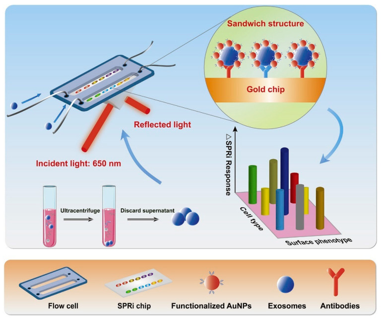
- Fan, 2020, High-sensitive and multiplex biosensing assay of NSCLC-derived exosomes via different recognition sites based on SPRi array, Biosens. Bioelectron., 154, 112066, 10.1016/j.bios.2020.112066 · DOI
- Picciolini, 2018, Detection and characterization of different brain-derived subpopulations of plasma exosomes by surface plasmon resonance imaging, Anal. Chem., 90, 8873, 10.1021/acs.analchem.8b00941 · DOI · Paper page
- Zhao, 2020, Aptamer-cholesterol-mediated proximity ligation assay for accurate identification of exosomes, Anal. Chem., 92, 5411, 10.1021/acs.analchem.0c00141 · DOI
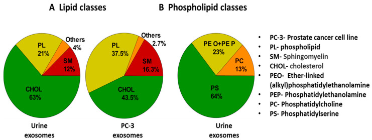
- Skotland, 2017, Molecular lipid species in urinary exosomes as potential prostate cancer biomarkers, Eur. J. Cancer, 70, 122, 10.1016/j.ejca.2016.10.011 · DOI
- Zhang, 2019, Sensitive multicolor visual detection of exosomes via dual signal amplification strategy of enzyme-catalyzed metallization of Au nanorods and hybridization chain reaction, ACS Sens., 4, 3210, 10.1021/acssensors.9b01644 · DOI
- Zhang, 2022, Label-free imaging and biomarker analysis of exosomes with plasmonic scattering microscopy, Chem. Sci., 13, 12760, 10.1039/D2SC05191E · DOI
- Chen, 2022, Label free imaging and deep tracking of single biological nanoparticles in free solution by reflection enhanced dark field scattering microscopy, Sens. Actuators B Chem., 355, 131317, 10.1016/j.snb.2021.131317 · DOI
- Cheng, 2015, Vibrational spectroscopic imaging of living systems: An emerging platform for biology and medicine, Science, 350, aaa8870, 10.1126/science.aaa8870 · DOI · Paper page
- Milane, 2015, Exosome mediated communication within the tumor microenvironment, J. Control. Release, 219, 278, 10.1016/j.jconrel.2015.06.029 · DOI
- Yu, 2014, Molecular scale origin of surface plasmon resonance biosensors, Anal. Chem., 86, 8992, 10.1021/ac501363z · DOI
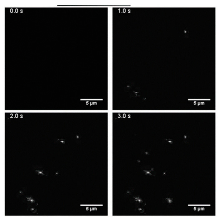
- Yang, 2018, Interferometric plasmonic imaging and detection of single exosomes, Proc. Natl. Acad. Sci. USA, 115, 10275, 10.1073/pnas.1804548115 · DOI
- Mehak, 2023, Surface modified iron-oxide based engineered nanomaterials for hyperthermia therapy of cancer cells, Biotechnol. Genet. Eng. Rev., 29, 1, 10.1080/02648725.2023.2169370 · DOI
- Pandey, 2021, Surface engineering of nano-sorbents for the removal of heavy metals: Interfacial aspects, J. Environ. Chem. Eng., 9, 104586, 10.1016/j.jece.2020.104586 · DOI
- Pandey, 2020, Surface engineering of personal protective equipments (PPEs) to prevent the contagious infections of SARS-CoV-2, Surf. Eng., 36, 901, 10.1080/02670844.2020.1801034 · DOI
- Chandra, P., and Pandey, L.M. (2020). Biointerface Engineering: Prospects in Medical Diagnostics and Drug Delivery, Springer Nature. · DOI
- Pandey, L.M., and Hasan, A. (2022). Nanoscale Engineering of Biomaterials: Properties and Applications, Springer Nature. · DOI
- Hasan, 2018, Surface Functionalization of Ti6Al4V via Self-assembled Monolayers for Improved Protein Adsorption and Fibroblast Adhesion, Langmuir, 34, 3494, 10.1021/acs.langmuir.7b03152 · DOI
- Hasan, 2016, Kinetic studies of attachment and re-orientation of octyltriethoxysilane for formation of self-assembled monolayer on a silica substrate, Mater. Sci. Eng. C, 68, 423, 10.1016/j.msec.2016.06.003 · DOI
- Pandey, 2023, Design of biocompatible and self-antibacterial titanium surfaces for biomedical applications, Curr. Opin. Biomed. Eng., 25, 100423, 10.1016/j.cobme.2022.100423 · DOI
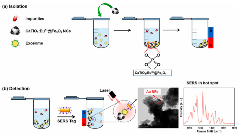
- Back, 2023, Rapid and simple isolation and detection of exosomes using CaTiO3:Eu3+@Fe3O4 multifunctional nanocomposites, Anal. Biochem., 673, 115161, 10.1016/j.ab.2023.115161 · DOI
- Wang, 2022, Magnetic guanidyl-functionalized covalent organic framework composite: A platform for specific capture and isolation of phosphopeptides and exosomes, Microchim. Acta, 189, 330, 10.1007/s00604-022-05394-3 · DOI
- Jang, 2023, Magnetic transferrin nanoparticles (MTNs) assay as a novel isolation approach for exosomal biomarkers in neurological diseases, Biomater. Res., 27, 1, 10.1186/s40824-023-00353-2 · DOI
- Farsani, 2023, Tailored design and preparation of magnetic nanocomposite particles for the isolation of exosomes, Nanotechnology, 34, 155603, 10.1088/1361-6528/acb2d2 · DOI
- Singh, 2022, Rapid immunomagnetic co-capture assay for quantification of lung cancer associated exosomes, J. Immunol. Methods, 508, 113324, 10.1016/j.jim.2022.113324 · DOI
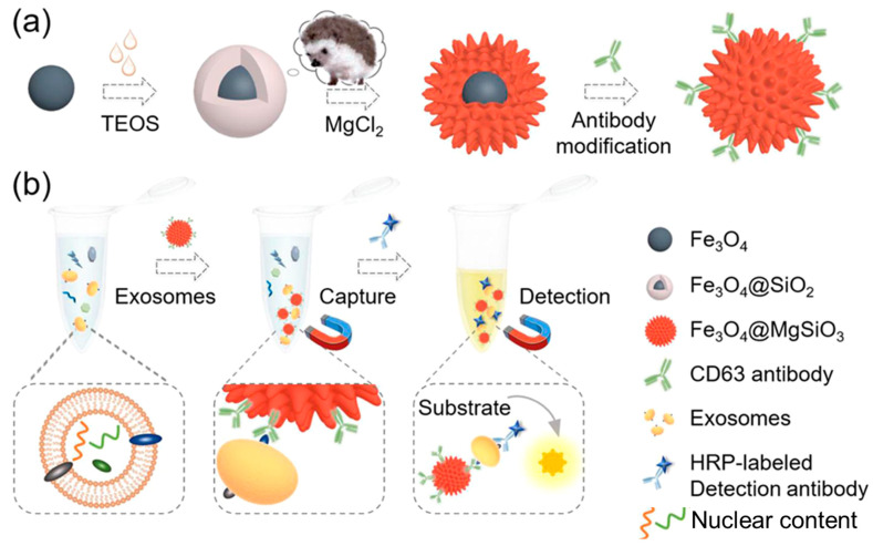
- Yang, 2021, Hedgehog-inspired magnetic nanoparticles for effectively capturing and detecting exosomes, NPG Asia Mater., 13, 78, 10.1038/s41427-021-00346-4 · DOI
- Pammi Guru, K.T., Praween, N., and Basu, P.K. (2023). Isolation of Exosomes from Human Serum Using Gold-Nanoparticle-Coated Silicon Surface. Nanomaterials, 13. · DOI
- Bhattacharyya, 2023, Brain-enriched miR-128: Reduced in exosomes from Parkinson’s patient plasma, improves synaptic integrity, and prevents 6-OHDA mediated neuronal apoptosis, Front. Cell Neurosci., 16, 680, 10.3389/fncel.2022.1037903 · DOI
- Gui, 2015, Altered microRNA profiles in cerebrospinal fluid exosome in Parkinson disease and Alzheimer disease, Oncotarget, 6, 37043, 10.18632/oncotarget.6158 · DOI · Paper page
- Cao, 2017, MicroRNA biomarkers of Parkinson’s disease in serum exosome-like microvesicles, Neurosci. Lett., 644, 94, 10.1016/j.neulet.2017.02.045 · DOI
- Yao, 2018, Circulating exosomal miRNAs as diagnostic biomarkers in Parkinson’s disease, Eur. Rev. Med. Pharm., 22, 5278
- Manna, 2021, Exosomal miRNA as peripheral biomarkers in Parkinson’s disease and progressive supranuclear palsy: A pilot study, Park. Relat. D, 93, 77, 10.1016/j.parkreldis.2021.11.020 · DOI
- Tomlinson, 2015, Identification of distinct circulating exosomes in Parkinson’s disease, Ann. Clin. Transl. Neurol., 2, 353, 10.1002/acn3.175 · DOI
- Sharma, 2021, Shear-induced aggregation of amyloid β (1–40) in a parallel plate geometry, J. Biomol. Struct. Dyn., 39, 6415, 10.1080/07391102.2020.1798814 · DOI
- Panda, C., Voelz, C., Habib, P., Mevissen, C., Pufe, T., Beyer, C., Gupta, S., and Slowik, A. (2021). Aggregated Tau-PHF6 (VQIVYK) Potentiates NLRP3 Inflammasome Expression and Autophagy in Human Microglial Cells. Cells, 10. · DOI
- Pandey, 2022, Physicochemical factors of bioprocessing impact the stability of therapeutic proteins, Biotechnol. Adv., 55, 107909, 10.1016/j.biotechadv.2022.107909 · DOI
- Xu, 2018, Comparison of the extraction and determination of serum exosome and miRNA in serum and the detection of miR-27a-3p in serum exosome of ALS patients, Intractable Rare Dis. Res., 7, 13, 10.5582/irdr.2017.01091 · DOI
- Katsu, 2019, MicroRNA expression profiles of neuron-derived extracellular vesicles in plasma from patients with amyotrophic lateral sclerosis, Neurosci. Lett., 708, 134176, 10.1016/j.neulet.2019.03.048 · DOI
- Lo, 2021, Extracellular Vesicles in Serum and Central Nervous System Tissues Contain microRNA Signatures in Sporadic Amyotrophic Lateral Sclerosis, Front. Mol. Neurosci., 14, 739016, 10.3389/fnmol.2021.739016 · DOI
- Kim, 2023, Small RNA sequencing of circulating small extracellular vesicles microRNAs in patients with amyotrophic lateral sclerosis, Sci. Rep., 13, 5528, 10.1038/s41598-023-32717-y · DOI
- Saucier, 2019, Identification of a circulating miRNA signature in extracellular vesicles collected from amyotrophic lateral sclerosis patients, Brain Res., 1708, 100, 10.1016/j.brainres.2018.12.016 · DOI · Paper page
- Cai, 2019, Long non-coding RNA LINC00467 regulates hepatocellular carcinoma progression by modulating miR-9-5p/PPARA expression, Open Biol., 9, 190074, 10.1098/rsob.190074 · DOI
- Bai, 2018, Liquid biopsy in tumors: Opportunities and challenges, Ann. Transl. Med., 6, S89, 10.21037/atm.2018.11.31 · DOI
- Li, 2021, Rapid and specific detection nanoplatform of serum exosomes for prostate cancer diagnosis, Mikrochim. Acta, 188, 283, 10.1007/s00604-021-04934-7 · DOI
- Lee, J., Lee, J.H., Mondal, J., Hwang, J., Kim, H.S., Kumar, V., Raj, A., Hwang, S.R., and Lee, Y.K. (2022). Magnetofluoro-Immunosensing Platform Based on Binary Nanoparticle-Decorated Graphene for Detection of Cancer Cell-Derived Exosomes. Int. J. Mol. Sci., 23. · DOI
- Li, 2021, A magnetic surface-enhanced Raman scattering platform for performing successive breast cancer exosome isolation and analysis, J. Mater. Chem. B, 9, 2709, 10.1039/D0TB02894K · DOI
- Qin, 2022, Simultaneous detection of cancerous exosomal miRNA-21 and PD-L1 with a sensitive dual-cycling nanoprobe, Biosens. Bioelectron., 216, 114636, 10.1016/j.bios.2022.114636 · DOI
- Hong, 2020, Label-free diagnosis for colorectal cancer through coffee ring-assisted surface-enhanced Raman spectroscopy on blood serum, J. Biophotonics, 13, e201960176, 10.1002/jbio.201960176 · DOI
- Chinnappan, 2023, An integrated lab-on-a-chip platform for pre-concentration and detection of colorectal cancer exosomes using anti-CD63 aptamer as a recognition element, Biosens. Bioelectron., 220, 114856, 10.1016/j.bios.2022.114856 · DOI
- Lee, 2009, Intravenous hMSCs Improve Myocardial Infarction in Mice because Cells Embolized in Lung Are Activated to Secrete the Anti-inflammatory Protein TSG-6, Cell Stem Cell, 5, 54, 10.1016/j.stem.2009.05.003 · DOI · Paper page
- Lai, 2010, Exosome secreted by MSC reduces myocardial ischemia/reperfusion injury, Stem Cell Res., 4, 214, 10.1016/j.scr.2009.12.003 · DOI · Paper page
- Hao, 2017, Stem cell-derived exosomes: A promising strategy for fracture healing, Cell Prolif., 50, e12359, 10.1111/cpr.12359 · DOI · Paper page
- Tang, 2015, Therapeutic potential of CAR-T cell-derived exosomes: A cell-free modality for targeted cancer therapy, Oncotarget, 6, 44179, 10.18632/oncotarget.6175 · DOI
- Lugini, 2012, Immune surveillance properties of human NK cell-derived exosomes, J. Immunol., 189, 2833, 10.4049/jimmunol.1101988 · DOI
- Vickers, 2014, MicroRNA-223 coordinates cholesterol homeostasis, Proc. Natl. Acad. Sci. USA, 111, 14518, 10.1073/pnas.1215767111 · DOI · Paper page
- Ibrahim, 2014, Exosomes as Critical Agents of Cardiac Regeneration Triggered by Cell Therapy, Stem Cell Rep., 2, 606, 10.1016/j.stemcr.2014.04.006 · DOI
- Agarwal, 2017, Experimental, systems, and computational approaches to understanding the microRNA-mediated reparative potential of cardiac progenitor cell–derived exosomes from pediatric patients, Circ. Res., 120, 701, 10.1161/CIRCRESAHA.116.309935 · DOI
- Zhang, 2022, Exosomes as Anticancer Drug Delivery Vehicles: Prospects and Challenges, Front. Biosci., 27, 293, 10.31083/j.fbl2710293 · DOI
- Butreddy, A., Kommineni, N., and Dudhipala, N. (2021). Exosomes as Naturally Occurring Vehicles for Delivery of Biopharmaceuticals: Insights from Drug Delivery to Clinical Perspectives. Nanomaterials, 11. · DOI
- Kim, H., Jang, H., Cho, H., Choi, J., Hwang, K.Y., Choi, Y., Kim, S.H., and Yang, Y. (2021). Recent Advances in Exosome-Based Drug Delivery for Cancer Therapy. Cancers, 13. · DOI · Paper page
200 of 203 shown
Cited by
Sorted by citations
Emerging role of exosomes in cancer therapy: progress and challenges
Li, J., Wang, J., & Chen, Z. (2025) · Molecular Cancer · doi:10.1186/s12943-024-02215-4 · Cited by 297
Metadata
- Authors
- Sonbhadra, S., Mehak, & Pandey, L. M.
- Affiliations
- Bio-Interface & Environmental Engineering Lab, Department of Biosciences and Bioengineering, Indian Institute of Technology Guwahati, Assam 781039, India
- Venue
- Biosensors
- Publisher
- MDPI AG
- Published
- 2023-08-10
- Volume
- 13
- Issue
- 8
- Pages
- 802
- ISSN
- 2079-6374
- PMC
- PMC10452587
- Cited by
- 97
- References
- 9
- Language
- en
About these checks
Registry, retraction, Cites and statistics results are computed, not judged, with one exception: a printed reference that no index lists is read once by a model before it counts as a citation error. Claims are extracted by a model and kept only when their quote appears verbatim in the text; each cited source is then checked, with a model’s help, for whether it says what the claim relies on. Those verdicts can be wrong — read the claim’s quote and the cited source before relying on one.
Statistics and arithmetic are recomputed, not judged.
Confidence bands are shown only for fields calibrated on our annotated test set. None is calibrated yet, so no band is shown.
What counts as a claim ›How we measure confidence ›How we check this ›
Are you an author, or do you see a problem with this page? Request a review
