

The DOI matches its Crossref record; 40 of the 40 references it cites were found in a registry or an index.
The Glycogen-Binding Domain on the AMPK β Subunit Allows the Kinase to Act as a Glycogen Sensor
Open accessAbstract
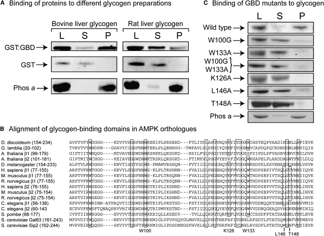
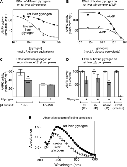
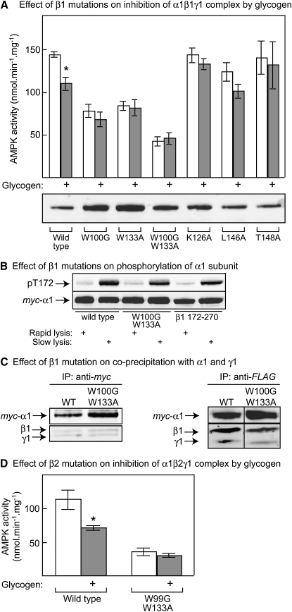
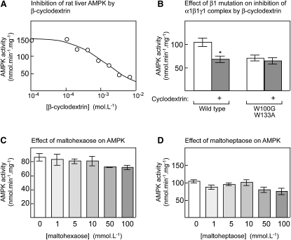
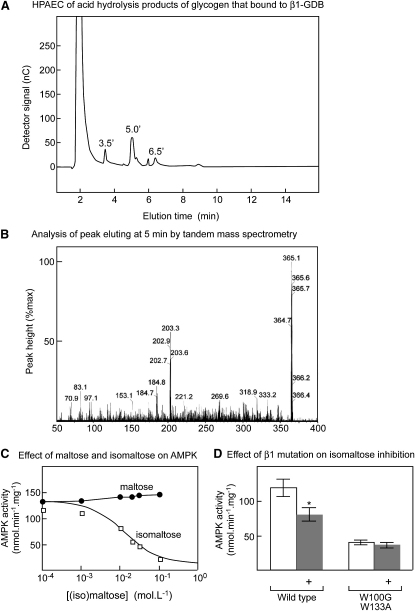
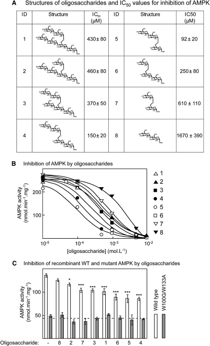
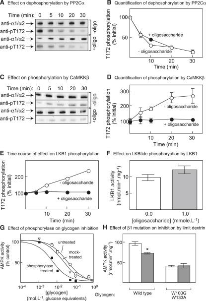
AMPK beta subunits contain a conserved domain that causes association with glycogen. Although glycogen availability is known to affect AMPK regulation in vivo, the molecular mechanism for this has not been clear. We now show that AMPK is inhibited by glycogen, particularly preparations with high branching content. We synthesized a series of branched oligosaccharides and show that those with a single alpha1-->6 branch are allosteric inhibitors that also inhibit phosphorylation by upstream kinases. Removal of the outer chains of glycogen using phosphorylase, thus exposing the outer branches, renders inhibition of AMPK more potent. Inhibition by all carbohydrates tested was dependent on the glycogen-binding domain being abolished by mutation of residues required for carbohydrate binding. Our results suggest the hypothesis that AMPK, as well as monitoring immediate energy availability by sensing AMP/ATP, may also be able to sense the status of cellular energy reserves in the form of glycogen.
Overview
The claims and statistics of this paper haven’t been checked yet.
Run the check — free, no account needed. It reads the open-access text and checks the sources it cites.
Integrity signals
Registry
Cites
Statistics
Figures
Verification
Claims the paper makes
The claims and statistics of this paper haven’t been checked yet.
Run the check — free, no account needed. It reads the open-access text and checks the sources it cites.
Claims other papers make about this one
Each is checked against this paper’s own text: does it say what the citing paper says it does?
The source backs part of the claim, not all of it — or backs it for a different population, condition or comparison.
In the citing paper
“there may even be benefits from deliberately withholding CHO after a high quality training session to minimise glycogen restoration and extend the period during which adaptive responses are elevated (65).”
From Postexercise muscle glycogen resynthesis in humans. (2017) · cites this paper as [65]
Experimental — accuracy not yet measuredThe cited source says what the claim attributes to it.
In the citing paper
“AMPK can be activated by low glycogen [66]”
From Using Molecular Biology to Maximize Concurrent Training (2014) · cites this paper as [66]
In the citing paper
“These data suggest that when glycogen is depleted AMPK is more active.”
From New strategies in sport nutrition to increase exercise performance (2016) · cites this paper as [20]
In the citing paper
“In addition to being a direct AMP:ATP ratio sensor, AMPK also has the capacity to be modulated by the glycogen storage status of a cell [20].The β-subunit of AMPK binds to specific branch points on glycogen inhibiting the enzyme and preventing it from being phosphorylated by upstream kinases [20].”
From New strategies in sport nutrition to increase exercise performance (2016) · cites this paper as [20]
In the citing paper
“Recently, mutations in the CBM have been shown to prevent glycogen binding and reduce total AMPK activity in cell-free assays [130] and in vivo [131].”
From AMPK and Exercise: Glucose Uptake and Insulin Sensitivity (2013) · cites this paper as [130]
In the citing paper
“Glycogen can also inhibit AMPK, suggesting that AMPK may regulate cellular energy status through glycogen sensing [130].”
From AMPK and Exercise: Glucose Uptake and Insulin Sensitivity (2013) · cites this paper as [130]
Self-check
Percentages, sums and reported differences re-computed from the paper’s own numbers.
The self-check needs the paper’s full text.
Citations
Cites
Kahn, B. B., Alquier, T., Carling, D., & Hardie, D. G. (2005) · Cell Metabolism · doi:10.1016/j.cmet.2004.12.003 · Cited by 2,799
AMP-activated/SNF1 protein kinases: conserved guardians of cellular energy
Hardie, D. G. (2007) · Nature Reviews Molecular Cell Biology · doi:10.1038/nrm2249 · Cited by 2,261
Hawley, S. A., Boudeau, J., Reid, J. L., Mustard, K. J., Udd, L., Mäkelä, T. P., Alessi, D. R., & Hardie, D. G. (2003) · Journal of Biology · doi:10.1186/1475-4924-2-28 · Cited by 1,694
LKB1 is the upstream kinase in the AMP-activated protein kinase cascade
Woods, A., Johnstone, S. R., Dickerson, K., Leiper, F. C., Fryer, L. G. D., Neumann, D., Schlattner, U., Wallimann, T., Carlson, M., & Carling, D. (2003) · Current Biology · doi:10.1016/j.cub.2003.10.031 · Cited by 1,650
Hawley, S. A., Pan, D. A., Mustard, K. J., Ross, L., Bain, J., Edelman, A. M., Frenguelli, B. G., & Hardie, D. G. (2005) · Cell Metabolism · doi:10.1016/j.cmet.2005.05.009 · Cited by 1,589
Self-citationLKB1 is a master kinase that activates 13 kinases of the AMPK subfamily, including MARK/PAR‐1
Lizcano, J. M., Göransson, O., Toth, R., Deak, M., Morrice, N. A., Boudeau, J., Hawley, S. A., Udd, L., Mäkelä, T. P., Hardie, D. G., et al. (2004) · The EMBO Journal · doi:10.1038/sj.emboj.7600110 · Cited by 1,381
Hawley, S. A., Davison, M., Woods, A., Davies, S. P., Beri, R. K., Carling, D., & Hardie, D. G. (1996) · Journal of Biological Chemistry · doi:10.1074/jbc.271.44.27879 · Cited by 1,264
Scott, J. W., Hawley, S. A., Green, K. A., Anis, M., Stewart, G., Scullion, G. A., Norman, D. G., & Hardie, D. G. (2004) · Journal of Clinical Investigation · doi:10.1172/jci19874 · Cited by 754
Self-citationStructural basis for AMP binding to mammalian AMP-activated protein kinase
Xiao, B., Heath, R., Saiu, P., Leiper, F. C., Leone, P., Jing, C., Walker, P. A., Haire, L., Eccleston, J. F., Davis, C. T., et al. (2007) · Nature · doi:10.1038/nature06161 · Cited by 591
Cited by
Sorted by citations
AMPK: Mechanisms of Cellular Energy Sensing and Restoration of Metabolic Balance
Garcia, D., & Shaw, R. J. (2017) · Molecular Cell · doi:10.1016/j.molcel.2017.05.032 · Cited by 2,061
The LKB1-AMPK pathway: metabolism and growth control in tumour suppression
Shackelford, D. B., & Shaw, R. J. (2009) · Nature Reviews Cancer · doi:10.1038/nrc2676 · Cited by 1,943
Steinberg, G. R., & Kemp, B. E. (2009) · Physiological Reviews · doi:10.1152/physrev.00011.2008 · Cited by 1,672
AMPK: Sensing Glucose as well as Cellular Energy Status
Lin, S., & Hardie, D. G. (2017) · Cell Metabolism · doi:10.1016/j.cmet.2017.10.009 · Cited by 1,198
Regulation and function of AMPK in physiology and diseases
Jeon, S. (2016) · Experimental & Molecular Medicine · doi:10.1038/emm.2016.81 · Cited by 1,149
AMPK: An Emerging Drug Target for Diabetes and the Metabolic Syndrome
Zhang, B. B., Zhou, G., & Li, C. (2009) · Cell Metabolism · doi:10.1016/j.cmet.2009.03.012 · Cited by 1,042
New insights into activation and function of the AMPK
Steinberg, G. R., & Hardie, D. G. (2022) · Nature Reviews Molecular Cell Biology · doi:10.1038/s41580-022-00547-x · Cited by 1,074
AMPK and TOR: The Yin and Yang of Cellular Nutrient Sensing and Growth Control
González, A., Hall, M. N., Lin, S.-C., & Hardie, D. G. (2020) · Cell Metabolism · doi:10.1016/j.cmet.2020.01.015 · Cited by 797
AMPK and mTOR in Cellular Energy Homeostasis and Drug Targets
Inoki, K., Kim, J., & Guan, K. (2011) · The Annual Review of Pharmacology and Toxicology · doi:10.1146/annurev-pharmtox-010611-134537 · Cited by 818
Starch turnover: pathways, regulation and role in growth
Stitt, M., & Zeeman, S. C. (2012) · Current Opinion in Plant Biology · doi:10.1016/j.pbi.2012.03.016 · Cited by 766
Glycogen and its metabolism: some new developments and old themes
Roach, P. J., DePaoli-Roach, A. A., Hurley, T. D., & Tagliabracci, V. S. (2012) · Biochemical Journal · doi:10.1042/bj20111416 · Cited by 728
Onken, B., & Driscoll, M. (2010) · PLoS ONE · doi:10.1371/journal.pone.0008758 · Cited by 671
AMPK—Sensing Energy while Talking to Other Signaling Pathways
Hardie, D. G. (2014) · Cell Metabolism · doi:10.1016/j.cmet.2014.09.013 · Cited by 597
Peripheral Chemoreceptors: Function and Plasticity of the Carotid Body
Kumar, P., & Prabhakar, N. R. (2012) · Comprehensive physiology · doi:10.1002/cphy.c100069 · Cited by 581
AMPK in skeletal muscle function and metabolism
Kjøbsted, R., Hingst, J. R., Fentz, J., Foretz, M., Sanz, M.-N., Pehmøller, C. K., Shum, M., Marette, A., Mounier, R., Treebak, J. T., et al. (2017) · The FASEB Journal · doi:10.1096/fj.201700442r · Cited by 517
AMPK and the biochemistry of exercise: implications for human health and disease
Richter, E. A., & Ruderman, N. B. (2009) · Biochemical Journal · doi:10.1042/bj20082055 · Cited by 481
Dzeja, P. P., & Terzic, A. (2009) · International Journal of Molecular Sciences · doi:10.3390/ijms10041729 · Cited by 497
AMP-activated protein kinase: nature's energy sensor
Carling, D., Mayer, F. V., Sanders, M. J., & Gamblin, S. J. (2011) · Nature Chemical Biology · doi:10.1038/nchembio.610 · Cited by 387
Favaro, E., Bensaad, K., Chong, M., Tennant, D. A., Ferguson, D. J. P., Snell, C. E., Steers, G., Turley, H., Li, J., Günther, U. L., et al. (2012) · Cell Metabolism · doi:10.1016/j.cmet.2012.10.017 · Cited by 391
AMPK inhibition in health and disease
Viollet, B., Horman, S., Leclerc, J., Lantier, L., Foretz, M., Billaud, M., Giri, S., & Andreelli, F. (2010) · Critical Reviews in Biochemistry and Molecular Biology · doi:10.3109/10409238.2010.488215 · Cited by 383
AMP-activated protein kinase: new regulation, new roles?
Carling, D., Thornton, C., Woods, A., & Sanders, M. J. (2012) · Biochemical Journal · doi:10.1042/bj20120546 · Cited by 390
AMP-Activated Protein Kinase: A Target for Drugs both Ancient and Modern
Hardie, D. G., Ross, F. A., & Hawley, S. A. (2012) · Chemistry & Biology · doi:10.1016/j.chembiol.2012.08.019 · Cited by 385
Sensing of energy and nutrients by AMP-activated protein kinase
Hardie, D. G. (2011) · The American Journal of Clinical Nutrition · doi:10.3945/ajcn.110.001925 · Cited by 372
Self-citationDevelopment of protein kinase activators: AMPK as a target in metabolic disorders and cancer
Fogarty, S., & Hardie, D. G. (2009) · Biochimica et Biophysica Acta (BBA) - Proteins and Proteomics · doi:10.1016/j.bbapap.2009.09.012 · Cited by 357
Regulation of starch biosynthesis in response to a fluctuating environment
Geigenberger, P. (2011) · Plant Physiology · doi:10.1104/pp.110.170399 · Cited by 348
Regulation of Tissue Growth through Nutrient Sensing
Hietakangas, V., & Cohen, S. M. (2009) · Annual Review of Genetics · doi:10.1146/annurev-genet-102108-134815 · Cited by 341
Glycogen accumulation and phase separation drives liver tumor initiation
Liu, Q., Li, J., Zhang, W., Xiao, C., Zhang, S., Nian, C., Li, J., Su, D., Chen, L., Zhao, Q. C., et al. (2021) · Cell · doi:10.1016/j.cell.2021.10.001 · Cited by 313
Mackenzie, R., & Elliott, B. (2014) · Diabetes Metabolic Syndrome and Obesity · doi:10.2147/dmso.s48260 · Cited by 322
AMPK as a mediator of hormonal signalling
Lim, C. T., Kola, B., & Korbonits, M. (2009) · Journal of Molecular Endocrinology · doi:10.1677/jme-09-0063 · Cited by 304
Srivastava, R. A. K., Pinkosky, S. L., Filippov, S. I., Hanselman, J. C., Cramer, C. T., & Newton, R. S. (2012) · Journal of Lipid Research · doi:10.1194/jlr.r025882 · Cited by 308
AMPK and Exercise: Glucose Uptake and Insulin Sensitivity
O'Neill, H. M. (2013) · Diabetes & Metabolism Journal · doi:10.4093/dmj.2013.37.1.1 · Cited by 295
Role of AMP-activated protein kinase in adipose tissue metabolism and inflammation
Bijland, S., Mancini, S. J., & Salt, I. P. (2013) · Clinical Science · doi:10.1042/cs20120536 · Cited by 296
AMP-activated protein kinase regulation and biological actions in the heart
Zaha, V. G., & Young, L. H. (2012) · Circulation Research · doi:10.1161/circresaha.111.255505 · Cited by 279
Falkowska, A., Gutowska, I., Goschorska, M., Nowacki, P., Chlubek, D., & Baranowska-Bosiacka, I. (2015) · International Journal of Molecular Sciences · doi:10.3390/ijms161125939 · Cited by 277
Regulation of glucose and glycogen metabolism during and after exercise
Jensen, T. E., & Richter, E. A. (2012) · The Journal of Physiology · doi:10.1113/jphysiol.2011.224972 · Cited by 274
Broeckx, T., Hulsmans, S., & Rolland, F. (2016) · Journal of Experimental Botany · doi:10.1093/jxb/erw416 · Cited by 279
Mechanisms Modulating Skeletal Muscle Phenotype
Blaauw, B., Schiaffino, S., & Reggiani, C. (2013) · Comprehensive physiology · doi:10.1002/cphy.c130009 · Cited by 298
Structure and Physiological Regulation of AMPK
Yan, Y., Zhou, X. E., Xu, H. E., & Melcher, K. (2018) · International Journal of Molecular Sciences · doi:10.3390/ijms19113534 · Cited by 254
Hearris, M., Hammond, K., Fell, J., & Morton, J. (2018) · Nutrients · doi:10.3390/nu10030298 · Cited by 252
AMP-activated protein kinase, stress responses and cardiovascular diseases
Wang, S., Song, P., & Zou, M. (2012) · Clinical Science · doi:10.1042/cs20110625 · Cited by 251
Emerging Role of cAMP/AMPK Signaling
Aslam, M., & Ladilov, Y. (2022) · Cells · doi:10.3390/cells11020308 · Cited by 241
Postexercise muscle glycogen resynthesis in humans.
Burke, L. M., van Loon, L. J. C., & Hawley, J. A. (2017) · Journal of Applied Physiology · doi:10.1152/japplphysiol.00860.2016 · Cited by 242
Mechanisms of regulation of SNF1/AMPK/SnRK1 protein kinases
Crozet, P., Margalha, L., Confraria, A., Rodrigues, A., Martinho, C., Adamo, M., Elias, A., & Gonzalez, E. B. (2014) · Frontiers in Plant Science · doi:10.3389/fpls.2014.00190 · Cited by 241
Carbohydrate availability and exercise training adaptation: Too much of a good thing?
Bartlett, J. D., Hawley, J. A., & Morton, J. P. (2015) · European Journal of Sport Science · doi:10.1080/17461391.2014.920926 · Cited by 230
Peng, C., Wang, Y., Liu, F., Ren, Y., Zhou, K., Lv, J., Zheng, M., Zhao, S., Zhang, L., Wang, C., et al. (2014) · The Plant Journal · doi:10.1111/tpj.12444 · Cited by 229
New strategies in sport nutrition to increase exercise performance
Close, G. L., Hamilton, D. L., Philp, A., Burke, L. M., & Morton, J. P. (2016) · Free Radical Biology and Medicine · doi:10.1016/j.freeradbiomed.2016.01.016 · Cited by 221
Gibala, M. J., Gillen, J. B., & Percival, M. E. (2014) · Sports Medicine · doi:10.1007/s40279-014-0259-6 · Cited by 249
SnRK1 and TOR: modulating growth–defense trade-offs in plant stress responses
Margalha, L., Confraria, A., & Gonzalez, E. B. (2019) · Journal of Experimental Botany · doi:10.1093/jxb/erz066 · Cited by 221
AMPK and autophagy in glucose/glycogen metabolism
Ha, J., Guan, K., & Kim, J. (2015) · Molecular Aspects of Medicine · doi:10.1016/j.mam.2015.08.002 · Cited by 219
Nutritional modulation of training-induced skeletal muscle adaptations
Hawley, J. A., Burke, L. M., Phillips, S. M., & Spriet, L. L. (2011) · Journal of Applied Physiology · doi:10.1152/japplphysiol.00949.2010 · Cited by 205
Pelletier, J., Bellot, G. L., Gounon, P., Lacas‐Gervais, S., Pouysségur, J., & Mazure, N. M. (2012) · Frontiers in Oncology · doi:10.3389/fonc.2012.00018 · Cited by 213
Seung, D., Soyk, S., Coiro, M., Maier, B. A., Eicke, S., & Zeeman, S. C. (2015) · PLoS Biology · doi:10.1371/journal.pbio.1002080 · Cited by 199
Miller, V. J., Villamena, F. A., & Volek, J. S. (2018) · Journal of Nutrition and Metabolism · doi:10.1155/2018/5157645 · Cited by 205
Redox implications of AMPK-mediated signal transduction beyond energetic clues
Cardaci, S., Filomeni, G., & Ciriolo, M. R. (2012) · Journal of Cell Science · doi:10.1242/jcs.095216 · Cited by 204
Structural insight into the autoinhibition mechanism of AMP-activated protein kinase
Chen, L., Jiao, Z., Zheng, L., Zhang, Y., Xie, S.-T., Wang, Z., & Wu, J. (2009) · Nature · doi:10.1038/nature08075 · Cited by 200
AMP-activated protein kinase (AMPK) activation regulates in vitro bone formation and bone mass
Shah, M., Kola, B., Bataveljic, A., Arnett, T. R., Viollet, B., Saxon, L., Korbonits, M., & Chenu, C. (2010) · Bone · doi:10.1016/j.bone.2010.04.596 · Cited by 196
Structural basis of AMPK regulation by adenine nucleotides and glycogen
Li, X., Wang, L., Zhou, X. E., Ke, J., de Waal, P. W., Gu, X., Tan, M. H. E., Wang, D., Wu, D., Xu, H. E., et al. (2014) · Cell Research · doi:10.1038/cr.2014.150 · Cited by 198
Keeping the home fires burning: AMP-activated protein kinase
Hardie, D. G. (2018) · Journal of The Royal Society Interface · doi:10.1098/rsif.2017.0774 · Cited by 195
AMP-activated protein kinase: Also regulated by ADP?
Hardie, D. G., Carling, D., & Gamblin, S. J. (2011) · Trends in Biochemical Sciences · doi:10.1016/j.tibs.2011.06.004 · Cited by 187
Self-citationBeauloye, C., Bertrand, L., Horman, S., & Hue, L. (2011) · Cardiovascular Research · doi:10.1093/cvr/cvr034 · Cited by 191
Maximizing Cellular Adaptation to Endurance Exercise in Skeletal Muscle
Hawley, J. A., Lundby, C., Cotter, J. D., & Burke, L. M. (2018) · Cell Metabolism · doi:10.1016/j.cmet.2018.04.014 · Cited by 182
Hardie, D. G. (2011) · Biochemical Society Transactions · doi:10.1042/bst0390001 · Cited by 180
Regulation of AMP-activated protein kinase by natural and synthetic activators
Hardie, D. G. (2015) · Acta Pharmaceutica Sinica B · doi:10.1016/j.apsb.2015.06.002 · Cited by 180
Muscle Glycogen Metabolism and High-Intensity Exercise Performance: A Narrative Review
Vigh-Larsen, J. F., Ørtenblad, N., Spriet, L. L., Overgaard, K., & Mohr, M. (2021) · Sports Medicine · doi:10.1007/s40279-021-01475-0 · Cited by 169
The role of skeletal muscle Akt in the regulation of muscle mass and glucose homeostasis
Jaiswal, N., Gavin, M. G., Quinn, W. J., Luongo, T. S., Gelfer, R. G., Baur, J. A., & Titchenell, P. M. (2019) · Molecular Metabolism · doi:10.1016/j.molmet.2019.08.001 · Cited by 167
Effects of AMP-Activated Protein Kinase in Cerebral Ischemia
Li, J., & McCullough, L. D. (2009) · Journal of Cerebral Blood Flow & Metabolism · doi:10.1038/jcbfm.2009.255 · Cited by 165
Steinberg, G. R., O'Neill, H. M., Dzamko, N. L., Galic, S., Naim, T., Koopman, R., Jørgensen, S. B., Honeyman, J., Hewitt, K., Chen, Z.-P., et al. (2010) · Journal of Biological Chemistry · doi:10.1074/jbc.m110.102434 · Cited by 164
AMPK in the brain: its roles in glucose and neural metabolism
Muraleedharan, R., & Dasgupta, B. (2021) · FEBS Journal · doi:10.1111/febs.16151 · Cited by 170
Targeting AMPK Signaling as a Neuroprotective Strategy in Parkinson’s Disease
Curry, D. W., Stutz, B., Andrews, Z. B., & Elsworth, J. D. (2018) · Journal of Parkinson s Disease · doi:10.3233/jpd-171296 · Cited by 160
Wu, S., Wu, Y., Wu, T.-P., & Wei, Y. (2013) · Biochimica et Biophysica Acta (BBA) - General Subjects · doi:10.1016/j.bbagen.2013.10.034 · Cited by 164
More than a store: regulatory roles for glycogen in skeletal muscle adaptation to exercise
Philp, A., Hargreaves, M., & Baar, K. (2012) · American Journal of Physiology-Endocrinology and Metabolism · doi:10.1152/ajpendo.00004.2012 · Cited by 156
Metabolic fuels: regulating fluxes to select mix
Weber, J. (2010) · Journal of Experimental Biology · doi:10.1242/jeb.047050 · Cited by 158
Bartlett, J. D., Louhelainen, J., Iqbal, Z., Cochran, A. J., Gibala, M. J., Gregson, W., Close, G. L., Drust, B., & Morton, J. P. (2013) · American Journal of Physiology-Regulatory, Integrative and Comparative Physiology · doi:10.1152/ajpregu.00498.2012 · Cited by 147
Energy sensing by the AMP-activated protein kinase and its effects on muscle metabolism
Hardie, D. G. (2011) · Proceedings of the Nutrition Society · doi:10.1017/s0029665110003915 · Cited by 146
Self-citationLong-chain fatty acyl-CoA esters regulate metabolism via allosteric control of AMPK β1 isoforms
Pinkosky, S. L., Scott, J. W., Desjardins, E. M., Smith, B. K., Day, E. A., Ford, R. J., Langendorf, C. G., Ling, N. X. Y., Nero, T. L., Loh, K., et al. (2020) · Nature Metabolism · doi:10.1038/s42255-020-0245-2 · Cited by 145
Homologs of PROTEIN TARGETING TO STARCH Control Starch Granule Initiation in Arabidopsis Leaves
Seung, D., Boudet, J., Monroe, J. D., Schreier, T. B., David, L. C., Abt, M. R., Lu, K., Zanella, M., & Zeeman, S. C. (2017) · The Plant Cell · doi:10.1105/tpc.17.00222 · Cited by 144
Exercise-induced AMPK activity in skeletal muscle: Role in glucose uptake and insulin sensitivity
Friedrichsen, M., Mortensen, B., Pehmøller, C., Birk, J. B., & Wojtaszewski, J. F. P. (2013) · Molecular and Cellular Endocrinology · doi:10.1016/j.mce.2012.06.013 · Cited by 143
Nutrient timing revisited: is there a post-exercise anabolic window?
Aragon, A. A., & Schoenfeld, B. J. (2013) · Journal of the International Society of Sports Nutrition · doi:10.1186/1550-2783-10-5 · Cited by 139
Cartee, G. D. (2015) · American Journal of Physiology-Endocrinology and Metabolism · doi:10.1152/ajpendo.00416.2015 · Cited by 146
AMP‐activated protein kinase enhances the phagocytic ability of macrophages and neutrophils
Bae, H., Zmijewski, J. W., Deshane, J. S., Tadié, J., Chaplin, D., Takashima, S., & Abraham, E. H. (2011) · The FASEB Journal · doi:10.1096/fj.11-190587 · Cited by 141
Glycogen controls Caenorhabditis elegans lifespan and resistance to oxidative stress
Gusarov, I., Pani, B., Gautier, L., Смоленцева, О. А., Eremina, S. Y., Shamovsky, I., Katkova-Zhukotskaya, O. A., Mironov, A. S., & Nudler, E. (2017) · Nature Communications · doi:10.1038/ncomms15868 · Cited by 143
The Systemic Control of GrowthPreprint
Boulan, L., Milán, M., & Léopold, P. (2015) · Cold Spring Harbor Perspectives in Biology · doi:10.1101/cshperspect.a019117 · Cited by 120
AMP-activated protein kinase phosphorylates and inactivates liver glycogen synthase
Bultot, L., Guigas, B., Von Wilamowitz-Moellendorff, A., Maisin, L., Vertommen, D., Hussain, N., Beullens, M., Guinovart, J. J., Foretz, M., Viollet, B., et al. (2012) · Biochemical Journal · doi:10.1042/bj20112026 · Cited by 131
Expanding roles for AMPK in skeletal muscle plasticity
Mounier, R., Théret, M., Lantier, L., Foretz, M., & Viollet, B. (2015) · Trends in Endocrinology and Metabolism · doi:10.1016/j.tem.2015.02.009 · Cited by 142
Expanding roles for AMP‐activated protein kinase in neuronal survival and autophagy
Poels, J., Spasić, M. R., Callaerts, P., & Norga, K. (2009) · BioEssays · doi:10.1002/bies.200900003 · Cited by 130
Role of Autophagy in Glycogen Breakdown and Its Relevance to Chloroquine Myopathy
Zirin, J. D., Nieuwenhuis, J., & Perrimon, N. (2013) · PLoS Biology · doi:10.1371/journal.pbio.1001708 · Cited by 132
AMP‐activated protein kinase – a sensor of glycogen as well as AMP and ATP?
McBride, A., & Hardie, D. G. (2009) · Acta Physiologica · doi:10.1111/j.1748-1716.2009.01975.x · Cited by 125
Yeo, W. K., McGee, S. L., Carey, A. L., Paton, C. D., Garnham, A. P., Hargreaves, M., & Hawley, J. A. (2010) · Experimental Physiology · doi:10.1113/expphysiol.2009.049353 · Cited by 117
Bond, P. (2016) · Journal of the International Society of Sports Nutrition · doi:10.1186/s12970-016-0118-y · Cited by 121
Evolving Lessons on the Complex Role of AMPK in Normal Physiology and Cancer
Dasgupta, B., & Chhipa, R. R. (2015) · Trends in Pharmacological Sciences · doi:10.1016/j.tips.2015.11.007 · Cited by 121
Funai, K., Schweitzer, G. G., Sharma, N., Kanzaki, M., & Cartee, G. D. (2009) · American Journal of Physiology-Endocrinology and Metabolism · doi:10.1152/ajpendo.00194.2009 · Cited by 119
Izumida, Y., Yahagi, N., Takeuchi, Y., Nishi, M., Shikama, A., Takarada, A., Masuda, Y., Kubota, M., Matsuzaka, T., Nakagawa, Y., et al. (2013) · Nature Communications · doi:10.1038/ncomms3316 · Cited by 121
Using Molecular Biology to Maximize Concurrent Training
Baar, K. (2014) · Sports Medicine · doi:10.1007/s40279-014-0252-0 · Cited by 135
Sanchez, A. M. J., Candau, R. B., Csibi, A., Pagano, A. F., Raibon, A., & Bernardi, H. (2012) · American Journal of Physiology-Cell Physiology · doi:10.1152/ajpcell.00125.2012 · Cited by 122
AICAR prevents heat-induced sudden death in RyR1 mutant mice independent of AMPK activation
Lanner, J. T., Georgiou, D. K., Dagnino-Acosta, A., Ainbinder, A., Cheng, Q., Joshi, A., Chen, Z., Yarotskyy, V., Oakes, J. M., Lee, C. S., et al. (2012) · Nature Medicine · doi:10.1038/nm.2598 · Cited by 121
AMP-activated protein kinase: A target for old drugs against diabetes and cancer
Russo, G. L., Russo, M., & Ungaro, P. (2013) · Biochemical Pharmacology · doi:10.1016/j.bcp.2013.05.023 · Cited by 116
Carbohydrate Availability and Training Adaptation: Effects on Cell Metabolism
Hawley, J. A., & Burke, L. M. (2010) · Exercise and Sport Sciences Reviews · doi:10.1097/jes.0b013e3181f44dd9 · Cited by 109
Impey, S. G., Hammond, K. M., Shepherd, S. O., Sharples, A. P., Stewart, C., Limb, M., Smith, K., Philp, A., Jeromson, S., Hamilton, D. L., et al. (2016) · Physiological Reports · doi:10.14814/phy2.12803 · Cited by 109
Muhammed Jamsheer, K., Jindal, S., & Laxmi, A. (2019) · Journal of Experimental Botany · doi:10.1093/jxb/erz107 · Cited by 107
The role of AMP-activated protein kinase in regulating white adipose tissue metabolism
Ceddia, R. B. (2012) · Molecular and Cellular Endocrinology · doi:10.1016/j.mce.2012.06.014 · Cited by 105
Metadata
- Authors
- McBride, A., Ghilagaber, S., Nikolaev, A., & Hardie, D. G.
- Affiliations
- University of Dundee (GB)
- Corresponding author
- Hardie, D. Grahame
- Venue
- Cell Metabolism
- Publisher
- Elsevier BV
- Published
- 2009-01
- Volume
- 9
- Issue
- 1
- Pages
- 23-34
- ISSN
- 1550-4131 (ISSN-L)
- PMC
- PMC2642990
- OpenAlex
- W2123429601
- Cited by
- 432
- References
- 40
- Citations by year
- 2026: 11 · 2025: 17 · 2024: 14 · 2023: 19 · 2022: 18 · 2021: 23 · 2020: 22 · 2019: 19 · 2018: 31 · 2017: 21 · 2016: 28 · 2015: 31 · 2014: 27 · 2013: 28 · 2012: 36
- FWCI
- 16.26
- Type
- journal article
- Language
- en
- Open access
- hybrid
- doi.org
- Licence
- Locations
- Cell Metabolism (publishedVersion, cc-by)
- PubMed Central (publishedVersion, cc-by)
- PubMed (publishedVersion)
- Discovery Research Portal (University of Dundee) (submittedVersion)
- Edinburgh Research Explorer (University of Edinburgh) (submittedVersion, other-oa)
- Funders
- Pfizer
- AstraZeneca
- GlaxoSmithKline
- Merck KGaA
- Wellcome Trust
- European Commission (LSHMCT-2004-005272)
- Topics
- Life Sciences › Biochemistry, Genetics and Molecular Biology › Molecular Biology › Metabolism, Diabetes, and Cancer
- Life Sciences › Biochemistry, Genetics and Molecular Biology › Biochemistry › Glycogen Storage Diseases and Myoclonus
- Life Sciences › Biochemistry, Genetics and Molecular Biology › Molecular Biology › Biochemical and Molecular Research
- Keywords
- AMPK, allosteric inhibition, branched oligosaccharides, phosphorylase, energy reserves, kinase regulation, carbohydrate binding
- MeSH terms
- AMP-Activated Protein Kinase Kinases, Allosteric Regulation, Amino Acid Sequence, Animals, Binding Sites, Cattle, Energy Metabolism, Glycogen, Glycogen Synthase, Molecular Sequence Data, Mutation, Oligosaccharides, Phosphorylation, Mutagenesis, Site-Directed, Protein Serine-Threonine Kinases, Protein Structure, Tertiary, Rats, Calcium-Calmodulin-Dependent Protein Kinase Kinase, AMP-Activated Protein Kinases
About these checks
Registry, retraction, Cites and statistics results are computed, not judged, with one exception: a printed reference that no index lists is read once by a model before it counts as a citation error. Claims are extracted by a model and kept only when their quote appears verbatim in the text; each cited source is then checked, with a model’s help, for whether it says what the claim relies on. Those verdicts can be wrong — read the claim’s quote and the cited source before relying on one.
Statistics and arithmetic are recomputed, not judged.
Confidence bands are shown only for fields calibrated on our annotated test set. None is calibrated yet, so no band is shown.
What counts as a claim ›How we measure confidence ›How we check this ›
Are you an author, or do you see a problem with this page? Request a review
