

The DOI matches its Crossref record; 233 of the 233 references it cites were found in a registry or an index.
Role of advanced glycation end products in cellular signaling
Open accessAbstract
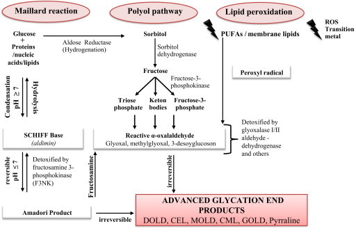
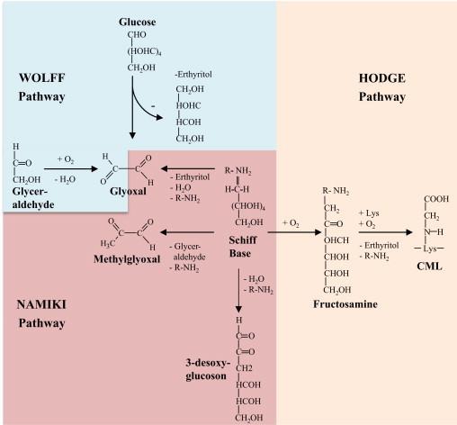
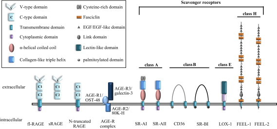
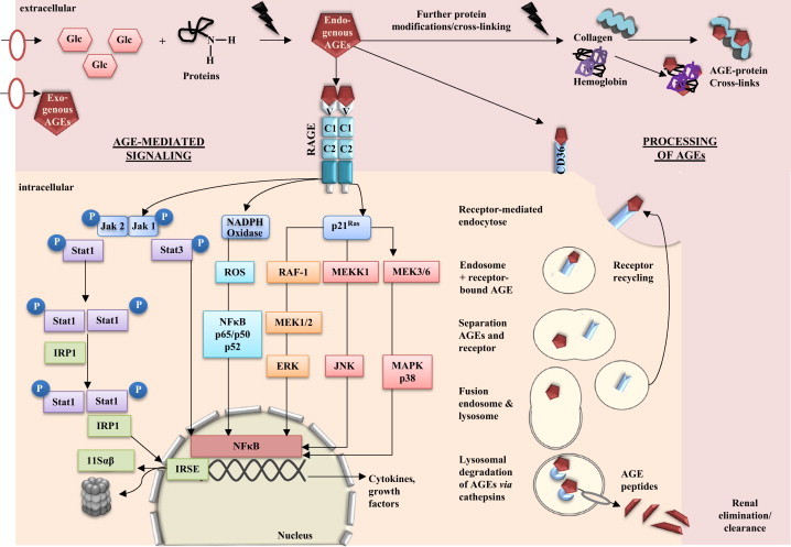
Improvements in health care and lifestyle have led to an elevated lifespan and increased focus on age-associated diseases, such as neurodegeneration, cardiovascular disease, frailty and arteriosclerosis. In all these chronic diseases protein, lipid or nucleic acid modifications are involved, including cross-linked and non-degradable aggregates, such as advanced glycation end products (AGEs). Formation of endogenous or uptake of dietary AGEs can lead to further protein modifications and activation of several inflammatory signaling pathways. This review will give an overview of the most prominent AGE-mediated signaling cascades, AGE receptor interactions, prevention of AGE formation and the impact of AGEs during pathophysiological processes.
Overview
The claims and statistics of this paper haven’t been checked yet.
Run the check — free, no account needed. It reads the open-access text and checks the sources it cites.
Integrity signals
Registry
Cites
Statistics
Figures
Verification
Claims the paper makes
The claims and statistics of this paper haven’t been checked yet.
Run the check — free, no account needed. It reads the open-access text and checks the sources it cites.
Claims other papers make about this one
Each is checked against this paper’s own text: does it say what the citing paper says it does?
The source backs part of the claim, not all of it — or backs it for a different population, condition or comparison.
In the citing paper
“In addition to lipid peroxide-related protein adducts, recent data suggest that sugar moiety-related protein adduct such as advanced glycation end products (AGEs) can be produced by high fat or excessive alcohol exposure or under pathological states such as diabetes, cardiovascular diseases and Alzheimer's disease, as recently reviewed [190], [191], [192], [193].”
From Mitochondrial dysfunction and tissue injury by alcohol, high fat, nonalcoholic substances and pathological conditions through post-translational protein modifications (2014) · cites this paper as [191]
Experimental — accuracy not yet measuredIn the citing paper
“These AGE cell surface receptors include AGE-R1, AGE-R2, AGE-R3, and scavenger receptors such as macrophage scavenger receptors, scavenger receptor class B type I and II (SR-BI, SR-BII), and cluster of differentiation 36 (CD36).”
From Advanced glycation end products (AGEs) and other adducts in aging-related diseases and alcohol-mediated tissue injury (2021) · cites this paper as [46]
In the citing paper
“The interaction of AGEs with these receptors can enhance their catabolism and clearance by modulating endocytosis and degradation45,46, suggesting a potentially adaptive defensive mechanism in the body to reduce the detrimental effects of increased glycation products.”
From Advanced glycation end products (AGEs) and other adducts in aging-related diseases and alcohol-mediated tissue injury (2021) · cites this paper as [46]
In the citing paper
“Notably, several conditions, including aging, diabetes, renal failure, tobacco smoking, and excessive alcohol consumption that induce AGE accumulation and AGE-RAGE interaction, are risk factors for increased bone fracture and osteoporosis46,85.”
From Advanced glycation end products (AGEs) and other adducts in aging-related diseases and alcohol-mediated tissue injury (2021) · cites this paper as [46]
Self-check
Percentages, sums and reported differences re-computed from the paper’s own numbers.
The self-check needs the paper’s full text.
Citations
Cites
Bundesgesundheitsblatt - Gesundheitsforschung - Gesundheitsschutz
(2013) · Bundesgesundheitsblatt - Gesundheitsforschung - Gesundheitsschutz · doi:10.1007/103.1437-1588 · Cited by 11,769
Pathways towards and Away from Alzheimer’s Disease
Mattson, M. P. (2004) · Nature · doi:10.1038/nature02621 · Cited by 3,052
Broken Limits to Life Expectancy
Oeppen, J., & Vaupel, J. W. (2002) · Science · doi:10.1126/science.1069675 · Cited by 2,819
Advanced glycation end-products: A review
Singh, R., Barden, A., Mori, T., & Beilin, L. (2001) · Diabetologia · doi:10.1007/s001250051591 · Cited by 2,597
Goldstein, J. L., Ho, Y. K., Basu, S. K., & Brown, M. S. (1979) · Proceedings of the National Academy of Sciences · doi:10.1073/pnas.76.1.333 · Cited by 2,447
Advanced Glycation End Products: Sparking the Development of Diabetic Vascular Injury
Goldin, A., Beckman, J. A., Schmidt, A. M., & Creager, M. A. (2006) · Circulation · doi:10.1161/circulationaha.106.621854 · Cited by 2,261
Identification of Scavenger Receptor SR-BI as a High Density Lipoprotein Receptor
Acton, S., Rigotti, A., Landschulz, K. T., Xu, S., Hobbs, H. H., & Krieger, M. (1996) · Science · doi:10.1126/science.271.5248.518 · Cited by 2,026
Understanding the odd science of aging
Kirkwood, T. B. L. (2005) · Cell · doi:10.1016/j.cell.2005.01.027 · Cited by 1,984
RAGE Mediates a Novel Proinflammatory Axis
Hofmann, M. A., Drury, S., Fu, C., Qu, W., Taguchi, A., Lu, Y., Avila, C., Kambham, N., Bierhaus, A., Nawroth, P., et al. (1999) · Cell · doi:10.1016/s0092-8674(00)80801-6 · Cited by 1,864
Steinbrecher, U. P., Parthasarathy, S., Leake, D. S., Witztum, J. L., & Steinberg, D. I. (1984) · Proceedings of the National Academy of Sciences · doi:10.1073/pnas.81.12.3883 · Cited by 1,626
Cited by
Sorted by citations
Items 401–500 of 1,000
Do advanced glycation end-products cause food allergy?
Smith, P. K. (2017) · Current Opinion in Allergy and Clinical Immunology · doi:10.1097/aci.0000000000000385 · Cited by 29
André, D. M., Calixto, M. C., Sollon, C. S., Alexandre, E. C., Tavares, E. B. G., Naime, A. C. A., Anhê, G. F., & Antunes, E. (2017) · Scientific Reports · doi:10.1038/s41598-017-17558-w · Cited by 28
Type 1 diabetes mellitus prevention: present and future
Henriques, F. L., Buckle, I., & Forbes, J. M. (2025) · Nature Reviews Endocrinology · doi:10.1038/s41574-025-01128-6 · Cited by 32
Chaplin, A. K., Chernukhin, I. V., & Bechtold, U. (2018) · Journal of Experimental Botany · doi:10.1093/jxb/ery389 · Cited by 27
Hu, N., Gong, X., Yin, S., Li, Q., Chen, H., Li, Y., Li, F., Qing, L., Yang, J., Zhu, S., et al. (2019) · Artificial Cells Nanomedicine and Biotechnology · doi:10.1080/21691401.2019.1647223 · Cited by 27
Physiological and pathogenic role of scavenger receptors in humans
Gusev, E., Zotova, N. V., Zhuravleva, Y. A., & Черешнев, В. А. (2020) · Medical Immunology (Russia) · doi:10.15789/1563-0625-pap-1893 · Cited by 28
miR‐21‐3p regulates AGE/RAGE signalling and improves diabetic atherosclerosis
Shao, M., Yu, M., Zhao, J., Mei, J., Pan, Y., Zhang, J., Wu, H., Yu, M., Liu, F., & Chen, G. (2020) · Cell Biochemistry and Function · doi:10.1002/cbf.3523 · Cited by 27
Deng, G., Zhang, L.-Y., Wang, C., Wang, S., Xu, J., Dong, J., Kang, Q., Zhai, X., Zhao, Y., & Shan, Z. (2020) · Experimental Gerontology · doi:10.1016/j.exger.2020.111088 · Cited by 26
Is carbonyl/AGE/RAGE stress a hallmark of the brain aging?
Semchyshyn, H. M. (2021) · Pflügers Archiv - European Journal of Physiology · doi:10.1007/s00424-021-02529-y · Cited by 27
Predicting Proteolysis in Complex Proteomes Using Deep Learning
Ozols, M., Eckersley, A., Platt, C. I., Stewart-McGuinness, C., Hibbert, S. A., Revote, J., Li, F., Griffiths, C. E. M., Watson, R. E. B., Song, J., et al. (2021) · International Journal of Molecular Sciences · doi:10.3390/ijms22063071 · Cited by 28
Wan, J., Feng, M., Pan, W., Zheng, X., Xie, X., Hu, B., Teng, C., Wang, Y., Liu, Z., Wu, J., et al. (2021) · Antioxidants · doi:10.3390/antiox10101513 · Cited by 27
Maciejczyk, M., Żebrowska, E., Nesterowicz, M., Żendzian‐Piotrowska, M., & Zalewska, A. (2022) · Oxidative Medicine and Cellular Longevity · doi:10.1155/2022/7450514 · Cited by 32
Annibal, A., Riemer, T., Jovanović, O., Westphal, D., Griesser, E., Pohl, E. E., Schiller, J., Hoffmann, R., & Fedorova, M. (2016) · Free Radical Biology and Medicine · doi:10.1016/j.freeradbiomed.2016.03.011 · Cited by 27
Limited field radiation therapy results in decreased bone fracture toughness in a murine model
Bartlow, C. M., Mann, K. A., Damron, T. A., & Oest, M. E. (2018) · PLoS ONE · doi:10.1371/journal.pone.0204928 · Cited by 31
Tao, D., Ni, N., Zhang, T., Li, C., Sun, Q., Wang, L., & Mei, Y. (2019) · Molecular Medicine Reports · doi:10.3892/mmr.2019.10590 · Cited by 27
Lim, S. C., Dorajoo, R., Zhang, X., Wang, L., Ang, S. F., Tan, C. S. H., Yeoh, L. Y., Ng, X. W., Li, N., Su, C., et al. (2016) · Nephrology Dialysis Transplantation · doi:10.1093/ndt/gfw263 · Cited by 26
Hwang, S. H., Kim, H.-Y., Quispe, Y. N. G., Wang, Z., Zuo, G., & Lim, S. S. (2019) · Molecules · doi:10.3390/molecules24102010 · Cited by 26
Obesity and Comorbidity: Could Simultaneous Targeting of esRAGE and sRAGE Be the Panacea?
Eleazu, C. O., Omar, N., Lim, O. Z., Yeoh, B. S., Hussain, N. H. N., & Mohamed, M. (2019) · Frontiers in Physiology · doi:10.3389/fphys.2019.00787 · Cited by 26
Yu, W., Fan, L., Wang, M., Cao, B., & Hu, X. (2021) · Molecular Nutrition & Food Research · doi:10.1002/mnfr.202100321 · Cited by 26
ADVANCED GLYCATION END PRODUCTS
ნ., M. N. Მ., პ., T. P. Თ., გ., M. G. Მ., მ., P. M. Პ., & ე., E. E. Ე. (2016) · TSMU COLLECTION OF SCIENTIFIC WORKS · doi:10.52340/csw.2016.50.0.81-84 · Cited by 26
Dietary Control of Inflammation and Resolution
Sears, B., & Saha, A. K. (2021) · Frontiers in Nutrition · doi:10.3389/fnut.2021.709435 · Cited by 30
Widiapradja, A., Kasparian, A. O., McCaffrey, S. L., Kolb, L. L., Imig, J. D., Lacey, J. L., Meléndez, G. C., & Levick, S. P. (2021) · Cells · doi:10.3390/cells10102659 · Cited by 27
Accumulation of advanced glycation end products in the rabbit blastocyst under maternal diabetes
Haucke, E., Santos, A. N., Simm, A., Henning, C., Glomb, M. A., Gürke, J., Schindler, M., Fischer, B., & Santos, A. N. (2014) · Reproduction · doi:10.1530/rep-14-0149 · Cited by 27
Jiang, X., Kuang, F., Kong, F., & Yan, C. (2016) · Carbohydrate Polymers · doi:10.1016/j.carbpol.2016.05.079 · Cited by 26
Deng, Y., Govers, C., Bastiaan‐Net, S., van der Hulst, N., Hettinga, K. A., & Wichers, H. J. (2019) · Food Research International · doi:10.1016/j.foodres.2019.01.038 · Cited by 26
ReactELISA method for quantifying methylglyoxal levels in plasma and cell cultures
Kold-Christensen, R., Jensen, K. K., Smedegård-Holmquist, E., Sørensen, L. K., Hansen, J., Jørgensen, K. A., Kristensen, P. K., & Johannsen, M. (2019) · Redox Biology · doi:10.1016/j.redox.2019.101252 · Cited by 26
Xie, F., Liu, B., Qiao, W., He, J.-Z., Cheng, J., Wang, Z., Hou, Y.-M., Zhang, X., Xu, B.-H., Zhang, Y., et al. (2024) · Nature Communications · doi:10.1038/s41467-024-49315-9 · Cited by 26
HMGB1: new biomarker and therapeutic target of autoimmune and autoinflammatory skin diseases
Fan, J., He, K., Zhang, Y., Li, R., Yi, X., & Li, S. (2025) · Frontiers in Immunology · doi:10.3389/fimmu.2025.1569632 · Cited by 25
Potential of glycative stress targeting for cancer prevention
Piperi, C., Adamopoulos, C., & Papavassiliou, A. G. (2017) · Cancer Letters · doi:10.1016/j.canlet.2017.01.020 · Cited by 25
Li, X., Jia, F., Zhu, Z., & Huang, L. (2019) · Artificial Cells Nanomedicine and Biotechnology · doi:10.1080/21691401.2019.1593996 · Cited by 25
Advanced Glycation End Products and Their Involvement in Cardiovascular Disease
Yubero‐Serrano, E. M., & Pérez-Martínez, P. (2020) · Angiology · doi:10.1177/0003319720916301 · Cited by 27
Teng, J., Li, J., Zhao, Y., & Wang, M. (2021) · Journal of Functional Foods · doi:10.1016/j.jff.2021.104480 · Cited by 25
AGE/Non-AGE Glycation: An Important Event in Rheumatoid Arthritis Pathophysiology
Monu, M., Agnihotri, P., & Biswas, S. (2021) · Inflammation · doi:10.1007/s10753-021-01589-7 · Cited by 27
Food-Related Carbonyl Stress in Cardiometabolic and Cancer Risk Linked to Unhealthy Modern Diet
Iacobini, C., Vitale, M., Haxhi, J., Pesce, C. M., Pugliese, G., & Menini, S. (2022) · Nutrients · doi:10.3390/nu14051061 · Cited by 31
Zeng, L., Lin, L., Xiao, W., & Li, Y. (2022) · European Journal of Pharmacology · doi:10.1016/j.ejphar.2022.175072 · Cited by 25
Receptor for advanced glycation end‐products: Biological significance and imaging applications
Dobrucki, I. T., Miskalis, A., Nelappana, M. B., Applegate, C. C., Woźniak, M., Czerwiński, A. A., Kalinowski, L., & Dobrucki, L. W. (2023) · Wiley Interdisciplinary Reviews Nanomedicine and Nanobiotechnology · doi:10.1002/wnan.1935 · Cited by 28
Lan, L., Han, F., Lang, X., & Chen, J. (2016) · PLoS ONE · doi:10.1371/journal.pone.0159964 · Cited by 25
Similarities and interactions between the ageing process and high chronic intake of added sugars
Gatineau, E., Polakof, S., Dardevet, D. P., & Mosoni, L. (2017) · Nutrition Research Reviews · doi:10.1017/s0954422417000051 · Cited by 27
Chen, H., Li, Y., Zhu, Y., Wu, L., Meng, J., Lin, N., Yang, D., Li, M., Ding, W., Tong, X., et al. (2017) · Medicine · doi:10.1097/md.0000000000007456 · Cited by 25
Cell Based Therapy for Type 1 Diabetes: Should We Take Hyperglycemia Into Account?
Grohová, A., Dáňová, K., Špíšek, R., & Palová-Jelínková, L. (2019) · Frontiers in Immunology · doi:10.3389/fimmu.2019.00079 · Cited by 25
Zhou, Q., Xu, H., Yu, W., Li, E., & Wang, M. (2019) · Oxidative Medicine and Cellular Longevity · doi:10.1155/2019/9026456 · Cited by 25
Dhananjayan, K., Gunawardena, D., Hearn, N., Sonntag, T., Moran, C., Gyengesi, E., Srikanth, V., & Münch, G. (2017) · Journal of Alzheimer s Disease · doi:10.3233/jad-161152 · Cited by 24
Targeting HMGB1 and Its Interaction with Receptors: Challenges and Future Directions
Shen, P., Zhang, L., Jiang, X., Yu, B., & Zhang, J. (2024) · Journal of Medicinal Chemistry · doi:10.1021/acs.jmedchem.4c01912 · Cited by 25
Yanar, K., Simsek, B., Çaylı, N., Bozkır, H. Ö., Mengi, M., Belce, A., Aydın, S., & Çakatay, U. (2018) · Journal of Food Biochemistry · doi:10.1111/jfbc.12740 · Cited by 24
Lee, D. E., Brown, J. L., Rosa‐Caldwell, M. E., Perry, R. A., Brown, L. A., Haynie, W. S., Washington, T. A., Wiggs, M. P., Rajaram, N., & Greene, N. P. (2020) · JCSM Rapid Communications · doi:10.1002/rco2.18 · Cited by 24
Sirangelo, I., Borriello, M., Liccardo, M., Scafuro, M., Russo, P., & Iannuzzi, C. (2021) · Antioxidants · doi:10.3390/antiox10071127 · Cited by 24
Cepas, V., Manig, F., Mayo, J. C., Hellwig, M., Collotta, D., Sanmartino, V., Carrocera, R., Collino, M., Henle, T., & Sainz, R. M. (2021) · Oxidative Medicine and Cellular Longevity · doi:10.1155/2021/9912240 · Cited by 23
Tombultürk, F. K., Seven, Z. G. T., Hüseyinbaş, Ö., Özyazgan, S., Ulutin, T., & Sultuybek, G. K. (2021) · Molecular Biology Reports · doi:10.1007/s11033-021-06843-7 · Cited by 24
Ahmad, S. S., Ahmed, F., Ali, R., Ghoneim, M. M., Alshehri, S., Najmi, A. K., Ahmad, S., Ahmad, M. Z., Ahmad, J., & Khan, M. A. (2022) · Immunotherapy · doi:10.2217/imt-2021-0282 · Cited by 25
Yuan, X., He, J., Su, H., Liu, H., & Sun, B. (2023) · Journal of Agricultural and Food Chemistry · doi:10.1021/acs.jafc.2c08777 · Cited by 24
Kothandan, D., Singh, D. S., Yerrakula, G., Backkiyashree, D., Pratibha, N., Vincy Santhana Sophia, B., Ramya, A., Sapthami Ramya, V. G., Keshavini, S., & Jagadheeshwari, M. (2024) · Cureus · doi:10.7759/cureus.61373 · Cited by 29
CaMKK2 Signaling in Metabolism and Skeletal Disease: a New Axis with Therapeutic Potential
Williams, J. N., & Sankar, U. (2019) · Current Osteoporosis Reports · doi:10.1007/s11914-019-00518-w · Cited by 24
Petriv, N., Neubert, L., Vatashchuk, M., Timrott, K., Suo, H., Hochnadel, I., Huber, R., Petzold, C., Hrushchenko, A. O., Yatsenko, A. S., et al. (2021) · OncoImmunology · doi:10.1080/2162402x.2021.1874159 · Cited by 24
Free Radicals and Reactive Oxygen Species
Siraki, A. G., Klotz, L.-O., & Kehrer, J. P. (2015) · Elsevier eBooks · doi:10.1016/b978-0-12-801238-3.01895-x · Cited by 31
Molecular Mechanisms Linking Diabetes with Increased Risk of Thrombosis
Batten, L., Sathyapalan, T., & Palmer, T. M. (2023) · International Journal of Molecular Sciences · doi:10.3390/ijms242417465 · Cited by 23
Liao, S., Liu, Z., Lv, W., Li, S., Tian, T., Wang, Y., Wu, H., Zhao, Z., & Lin, Y. (2024) · ACS Applied Materials & Interfaces · doi:10.1021/acsami.4c11089 · Cited by 25
Zenker, H. E., Teodorowicz, M., Ewaz, A., van Neerven, R. J. J., Savelkoul, H. F. J., de Jong, N. W., Wichers, H. J., & Hettinga, K. A. (2020) · International Journal of Molecular Sciences · doi:10.3390/ijms21124567 · Cited by 24
Santos, H. O., & Penha‐Silva, N. (2021) · European Journal of Clinical Nutrition · doi:10.1038/s41430-021-01028-8 · Cited by 22
The RAGE/multiligand axis: a new actor in tumor biology
Armando Rojas, R., Schneider, I., Lindner, C., González, I., & Morales, M. A. (2022) · Bioscience Reports · doi:10.1042/bsr20220395 · Cited by 24
The role of Sirtuin 1 in the pathophysiology of polycystic ovary syndrome
Wu, M., Zhang, J., Gu, R., Dai, F., Yang, D., Zheng, Y., Tan, W., Jia, Y., Li, B., & Cheng, Y. (2022) · European journal of medical research · doi:10.1186/s40001-022-00746-4 · Cited by 24
Jansen, F. A. C., Fogliano, V., Rubert, J., & Hoppenbrouwers, T. (2023) · Molecular Metabolism · doi:10.1016/j.molmet.2023.101734 · Cited by 24
Li, P., Qin, D., Chen, T., Hou, W., Song, X., Yin, S., Song, M., Fernando, W., Chen, X., Sun, Y., et al. (2023) · Cardiovascular Diabetology · doi:10.1186/s12933-023-01894-5 · Cited by 23
Cooksley, G., Nam, M., Nahomi, R. B., Rankenberg, J., Smith, A. J. O., Wormstone, Y. M., Wormstone, I. M., & Nagaraj, R. H. (2024) · Aging Cell · doi:10.1111/acel.14249 · Cited by 22
Kheniser, K. G., Kashyap, S. R., & Kasumov, T. (2018) · Obesity Science & Practice · doi:10.1002/osp4.309 · Cited by 23
Wang, A., Xiao, C., Zheng, J., Ye, C., Dai, Z., Wu, Q., Liu, J., Strappe, P., & Zhou, Z. (2020) · Journal of Functional Foods · doi:10.1016/j.jff.2020.104142 · Cited by 24
Mahmoud, A. M., & Ali, M. M. (2021) · Cells · doi:10.3390/cells10082098 · Cited by 23
Hansen, F., Battú, C. E., Dutra, M. F., Galland, F., Pedroso, F. L., Cunha, N. B., Nardin, P., & Gonçalves, C. (2015) · Amino Acids · doi:10.1007/s00726-015-2091-1 · Cited by 22
Liang, Q., Li, F., Li, Y., Liu, Y., Lan, M., Wu, S.-H., Wu, X., Ji, Y. H., Zhang, R., & Yin, L. (2019) · Biomaterials Science · doi:10.1039/c9bm00719a · Cited by 22
Kjerulf, D. G., Wang, S., Omer, M., Pathak, A., Subramanian, S., Han, C. Y., Tang, C., den Hartigh, L. J., Shao, B., & Chait, A. (2020) · Journal of Diabetes and its Complications · doi:10.1016/j.jdiacomp.2020.107693 · Cited by 22
Contribution of RAGE axis activation to the association between metabolic syndrome and cancer
Garay‐Sevilla, M. E., Gómez-Ojeda, A., González, I., Luévano‐Contreras, C., & Armando Rojas, R. (2021) · Molecular and Cellular Biochemistry · doi:10.1007/s11010-020-04022-z · Cited by 22
Inhibitory effect of sea buckthorn extracts on advanced glycation endproduct formation
Lee, H. H. L., Lee, C. J., Choi, S. Y., Kim, Y., & Hur, J. (2021) · Food Chemistry · doi:10.1016/j.foodchem.2021.131364 · Cited by 22
Búfalo, M. C., de Almeida, M. E. S., Jensen, J. R., DeOcesano-Pereira, C., Lichtenstein, F., Picolo, G., Chudzinski‐Tavassi, A. M., Sampaio, S. C., Cury, Y., & Zambelli, V. O. (2022) · Cells · doi:10.3390/cells11020247 · Cited by 22
Santolla, M. F., Talia, M., Cirillo, F., Scordamaglia, D., Rosis, S. D., Spinelli, A., Miglietta, A., Nardo, B., Filippelli, G., Francesco, E. M. D., et al. (2022) · Cells · doi:10.3390/cells11152402 · Cited by 23
Bangar, N. S., Gvalani, A., Ahmad, S., Khan, M. S., & Tupe, R. S. (2022) · Glycobiology · doi:10.1093/glycob/cwac060 · Cited by 22
RAGE engagement by SARS-CoV-2 enables monocyte infection and underlies COVID-19 severity
Angioni, R., Bonfanti, M., Caporale, N., Sánchez‐Rodríguez, R., Munari, F., Savino, A., Pasqualato, S., Buratto, D., Pagani, I., Bertoldi, N., et al. (2023) · Cell Reports Medicine · doi:10.1016/j.xcrm.2023.101266 · Cited by 24
Kim, S., & Lee, S. (2017) · Inflammation · doi:10.1007/s10753-017-0632-3 · Cited by 22
Jiménez, I. U., Díaz‐Díaz, E., Salmerón, J., Ramos, J. P., León, M. C., Ríos, J. A. A., Bautista, J. C. A., Correa‐Rotter, R., Aguilar-Salinas, C. A., & Larrea, F. (2017) · Archives of Medical Research · doi:10.1016/j.arcmed.2017.07.001 · Cited by 22
Hagiwara, S., Sourris, K. C., Ziemann, M., Wu, T., Mohan, M., McClelland, A., Brennan, E. P., Forbes, J. M., Coughlan, M. T., Harcourt, B. E., et al. (2018) · Diabetes · doi:10.2337/db17-0538 · Cited by 22
Alam, S., Réhman, S. U., & Kim, M. O. (2019) · Journal of Clinical Medicine · doi:10.3390/jcm8020271 · Cited by 22
Vélayoudom‐Céphise, F., Cano-Sanchez, M., Bercion, S., Tessier, F. J., Yu, Y.-C., Boulanger, É., & Nevière, R. (2020) · Applied Physiology Nutrition and Metabolism · doi:10.1139/apnm-2019-0936 · Cited by 22
Humors Effect on Short-term Memory in Healthy and Diabetic Older Adults.
Bains, G. S., Berk, L., Lohman, E., Daher, N. S., PETROFSKY, J. S., Schwab, E., & Deshpande, P. (2015) · PubMed · Cited by 22
Preserving Brain Function in Aging: The Anti-glycative Potential of Berry Fruit
Thangthaeng, N., Poulose, S. M., Miller, M. G., & Shukitt‐Hale, B. (2016) · NeuroMolecular Medicine · doi:10.1007/s12017-016-8400-3 · Cited by 22
Advanced glycation end products regulate the receptor of AGEs epigenetically
Wu, X., Shi, X., Chen, X., & Yin, Z. (2023) · Frontiers in Cell and Developmental Biology · doi:10.3389/fcell.2023.1062229 · Cited by 21
The role of AGEs in muscle ageing and sarcopenia
Guo, Z.-J., Li, H., Jiang, S., Rahmati, M., Su, J., Yang, S., Wu, Y., Li, Y., & Deng, Z. (2025) · Bone and Joint Research · doi:10.1302/2046-3758.143.bjr-2024-0252.r1 · Cited by 21
Awasthi, S., & Saraswathi, N. (2016) · RSC Advances · doi:10.1039/c5ra27460e · Cited by 19
White, D. L., Hoogeveen, R. C., Chen, L., Richardson, P., Ravishankar, M., Shah, P., Tinker, L. F., Rohan, T. E., Whitsel, E. A., El‐Serag, H. B., et al. (2018) · Cancer Medicine · doi:10.1002/cam4.1426 · Cited by 20
Wu, Q., Liu, H., & Zhou, M. (2019) · Medical Science Monitor · doi:10.12659/msm.912927 · Cited by 21
Senavirathna, L. K., Ma, C., Chen, R., & Pan, S. (2021) · Cells · doi:10.3390/cells10051005 · Cited by 21
Yakovleva, M. A., Dontsov, A. E., Трофимова, Н. Н., Sakina, N. L., Kononikhin, A. S., Aybush, A. V., Gulin, A. A., Feldman, T. B., & Оstrovsky, М. А. (2021) · International Journal of Molecular Sciences · doi:10.3390/ijms23010222 · Cited by 21
Anti-obesity pharmacotherapy in adults with chronic kidney disease
Taber-Hight, E., Gilmore, A. C., & Friedman, A. N. (2023) · Kidney International · doi:10.1016/j.kint.2023.10.014 · Cited by 21
Logan, S. M., & Storey, K. B. (2018) · PeerJ · doi:10.7717/peerj.4911 · Cited by 24
RAGE and TLRs as Key Targets for Antiatherosclerotic Therapy
Olejarz, W., Łacheta, D., Głuszko, A., Migacz, E., Kukwa, W., Szczepański, M. J., Tomaszewski, P., & Nowicka, G. (2018) · BioMed Research International · doi:10.1155/2018/7675286 · Cited by 21
Inflammatory effects of in vivo glycated albumin from cardiovascular patients
Paradela‐Dobarro, B., Bravo, S. B., Rozados-Luís, A., González‐Peteiro, M., Roman, A. V., González‐Juanatey, J. R., García‐Seara, J., & Álvarez, E. (2019) · Biomedicine & Pharmacotherapy · doi:10.1016/j.biopha.2019.108763 · Cited by 21
Male infertility: A proximate look at the advanced glycation end products
Omolaoye, T. S., & du Plessis, S. S. (2020) · Reproductive Toxicology · doi:10.1016/j.reprotox.2020.02.002 · Cited by 21
Chen, Y., Filipov, N. M., & Guo, T. L. (2018) · Molecular Nutrition & Food Research · doi:10.1002/mnfr.201700641 · Cited by 20
Jin, J., Yang, Y., Yang, J., Sun, Z., Wang, D., Qin, Y., Ruan, C., Li, D., Pan, Y., Wu, J., et al. (2024) · International Journal of Biological Macromolecules · doi:10.1016/j.ijbiomac.2024.134830 · Cited by 20
Mancini, Ö. K., Huynh, D. N., Ménard, L., Tim, D. S., Ong, H., Marleau, S., Colmegna, I., & Servant, M. J. (2020) · Cardiovascular Research · doi:10.1093/cvr/cvaa118 · Cited by 20
Aglago, E. K., Mayén, A., Knaze, V., Freisling, H., Fedirko, V., Hughes, D. J., Jiao, L., Eriksen, A. K., Tjønneland, A. M., Boutron‐Ruault, M., et al. (2021) · Nutrients · doi:10.3390/nu13093132 · Cited by 20
Rochín‐Hernández, L. S., Rochín-Hernández, L. J., & Flores‐Cotera, L. B. (2022) · Molecules · doi:10.3390/molecules27144469 · Cited by 20
Sun, J., Akıllıoğlu, H. G., Aasmul‐Olsen, K., Ye, Y., Lund, P., Zhao, X., Brunse, A., Nielsen, C. F., Chatterton, D. E. W., Sangild, P. T., et al. (2022) · Molecular Nutrition & Food Research · doi:10.1002/mnfr.202200132 · Cited by 19
Metadata
- Authors
- Ott, C., Jacobs, K., Haucke, E., Navarrete Santos, A., Grune, T., & Simm, A.
- Affiliations
- Friedrich Schiller University Jena (DE)
- University Hospital in Halle (DE)
- Martin Luther University Halle-Wittenberg (DE)
- Corresponding author
- Simm, Andreas
- Venue
- Redox Biology
- Publisher
- Elsevier BV
- Published
- 2014
- Volume
- 2
- Pages
- 411-429
- ISSN
- 2213-2317 (ISSN-L)
- PMC
- PMC3949097
- OpenAlex
- W2086327676
- Cited by
- 1,150
- References
- 233
- Citations by year
- 2026: 62 · 2025: 82 · 2024: 105 · 2023: 121 · 2022: 108 · 2021: 131 · 2020: 107 · 2019: 106 · 2018: 86 · 2017: 94 · 2016: 70 · 2015: 52 · 2014: 21
- FWCI
- 10.15
- Type
- journal article
- Language
- en
- Open access
- gold
- doi.org
- Licence
- Locations
- Redox Biology (publishedVersion, cc-by-nc-sa)
- Europe PMC (PubMed Central) (publishedVersion)
- PubMed Central (publishedVersion, cc-by-nc-sa)
- PubMed (publishedVersion)
- DOAJ (DOAJ: Directory of Open Access Journals) (submittedVersion)
- Topics
- Life Sciences › Biochemistry, Genetics and Molecular Biology › Clinical Biochemistry › Advanced Glycation End Products research
- Life Sciences › Agricultural and Biological Sciences › Food Science › Potato Plant Research
- Life Sciences › Biochemistry, Genetics and Molecular Biology › Aging › Genetics, Aging, and Longevity in Model Organisms
- Keywords
- advanced glycation end products, AGE receptor, signal transduction, protein modification, inflammatory signaling, age-related diseases, neurodegeneration, cardiovascular disease, arteriosclerosis, frailty, dietary AGEs
- MeSH terms
- Receptor for Advanced Glycation End Products, Aging, Animals, Bone and Bones, Dietary Proteins, Humans, Hyperglycemia, Immune System, Inflammation, Lung, Models, Biological, Neurons, Polymers, Receptors, Immunologic, Lipid Peroxidation, Signal Transduction, Maillard Reaction, NF-kappa B, Glycation End Products, Advanced, Reactive Oxygen Species, Oxidative Stress, Protein Transport, Reactive Nitrogen Species, Receptors, Scavenger, Protein Aggregates
About these checks
Registry, retraction, Cites and statistics results are computed, not judged, with one exception: a printed reference that no index lists is read once by a model before it counts as a citation error. Claims are extracted by a model and kept only when their quote appears verbatim in the text; each cited source is then checked, with a model’s help, for whether it says what the claim relies on. Those verdicts can be wrong — read the claim’s quote and the cited source before relying on one.
Statistics and arithmetic are recomputed, not judged.
Confidence bands are shown only for fields calibrated on our annotated test set. None is calibrated yet, so no band is shown.
What counts as a claim ›How we measure confidence ›How we check this ›
Are you an author, or do you see a problem with this page? Request a review
