

The DOI matches its Crossref record; 7 of the 7 references it cites were found in a registry or an index.
Small molecule regulators of microRNAs identified by high-throughput screen coupled with high-throughput sequencing
Published version of a preprint
Abstract
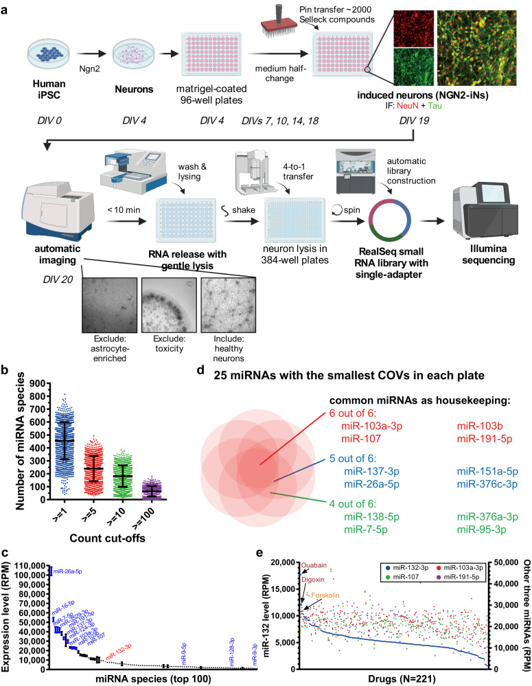
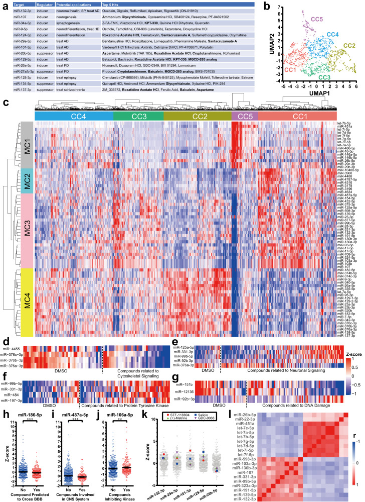
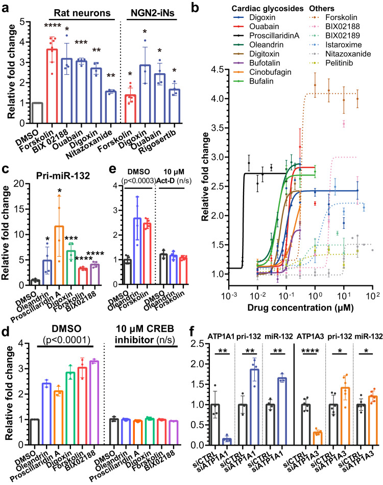
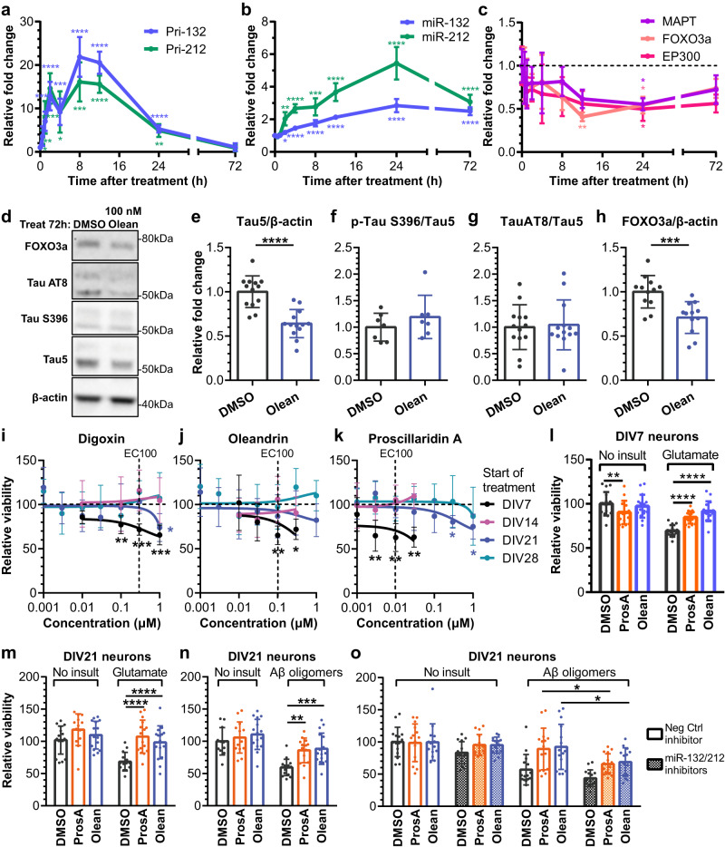
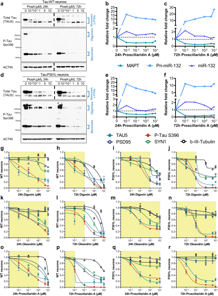
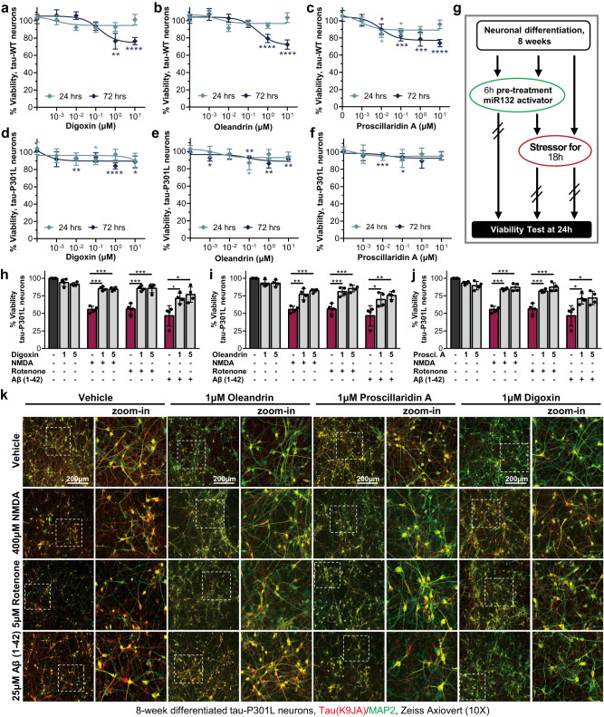
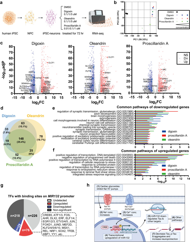
MicroRNAs (miRNAs) regulate fundamental biological processes by silencing mRNA targets and are dysregulated in many diseases. Therefore, miRNA replacement or inhibition can be harnessed as potential therapeutics. However, existing strategies for miRNA modulation using oligonucleotides and gene therapies are challenging, especially for neurological diseases, and none have yet gained clinical approval. We explore a different approach by screening a biodiverse library of small molecule compounds for their ability to modulate hundreds of miRNAs in human induced pluripotent stem cell-derived neurons. We demonstrate the utility of the screen by identifying cardiac glycosides as potent inducers of miR-132, a key neuroprotective miRNA downregulated in Alzheimer’s disease and other tauopathies. Coordinately, cardiac glycosides downregulate known miR-132 targets, including Tau, and protect rodent and human neurons against various toxic insults. More generally, our dataset of 1370 drug-like compounds and their effects on the miRNome provides a valuable resource for further miRNA-based drug discovery.
Overview
The claims and statistics of this paper haven’t been checked yet.
Run the check — free, no account needed. It reads the open-access text and checks the sources it cites.
Integrity signals
Registry
Cites
Statistics
Figures
Verification
Claims the paper makes
The claims and statistics of this paper haven’t been checked yet.
Run the check — free, no account needed. It reads the open-access text and checks the sources it cites.
Claims other papers make about this one
Each is checked against this paper’s own text: does it say what the citing paper says it does?
In the citing paper
“We confirmed that miR-132 levels increased in the hippocampus (Fig. 6B), which led to the in vivo repression of one of the most often replicated neuronal miR-132 targets, Mapt [96] (Fig. 6C).”
From microRNA-132 attenuates inflammation in induced pluripotent stem cell-derived microglia from Alzheimer’s disease patients (2026) · cites this paper as [96]
In the citing paper
“This method has shown promising results, with a dataset of 1,370 effective drug-like compounds providing a valuable resource for further microRNA-based drug discovery.205”
From MiR-128-3p – a gray eminence of the human central nervous system (2024) · cites this paper as [205]
In the citing paper
“Recently, a new approach has been developed, which involves screening a biodiverse library of small molecule compounds to determine their ability to modulate hundreds of microRNAs.”
From MiR-128-3p – a gray eminence of the human central nervous system (2024) · cites this paper as [205]
Self-check
Percentages, sums and reported differences re-computed from the paper’s own numbers.
The self-check needs the paper’s full text.
Citations
Cites
Synapse Loss and Microglial Activation Precede Tangles in a P301S Tauopathy Mouse Model
Yoshiyama, Y., Higuchi, M., Zhang, B., Huang, S.-M., Iwata, N., Saido, T. C., Maeda, J., Suhara, T., Trojanowski, J. Q., & Lee, V. M.-Y. (2007) · Neuron · doi:10.1016/j.neuron.2007.01.010 · Cited by 2,208
Seeding of Normal Tau by Pathological Tau Conformers Drives Pathogenesis of Alzheimer-like Tangles
Guo, J. L., & Lee, V. M.-Y. (2011) · Journal of Biological Chemistry · doi:10.1074/jbc.m110.209296 · Cited by 636
Homeostatic regulation of MeCP2 expression by a CREB-induced microRNA
Klein, M. E., Lioy, D. T., Ma, L., Impey, S., Mandel, G., & Goodman, R. H. (2007) · Nature Neuroscience · doi:10.1038/nn2010 · Cited by 444
Lekka, E., & Hall, J. (2018) · FEBS Letters · doi:10.1002/1873-3468.13182 · Cited by 251
Evidence for miR‐181 involvement in neuroinflammatory responses of astrocytes
Hutchison, E. R., Kawamoto, E. M., Taub, D. D., Lal, A., Abdelmohsen, K., Zhang, Y., Wood, W. H., Lehrmann, E., Camandola, S., Becker, K. G., et al. (2013) · Glia · doi:10.1002/glia.22483 · Cited by 246
DRUG-seq for miniaturized high-throughput transcriptome profiling in drug discovery
Ye, C., Ho, D. J., Neri, M., Yang, C., Kulkarni, T., Randhawa, R., Henault, M., Mostacci, N., Farmer, P., Renner, S., et al. (2018) · Nature Communications · doi:10.1038/s41467-018-06500-x · Cited by 222
Lagomarsino, V. N., Pearse, R. V., Liu, L., Hsieh, Y.-C., Fernandez, M. A., Vinton, E. A., Paull, D., Felsky, D., Tasaki, S., Gaiteri, C., et al. (2021) · Neuron · doi:10.1016/j.neuron.2021.08.003 · Cited by 151 · cited 4×
“Induced pluripotent stem cell (iPSC) lines were retrieved and differentiated into neurons with NGN2 expression, as previously reported 27 .”
InfluentialSelf-citation
Research works printed but not linked: 74
- Wang, F., Zuroske, T. & Watts, J. K. RNA therapeutics on the rise. Nat. Rev. Drug Discov. 19, 441–442 (2020). · DOI
- Warner, K. D., Hajdin, C. E. & Weeks, K. M. Principles for targeting RNA with drug-like small molecules. Nat. Rev. Drug Discov. 17, 547–558 (2018). · DOI
- Gebert, L. F. R. & MacRae, I. J. Regulation of microRNA function in animals. Nat. Rev. Mol. Cell Biol. 20, 21–37 (2019). · DOI · Paper page
- Mendell, J. T. & Olson, E. N. MicroRNAs in stress signaling and human disease. Cell 148, 1172–1187 (2012). · DOI · Paper page
- Chen, Y., Gao, D. Y. & Huang, L. In vivo delivery of miRNAs for cancer therapy: challenges and strategies. Adv. Drug Deliv. Rev. 81, 128–141 (2015). · DOI
- Garzon, R., Marcucci, G. & Croce, C. M. Targeting microRNAs in cancer: rationale, strategies and challenges. Nat. Rev. Drug Discov. 9, 775–789 (2010). · DOI
- Watts, J. K., Brown, R. H. & Khvorova, A. Nucleic acid therapeutics for neurological diseases. Neurotherapeutics 16, 245–247 (2019). · DOI
- Nguyen, L. D., Chau, R. K. & Krichevsky, A. M. Small molecule drugs targeting non-coding RNAs as treatments for Alzheimer’s disease and related dementias. Genes 12, 2005 (2021). · DOI
- Gumireddy, K. et al. Small-molecule inhibitors of microrna miR-21 function. Angew. Chem. Int. Ed. Engl. 47, 7482–7484 (2008). · DOI
- Young, D. D., Connelly, C. M., Grohmann, C. & Deiters, A. Small molecule modifiers of microRNA miR-122 function for the treatment of hepatitis C virus infection and hepatocellular carcinoma. J. Am. Chem. Soc. 132, 7976–7981 (2010). · DOI
- Velagapudi, S. P., Gallo, S. M. & Disney, M. D. Sequence-based design of bioactive small molecules that target precursor microRNAs. Nat. Chem. Biol. 10, 291–297 (2014). · DOI
- Connelly, C. M., Boer, R. E., Moon, M. H., Gareiss, P. & Schneekloth, J. S. Jr Discovery of inhibitors of microRNA-21 processing using small molecule microarrays. ACS Chem. Biol. 12, 435–443 (2017). · DOI
- Van Meter, E. N., Onyango, J. A. & Teske, K. A. A review of currently identified small molecule modulators of microRNA function. Eur. J. Med. Chem. 188, 112008 (2020). · DOI
- Velagapudi, S. P. et al. Design of a small molecule against an oncogenic noncoding RNA. Proc. Natl Acad. Sci. USA 113, 5898–5903 (2016). · DOI
- Salta, E. & De Strooper, B. microRNA-132: a key noncoding RNA operating in the cellular phase of Alzheimer’s disease. FASEB J. 31, 424–433 (2017). · DOI · Paper page
- Pichler, S. et al. The miRNome of Alzheimer’s disease: consistent downregulation of the miR-132/212 cluster. Neurobiol. Aging 50, 167 e161–167.e110 (2017). · DOI · Paper page
- Cha, D. J. et al. miR-212 and miR-132 are downregulated in neurally derived plasma exosomes of Alzheimer’s patients. Front. Neurosci. 13, 1208 (2019). · DOI · Paper page
- Lau, P. et al. Alteration of the microRNA network during the progression of Alzheimer’s disease. EMBO Mol. Med. 5, 1613–1634 (2013). · DOI · Paper page
- Wong, H. K. et al. De-repression of FOXO3a death axis by microRNA-132 and -212 causes neuronal apoptosis in Alzheimer’s disease. Hum. Mol. Genet. 22, 3077–3092 (2013). · DOI · Paper page
- Hernandez-Rapp, J. et al. microRNA-132/212 deficiency enhances Abeta production and senile plaque deposition in Alzheimer’s disease triple transgenic mice. Sci. Rep. 6, 30953 (2016). · DOI
- Salta, E., Sierksma, A., Vanden Eynden, E. & De Strooper, B. miR-132 loss de-represses ITPKB and aggravates amyloid and TAU pathology in Alzheimer’s brain. EMBO Mol. Med. 8, 1005–1018 (2016). · DOI · Paper page
- Smith, P. Y. et al. miR-132/212 deficiency impairs tau metabolism and promotes pathological aggregation in vivo. Hum. Mol. Genet. 24, 6721–6735 (2015). · DOI · Paper page
- Wang, Y. et al. Downregulation of miR-132/212 impairs S-nitrosylation balance and induces tau phosphorylation in Alzheimer’s disease. Neurobiol. Aging 51, 156–166 (2017). · DOI · Paper page
- El Fatimy, R. et al. MicroRNA-132 provides neuroprotection for tauopathies via multiple signaling pathways. Acta Neuropathol. 136, 537–555 (2018). · DOI · Paper page
- Walgrave, H. et al. Restoring miR-132 expression rescues adult hippocampal neurogenesis and memory deficits in Alzheimer’s disease. Cell Stem Cell 28, 1805–1821.e1808 (2021). · DOI · Paper page
- Vo, N. et al. A cAMP-response element binding protein-induced microRNA regulates neuronal morphogenesis. Proc. Natl Acad. Sci. USA 102, 16426–16431 (2005). · DOI · Paper page
- van Zonneveld, A. J. et al. MicroRNA-132 regulates salt-dependent steady-state renin levels in mice. Commun. Biol. 3, 238 (2020). · DOI
- Barberan-Soler, S. et al. Decreasing miRNA sequencing bias using a single adapter and circularization approach. Genome Biol. 19, 105 (2018). · DOI
- Zhang, Y., Parmigiani, G. & Johnson, W. E. ComBat-seq: batch effect adjustment for RNA-seq count data. NAR Genom. Bioinform. 2, lqaa078 (2020). · DOI · Paper page
- Zhang, C. et al. Lnc00462717 regulates the permeability of the blood-brain tumor barrier through interaction with PTBP1 to inhibit the miR-186-5p/Occludin signaling pathway. FASEB J. 34, 9941–9958 (2020). · DOI
- Wolfle, U. et al. Salicin from Willow Bark can modulate neurite outgrowth in human neuroblastoma SH-SY5Y cells. Phytother Res. 29, 1494–1500 (2015). · DOI
- Park, J. H. et al., Neuroprotective effects of salicin in a gerbil model of transient forebrain ischemia by attenuating oxidative stress and activating PI3K/Akt/GSK3beta Pathway. Antioxidants 10, 629 (2021). · DOI
- Tanabe, N., Kuboyama, T., Kazuma, K., Konno, K. & Tohda, C. The extract of roots of sophora flavescens enhances the recovery of motor function by axonal growth in mice with a spinal cord injury. Front. Pharmacol. 6, 326 (2015).
- Tanabe, N., Kuboyama, T. & Tohda, C. Matrine promotes neural circuit remodeling to regulate motor function in a mouse model of chronic spinal cord injury. Neural Regen Res. 14, 1961–1967 (2019). · DOI
- Absalon, S. et al. MiR-26b, upregulated in Alzheimer’s disease, activates cell cycle entry, tau-phosphorylation, and apoptosis in postmitotic neurons. J. Neurosci. 33, 14645 (2023). · DOI · Paper page
- Lehmann, S. M. et al. An unconventional role for miRNA: let-7 activates Toll-like receptor 7 and causes neurodegeneration. Nat. Neurosci. 15, 827–835 (2012). · DOI · Paper page
- Khan, H. et al. Istaroxime, a first in class new chemical entity exhibiting SERCA-2 activation and Na-K-ATPase inhibition: a new promising treatment for acute heart failure syndromes? Heart Fail Rev. 14, 277–287 (2009). · DOI
- Magill, S. T. et al. microRNA-132 regulates dendritic growth and arborization of newborn neurons in the adult hippocampus. Proc. Natl Acad. Sci. USA 107, 20382–20387 (2010). · DOI · Paper page
- Vandesompele, J. et al. Accurate normalization of real-time quantitative RT-PCR data by geometric averaging of multiple internal control genes. Genome Biol. 3, RESEARCH0034 (2002). · DOI · Paper page
- Brines, M. L., Dare, A. O. & de Lanerolle, N. C. The cardiac glycoside ouabain potentiates excitotoxic injury of adult neurons in rat hippocampus. Neurosci. Lett. 191, 145–148 (1995). · DOI
- Sun, Y., Dong, Z., Khodabakhsh, H., Chatterjee, S. & Guo, S. Zebrafish chemical screening reveals the impairment of dopaminergic neuronal survival by cardiac glycosides. PLoS One 7, e35645 (2012). · DOI
- Silva, M. C. et al. Prolonged tau clearance and stress vulnerability rescue by pharmacological activation of autophagy in tauopathy neurons. Nat. Commun. 11, 3258 (2020). · DOI · Paper page
- Silva, M. C. et al. Discovery and optimization of tau targeted protein degraders enabled by patient induced pluripotent stem cells-derived neuronal models of tauopathy. Front. Cell Neurosci. 16, 801179 (2022). · DOI
- Silva, M. C. et al. Targeted degradation of aberrant tau in frontotemporal dementia patient-derived neuronal cell models. Elife 8, e45457 (2019). · DOI · Paper page
- Silva, M. C. et al. Human iPSC-derived neuronal model of tau-A152T frontotemporal dementia reveals tau-mediated mechanisms of neuronal vulnerability. Stem Cell Reports 7, 325–340 (2016). · DOI · Paper page
- Ni, D. et al. Murine pharmacokinetics and metabolism of oleandrin, a cytotoxic component of Nerium oleander. J. Exp. Ther. Oncol. 2, 278–285 (2002). · DOI
- Dunn, D. E. et al. In vitro and in vivo neuroprotective activity of the cardiac glycoside oleandrin from Nerium oleander in brain slice-based stroke models. J. Neurochem. 119, 805–814 (2011). · DOI
- Van Kanegan, M. J. et al. BDNF mediates neuroprotection against oxygen-glucose deprivation by the cardiac glycoside oleandrin. J. Neurosci. 34, 963–968 (2014). · DOI
- Pastuzyn, E. D. et al. The neuronal gene arc encodes a repurposed retrotransposon gag protein that mediates intercellular RNA transfer. Cell 172, 275–288.e218 (2018). · DOI · Paper page
- Ashley, J. et al. Retrovirus-like gag protein Arc1 binds RNA and traffics across synaptic boutons. Cell 172, 262–274.e211 (2018). · DOI · Paper page
- Aruga, J. & Mikoshiba, K. Identification and characterization of Slitrk, a novel neuronal transmembrane protein family controlling neurite outgrowth. Mol. Cell Neurosci. 24, 117–129 (2003). · DOI
- Khvorova, A. & Watts, J. K. The chemical evolution of oligonucleotide therapies of clinical utility. Nat. Biotechnol. 35, 238–248 (2017). · DOI · Paper page
- Readhead, B. et al. Expression-based drug screening of neural progenitor cells from individuals with schizophrenia. Nat. Commun. 9, 4412 (2018). · DOI
- Norkin, M., Ordonez-Moran, P. & Huelsken, J. High-content, targeted RNA-seq screening in organoids for drug discovery in colorectal cancer. Cell Rep. 35, 109026 (2021). · DOI
- Liu, X. et al. ARIH1 activates STING-mediated T-cell activation and sensitizes tumors to immune checkpoint blockade. Nat. Commun. 14, 4066 (2023). · DOI
- Reyes Gaido, O. E. et al. An improved reporter identifies ruxolitinib as a potent and cardioprotective CaMKII inhibitor. Sci. Transl. Med. 15, eabq7839 (2023). · DOI
- Majd, H. et al. Deriving Schwann cells from hPSCs enables disease modeling and drug discovery for diabetic peripheral neuropathy. Cell Stem Cell 30, 632–647.e610 (2023). · DOI
- Currie, G. M. et al. Pharmacokinetic considerations for digoxin in older people. Open Cardiovasc. Med. J. 5, 130–135 (2011). · DOI
- Wang, J. K. T. et al. Cardiac glycosides provide neuroprotection against ischemic stroke: discovery by a brain slice-based compound screening platform. Proc. Natl Acad. Sci. USA 103, 10461–10466 (2006). · DOI
- Dvela-Levitt, M., Ami, H. C., Rosen, H., Shohami, E. & Lichtstein, D. Ouabain improves functional recovery following traumatic brain injury. J. Neurotrauma 31, 1942–1947 (2014). · DOI
- Kinoshita, P. F. et al. Signaling function of Na,K-ATPase induced by ouabain against LPS as an inflammation model in hippocampus. J. Neuroinflammation 11, 218 (2014). · DOI
- Song, H. L., Demirev, A. V., Kim, N. Y., Kim, D. H. & Yoon, S. Y. Ouabain activates transcription factor EB and exerts neuroprotection in models of Alzheimer’s disease. Mol. Cell Neurosci. 95, 13–24 (2019). · DOI
- Mann, C. N. et al. Astrocytic alpha2-Na(+)/K(+) ATPase inhibition suppresses astrocyte reactivity and reduces neurodegeneration in a tauopathy mouse model. Sci. Transl. Med. 14, eabm4107 (2022). · DOI
- Laudisio, A. et al. Digoxin and cognitive performance in patients with heart failure: a cohort, pharmacoepidemiological survey. Drugs Aging 26, 103–112 (2009). · DOI
- Johansson, S. et al. Cytotoxicity of digitoxin and related cardiac glycosides in human tumor cells. Anticancer Drugs 12, 475–483 (2001). · DOI
- Paula, S., Tabet, M. R. & Ball, W. J. Jr Interactions between cardiac glycosides and sodium/potassium-ATPase: three-dimensional structure-activity relationship models for ligand binding to the E2-Pi form of the enzyme versus activity inhibition. Biochemistry 44, 498–510 (2005). · DOI
- Hong, D. S. et al. First-in-human study of pbi-05204, an oleander-derived inhibitor of akt, fgf-2, nf-kappaBeta and p70s6k, in patients with advanced solid tumors. Invest. New Drugs 32, 1204–1212 (2014). · DOI
- Benarroch, E. E. Na+, K+-ATPase: functions in the nervous system and involvement in neurologic disease. Neurology 76, 287–293 (2011). · DOI
- Hansen, K. F. et al. miRNA-132: a dynamic regulator of cognitive capacity. Brain Struct. Funct. 218, 817–831 (2013). · DOI · Paper page
- Schlingmann, K. P. et al. Germline de novo mutations in ATP1A1 cause renal hypomagnesemia, refractory seizures, and intellectual disability. Am. J. Hum. Genet. 103, 808–816 (2018). · DOI
- Dohrn, M. F. et al. De novo ATP1A1 variants in an early-onset complex neurodevelopmental syndrome. Neurology 98, 440–445 (2022). · DOI
- Vezyroglou, A. et al. The phenotypic continuum of ATP1A3-related disorders. Neurology 99, e1511–e1526 (2022). · DOI
- Stoilov, P., Lin, C. H., Damoiseaux, R., Nikolic, J. & Black, D. L. A high-throughput screening strategy identifies cardiotonic steroids as alternative splicing modulators. Proc. Natl Acad. Sci. USA 105, 11218–11223 (2008). · DOI
- Langmead, B., Trapnell, C., Pop, M. & Salzberg, S. L. Ultrafast and memory-efficient alignment of short DNA sequences to the human genome. Genome Biol. 10, R25 (2009). · DOI · Paper page
Cited by
Sorted by citations
MiR-128-3p – a gray eminence of the human central nervous system
Kiel, K., Król, S. K., Bronisz, A., & Godlewski, J. (2024) · Molecular Therapy Nucleic Acids · doi:10.1016/j.omtn.2024.102141 · Cited by 15
Penning, A., Snoeck, S., Ormaechea, O. R., Ayyildiz, D., Polzer, O., Buitrago-Arango, M., Capobianco, R., de Winter, F., Balusu, S., Verhaagen, J., et al. (2026) · Acta Neuropathologica Communications · doi:10.1186/s40478-026-02228-8 · Cited by 6
Metadata
- Authors
- Nguyen, L. D., Wei, Z., Silva, M. C., Barberán-Soler, S., Zhang, J., Rabinovsky, R., Muratore, C. R., Stricker, J. M. S., Hortman, C., Young-Pearse, T. L., Haggarty, S. J., & Krichevsky, A. M.
- ORCID
- Venue
- Nature Communications
- Published
- 2023-11-21
- Volume
- 14
- Issue
- 1
- ISSN
- 2041-1723
- PMC
- PMC10663445
- Cited by
- 50
- References
- 7
- Language
- en
About these checks
Registry, retraction, Cites and statistics results are computed, not judged, with one exception: a printed reference that no index lists is read once by a model before it counts as a citation error. Claims are extracted by a model and kept only when their quote appears verbatim in the text; each cited source is then checked, with a model’s help, for whether it says what the claim relies on. Those verdicts can be wrong — read the claim’s quote and the cited source before relying on one.
Statistics and arithmetic are recomputed, not judged.
Confidence bands are shown only for fields calibrated on our annotated test set. None is calibrated yet, so no band is shown.
What counts as a claim ›How we measure confidence ›How we check this ›
Are you an author, or do you see a problem with this page? Request a review
