

The DOI matches its Crossref record; 61 of the 63 references it cites were found in a registry or an index; 27 of the 50 sources its claims cite were checked for support.
Localized Biphasic Changes in Phosphatidylinositol-4,5-Bisphosphate at Sites of Phagocytosis
Open accessAbstract
Phagocytosis requires localized and transient remodeling of actin filaments. Phosphoinositide signaling is believed to play an important role in cytoskeletal organization, but it is unclear whether lipids, which can diffuse along the membrane, can mediate the focal actin assembly required for phagocytosis. We used imaging of fluorescent chimeras of pleckstrin homology and C1 domains in live macrophages to monitor the distribution of phosphatidylinositol-4,5-bisphosphate (4,5-PIP2) and diacylglycerol, respectively, during phagocytosis. Our results reveal a sequence of exquisitely localized, coordinated steps in phospholipid metabolism: a focal, rapid accumulation of 4,5-PIP2 accompanied by recruitment of type Iα phosphatidylinositol phosphate kinase to the phagosomal cup, followed by disappearance of the phosphoinositide as the phagosome seals. Loss of 4,5-PIP2 correlated with mobilization of phospholipase Cγ (PLCγ) and with the localized formation of diacylglycerol. The presence of 4,5-PIP2 and active PLCγ at the phagosome was shown to be essential for effective particle ingestion. The temporal sequence of phosphoinositide metabolism suggests that accumulation of 4,5-PIP2 is involved in the initial recruitment of actin to the phagocytic cup, while its degradation contributes to the subsequent cytoskeletal remodeling.
Overview
AI snapshot
Written from the paper’s full text.
TL;DR
In live RAW macrophages, phosphatidylinositol-4,5-bisphosphate (4,5-PIP2) monitored with PLCδ-PH-GFP undergoes a localized biphasic change during FcγR-mediated phagocytosis: accumulation at the phagocytic cup followed by loss as the phagosome seals. This is accompanied by PIPKIα recruitment, PLCγ mobilization, and local diacylglycerol production, and interfering with PIP2 or PLCγ impairs particle ingestion.
Key findings
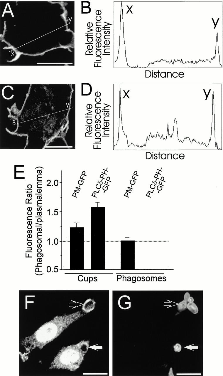
- PLCδ-PH-GFP accumulated in phagocytic cups and then dissociated from sealed phagosomes; peak phagosomal-cup intensity was 1.3–2.3× plasma membrane, mean 1.61 ± 0.08 (n = 64), significantly greater than PM-GFP (mean 1.25 ± 0.08, n = 35; P < 0.005).
- PIPKIα transiently concentrated at nascent phagosomes and dissociated after completion; PLCγ2 and PLCγ-SH2-GFP concentrated at phagocytic cups, and the DAG probe C1δ-YFP was recruited as PIP2 disappeared, strongest immediately after phagosome closure.
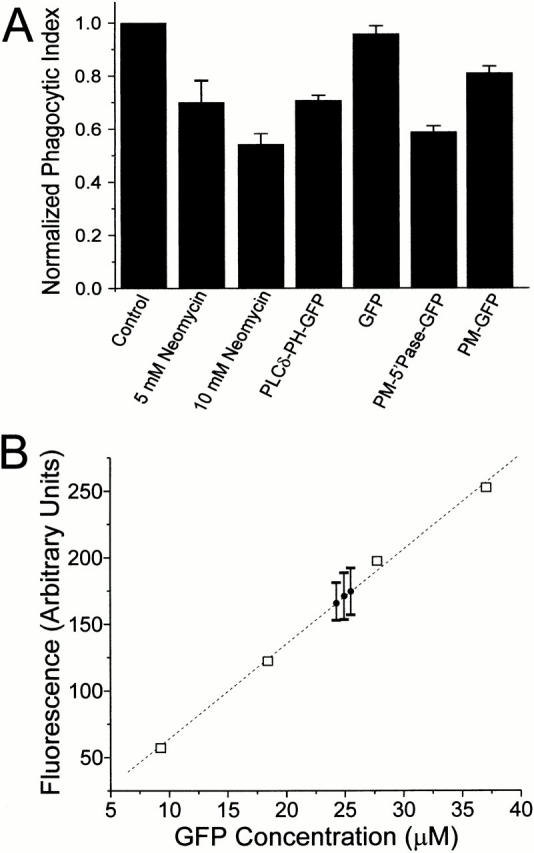
- Reducing PIP2 availability inhibited phagocytosis: neomycin at 5 and 10 mM reduced the phagocytic index by 30 ± 8% and 45 ± 5% (n = 15, P < 0.001); PLCδ-PH-GFP overexpression reduced it by 30 ± 2% (n = 12, P < 0.0001); PM-5′-phosphatase-GFP reduced it by 42 ± 2% (n = 12, P < 0.0001).
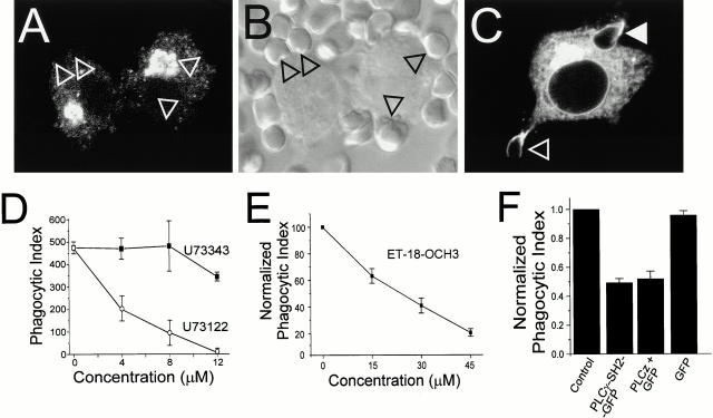
- PLC inhibition blocked phagocytosis and actin-cup/pseudopod formation: U73122 had half-maximal effects at ≈3 μM, ET-18-OCH3 at ≈25 μM, and dominant-negative PLCγ constructs (PLCγ-SH2-GFP or PLCz) reduced the phagocytic index by >50% (n = 12, P < 0.0001).
Methods
- Live-cell confocal imaging of RAW 264.7 macrophages transiently transfected with GFP/CFP/YFP chimeras: PLCδ-PH for 4,5-PIP2, C1δ for DAG, PM-GFP, PM-5′-phosphatase-GFP, PLCγ-SH2-GFP, and PLCz.
- Phagocytosis assays used IgG-opsonized sheep RBCs, zymosan, or 8-μm latex beads; phagocytic index was estimated by DIC microscopy, with synchronized binding on ice followed by warming. Immunofluorescence was used for PIPKIα and PLCγ2, and SEM/TEM for pseudopod morphology.
- Main analysis was confocal microscopy with fluorescence quantitation by line scanning and t tests; pharmacological inhibitors included neomycin, U73122/U73343, and ET-18-OCH3.
Limitations
- Stated: the authors note that PLCγ-SH2-GFP or PLCz could bind phosphotyrosine groups unrelated to those that anchor PLCγ, so the dominant-negative specificity is not absolute.
- Stated: the PM-5′-phosphatase-GFP effect may not be entirely attributable to phosphatase activity, since PM-GFP alone produced a modest, reproducible inhibition of phagocytosis, possibly by displacing acylated Src family kinases.
- Evident: conclusions rely on fluorescent lipid-binding probes and pharmacological inhibitors rather than direct lipid measurements; PLCδ-PH has measurable low-affinity binding to 3′-phosphorylated inositides in vitro, and DAG effects may involve C1-domain proteins other than PKC.
Model-written summary. Check it against the paper before relying on it.
Integrity signals
Registry
Cites
Statistics
Figures
Verification
Claims the paper makes
64 Own result · 35 Own interpretation · 13 Partly supported · 8 No full text of the sources · 3 Supported so far · 2 Supported · 1 Sources not found · 1 Not checked yet · 1 Own method · 1 No basis given · accuracy not yet measured
The cited sources support only part of this claim — read each source’s verdict below.
In the paper
“In vitro, the PH domain of PLCδ can also bind to phosphatidylinositol-3,4-bisphosphate (3,4-PIP2) and phosphatidylinositol-3,4,5-trisphosphate (3,4,5-PIP3), though with comparatively low affinity (Kavran et al. 1998).”
§ Localized Biphasic Changes in Phosphatidylinositol-4,5-Bisphosphate at Sites of Phagocytosis > Results > PLCδ-PH-GFP Is a Specific Probe for 4,5-PIP2 in Macrophages
What the sources say
- Partly supportedcites Kavran et al. 1998 sourceExperimental — accuracy not yet measured
The section on relative binding affinities says PLCδ-PH does not select against other phosphoinositides as strongly as some PH domains, which broadly supports binding beyond its preferred ligand. But the Discussion reports that, in vesicles containing 33% phosphatidylserine, PLCδ-PH bound PtdIns(3,4,5)P3 slightly more strongly than its preferred PtdIns(4,5)P2. The copy does not establish comparatively low affinity for both lipids named in the claim. (LLM, conf=0.82)
plaincorrelational- Asserted: passes — The authors put it forward as true.
- Checkable: passes — It can be checked against evidence.
- Load-bearing: passes — The paper’s conclusions depend on it.
- Based: passes — The paper gives a reason why it holds.
- Partly supported
The cited sources support only part of this claim — read each source’s verdict below.
In the paper
“This kinase appears to be essential for phagocytosis since inhibitors such as wortmannin impair particle engulfment (Ninomiya et al. 1994; Araki et al. 1996; Cox et al. 1999).”
§ Localized Biphasic Changes in Phosphatidylinositol-4,5-Bisphosphate at Sites of Phagocytosis > Introduction
What the sources say
- Supportedcites Ninomiya et al. 1994 source
The FIG. 1 results report that wortmannin markedly inhibited engulfment of IgG-coated erythrocytes by guinea pig neutrophils without preventing binding. The Discussion interprets this inhibition as evidence for an indispensable role of PI3K activation in Fcγ receptor-mediated phagocytosis; the study's conclusion is specific to that pathway. (LLM, conf=0.93)
- Supportedcites Araki et al. 1996 source
The Abstract reports that wortmannin and LY294002 inhibited Fc receptor-mediated phagocytosis in macrophages. The Discussion specifies that PI 3-kinase is necessary for completion of phagocytosis: the inhibitors prevented pseudopodia from closing around opsonized erythrocytes, rather than preventing initial binding or extension. (LLM, conf=0.94)
- Partly supportedcites Cox et al. 1999 sourceExperimental — accuracy not yet measured
The Results section, “Wortmannin and LY294002 Inhibit Fcγ-mediated Phagocytosis,” reports concentration-dependent inhibition of particle engulfment by both PI3K inhibitors. But the paper finds weaker inhibition for smaller beads and cautions against generalizing to other phagocytic targets. It therefore supports the inhibitor finding, but not an unqualified requirement for PI3K in phagocytosis generally. (LLM, conf=0.84)
hedgeddirect- Asserted: passes — The authors put it forward as true.
- Checkable: passes — It can be checked against evidence.
- Load-bearing: unclear — It is unclear whether the paper’s conclusions depend on it.
- Based: passes — The paper gives a reason why it holds.
- Supported
The cited sources support only part of this claim — read each source’s verdict below.
In the paper
“The PH domain of PLCδ was shown to bind to 4,5-PIP2 in vitro and in vivo with high affinity and specificity (Lemmon et al. 1995).”
§ Localized Biphasic Changes in Phosphatidylinositol-4,5-Bisphosphate at Sites of Phagocytosis > Results > PLCδ-PH-GFP Is a Specific Probe for 4,5-PIP2 in Macrophages
What the sources say
- Partly supportedcites Lemmon et al. 1995 sourceExperimental — accuracy not yet measured
The Abstract reports that the isolated PLCδ1 PH domain binds PtdIns(4,5)P2 with high affinity and specificity. It does not report binding demonstrated in vivo, so the cited paper supports the binding claim but not that setting. (LLM, conf=0.88)
boostedcorrelational- Asserted: passes — The authors put it forward as true.
- Checkable: passes — It can be checked against evidence.
- Load-bearing: passes — The paper’s conclusions depend on it.
- Based: passes — The paper gives a reason why it holds.
- Partly supported
The sources with a verdict support this claim; the rest weren’t checked yet, have no full text we could read, or their verdicts aren’t rated yet.
In the paper
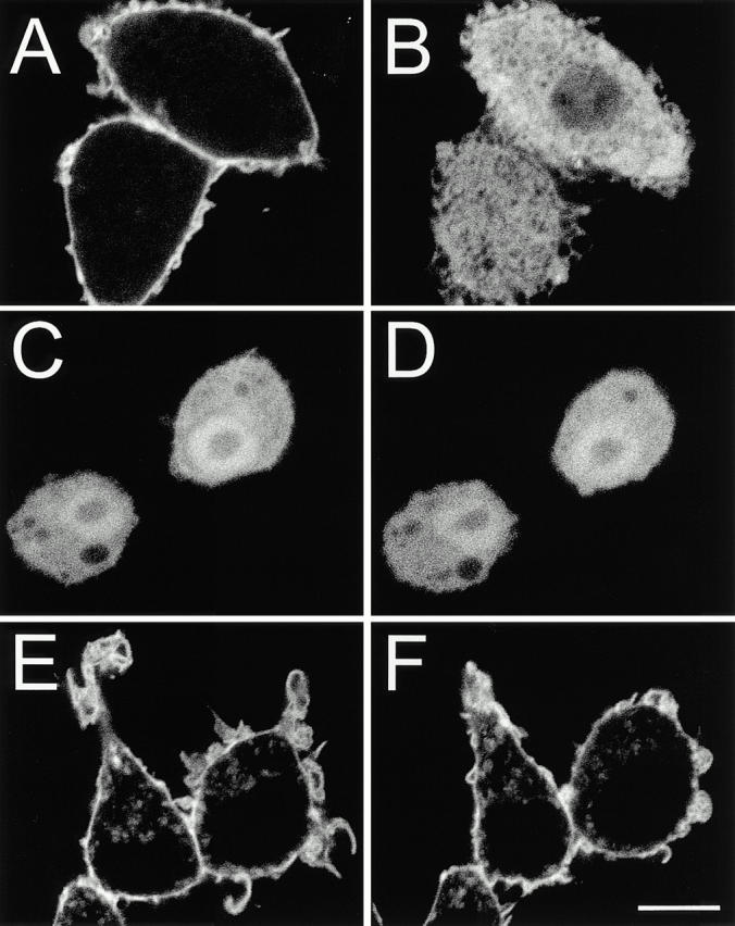
“As reported for other cell types (Stauffer et al. 1998; Varnai and Balla 1998), PLCδ-PH-GFP associated predominantly with the plasmalemma of RAW cells, with little binding to organellar membranes (Fig. 1 A).”
§ Localized Biphasic Changes in Phosphatidylinositol-4,5-Bisphosphate at Sites of Phagocytosis > Results > PLCδ-PH-GFP Is a Specific Probe for 4,5-PIP2 in Macrophages
What the sources say
- Not checked — no full text of the sourcecites Stauffer et al. 1998 source
- Supportedcites Varnai and Balla 1998 source
The Results section (‘Plasma Membrane Localization of the PLCδ PH Domain Requires Interaction with PtdIns(4,5)P2,’ Fig. 1) reports that PLCδ-PH-GFP localized to the plasma membrane in various cell types, with very little fluorescence in internal membranes. The citation supports that comparison; the RAW-cell observation is presented as the citing authors’ own result. (LLM, conf=0.93)
plaincorrelational- Asserted: passes — The authors put it forward as true.
- Checkable: passes — It can be checked against evidence.
- Load-bearing: passes — The paper’s conclusions depend on it.
- Based: passes — The paper gives a reason why it holds.
The cited sources support only part of this claim — read each source’s verdict below.
In the paper
“PLCδ-PH-GFP Is a Specific Probe for 4,5-PIP2 in Macrophages”
- Also stated in: § PLCδ-PH-GFP Is a Specific Probe for 4,5-PIP2 in Macrophages · plain
“Therefore, we used a fusion of this domain with GFP (PLCδ-PH-GFP) to analyze phospholipid metabolism in RAW 264.7 cells.”
§ Localized Biphasic Changes in Phosphatidylinositol-4,5-Bisphosphate at Sites of Phagocytosis > Results > PLCδ-PH-GFP Is a Specific Probe for 4,5-PIP2 in Macrophages
What the sources say
- Partly supportedcites Lemmon et al. 1995 sourceExperimental — accuracy not yet measured
The Abstract reports that the isolated PLCδ1 PH domain binds PtdIns(4,5)P2 with high affinity and binding specificity. It does not evaluate a GFP-tagged probe or establish that it specifically detects PtdIns(4,5)P2 in macrophages; it also reports binding to Ins(1,4,5)P3. (LLM, conf=0.87)
plaincorrelational- Asserted: passes — The authors put it forward as true.
- Checkable: passes — It can be checked against evidence.
- Load-bearing: passes — The paper’s conclusions depend on it.
- Based: passes — The paper gives a reason why it holds.
- Also stated in: § PLCδ-PH-GFP Is a Specific Probe for 4,5-PIP2 in Macrophages · plain
The cited sources support only part of this claim — read each source’s verdict below.
In the paper
“Experiments using clostridial toxins or transfection of dominant-negative constructs have convincingly demonstrated that actin assembly during phagocytosis is mediated by Rho family GTPases (Cox et al. 1997; Hackam et al. 1997; Caron and Hall 1998; Massol et al. 1998).”
§ Localized Biphasic Changes in Phosphatidylinositol-4,5-Bisphosphate at Sites of Phagocytosis > Introduction
What the sources say
- Supportedcites Cox et al. 1997 source
The Abstract reports that dominant-negative Rac1 and Cdc42 blocked phagocytosis and partially inhibited F-actin–rich phagocytic cups. The Results section confirms that these constructs inhibited the formation of distinct F-actin–rich cups during phagocytosis, supporting the attributed role of Rho family GTPases in actin assembly. (LLM, conf=0.93)
- Partly supportedcites Hackam et al. 1997 sourceExperimental — accuracy not yet measured
The Results report that inactivating Rho eliminated F-actin cups during Fcγ receptor–mediated phagocytosis, supporting Rho's role in actin assembly. The Discussion confines the conclusions to transformed cell lines; this paper alone does not establish the claim for phagocytosis generally. (LLM, conf=0.85)
- Not checked — no full text of the sourcecites Caron and Hall 1998 source
boosteddirect- Asserted: passes — The authors put it forward as true.
- Checkable: passes — It can be checked against evidence.
- Load-bearing: passes — The paper’s conclusions depend on it.
- Based: passes — The paper gives a reason why it holds.
- Supported
The cited sources support only part of this claim — read each source’s verdict below.
In the paper
“Recruitment and activation of this tyrosine kinase is critical for FcγR-mediated phagocytosis, as demonstrated by the inability of macrophages from Syk−/− mice to internalize IgG-opsonized particles (Crowley et al. 1997; Kiefer et al. 1998).”
§ Localized Biphasic Changes in Phosphatidylinositol-4,5-Bisphosphate at Sites of Phagocytosis > Introduction
What the sources say
- Supportedcites Crowley et al. 1997 source
The Results section reports that Syk-deficient macrophages bound IgG-coated erythrocytes but failed to internalize them, demonstrating a requirement for Syk in FcγR-mediated phagocytosis. The Discussion also describes recruitment and activation of Syk in FcγR signaling. Page numbers are not provided in our copy. (LLM, conf=0.94)
- Partly supportedcites Kiefer et al. 1998 sourceExperimental — accuracy not yet measured
The Results report that Syk is important for FcγR-mediated uptake of IgG-opsonized red blood cells, but they do not show an inability to internalize them: 15% of Syk-deficient macrophages completed phagocytosis after 10 minutes. The Discussion likewise describes a significant reduction, not a complete loss. Page numbers were not available in our copy. (LLM, conf=0.90)
boosteddirect- Asserted: passes — The authors put it forward as true.
- Checkable: passes — It can be checked against evidence.
- Load-bearing: unclear — It is unclear whether the paper’s conclusions depend on it.
- Based: passes — The paper gives a reason why it holds.
- Supported
Every source the paper cites for this claim was checked and supports it.
In the paper
“The increase in 3,4,5-PIP3 predicted to occur during phagocytosis, based on the reported activation of PI3K (Araki et al. 1996; Cox et al. 1999), did not affect the distribution of PLCδ-PH-GFP.”
§ Localized Biphasic Changes in Phosphatidylinositol-4,5-Bisphosphate at Sites of Phagocytosis > Discussion > Suitability of GFP Chimeras as Probes
What the sources say
- Supportedcites Araki et al. 1996 source
The citation is attached to the reported role of PI3K in phagocytosis, not to the citing authors’ observation about PLCδ-PH-GFP. The Introduction identifies 3,4,5-PIP3 as a product of PI3K, and the Discussion reports that PI3K functions in Fc receptor-mediated phagocytosis. Together, these support the citing authors’ predicted increase; this paper does not report the GFP distribution. (LLM, conf=0.88)
- Supportedcites Cox et al. 1999 source
The citation is attached to the reported activation of PI3K during phagocytosis, not to the citing paper’s observation about PLCδ-PH-GFP distribution. In the “Fcγ-mediated Phagocytosis Is Accompanied by Enhanced Production of PIP3” section (page not supplied), Cox et al. report a rapid rise in PIP3 after Fcγ receptor ligation, consistent with increased PI3K activity. (LLM, conf=0.92)
plain- Asserted: passes — The authors put it forward as true.
- Checkable: passes — It can be checked against evidence.
- Load-bearing: passes — The paper’s conclusions depend on it.
- Based: passes — The paper gives a reason why it holds.
- Supported
The cited sources support only part of this claim — read each source’s verdict below.
In the paper
“Hence, consumption of 4,5-PIP2 by PI3K must contribute to the dissociation of PLCδ-PH-GFP.”
§ Localized Biphasic Changes in Phosphatidylinositol-4,5-Bisphosphate at Sites of Phagocytosis > Discussion > Biphasic Changes in 4,5-PIP2 during Phagocytosis
What the sources say
- Partly supportedcites Araki et al. 1996 sourceExperimental — accuracy not yet measured
The Introduction (around p. 1250) says PI3K phosphorylates phosphoinositides to produce phosphatidylinositol (3,4,5)-trisphosphate, and the paper links PI3K activity to completion of phagocytosis. It does not examine PLCδ-PH-GFP or show that PI3K’s consumption of 4,5-PIP2 causes that reporter to dissociate from phagosomes; that causal conclusion goes beyond this source. (LLM, conf=0.84)
- Partly supportedcites Cox et al. 1999 sourceExperimental — accuracy not yet measured
The Results (page unavailable) report rapid PIP3 accumulation during Fcγ receptor-mediated phagocytosis, consistent with increased PI3K activity. The Experimental Procedures describe generating PIP3 from 4,5-PIP2 for a laboratory standard. Neither passage establishes that PI3K consumes phagosomal 4,5-PIP2 or causes PLCδ-PH-GFP to dissociate. (LLM, conf=0.83)
boosteddirect- Asserted: passes — The authors put it forward as true.
- Checkable: passes — It can be checked against evidence.
- Load-bearing: passes — The paper’s conclusions depend on it.
- Based: passes — The paper gives a reason why it holds.
- Partly supported
A result the paper reports from its own data — it cites no other source for it, so there is none to check. The quote shows it is in the paper.
In the paper
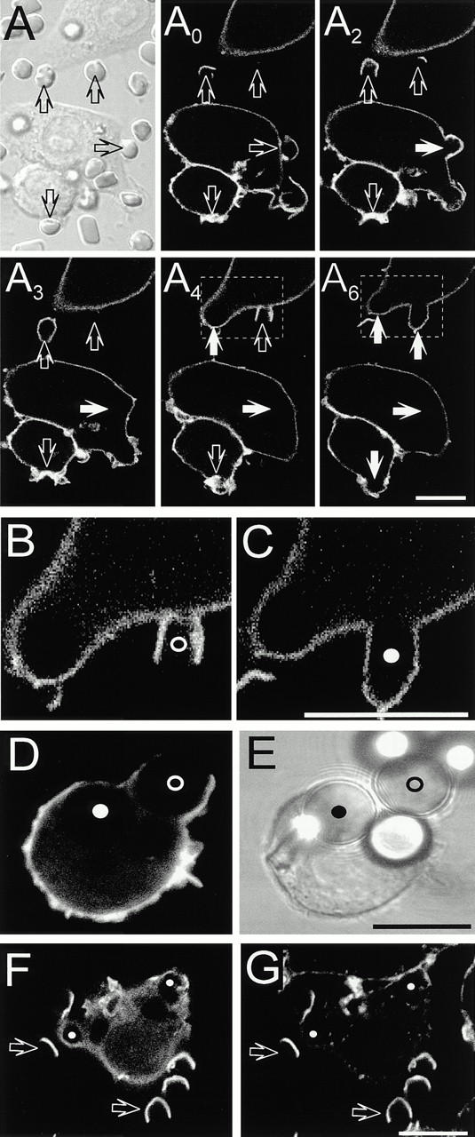
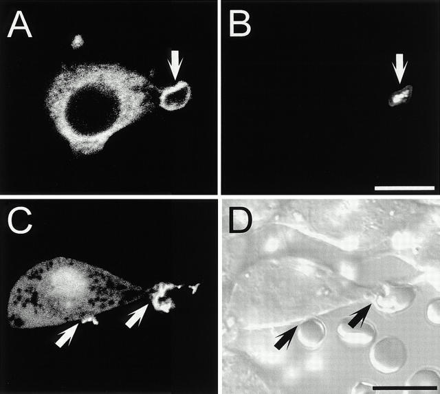
“During pseudopod extension PLCδ-PH-GFP appeared to accumulate in phagocytic cups compared with the rest of the plasmalemma (Fig. 2 A, see also Fig. 4).”
§ Localized Biphasic Changes in Phosphatidylinositol-4,5-Bisphosphate at Sites of Phagocytosis > Results > Redistribution of PLCδ-PH-GFP during Phagocytosis
hedgedcorrelational- Asserted: passes — The authors put it forward as true.
- Checkable: passes — It can be checked against evidence.
- Load-bearing: passes — The paper’s conclusions depend on it.
- Based: passes — The paper gives a reason why it holds.
- Probably not a substantial claim. 9
- It may state two separate claims as one. 33
- The quote only partly states this claim. 1
Every source the paper cites for this claim was checked and supports it.
In the paper
“As reported previously, F-actin accumulated transiently underneath the phagosomal cup, where it is thought to propel the extension of pseudopods (Greenberg et al. 1991).”
§ Localized Biphasic Changes in Phosphatidylinositol-4,5-Bisphosphate at Sites of Phagocytosis > Results > Redistribution of PLCδ-PH-GFP during Phagocytosis
What the sources say
- Supportedcites Greenberg et al. 1991 source
The Results (Fig. 3) describe F-actin around forming phagosomes at early time points that was barely visible by 30 minutes. The Discussion (around p. 765) describes F-actin accumulating at the bases of phagocytic cups and proposes that growth of the actin network pushes pseudopods outward. The citing sentence appropriately presents that mechanism as a hypothesis. (LLM, conf=0.92)
hedgedconditional- Asserted: passes — The authors put it forward as true.
- Checkable: passes — It can be checked against evidence.
- Load-bearing: fails — The paper’s conclusions may not depend on it.
- Based: passes — The paper gives a reason why it holds.
Probably not a substantial claim. The paper’s conclusions may not depend on it.
- Supported
The sources with a verdict support this claim; the rest weren’t checked yet, have no full text we could read, or their verdicts aren’t rated yet.
In the paper
“To assess the relative abundance of these lipids, RAW macrophages were transfected with the GFP-tagged PH domains of either Akt (Fig. 1C and Fig. D) or Btk (not illustrated), which recognize specifically 3,4-PIP2 and 3,4,5-PIP3 (Kavran et al. 1998; Varnai et al. 1999).”
§ Localized Biphasic Changes in Phosphatidylinositol-4,5-Bisphosphate at Sites of Phagocytosis > Results > PLCδ-PH-GFP Is a Specific Probe for 4,5-PIP2 in Macrophages
What the sources say
- Supportedcites Kavran et al. 1998 source
The Results report that the Akt/PKB PH domain recognizes PtdIns(3,4)P2 and PtdIns(3,4,5)P3. In “PH Domains Can Be Grouped According to Their Likely Physiological Ligand(s),” the paper places Btk’s PH domain among those that bind specifically to PtdIns(3,4,5)P3. The paper does not say Akt recognizes only PtdIns(3,4)P2; it recognizes both lipids. (LLM, conf=0.88)
- Not checked — no full text of the sourcecites Varnai et al. 1999 source
plaincorrelational- Asserted: passes — The authors put it forward as true.
- Checkable: passes — It can be checked against evidence.
- Load-bearing: passes — The paper’s conclusions depend on it.
- Based: passes — The paper gives a reason why it holds.
It may state two separate claims as one.
- Supported
The cited sources support only part of this claim — read each source’s verdict below.
In the paper
“In addition to its role as a direct docking site for skeletal proteins, 4,5-PIP2 is also the primary substrate of phospholipase Cγ (PLCγ), which was shown to become activated upon engagement of FcγRs (Azzoni et al. 1992; Liao et al. 1992).”
§ Localized Biphasic Changes in Phosphatidylinositol-4,5-Bisphosphate at Sites of Phagocytosis > Introduction
What the sources say
- Partly supportedcites Azzoni et al. 1992 sourceExperimental — accuracy not yet measured
The discussion following Fig. 4 (p. 1748) says FcγRIIIA stimulation induces PLCγ1 tyrosine phosphorylation and strongly suggests that PLCγ1 activation may account for the observed PIP2 hydrolysis. This supports the proposed link, but the paper does not establish PLCγ1 activation directly or identify PIP2 as its primary substrate; its experiments concern FcγRIIIA rather than FcγRs generally. (LLM, conf=0.86)
- Partly supportedcites Liao et al. 1992 sourceExperimental — accuracy not yet measured
The paper reports that cross-linking FcγRI or FcγRII in U937 cells leads to PLCγ1 phosphorylation and hydrolysis of phosphatidylinositol 4,5-bisphosphate (abstract, p. 3659). This supports receptor-linked PLCγ activity and identifies 4,5-PIP2 as a substrate, but the passage does not establish that it is PLCγ’s primary substrate. (LLM, conf=0.84)
boosteddirect- Asserted: passes — The authors put it forward as true.
- Checkable: passes — It can be checked against evidence.
- Load-bearing: passes — The paper’s conclusions depend on it.
- Based: passes — The paper gives a reason why it holds.
It may state two separate claims as one.
- Partly supported
The cited sources support only part of this claim — read each source’s verdict below.
In the paper
“Clustering of FcγRs on the surface of opsonized particles is associated with the phosphorylation of tyrosine residues within a defined region known as the immunoreceptor tyrosine-based activation motif (Greenberg et al. 1993, Greenberg et al. 1994).”
§ Localized Biphasic Changes in Phosphatidylinositol-4,5-Bisphosphate at Sites of Phagocytosis > Introduction
What the sources say
- Partly supportedcites Greenberg et al. 1993 sourceExperimental — accuracy not yet measured
Figure 5 (p. 532) reports that tyrosine phosphoproteins accumulate where ligand density is high on phagocytic particles, supporting an association between particle-bound ligand and tyrosine phosphorylation. The paper does not identify the phosphorylated residues as being within an immunoreceptor tyrosine-based activation motif; it describes the possible role of an Fc receptor-associated subunit in signaling as speculative. (LLM, conf=0.88)
- Not checked — no full text of the sourcecites Greenberg et al. 1994 source
plaincorrelational- Asserted: passes — The authors put it forward as true.
- Checkable: passes — It can be checked against evidence.
- Load-bearing: fails — The paper’s conclusions may not depend on it.
- Based: passes — The paper gives a reason why it holds.
Probably not a substantial claim. The paper’s conclusions may not depend on it.
- Partly supported
The sources with a verdict support this claim; the rest weren’t checked yet, have no full text we could read, or their verdicts aren’t rated yet.
In the paper
“These probes had been validated in other systems (Oancea et al. 1998; Stauffer et al. 1998; Varnai and Balla 1998) and their suitability was confirmed in RAW macrophages.”
§ Localized Biphasic Changes in Phosphatidylinositol-4,5-Bisphosphate at Sites of Phagocytosis > Discussion > Suitability of GFP Chimeras as Probes
What the sources say
- Not matchedcites Oancea et al. 1998
Couldn’t be matched to a paper in Crossref, so it can’t be checked.
- Not checked — no full text of the sourcecites Stauffer et al. 1998 source
- Supportedcites Varnai and Balla 1998 source
The Results section, under “Plasma Membrane Localization of the PLCδ PH Domain Requires Interaction with PtdIns(4,5)P2,” reports that the PLCδ-PH–GFP probe localized to the plasma membrane in various cell types and that mutations disrupting its PtdIns(4,5)P2 binding prevented that localization. This supports the citation’s role in the multi-source statement that the probes had been validated in other systems; the RAW-macrophage confirmation is the citing authors’ own finding. (LLM, conf=0.91)
plain- Asserted: passes — The authors put it forward as true.
- Checkable: passes — It can be checked against evidence.
- Load-bearing: passes — The paper’s conclusions depend on it.
- Based: passes — The paper gives a reason why it holds.
It may state two separate claims as one.
- Not matched
The cited sources support only part of this claim — read each source’s verdict below.
In the paper
“PLCδ-PH-GFP can be used not only to monitor the distribution of 4,5-PIP2, but when expressed at sufficiently high levels, also to competitively inhibit other 4,5-PIP2–dependent processes.”
§ Localized Biphasic Changes in Phosphatidylinositol-4,5-Bisphosphate at Sites of Phagocytosis > Results > Reduced Availability of 4,5-PIP2 Attenuates FcγR-mediated Phagocytosis
What the sources say
- Partly supportedcites Varnai and Balla 1998 sourceExperimental — accuracy not yet measured
The Results section (‘PH Domains Can Inhibit the Agonist-induced Hydrolysis…’; page unavailable) reports that overexpressed PLCδ PH-domain–GFP greatly inhibited agonist-stimulated inositol phosphate formation. The Discussion proposes masking PtdIns(4,5)P2 or competing with PLC enzymes as possible mechanisms, but does not establish competitive inhibition or demonstrate inhibition of other PtdIns(4,5)P2-dependent processes beyond the PLC response tested. (LLM, conf=0.84)
- Not checked — no full text of the sourcecites Raucher et al. 2000 source
plaindirect- Asserted: passes — The authors put it forward as true.
- Checkable: passes — It can be checked against evidence.
- Load-bearing: passes — The paper’s conclusions depend on it.
- Based: passes — The paper gives a reason why it holds.
It may state two separate claims as one.
- Partly supported
The cited sources support only part of this claim — read each source’s verdict below.
In the paper
“PLCγ is known to be recruited to the vicinity of activated receptors through interaction of its dual SH2 domains with phosphotyrosine residues on receptor–adaptor complexes (Rhee and Bae 1997).”
§ Localized Biphasic Changes in Phosphatidylinositol-4,5-Bisphosphate at Sites of Phagocytosis > Results > PLCγ Localizes to Phagocytic Cups
What the sources say
- Partly supportedcites Rhee and Bae 1997 sourceExperimental — accuracy not yet measured
In “Activation of PLC-γ by Protein Tyrosine Kinases” (page unavailable), the paper says PLC-γ binds phosphorylated receptors or receptor components through its SH2 domains. “PLC Isoforms and Structural Organization” (page unavailable) confirms that PLC-γ has two SH2 domains. The copy does not establish binding to receptor–adaptor complexes or that both SH2 domains participate in a given interaction. (LLM, conf=0.82)
boosteddirect- Asserted: passes — The authors put it forward as true.
- Checkable: passes — It can be checked against evidence.
- Load-bearing: passes — The paper’s conclusions depend on it.
- Based: passes — The paper gives a reason why it holds.
Probably not a substantial claim.
- Partly supported
The cited sources support only part of this claim — read each source’s verdict below.
In the paper
“While wortmannin-treated cells fail to internalize particles, they nevertheless display marked actin reassembly under the adherent RBCs (Araki et al. 1996; Cox et al. 1999; and our own observations, not shown).”
§ Localized Biphasic Changes in Phosphatidylinositol-4,5-Bisphosphate at Sites of Phagocytosis > Results > Inactivation of PLC Abolishes Pseudopod and Actin Cup Formation
What the sources say
- Supportedcites Araki et al. 1996 source
The Results (p. 1254) report that wortmannin inhibited internalization of erythrocytes, while treated macrophages extended actin-rich pseudopodia partway around them. The paper also reports more F-actin-rich phagocytic cups in treated cells than in controls, supporting the claim about actin beneath adherent RBCs. (LLM, conf=0.93)
- Partly supportedcites Cox et al. 1999 sourceExperimental — accuracy not yet measured
In “Actin Assembly in Response to Fc” (page unavailable), the paper reports that wortmannin did not visibly reduce F-actin accumulation beneath attached particles, including IgG-coated erythrocytes. It also reports inhibited phagocytosis, but not a complete failure to internalize particles: inhibition depended on particle size and was less effective for smaller beads. (LLM, conf=0.86)
plaincorrelational- Asserted: passes — The authors put it forward as true.
- Checkable: passes — It can be checked against evidence.
- Load-bearing: passes — The paper’s conclusions depend on it.
- Based: passes — The paper gives a reason why it holds.
It may state two separate claims as one.
- Supported
The cited sources support only part of this claim — read each source’s verdict below.
In the paper
“both PKCα and PKCδ copurify with isolated phagosomal membranes (Allen and Aderem 1995; Brumell et al. 1999) and some inhibitors of PKC activity have been reported to interfere with phagocytosis (Zheleznyak and Brown 1992; Karimi et al. 1999).”
§ Localized Biphasic Changes in Phosphatidylinositol-4,5-Bisphosphate at Sites of Phagocytosis > Discussion > Localized Phosphoinositide Metabolism Is Required for Phagocytosis
What the sources say
- Partly supportedcites Allen and Aderem 1995 sourceExperimental — accuracy not yet measured
The Discussion reports that PKCα associates with forming phagosomes and remains on maturing phagosomes. It does not report PKCδ or show that both isoforms copurify with isolated phagosomal membranes. The separate claim about PKC inhibitors is cited to other papers in the citing sentence, so it is not assessed against Allen and Aderem. (LLM, conf=0.85)
- Partly supportedcites Brumell et al. 1999 sourceExperimental — accuracy not yet measured
The passage on phagosomal membranes reports that PKCδ accumulates on purified phagosomal membranes. It says conventional PKC isoforms have been found on phagosomes, but does not identify PKCα or establish its copurification. The inhibitor clause is attributed to other citations, not Brumell et al. (1999). (LLM, conf=0.87)
- Not checked — no full text of the sourcecites Zheleznyak and Brown 1992 source
plaincorrelational- Asserted: passes — The authors put it forward as true.
- Checkable: passes — It can be checked against evidence.
- Load-bearing: unclear — It is unclear whether the paper’s conclusions depend on it.
- Based: passes — The paper gives a reason why it holds.
It may state two separate claims as one.
- Partly supported
A result the paper reports from its own data — it cites no other source for it, so there is none to check. The quote shows it is in the paper.
In the paper
“The average ratio in this case was 1.25 ± 0.08 (n = 35, Fig. 4 E), which is significantly different from unity (t test, P ≤ 0.005).”
§ Localized Biphasic Changes in Phosphatidylinositol-4,5-Bisphosphate at Sites of Phagocytosis > Results > Accumulation of PLCδ-PH-GFP in Nascent Phagosomes
plainn = 35- Asserted: passes — The authors put it forward as true.
- Checkable: passes — It can be checked against evidence.
- Load-bearing: passes — The paper’s conclusions depend on it.
- Based: passes — The paper gives a reason why it holds.
It may state two separate claims as one.
A result the paper reports from its own data — it cites no other source for it, so there is none to check. The quote shows it is in the paper.
In the paper
“PM-GFP was preferentially targeted to the plasma membrane, although internal structures were also decorated (Fig. 1 E).”
§ Localized Biphasic Changes in Phosphatidylinositol-4,5-Bisphosphate at Sites of Phagocytosis > Results > PLCδ-PH-GFP Is a Specific Probe for 4,5-PIP2 in Macrophages
plain- Asserted: passes — The authors put it forward as true.
- Checkable: passes — It can be checked against evidence.
- Load-bearing: unclear — It is unclear whether the paper’s conclusions depend on it.
- Based: passes — The paper gives a reason why it holds.
It may state two separate claims as one.
A result the paper reports from its own data — it cites no other source for it, so there is none to check. The quote shows it is in the paper.
In the paper
“In this system, fluorescence was seen to surround the phagosome throughout the sealing process and to remain therein long after (Fig. 3D and Fig. E, open and filled circles, respectively).”
§ Localized Biphasic Changes in Phosphatidylinositol-4,5-Bisphosphate at Sites of Phagocytosis > Results > Distribution of PM-GFP during Phagocytosis
plain- Asserted: passes — The authors put it forward as true.
- Checkable: passes — It can be checked against evidence.
- Load-bearing: passes — The paper’s conclusions depend on it.
- Based: passes — The paper gives a reason why it holds.
It may state two separate claims as one.
A result the paper reports from its own data — it cites no other source for it, so there is none to check. The quote shows it is in the paper.
In the paper
“In this case, the density of phagosomal and plasmalemmal PM-GFP was virtually identical, whereas PLCδ-PH-GFP was essentially undetectable in the phagosome (Fig. 4 E).”
§ Localized Biphasic Changes in Phosphatidylinositol-4,5-Bisphosphate at Sites of Phagocytosis > Results > Accumulation of PLCδ-PH-GFP in Nascent Phagosomes
plain- Asserted: passes — The authors put it forward as true.
- Checkable: passes — It can be checked against evidence.
- Load-bearing: passes — The paper’s conclusions depend on it.
- Based: passes — The paper gives a reason why it holds.
It may state two separate claims as one.
A result the paper reports from its own data — it cites no other source for it, so there is none to check. The quote shows it is in the paper.
In the paper
“Soon after addition of the opsonized particles, pseudopods labeled with PLCδ-PH-GFP extended from the macrophage surface and made contact with the RBCs (Fig. 2 A, open arrows).”
§ Localized Biphasic Changes in Phosphatidylinositol-4,5-Bisphosphate at Sites of Phagocytosis > Results > Redistribution of PLCδ-PH-GFP during Phagocytosis
plaincorrelational- Asserted: passes — The authors put it forward as true.
- Checkable: passes — It can be checked against evidence.
- Load-bearing: passes — The paper’s conclusions depend on it.
- Based: passes — The paper gives a reason why it holds.
It may state two separate claims as one.
A result the paper reports from its own data — it cites no other source for it, so there is none to check. The quote shows it is in the paper.
In the paper
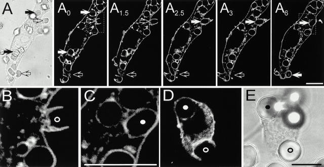
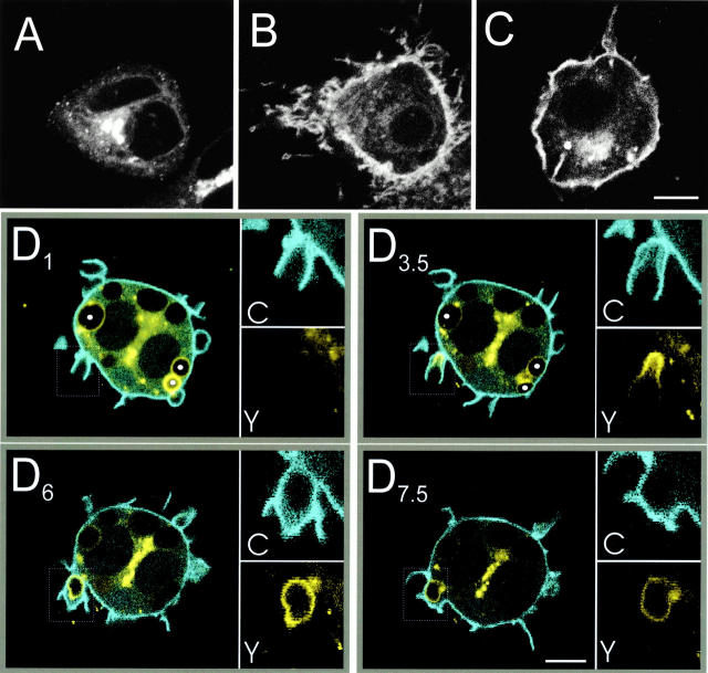
“Importantly, C1δ-YFP labeling was usually strongest immediately after phagosome closure, at which time PLCδ-PH-CFP was completely absent from the phagosome (Fig. 6 D6).”
§ Localized Biphasic Changes in Phosphatidylinositol-4,5-Bisphosphate at Sites of Phagocytosis > Results > Localized Generation of Diacylglycerol during Phagocytosis
plaincorrelational- Asserted: passes — The authors put it forward as true.
- Checkable: passes — It can be checked against evidence.
- Load-bearing: passes — The paper’s conclusions depend on it.
- Based: passes — The paper gives a reason why it holds.
It may state two separate claims as one.
A result the paper reports from its own data — it cites no other source for it, so there is none to check. The quote shows it is in the paper.
In the paper
“The initial accumulation of PLCδ-PH-GFP was transient and was followed immediately by its abrupt dissociation from formed phagosomes (Fig. 2, see also Fig. 6 D).”
§ Localized Biphasic Changes in Phosphatidylinositol-4,5-Bisphosphate at Sites of Phagocytosis > Discussion > Biphasic Changes in 4,5-PIP2 during Phagocytosis
plaincorrelational- Asserted: passes — The authors put it forward as true.
- Checkable: passes — It can be checked against evidence.
- Load-bearing: passes — The paper’s conclusions depend on it.
- Based: passes — The paper gives a reason why it holds.
It may state two separate claims as one.
A result the paper reports from its own data — it cites no other source for it, so there is none to check. The quote shows it is in the paper.
In the paper
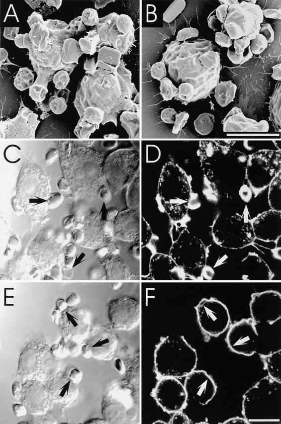
“In contrast, cells treated with U73122 were unable to extend pseudopods or form pedestals, even though RBCs appeared tightly attached to the surface of the macrophages (Fig. 9 B).”
§ Localized Biphasic Changes in Phosphatidylinositol-4,5-Bisphosphate at Sites of Phagocytosis > Results > Inactivation of PLC Abolishes Pseudopod and Actin Cup Formation
plaindirect- Asserted: passes — The authors put it forward as true.
- Checkable: passes — It can be checked against evidence.
- Load-bearing: passes — The paper’s conclusions depend on it.
- Based: passes — The paper gives a reason why it holds.
It may state two separate claims as one.
A result the paper reports from its own data — it cites no other source for it, so there is none to check. The quote shows it is in the paper.
In the paper
“In brief, PLCδ-PH-GFP localized almost exclusively to the plasmalemma of untreated cells, but redistributed to the cytosol when PLC was activated (Fig. 1A and Fig. B).”
§ Localized Biphasic Changes in Phosphatidylinositol-4,5-Bisphosphate at Sites of Phagocytosis > Discussion > Suitability of GFP Chimeras as Probes
plaindirect- Asserted: passes — The authors put it forward as true.
- Checkable: passes — It can be checked against evidence.
- Load-bearing: passes — The paper’s conclusions depend on it.
- Based: passes — The paper gives a reason why it holds.
It may state two separate claims as one.
A result the paper reports from its own data — it cites no other source for it, so there is none to check. The quote shows it is in the paper.
In the paper
“In contrast, U73343 had no significant effect at up to 6 μM and had only a minor antagonistic effect at 12 μM (Fig. 8 D).”
§ Localized Biphasic Changes in Phosphatidylinositol-4,5-Bisphosphate at Sites of Phagocytosis > Results > Inhibition of PLC Blocks Diacylglycerol Synthesis and Ablates Phagocytosis
plain- Asserted: passes — The authors put it forward as true.
- Checkable: passes — It can be checked against evidence.
- Load-bearing: passes — The paper’s conclusions depend on it.
- Based: passes — The paper gives a reason why it holds.
It may state two separate claims as one.
A result the paper reports from its own data — it cites no other source for it, so there is none to check. The quote shows it is in the paper.
In the paper
“The accumulation of the 4,5-PIP2 probe exceeded that of PM-GFP, implying that it is not simply due to folding of the membrane during pseudopod extension (Fig. 2 Fig. 3 Fig. 4).”
§ Localized Biphasic Changes in Phosphatidylinositol-4,5-Bisphosphate at Sites of Phagocytosis > Discussion > Biphasic Changes in 4,5-PIP2 during Phagocytosis
plaindirect- Asserted: passes — The authors put it forward as true.
- Checkable: passes — It can be checked against evidence.
- Load-bearing: passes — The paper’s conclusions depend on it.
- Based: passes — The paper gives a reason why it holds.
It may state two separate claims as one.
A result the paper reports from its own data — it cites no other source for it, so there is none to check. The quote shows it is in the paper.
In the paper
“Treatment with 6 μM U73122 for 15 min precluded accumulation of C1δ-GFP at the sites of RBC attachment (Fig. 8A and Fig. B).”
§ Localized Biphasic Changes in Phosphatidylinositol-4,5-Bisphosphate at Sites of Phagocytosis > Results > Inhibition of PLC Blocks Diacylglycerol Synthesis and Ablates Phagocytosis
plaindirect- Asserted: passes — The authors put it forward as true.
- Checkable: passes — It can be checked against evidence.
- Load-bearing: passes — The paper’s conclusions depend on it.
- Based: passes — The paper gives a reason why it holds.
It may state two separate claims as one.
A result the paper reports from its own data — it cites no other source for it, so there is none to check. The quote shows it is in the paper.
In the paper
“It redistributed to the plasma membrane when endogenous PLC was activated with ionomycin, leading to production of DAG synthesis (Fig. 6A and Fig. B), as well as upon addition of phorbol esters (Fig. 6 C) or exogenous DAG (not shown).”
§ Localized Biphasic Changes in Phosphatidylinositol-4,5-Bisphosphate at Sites of Phagocytosis > Discussion > Suitability of GFP Chimeras as Probes
plaindirect- Asserted: passes — The authors put it forward as true.
- Checkable: passes — It can be checked against evidence.
- Load-bearing: passes — The paper’s conclusions depend on it.
- Based: passes — The paper gives a reason why it holds.
It may state two separate claims as one.
A result the paper reports from its own data — it cites no other source for it, so there is none to check. The quote shows it is in the paper.
In the paper
“our own unpublished observations also failed to demonstrate an absolute requirement for PKC.”
§ Localized Biphasic Changes in Phosphatidylinositol-4,5-Bisphosphate at Sites of Phagocytosis > Discussion > Localized Phosphoinositide Metabolism Is Required for Phagocytosis
plain- Asserted: passes — The authors put it forward as true.
- Checkable: passes — It can be checked against evidence.
- Load-bearing: fails — The paper’s conclusions may not depend on it.
- Based: passes — The paper gives a reason why it holds.
Probably not a substantial claim. The paper’s conclusions may not depend on it.
A result the paper reports from its own data — it cites no other source for it, so there is none to check. The quote shows it is in the paper.
In the paper
“We found that phagocytosis was markedly inhibited when 4,5-PIP2 was sequestered by addition of neomycin or by overexpression of PLCδ-PH domain. Similarly, enzymatic degradation of 4,5-PIP2 also resulted in diminution of the phagocytic index (Fig. 7).”
§ Localized Biphasic Changes in Phosphatidylinositol-4,5-Bisphosphate at Sites of Phagocytosis > Discussion > Localized Phosphoinositide Metabolism Is Required for Phagocytosis
plaindirect- Asserted: passes — The authors put it forward as true.
- Checkable: passes — It can be checked against evidence.
- Load-bearing: passes — The paper’s conclusions depend on it.
- Based: passes — The paper gives a reason why it holds.
It may state two separate claims as one.
In the paper
“During this process phagocytes internalize opsonized particles into a vacuole by an actin-dependent mechanism (Kwiatkowska and Sobota 1999).”
§ Localized Biphasic Changes in Phosphatidylinositol-4,5-Bisphosphate at Sites of Phagocytosis > Introduction
What the sources say
- Not checked — no full text of the sourcecites Kwiatkowska and Sobota 1999 source
plaindirect- Asserted: passes — The authors put it forward as true.
- Checkable: passes — It can be checked against evidence.
- Load-bearing: fails — The paper’s conclusions may not depend on it.
- Based: passes — The paper gives a reason why it holds.
Probably not a substantial claim. The paper’s conclusions may not depend on it.
A claim is checked only against the cited paper’s full text, never its abstract, and none is available for its sources.
In the paper
“This phospholipid is thought to govern the distribution and function of a variety of cytoskeletal and signaling proteins that bear pleckstrin homology (PH) domains or lysine/arginine-rich hydrophobic motifs, which interact directly and specifically with the headgroup of 4,5-PIP2 (Bottomley et al. 1998; Hinchliffe et al. 1998; Martin 1998; Czech 2000).”
§ Localized Biphasic Changes in Phosphatidylinositol-4,5-Bisphosphate at Sites of Phagocytosis > Introduction
What the sources say
hedgedconditional- Asserted: passes — The authors put it forward as true.
- Checkable: passes — It can be checked against evidence.
- Load-bearing: passes — The paper’s conclusions depend on it.
- Based: passes — The paper gives a reason why it holds.
Probably not a substantial claim.
In the paper
“After internalization, the phagosomal vacuole is converted into an effective microbicidal organelle by progressive fusion with endosomes and lysosomes (Beron et al. 1995).”
§ Localized Biphasic Changes in Phosphatidylinositol-4,5-Bisphosphate at Sites of Phagocytosis > Introduction
What the sources say
- Not checked — no full text of the sourcecites Beron et al. 1995 source
plaindirect- Asserted: passes — The authors put it forward as true.
- Checkable: passes — It can be checked against evidence.
- Load-bearing: fails — The paper’s conclusions may not depend on it.
- Based: passes — The paper gives a reason why it holds.
Probably not a substantial claim. The paper’s conclusions may not depend on it.
The paper states this without a source or data behind it here, so there is nothing to check it against. The quote shows it is in the paper.
In the paper
“These properties place constraints on the effectiveness of conventional biochemical analysis, since in a population of cells, receptor clustering and particle internalization occur asynchronously and only in a subdomain of the plasmalemma.”
§ Localized Biphasic Changes in Phosphatidylinositol-4,5-Bisphosphate at Sites of Phagocytosis > Introduction
plain- Asserted: passes — The authors put it forward as true.
- Checkable: passes — It can be checked against evidence.
- Load-bearing: passes — The paper’s conclusions depend on it.
- Based: fails — The paper may give no reason why it holds.
Probably not a substantial claim. The paper may give no reason why it holds. It may state two separate claims as one.
In the paper
“These include the Fcγ receptors (FcγRs), that bind specifically to the IgG found on antibody-coated particles (Daeron 1997).”
§ Localized Biphasic Changes in Phosphatidylinositol-4,5-Bisphosphate at Sites of Phagocytosis > Introduction
What the sources say
- Not checked — no full text of the sourcecites Daeron 1997 source
plaincorrelational- Asserted: passes — The authors put it forward as true.
- Checkable: passes — It can be checked against evidence.
- Load-bearing: fails — The paper’s conclusions may not depend on it.
- Based: passes — The paper gives a reason why it holds.
Probably not a substantial claim. The paper’s conclusions may not depend on it.
A conclusion the paper draws from its own results — it cites no other source for it, so there is none to check. The quote shows it is in the paper.
In the paper
“As depicted for Akt-PH-GFP, these fusion proteins were almost entirely in the cytosol before and after treatment with ionomycin (Fig. 1C and Fig. D), implying that 3′-phosphorylated inositides are not abundant in unstimulated macrophages and that their contribution to the distribution of PLCδ-PH-GFP is likely negligible.”
§ Localized Biphasic Changes in Phosphatidylinositol-4,5-Bisphosphate at Sites of Phagocytosis > Results > PLCδ-PH-GFP Is a Specific Probe for 4,5-PIP2 in Macrophages
hedged- Asserted: passes — The authors put it forward as true.
- Checkable: passes — It can be checked against evidence.
- Load-bearing: passes — The paper’s conclusions depend on it.
- Based: passes — The paper gives a reason why it holds.
It may state two separate claims as one.
A conclusion the paper draws from its own results — it cites no other source for it, so there is none to check. The quote shows it is in the paper.
In the paper
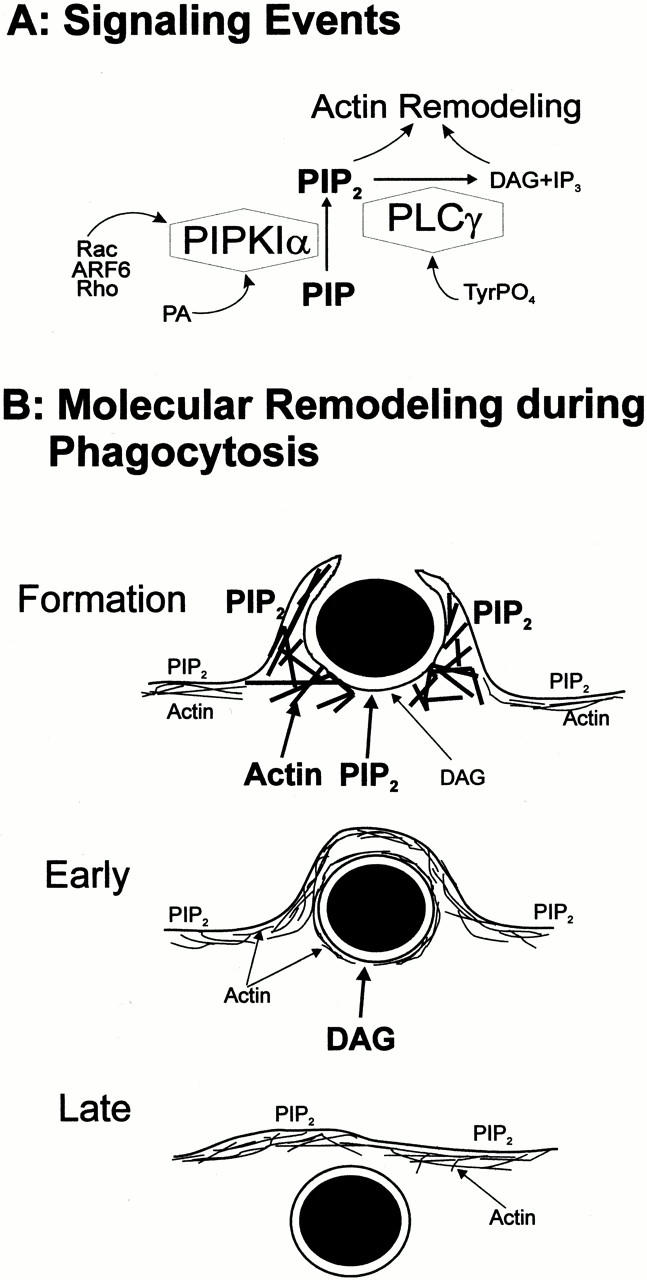
“These observations suggest that DAG is locally synthesized by PLC-mediated hydrolysis of 4,5-PIP2 during phagosome formation and is subsequently converted in the sealed phagosome to other metabolites.”
§ Localized Biphasic Changes in Phosphatidylinositol-4,5-Bisphosphate at Sites of Phagocytosis > Results > Localized Generation of Diacylglycerol during Phagocytosis
hedgeddirect- Asserted: passes — The authors put it forward as true.
- Checkable: passes — It can be checked against evidence.
- Load-bearing: passes — The paper’s conclusions depend on it.
- Based: passes — The paper gives a reason why it holds.
It may state two separate claims as one.
A conclusion the paper draws from its own results — it cites no other source for it, so there is none to check. The quote shows it is in the paper.
In the paper
“We conclude that inhibition of PLC arrests phagocytosis at an early stage, and is required for actin remodeling during pseudopod formation.”
§ Localized Biphasic Changes in Phosphatidylinositol-4,5-Bisphosphate at Sites of Phagocytosis > Results > Inactivation of PLC Abolishes Pseudopod and Actin Cup Formation
plaindirect- Asserted: passes — The authors put it forward as true.
- Checkable: passes — It can be checked against evidence.
- Load-bearing: passes — The paper’s conclusions depend on it.
- Based: passes — The paper gives a reason why it holds.
It may state two separate claims as one.
A conclusion the paper draws from its own results — it cites no other source for it, so there is none to check. The quote shows it is in the paper.
In the paper
“This behavior contrasted strikingly with that of PM-GFP, which remained associated with phagosomes for the duration of our analysis (Fig. 3), ruling out rapid membrane turnover as the process responsible for the loss of PLCδ-PH-GFP.”
§ Localized Biphasic Changes in Phosphatidylinositol-4,5-Bisphosphate at Sites of Phagocytosis > Discussion > Biphasic Changes in 4,5-PIP2 during Phagocytosis
plaindirect- Asserted: passes — The authors put it forward as true.
- Checkable: passes — It can be checked against evidence.
- Load-bearing: passes — The paper’s conclusions depend on it.
- Based: passes — The paper gives a reason why it holds.
It may state two separate claims as one.
A conclusion the paper draws from its own results — it cites no other source for it, so there is none to check. The quote shows it is in the paper.
In the paper
“The concomitant appearance of DAG argues in favor of a central role of PLC in the depletion of 4,5-PIP2 from phagosomes (Fig. 6 D).”
§ Localized Biphasic Changes in Phosphatidylinositol-4,5-Bisphosphate at Sites of Phagocytosis > Discussion > Biphasic Changes in 4,5-PIP2 during Phagocytosis
hedgeddirect- Asserted: passes — The authors put it forward as true.
- Checkable: passes — It can be checked against evidence.
- Load-bearing: passes — The paper’s conclusions depend on it.
- Based: passes — The paper gives a reason why it holds.
It may state two separate claims as one. The quote only partly states this claim.
A conclusion the paper draws from its own results — it cites no other source for it, so there is none to check. The quote shows it is in the paper.
In the paper
“Instead, we attribute the concentration of PLCδ-PH-GFP in the phagocytic cups to increased local synthesis of 4,5-PIP2. In support of this conclusion, we noticed local recruitment of PIPKIα to phagocytic cups (Fig. 4).”
§ Localized Biphasic Changes in Phosphatidylinositol-4,5-Bisphosphate at Sites of Phagocytosis > Discussion > Biphasic Changes in 4,5-PIP2 during Phagocytosis
plaindirect- Asserted: passes — The authors put it forward as true.
- Checkable: passes — It can be checked against evidence.
- Load-bearing: passes — The paper’s conclusions depend on it.
- Based: passes — The paper gives a reason why it holds.
It may state two separate claims as one.
A conclusion the paper draws from its own results — it cites no other source for it, so there is none to check. The quote shows it is in the paper.
In the paper
“A similar mechanism is likely involved during phagocytosis since, when transfected into RAW cells, the dual SH2 domains of PLCγ1 fused to GFP (PLCγ-SH2-GFP) were often seen to concentrate around phagocytic cups (Fig. 5C and Fig. D).”
§ Localized Biphasic Changes in Phosphatidylinositol-4,5-Bisphosphate at Sites of Phagocytosis > Results > PLCγ Localizes to Phagocytic Cups
hedgedconditional- Asserted: passes — The authors put it forward as true.
- Checkable: passes — It can be checked against evidence.
- Load-bearing: passes — The paper’s conclusions depend on it.
- Based: passes — The paper gives a reason why it holds.
It may state two separate claims as one.
What the paper did — it cites no source for this, so there is none to check. The quote shows it is in the paper.
In the paper
“The inability of cells to form pseudopods and actin cups upon incubation with U73122 and ET-18-OCH3 is not the result of a detrimental effect of the drug on cellular viability. These effects were fully reversible. Moreover, in the presence of the drug, pinocytosis of FITC-dextran appeared to be unimpaired (data not shown).”
§ Localized Biphasic Changes in Phosphatidylinositol-4,5-Bisphosphate at Sites of Phagocytosis > Results > Inactivation of PLC Abolishes Pseudopod and Actin Cup Formation
hedged- Asserted: passes — The authors put it forward as true.
- Checkable: passes — It can be checked against evidence.
- Load-bearing: passes — The paper’s conclusions depend on it.
- Based: passes — The paper gives a reason why it holds.
It may state two separate claims as one.
In the paper
“The latter results, at least in part, from activation of PLC, although a contribution of phospholipase D and phosphatidate phosphohydrolase is also possible (Kusner et al. 1999).”
§ Localized Biphasic Changes in Phosphatidylinositol-4,5-Bisphosphate at Sites of Phagocytosis > Discussion > Biphasic Changes in 4,5-PIP2 during Phagocytosis
What the sources say
- Not checked — no full text of the sourcecites Kusner et al. 1999 source
hedgedconditional- Asserted: passes — The authors put it forward as true.
- Checkable: passes — It can be checked against evidence.
- Load-bearing: passes — The paper’s conclusions depend on it.
- Based: passes — The paper gives a reason why it holds.
It may state two separate claims as one.
In the paper
“Accordingly, addition of exogenous DAG or its analogues augments phagocytosis (Karimi et al. 1999), an effect that has been attributed to PKC isoforms.”
§ Localized Biphasic Changes in Phosphatidylinositol-4,5-Bisphosphate at Sites of Phagocytosis > Discussion > Localized Phosphoinositide Metabolism Is Required for Phagocytosis
What the sources say
- Not checked — no full text of the sourcecites Karimi et al. 1999 source
plaindirect- Asserted: passes — The authors put it forward as true.
- Checkable: passes — It can be checked against evidence.
- Load-bearing: unclear — It is unclear whether the paper’s conclusions depend on it.
- Based: passes — The paper gives a reason why it holds.
It may state two separate claims as one.
A claim is checked only against the cited paper’s full text, never its abstract, and none is available for its sources.
In the paper
“This phospholipid is a regulator of actin capping, nucleation of de novo F-actin, and anchoring, cross-linking, and severing proteins (Hinchliffe et al. 1998), and actin remodeling is essential to the phagocytic process (Kwiatkowska and Sobota 1999).”
§ Localized Biphasic Changes in Phosphatidylinositol-4,5-Bisphosphate at Sites of Phagocytosis > Discussion > Localized Phosphoinositide Metabolism Is Required for Phagocytosis
What the sources say
plaindirect- Asserted: passes — The authors put it forward as true.
- Checkable: passes — It can be checked against evidence.
- Load-bearing: passes — The paper’s conclusions depend on it.
- Based: passes — The paper gives a reason why it holds.
It may state two separate claims as one.
A conclusion the paper draws from its own results — it cites no other source for it, so there is none to check. The quote shows it is in the paper.
In the paper
“Thus we tentatively postulate that local accumulation of 4,5-PIP2 might promote the recruitment of cytoskeletal proteins, stimulating F-actin anchorage and remodeling and contributing to the formation of the phagocytic cup (Fig. 10).”
§ Localized Biphasic Changes in Phosphatidylinositol-4,5-Bisphosphate at Sites of Phagocytosis > Discussion > Localized Phosphoinositide Metabolism Is Required for Phagocytosis
hedgedconditional- Asserted: passes — The authors put it forward as true.
- Checkable: passes — It can be checked against evidence.
- Load-bearing: passes — The paper’s conclusions depend on it.
- Based: passes — The paper gives a reason why it holds.
It may state two separate claims as one.
The paper relies on this without support.
At 15°C, vesicular traffic essentially ceases in mammalian cells.
“since at this temperature vesicular traffic essentially ceases in mammalian cells.”
The paper relies on this without support.
Neomycin sulfate can compete with endogenous ligands for binding to 4,5-PIP2.
“Neomycin sulfate is a somewhat permeant divalent cation that can compete with endogenous ligands for binding to 4,5-PIP2.”
The paper relies on this without support.
In resting cells, the C1 domain of PKCγ is almost exclusively in the cytosol.
“In resting cells this domain is almost exclusively in the cytosol, ruling out translocation by vesicular fusion.”
The paper relies on this without support.
4,5-PIP2 exists in the micromolar range in cells.
“which can be estimated to exist in the micromolar range, based on existing determinations of phospholipid content in several cell types.”
Citations
Cites
Daëron, M. (1997) · Annual Review of Immunology · doi:10.1146/annurev.immunol.15.1.203 · Cited by 1,227 · cited 2×
“These include the Fc g receptors (Fc g Rs), 1 that bind specifically to the IgG found on antibody-coated particles (Daeron, 1997).”
BackgroundIdentification of two distinct mechanisms of phagocytosis controlled by different Rho GTPases
Caron, E., & Hall, A. (1998) · Science · doi:10.1126/science.282.5394.1717 · Cited by 1,019 · cited 6×
“Experiments using clostridial toxins or transfection of dominant-negative constructs have convincingly demonstrated that actin assembly during phagocytosis is mediated by Rho family GTPases (Cox et al., 1997; Hackam et al., 1997; Caron and Hall, 1998; Massol et al., 1998).”
BackgroundInfluentialAraki, N., Johnson, M. T., & Swanson, J. A. (1996) · The Journal of cell biology · doi:10.1083/jcb.135.5.1249 · Cited by 949 · cited 11×
“This kinase appears to be essential for phagocytosis since inhibitors such as wortmannin impair particle engulfment (Ninomiya et al., 1994; Araki et al., 1996; Cox et al., 1999).”
BackgroundInfluentialRegulation of Phosphoinositide-specific Phospholipase C Isozymes
Rhee, S. G., & Bae, Y. S. (1997) · Journal of Biological Chemistry · doi:10.1074/jbc.272.24.15045 · Cited by 876 · cited 1×
“PLCg is known to be recruited to the vicinity of activated receptors through interaction of its dual SH2 domains with phosphotyrosine residues on receptor–adaptor complexes (Rhee and Bae, 1997).”
BackgroundVárnai, P., & Balla, T. (1998) · The Journal of Cell Biology · doi:10.1083/jcb.143.2.501 · Cited by 859 · cited 6×
“As reported for other cell types (Stauffer et al., 1998; Varnai and Balla, 1998), PLC d - PH-GFP associated predominantly with the plasmalemma of RAW cells, with little binding to organellar membranes (Fig.”
BackgroundMethodInfluentialHonda, A., Nogami, M., Yokozeki, T., Yamazaki, M., Nakamura, H., Watanabe, H., Kawamoto, K., Nakayama, K., Morris, A. J., Frohman, M. A., et al. (1999) · Cell · doi:10.1016/s0092-8674(00)81540-8 · Cited by 798
Stauffer, T. P., Ahn, S., & Meyer, T. (1998) · Current Biology · doi:10.1016/s0960-9822(98)70135-6 · Cited by 761 · cited 7×
“Key words: Fc g receptors • phospholipase C • PH domain • diacylglycerol • phosphatidylinositol”
BackgroundMethodInfluentialSelf-citationRaucher, D., Stauffer, T., Chen, W., Shen, K., Guo, S., York, J. D., Sheetz, M. P., & Meyer, T. (2000) · Cell · doi:10.1016/s0092-8674(00)81560-3 · Cited by 675 · cited 8×
“The mammalian expression vectors pEGFP::AktPH, pEGFP::PLC d PH, pEGFP::PLC g SH2, and pEGFP::C1 d encode the PH domain of Akt (Kontos et al., 1998), the PH domain of PLC d (Stauffer et al., 1998), the dual SH2 domains of PLC g 1 (Stauffer and Meyer, 1997), and the C1 domain of PKC d , respectively, fused to EGFP. pCFP…”
BackgroundInfluentialSelf-citationChong, L. D., Traynor‐Kaplan, A., Bokoch, G., & Schwartz, M. A. (1994) · Cell · doi:10.1016/0092-8674(94)90259-3 · Cited by 641
Protein Kinase C as a Molecular Machine for Decoding Calcium and Diacylglycerol Signals
Oancea, E., & Meyer, T. (1998) · Cell · doi:10.1016/s0092-8674(00)81763-8 · Cited by 640
Self-citation
Cited by
Sorted by citations
Phosphoinositides: Tiny Lipids With Giant Impact on Cell Regulation
Balla, T. (2013) · Physiological Reviews · doi:10.1152/physrev.00028.2012 · Cited by 1,638
“Hydrolysis of PtdIns(4,5)P2 by PLC is necessary for completion of phagosome formation (172, 1370).”
BackgroundInfluentialInnate Immune Pattern Recognition: A Cell Biological Perspective
Brubaker, S. W., Bonham, K. S., Zanoni, I., & Kagan, J. C. (2015) · Annual Review of Immunology · doi:10.1146/annurev-immunol-032414-112240 · Cited by 1,475
“Additionally, Dectin-1 activates PLCγ2, which is a regulator of phagocytosis and may contribute to the Dectin-1-mediated phagocytic process (130, 138, 139).”
The State of Lipid Rafts: From Model Membranes to Cells
Edidin, M. A. (2003) · Annual Review of Biophysics and Biomolecular Structure · doi:10.1146/annurev.biophys.32.110601.142439 · Cited by 1,357
The emerging field of lipidomics
Wenk, M. R. (2005) · Nature Reviews Drug Discovery · doi:10.1038/nrd1776 · Cited by 1,362
“In particular phosphoinositide and cell-signalling research has profited from the many available phosphoinositide-binding protein modules (for example, PH, PX, FYVE, epsin N-terminal homology (ENTH) domains and so on), which enable studies of the kinetics of lipid metabolism in living cell”
BackgroundTRAM couples endocytosis of Toll-like receptor 4 to the induction of interferon-β
Kagan, J. C., Su, T., Horng, T., Chow, A. Y., Akira, S., & Medzhitov, R. M. (2008) · Nature Immunology · doi:10.1038/ni1569 · Cited by 1,310
The Cell Biology of Phagocytosis
Flannagan, R. S., Jaumouillé, V., & Grinstein, S. (2011) · Annual Review of Pathology Mechanisms of Disease · doi:10.1146/annurev-pathol-011811-132445 · Cited by 1,096
Self-citationShaping cups into phagosomes and macropinosomes
Swanson, J. A. (2008) · Nature Reviews Molecular Cell Biology · doi:10.1038/nrm2447 · Cited by 941
CD14 Controls the LPS-Induced Endocytosis of Toll-like Receptor 4
Zanoni, I., Ostuni, R., Marek, L. R., Barresi, S., Barbalat, R., Barton, G. M., Granucci, F., & Kagan, J. C. (2011) · Cell · doi:10.1016/j.cell.2011.09.051 · Cited by 997
“PLCg2 is a regulator of endocytosis (Botelho et al., 2000; Depoil et al., 2009) and is thus a candidate regulator of TLR4 endocytosis.”
BackgroundPlasma membrane phosphoinositide organization by protein electrostatics
McLaughlin, S., & Murray, D. (2005) · Nature · doi:10.1038/nature04398 · Cited by 868
Phagocytosis of Microbes: Complexity in Action
Underhill, D. M., & Ozinsky, A. (2002) · Annual Review of Immunology · doi:10.1146/annurev.immunol.20.103001.114744 · Cited by 1,124
PIP2and Proteins: Interactions, Organization, and Information Flow
McLaughlin, S., Wang, J., Gambhir, A., & Murray, D. (2002) · Annual Review of Biophysics and Biomolecular Structure · doi:10.1146/annurev.biophys.31.082901.134259 · Cited by 837
“(20) discuss the (still imperfectly understood) roles that PIP 2,”
BackgroundPhagocytosis and the actin cytoskeleton
May, R. C., & Machesky, L. M. (2001) · Journal of Cell Science · doi:10.1242/jcs.114.6.1061 · Cited by 715
Endoplasmic Reticulum-Mediated Phagocytosis Is a Mechanism of Entry into Macrophages
Gagnon, É., Duclos, S., Rondeau, C., Chevet, É., Cameron, P. H., Steele‐Mortimer, O., Paiement, J. M., Bergeron, J. J. M., & Desjardins, M. (2002) · Cell · doi:10.1016/s0092-8674(02)00797-3 · Cited by 683
Phagocytosis: Our Current Understanding of a Universal Biological Process
Uribe-Querol, E., & Rosales, C. (2020) · Frontiers in Immunology · doi:10.3389/fimmu.2020.01066 · Cited by 651
“Reduction of PI(4,5)P 2 in the membrane is also mediated by the action of PLC γ , which produces diacylglycerol (DAG) (91).”
BackgroundInfluentialPhosphoinositide Regulation of the Actin Cytoskeleton
Yin, H. L., & Janmey, P. A. (2003) · Annual Review of Physiology · doi:10.1146/annurev.physiol.65.092101.142517 · Cited by 672
The PX domains of p47phox and p40phox bind to lipid products of PI(3)K
Kanai, F., Liu, H., Field, S. J., Akbary, H., Matsuo, T., Brown, G. E., Cantley, L. C., & Yaffe, M. B. (2001) · Nature Cell Biology · doi:10.1038/35083070 · Cited by 611
The role of IgG Fc receptors in antibody-dependent enhancement
Bournazos, S., Gupta, A., & Ravetch, J. V. (2020) · Nature reviews. Immunology · doi:10.1038/s41577-020-00410-0 · Cited by 619
Barreiro, O., Yáñez‐Mó, M., Serrador, J. M., Montoya, M. C., Vicente‐Manzanares, M., Tejedor, R., Furthmayr, H., & Sánchez‐Madrid, F. (2002) · The Journal of Cell Biology · doi:10.1083/jcb.200112126 · Cited by 594
“Interestingly, these regulatory molecules are also important in phagocytosis (Botelho et al., 2000; Chimini and Chavrier, 2000).”
BackgroundPhospholipid-Based Signaling in Plants
Meijer, H. J. G., & Munnik, T. (2003) · Annual Review of Plant Biology · doi:10.1146/annurev.arplant.54.031902.134748 · Cited by 588
Phagocytosis and innate immunity
Greenberg, S., & Grinstein, S. (2002) · Current Opinion in Immunology · doi:10.1016/s0952-7915(01)00309-0 · Cited by 581
Self-citationPhagocytosis: A Fundamental Process in Immunity
Rosales, C., & Uribe‐Querol, E. (2017) · BioMed Research International · doi:10.1155/2017/9042851 · Cited by 618
“PLC𝛾 is phosphorylated and recruited to the phagocytic cup in a Sykdependent manner, probably by interaction with LAT [83, 85].”
BackgroundInfluentialTsai, R. K., & Discher, D. E. (2008) · The Journal of Cell Biology · doi:10.1083/jcb.200708043 · Cited by 486
“…human CD47, human macrophages might be expected to effi ciently phagocytose opsonized sheep red blood cells (RBCs), which is certainly consistent with years of data showing sheep RBCs are readily phagocytosed by human macrophages ( Lowry et al., 1998 ; Botelho et al., 2000 ; Cooney et al., 2001 ).”
BackgroundPhosphoinositides as membrane organizers
Posor, Y., Jang, W., & Haucke, V. (2022) · Nature Reviews Molecular Cell Biology · doi:10.1038/s41580-022-00490-x · Cited by 469
Regulation of the Actin Cytoskeleton-Plasma Membrane Interplay by Phosphoinositides
Saarikangas, J., Zhao, H., & Lappalainen, P. (2010) · Physiological Reviews · doi:10.1152/physrev.00036.2009 · Cited by 515
“PI(4,5)P2 accumulates to early phagosomes via local activation of PIPK-I and plays a crucial role in actin remodeling through recruitment/activation of actin-binding proteins at phagosome membrane (26, 58).”
BackgroundThe role of phosphoinositides in membrane transport
Simonsen, A., Wurmser, A. E., Emr, S. D., & Stenmark, H. (2001) · Current Opinion in Cell Biology · doi:10.1016/s0955-0674(00)00240-4 · Cited by 407
The Complex and Intriguing Lives of PIP2 with Ion Channels and Transporters
Hilgemann, D. W., Feng, S., & Nasuhoglu, C. (2001) · Science Signaling · doi:10.1126/scisignal.1112001re19 · Cited by 463
The Complex and Intriguing Lives of PIP 2 with Ion Channels and Transporters
Hilgemann, D. W., Feng, S., & Nasuhoglu, C. (2001) · Science s STKE · doi:10.1126/stke.2001.111.re19 · Cited by 453
CELL BIOLOGY OF MYCOBACTERIUM TUBERCULOSIS PHAGOSOME
Vergne, I., Chua, J., Singh, S., & Deretic, V. P. (2004) · Annual Review of Cell and Developmental Biology · doi:10.1146/annurev.cellbio.20.010403.114015 · Cited by 440
Phosphatidylinositol 4,5-bisphosphate and Arf6-regulated membrane traffic
Brown, F. D., Rozelle, A. L., Yin, H. L., Balla, T., & Donaldson, J. G. (2001) · The Journal of Cell Biology · doi:10.1083/jcb.200103107 · Cited by 435
“However, low level expression of the chimera as used here is a useful indicator of PIP2 distribution (Botelho et al., 2000; Tall et al., 2000).”
BackgroundLee, W. L., Harrison, R. E., & Grinstein, S. (2003) · Microbes and Infection · doi:10.1016/j.micinf.2003.09.014 · Cited by 433
Self-citationContinuous T cell receptor signaling required for synapse maintenance and full effector potential
Huppa, J. B., Gleimer, M., Sumen, C., & Davis, M. M. (2003) · Nature Immunology · doi:10.1038/ni951 · Cited by 393
Fc-receptor-mediated phagocytosis is regulated by mechanical properties of the target
Beningo, K. A., & Wang, Y.-L. (2002) · Journal of Cell Science · doi:10.1242/jcs.115.4.849 · Cited by 389
“Components of all three pathways have been localized to the phagocytic cup (Dharmawardhane et al., 1999; May et al., 2000; Botelho et al., 2000).”
BackgroundPI(4,5)P2 regulation of surface membrane traffic
Martin, T. F. J. (2001) · Current Opinion in Cell Biology · doi:10.1016/s0955-0674(00)00241-6 · Cited by 381
Cdc42, Rac1, and Rac2 Display Distinct Patterns of Activation during Phagocytosis
Hoppe, A. D., & Swanson, J. A. (2004) · Molecular Biology of the Cell · doi:10.1091/mbc.e03-11-0847 · Cited by 365
“Rac1 also could regulate actin polymerization without the aid of Cdc42, through activation of phosphatidylinositol-4-phosphate 5-kinase (Bishop and Hall, 2000; Botelho et al., 2000).”
BackgroundWatt, S. A., Kular, G. S., Fleming, I. N., Downes, C. P., & Lucocq, J. M. (2002) · Biochemical Journal · doi:10.1042/0264-6021:3630657 · Cited by 353
Matteis, M. A. D., & Godi, A. (2004) · Nature Cell Biology · doi:10.1038/ncb0604-487 · Cited by 351
“; phosphatidylinositol-3,4,5-trisphosphate (PtdIns(3,4,5)P 3 ) and PtdIns(4,5)P 2 that are found at sites of phagocytosi”
BackgroundThe coordination of signaling during Fc receptor-mediated phagocytosis
Swanson, J. A., & Hoppe, A. D. (2004) · Journal of Leukocyte Biology · doi:10.1189/jlb.0804439 · Cited by 339
“Although little is known about the regulation of PI4P5K activity during phagocytosis, it has been shown in separate systems that Rac can stimulate PI4P5K activity [57].”
BackgroundInfluentialPhagosome dynamics during phagocytosis by neutrophils
Nordenfelt, P., & Tapper, H. (2011) · Journal of Leukocyte Biology · doi:10.1189/jlb.0810457 · Cited by 350
“They have a central role in signal transduction during phagocytosis [147], where PI(4,5)P2 is present at the phagocytic cup, is enriched during phagosome formation, and disappears during sealing [148].”
BackgroundCross-talk unfolded: MARCKS proteins
Arbuzova, A., Schmitz, A. A. P., & Vergères, G. (2002) · Biochemical Journal · doi:10.1042/0264-6021:3620001 · Cited by 323
The life cycle of phagosomes: formation, maturation, and resolution
Levin‐Konigsberg, R., Grinstein, S., & Canton, J. (2016) · Immunological Reviews · doi:10.1111/imr.12439 · Cited by 328
“One of the first major lipid-remodeling events to occur during phagocytic receptor signaling is a local increase in phosphatidylinositol 4,5-bisphosphate [PtdIns(4,5)P2] levels (108).”
BackgroundSelf-citationActin Depletion Initiates Events Leading to Granule Secretion at the Immunological Synapse
Ritter, A. T., Asano, Y., Stinchcombe, J. C., et al. (2015) · Immunity · doi:10.1016/j.immuni.2015.04.013 · Cited by 315
“Given that phosphatidyl inositide-4,5-bisphosphate (PIP2) plays a role in recruitment of cortical actin to membranes (Botelho et al., 2000; Janmey and Lindberg, 2004; Raucher et al., 2000), we examined PIP2 localization across the synapse when cortical actin levels were depleted (Figure 7C).”
BackgroundElectrostatic Sequestration of PIP2 on Phospholipid Membranes by Basic/Aromatic Regions of Proteins
Gambhir, A., Hangyás-Mihályné, G., Zaitseva, I., Cafiso, D. S., Wang, J., Murray, D., Pentyala, S., Smith, S. O., & McLaughlin, S. (2004) · Biophysical Journal · doi:10.1016/s0006-3495(04)74278-2 · Cited by 318
“It is based on experiments that use a fluorescent construct of the PH domain of PLC-d1 to detect PIP2 (Botelho et al., 2000; Tall et al., 2000).”
BackgroundInfluentialRole of Phospholipids in Endocytosis, Phagocytosis, and Macropinocytosis
Bohdanowicz, M., & Grinstein, S. (2013) · Physiological Reviews · doi:10.1152/physrev.00002.2012 · Cited by 326
Self-citationLocalisation and trafficking of Toll-like receptors: an important mode of regulation
McGettrick, A. F., & O'Neill, L. A. J. (2010) · Current Opinion in Immunology · doi:10.1016/j.coi.2009.12.002 · Cited by 316
Weißmann, N., Dietrich, A., Fuchs, B., Kalwa, H., Ay, M., Dumitrascu, R., Olschewski, A., Storch, U., y Schnitzler, M. M., Ghofrani, H. A., et al. (2006) · Proceedings of the National Academy of Sciences · doi:10.1073/pnas.0606728103 · Cited by 304
Phosphoinositides in Control of Membrane Dynamics
Schink, K. O., Tan, K.-W., & Stenmark, H. (2016) · Annual Review of Cell and Developmental Biology · doi:10.1146/annurev-cellbio-111315-125349 · Cited by 283
Marshall, J. G., Booth, J. W., Stambolic, V., Mak, T. W., Balla, T., Schreiber, A. D., Meyer, T., & Grinstein, S. (2001) · The Journal of Cell Biology · doi:10.1083/jcb.153.7.1369 · Cited by 281
“As reported previously (Botelho et al., 2000), PI(4,5)P 2 undergoes a biphasic change: a small transient increase followed immediately by its virtual disappearance from the phagosomal membrane.”
BackgroundInfluentialSelf-citationSheetz, M. P., Sable, J. E., & Döbereiner, J. (2006) · Annual Review of Biophysics and Biomolecular Structure · doi:10.1146/annurev.biophys.35.040405.102017 · Cited by 286
Terebiznik, M. R., Vieira, O. V., Marcus, S. L., Slade, A. L., Yip, C. M., Trimble, W. S., Meyer, T., Finlay, B. B., & Grinstein, S. (2002) · Nature Cell Biology · doi:10.1038/ncb854 · Cited by 278
“The C1 domain, which associates with juxtanuclear membranes in resting cells, translocated to the plasma membrane on addition of exogenous diacylglycerol (data not shown; see ref”
BackgroundSelf-citationMembrane ruffling and macropinocytosis in A431 cells require cholesterol
Grimmer, S., van Deurs, B., & Sandvig, K. (2002) · Journal of Cell Science · doi:10.1242/jcs.115.14.2953 · Cited by 278
Barnett, K. C., Coronas-Serna, J. M., Zhou, W., Ernandes, M. J., Cao, A., Kranzusch, P. J., & Kagan, J. C. (2019) · Cell · doi:10.1016/j.cell.2019.01.049 · Cited by 275
Phosphatidylinositol Phosphate Kinases Put PI4,5P 2 in Its Place
Doughman, R. L., Firestone, A. J., & Anderson, R. A. (2003) · The Journal of Membrane Biology · doi:10.1007/s00232-003-2027-7 · Cited by 258
“However, the PH-PLCd antibody only detects PI4,5P2 changes on the plasma membrane, suggesting a bias towards PI4,5P2 detection using this probe.”
BackgroundInfluentialSelf-citationPhosphatidylinositol-4,5- bis phosphate hydrolysis directs actin remodeling during phagocytosis
Scott, C. C., Dobson, W., Botelho, R. J., Coady-Osberg, N., Chavrier, P., Knecht, D. A., Heath, C. M., Stahl, P. D., & Grinstein, S. (2005) · The Journal of Cell Biology · doi:10.1083/jcb.200412162 · Cited by 252
“A chimeric construct consisting of GFP attached to the PH domain of PLC (PH PLC -GFP; Stauffer et al., 1998; Varnai and Balla, 1998; Botelho et al., 2000) was used to monitor PI(4,5)P 2 in live macrophages during the course of particle ingestion.”
BackgroundMethodInfluentialSelf-citationSignal transduction during Fc receptor-mediated phagocytosis
García-García, E., & Rosales, C. (2002) · Journal of Leukocyte Biology · doi:10.1189/jlb.72.6.1092 · Cited by 249
“vation of ERK has been described in monocytes [61, 79, 105],”
BackgroundInfluentialRougerie, P., Miskolci, V., & Cox, D. (2013) · Immunological Reviews · doi:10.1111/imr.12118 · Cited by 237
“PI(4,5)P2 is generated upon FccR binding by PIPKIa (51), and the inhibition of PIPKIa decreases PI(4,5)P2 accumulation.”
BackgroundInfluentialLocation, Location, Location: Membrane Targeting Directed by PX Domains
Sato, T. K., Overduin, M., & Emr, S. D. (2001) · Science · doi:10.1126/science.1065763 · Cited by 236
Stefan, C. J., Audhya, A., & Emr, S. D. (2002) · Molecular Biology of the Cell · doi:10.1091/mbc.01-10-0476 · Cited by 235
“The PH domain of phospholipase C 1 (PLC) binds phosphatidylinositol (4,5)-bisphosphate (PtdIns(4,5)-P2) with high specificity and is efficiently recruited to the plasma membrane (Kavran et al., 1998; Stauffer et al., 1998; Honda et al., 1999; Botelho et al., 2000 ).”
BackgroundA polarized Ca2+, diacylglycerol and STIM1 signalling system regulates directed cell migration
Tsai, F., Seki, A., Yang, H. W., Hayer, A., Carrasco, S., Malmersjö, S., & Meyer, T. (2014) · Nature Cell Biology · doi:10.1038/ncb2906 · Cited by 241
Self-citationMasters, T. A., Pontes, B., Viasnoff, V., Li, Y., & Gauthier, N. C. (2013) · Proceedings of the National Academy of Sciences · doi:10.1073/pnas.1301766110 · Cited by 228
“Conclusion During phagocytosis, particularly of large particles, cortical tension (34), contractility (35), and phosphoinositide signaling (36) have been reported to display biphasic behavior.”
BackgroundMilošević, I., Sørensen, J. B., Lang, T., Krauss, M. I., Nagy, G., Haucke, V., Jahn, R., & Neher, E. (2005) · Journal of Neuroscience · doi:10.1523/jneurosci.3761-04.2005 · Cited by 231
“Some later studies that revealed local accumulations in plasma membrane “ruffles” (Honda et al., 1999; Botelho et al., 2000; Tall et al., 2000) were recently reinterpreted after the demonstration of colocalization with lipophilic membrane dyes, implying that the apparent PI(4,5)P2 enrichments might have been local mem…”
BackgroundFreeman, S. A., Vega, A. R., Riedl, M., Collins, R. F., Ostrowski, P. P., Woods, E. C., Bertozzi, C. R., Tammi, M., Lidke, D. S., Johnson, P., et al. (2018) · Cell · doi:10.1016/j.cell.2017.12.023 · Cited by 237
Self-citationER-mediated phagocytosis: a new membrane for new functions
Desjardins, M. (2003) · Nature reviews. Immunology · doi:10.1038/nri1053 · Cited by 230
“In addition to its role in early phagocytosis, PI3K seems to be important in phagosome maturatio”
BackgroundAntibody-Dependent Cellular Phagocytosis in Antiviral Immune Responses
Tay, M. Z., Wiehe, K. J., & Pollara, J. (2019) · Frontiers in Immunology · doi:10.3389/fimmu.2019.00332 · Cited by 242
“In Fc receptor-mediated phagocytosis, ligated and aggregated Fc receptor become phosphorylated via Src family tyrosine kinases (54) on their ITAM domains (either their own or from an associated γ-subunit), forming a docking site for Syk (55) and triggering a signaling cascade involving PKC (56, 57), PI3K (58–60), and…”
BackgroundMolecular mechanisms of phagocytic uptake in mammalian cells
Groves, E., Dart, A., Covarelli, V., & Caron, E. (2008) · Cellular and Molecular Life Sciences · doi:10.1007/s00018-008-7578-4 · Cited by 238
Aikawa, Y., & Martin, T. F. J. (2003) · The Journal of Cell Biology · doi:10.1083/jcb.200212142 · Cited by 226
“, 2002), phagocytosis (Botelho et al., 2000), pinocytosis (Schafer et al.”
Bonham, K. S., Orzalli, M. H., Hayashi, K., Wolf, A. I., Glanemann, C., Weninger, W., Iwasaki, A., Knipe, D. M., & Kagan, J. C. (2014) · Cell · doi:10.1016/j.cell.2014.01.019 · Cited by 232
“We transfected these cells with plasmids encoding the GFP-tagged lipid-binding domain of TIRAP (TIRAP-loc) along with a plasmid encoding a cherrytagged pleckstrin homology (PH) domain from PLCd1 (PLC), which binds to PI(4,5)P2 uniquely (Botelho et al., 2000).”
MethodFratti, R. A., Jun, Y., Merz, A. J., Margolis, N., & Wickner, W. T. (2004) · The Journal of Cell Biology · doi:10.1083/jcb.200409068 · Cited by 217
“Isogenic strains containing the ACT1 wild type and act1-113 mutant alleles were a gift from D. Drubin (University of California at Berkeley, Berkeley, CA; Ayscough, 2000).”
BackgroundMethodInfluentialWang, J., Gambhir, A., Hangyás-Mihályneá, G. ̈., Murray, D., Golebiewska, U. P., & McLaughlin, S. (2002) · Journal of Biological Chemistry · doi:10.1074/jbc.m203954200 · Cited by 220
Characterization of the megakaryocyte demarcation membrane system and its role in thrombopoiesis
Schulze, H., Korpal, M., Hurov, J., Kim, S.-W., Zhang, J., Cantley, L. C., Graf, T., & Shivdasani, R. A. (2006) · Blood · doi:10.1182/blood-2005-07-2755 · Cited by 211
Integration of Phosphoinositide- and Calmodulin-Mediated Regulation of TRPC6
Kwon, Y. V., Hofmann, T., & Montell, C. (2007) · Molecular Cell · doi:10.1016/j.molcel.2007.01.021 · Cited by 206
“Detection of AKT-PH-GFP AKT-PH-GFP (Botelho et al., 2000) and AKT-PHR25C-GFP (Franke et al., 1997) were transfected together with the H1 receptor in HEK293T cells.”
BackgroundInfluentialA Polybasic Motif Allows N-WASP to Act as a Sensor of PIP2 Density
Papayannopoulos, V., Co, C., Prehoda, K. E., Snapper, S. B., Taunton, J., & Lim, W. A. (2005) · Molecular Cell · doi:10.1016/j.molcel.2004.11.054 · Cited by 205
Cebecauer, M., Amaro, M., Jurkiewicz, P., Sarmento, M. J., Šachl, R., Cwiklik, L., & Hof, M. (2018) · Chemical Reviews · doi:10.1021/acs.chemrev.8b00322 · Cited by 199
Understanding early TLR signaling through the Myddosome
Balka, K. R., & Nardo, D. D. (2018) · Journal of Leukocyte Biology · doi:10.1002/jlb.mr0318-096r · Cited by 201
Phosphatidylinositol‐(4,5)‐bisphosphate enables efficient secretion of HIV‐1 Tat by infected T‐cells
Rayne, F., Debaisieux, S., Yezid, H., Lin, Y., Mettling, C., Konate, K., Chazal, N., Arold, S. T., Pugnière, M., Sanchez, F., et al. (2010) · The EMBO Journal · doi:10.1038/emboj.2010.32 · Cited by 201
“Indeed, overexpression of PHPLCd prevents the PI(4,5)P2-mediated recruitment at the plasma membrane of cell machineries that enable clathrin-mediated endocytosis (Jost et al, 1998), phagocytosis (Botelho et al, 2000) or exocytosis (Holz et al, 2000) and inhibits these processes.”
BackgroundAraki, N., Hatae, T., Furukawa, A., & Swanson, J. A. (2002) · Journal of Cell Science · doi:10.1242/jcs.00235 · Cited by 190
“…induce F-actin polymerization and reorganization (Cox et al., 1997; Massol et al., 1998; Castellano et al., 2000; Chimini and Chavrier, 2000), changes which are also regulated by phosphoinositides such as PI(4,5)P2 and PI(3,4,5)P3 (Hackam et al., 1997; Botelho et al., 2000; Marshall et al., 2001).”
BackgroundIntegrins Form an Expanding Diffusional Barrier that Coordinates Phagocytosis
Freeman, S. A., Goyette, J., Furuya, W., Woods, E. C., Bertozzi, C. R., Bergmeier, W., Hinz, B., van der Merwe, P. A., Das, R., & Grinstein, S. (2016) · Cell · doi:10.1016/j.cell.2015.11.048 · Cited by 192
Self-citationActivation of TRPC6 channels is essential for lung ischaemia–reperfusion induced oedema in mice
Weißmann, N., Sydykov, A. S., Kalwa, H., Storch, U., Fuchs, B., y Schnitzler, M. M., Brandes, R. P., Grimminger, F., Meissner, M., Freichel, M., et al. (2012) · Nature Communications · doi:10.1038/ncomms1660 · Cited by 190
“We took advantage of a fluorescent DAG-sensor, consisting of the DAG-binding C1 domain of protein kinase Cδ”
MethodThe PX domain: a new phosphoinositide-binding module
Ellson, C. D., Andrews, S., Stephens, L., & Hawkins, P. T. (2002) · Journal of Cell Science · doi:10.1242/jcs.115.6.1099 · Cited by 189
Fujita, A., Cheng, J., Tauchi-Sato, K., Takenawa, T., & Fujimoto, T. (2009) · Proceedings of the National Academy of Sciences · doi:10.1073/pnas.0900216106 · Cited by 196
Phosphatidylserine dynamics in cellular membranes
Kay, J. G., Koivusalo, M., Ma, X., Wohland, T., & Grinstein, S. (2012) · Molecular Biology of the Cell · doi:10.1091/mbc.e11-11-0936 · Cited by 184
“PS is abundant—the average cellular concentration is estimated to be well in excess of 100 μM (Blom et al., 2001; Junqueira et al., 1957; Kay and Grinstein, 2011)—whereas GFP-tagged fluorescent probes like LactC2-GFP are generally expressed at low micromolar concentrations (Botelho et al., 2000).”
BackgroundSelf-citationThe role of electrostatics in protein–membrane interactions
Mulgrew-Nesbitt, A., Diraviyam, K., Wang, J., Singh, S. P., MURRAY, P., LI, Z., ROGERS, L., MIRKOVIC, N., & MURRAY, D. (2006) · Biochimica et Biophysica Acta (BBA) - Molecular and Cell Biology of Lipids · doi:10.1016/j.bbalip.2006.07.002 · Cited by 190
Aksoy, E., Taboubi, S., Torres, D., Delbauve, S., Hachani, A., Whitehead, M. A., Pearce, W. P., Berenjeno, I. M., Nock, G. E., Filloux, A., et al. (2012) · Nature Immunology · doi:10.1038/ni.2426 · Cited by 194
Lipid signaling and the modulation of surface charge during phagocytosis
Yeung, T., & Grinstein, S. (2007) · Immunological Reviews · doi:10.1111/j.1600-065x.2007.00546.x · Cited by 182
“Using this approach, PI(4,5)P2 was found to undergo a modest and transient accumulation at the forming phagosome (108) (Fig.”
BackgroundMethodInfluentialSelf-citationActin dynamics during phagocytosis
Castellano, F., Chavrier, P., & Caron, E. (2001) · Seminars in Immunology · doi:10.1006/smim.2001.0331 · Cited by 181
PIP2 Hydrolysis and Calcium Release Are Required for Cytokinesis in Drosophila Spermatocytes
Wong, R., Hadjiyanni, I., Wei, H.-C., Polevoy, G., McBride, R., Sem, K.-P., & Brill, J. A. (2005) · Current Biology · doi:10.1016/j.cub.2005.06.060 · Cited by 177
Antonescu, C. N., Aguet, F., Danuser, G. M., & Schmid, S. L. (2011) · Molecular Biology of the Cell · doi:10.1091/mbc.e11-04-0362 · Cited by 180
“This approach has been used to reveal PIP2 enrichment in larger structures such as sites of phagocytosis (Botelho et al., 2000) and the cleavage furrows of dividing Drosophila spermatocytes (Wong et al., 2005).”
MethodModular phosphoinositide-binding domains – their role in signalling and membrane trafficking
Cullen, P. J., Cozier, G. E., Banting, G., & Mellor, H. (2001) · Current Biology · doi:10.1016/s0960-9822(01)00523-1 · Cited by 150
Mosaic Organization of the Endocytic Pathway
Miączyńska, M., & Zerial, M. (2002) · Experimental Cell Research · doi:10.1006/excr.2001.5401 · Cited by 173
There Is No Simple Model of the Plasma Membrane Organization
de la Serna, J. B., Schütz, G. J., Eggeling, C., & Cebecauer, M. (2016) · Frontiers in Cell and Developmental Biology · doi:10.3389/fcell.2016.00106 · Cited by 180
“In cells, binding of proteins to charged headgroups of inner leaflet lipids is well-documented to control important cellular processes, e.g., phagocytosis (Botelho et al., 2000).”
BackgroundEsselens, C. W., Oorschot, V. M. J., Baert, V., Raemaekers, T., Spittaels, K., Serneels, L., Zheng, H., Säftig, P., Strooper, B. D., Klumperman, J., et al. (2004) · The Journal of Cell Biology · doi:10.1083/jcb.200406060 · Cited by 170
“This may reflect different stages of phagosome formation because PIP2 is rapidly lost upon phagosome sealing and completion due to the recruitment of PLC (Botelho et al., 2000).”
BackgroundLipids in host–pathogen interactions: Pathogens exploit the complexity of the host cell lipidome
van der Meer-Janssen, Y. P. M., van Galen, J., Batenburg, J. J., & Helms, J. B. (2009) · Progress in Lipid Research · doi:10.1016/j.plipres.2009.07.003 · Cited by 164
“brief: at the phagocytic cup, rapid interconversions between PI(4)P, PI(3,4)P2, PI(4,5)P2, and PI(3,4,5)P3 take place [109,110].”
BackgroundRatiometric biosensors based on dimerization-dependent fluorescent protein exchange
Ding, Y., Li, J., Enterina, J. R., Shen, Y., Zhang, I., Tewson, P. H., Mo, G. C. H., Zhang, J., Quinn, A. M., Hughes, T. E., et al. (2015) · Nature Methods · doi:10.1038/nmeth.3261 · Cited by 159
Dynamics of Phosphoinositides in Membrane Retrieval and Insertion
Czech, M. (2003) · Annual Review of Physiology · doi:10.1146/annurev.physiol.65.092101.142522 · Cited by 157
Deciphering the complexity of Toll-like receptor signaling
Ostuni, R., Zanoni, I., & Granucci, F. (2010) · Cellular and Molecular Life Sciences · doi:10.1007/s00018-010-0464-x · Cited by 162
“However, a subsequent drop in the local concentration of this phospholipid is required for the severing and the cytosolic release of a vesicle [76, 83] containing LPS as well as TLR4 and CD14.”
BackgroundGolub, T., & Caroni, P. (2005) · The Journal of Cell Biology · doi:10.1083/jcb.200407058 · Cited by 154
“…O G Y © The Rockefeller University Press $8.00 The Journal of Cell Biology, Vol. 169, No. 1, April 11, 2005 151–165 http://www.jcb.org/cgi/doi/10.1083/jcb.200407058 JCB: ARTICLE JCB 151 PI(4,5)P 2 -dependent microdomain assemblies capture microtubules to promote and control leading edge motility”
BackgroundInfluentialCardelli, J. (2001) · Traffic · doi:10.1034/j.1600-0854.2001.002005311.x · Cited by 150
Dynamics of Cytoskeletal Proteins during Fcγ Receptor-mediated Phagocytosis in Macrophages
Diakonova, M., Bokoch, G., & Swanson, J. A. (2002) · Molecular Biology of the Cell · doi:10.1091/mbc.01-05-0273 · Cited by 151
“Fluorescent molecular probes observed in single living macrophages have revealed much about molecular timing and localization during phagocytosis (Swanson et al., 1999; Botelho et al., 2000 ).”
BackgroundCanton, J., Schlam, D., Breuer, C., Gütschow, M., Glogauer, M., & Grinstein, S. (2016) · Nature Communications · doi:10.1038/ncomms11284 · Cited by 148
Self-citationNasuhoglu, C., Feng, S., Mao, J., Yamamoto, M., Yin, H. L., Earnest, S. A., Baryłko, B., Albanesi, J. P., & Hilgemann, D. W. (2002) · Analytical Biochemistry · doi:10.1006/abio.2001.5489 · Cited by 150
Phosphoinositides in phagocytosis and macropinocytosis
Levin‐Konigsberg, R., Grinstein, S., & Schlam, D. (2014) · Biochimica et Biophysica Acta (BBA) - Molecular and Cell Biology of Lipids · doi:10.1016/j.bbalip.2014.09.005 · Cited by 151
Self-citation
Metadata
- Authors
- Botelho, R. J., Teruel, M., Dierckman, R., Anderson, R., Wells, A., York, J. D., Meyer, T., & Grinstein, S.
- Affiliations
- University of Toronto (CA)
- Duke University (US)
- University of Wisconsin–Madison (US)
- University of Pittsburgh (US)
- ORCID
- Venue
- The Journal of Cell Biology
- Publisher
- Rockefeller University Press
- Published
- 2000-12-25
- Volume
- 151
- Issue
- 7
- Pages
- 1353-1368
- ISSN
- 0021-9525 (ISSN-L)
- 1540-8140
- PMC
- PMC2150667
- OpenAlex
- W2165448884
- Semantic Scholar
- 302bad004b83031bcf764474ff359f2e92fbe252
- Cited by
- 539
- References
- 63
- Citations by year
- 2026: 4 · 2025: 13 · 2024: 11 · 2023: 12 · 2022: 19 · 2021: 18 · 2020: 21 · 2019: 18 · 2018: 15 · 2017: 10 · 2016: 23 · 2015: 26 · 2014: 11 · 2013: 23 · 2012: 28
- FWCI
- 10.71
- Type
- journal article
- Language
- en
- Open access
- green
- www.ncbi.nlm.nih.gov
- Licence
- cc-by-nc-sa
- Locations
- The Journal of Cell Biology (publishedVersion)
- PubMed (publishedVersion)
- PubMed Central (submittedVersion, cc-by-nc-sa)
- Topics
- Life Sciences › Biochemistry, Genetics and Molecular Biology › Cell Biology › Cellular transport and secretion
- Life Sciences › Biochemistry, Genetics and Molecular Biology › Molecular Biology › Protein Kinase Regulation and GTPase Signaling
- Life Sciences › Immunology and Microbiology › Immunology › Phagocytosis and Immune Regulation
- Keywords
- phagocytosis, phosphatidylinositol 4,5-bisphosphate, diacylglycerol, phosphoinositide signaling, phosphatidylinositol phosphate kinase, phospholipase C-gamma, actin cytoskeleton remodeling, phagosome, macrophages, phagocytic cup, membrane lipid metabolism
- MeSH terms
- Actins, Animals, Cell Line, Diglycerides, Fluorescent Antibody Technique, Genes, Dominant, Isoenzymes, Macrophages, Microscopy, Electron, Models, Biological, Phagocytosis, Phagosomes, Type C Phospholipases, Pseudopodia, Recombinant Fusion Proteins, Transfection, Signal Transduction, Protein Structure, Tertiary, Receptors, IgG, Phosphotransferases (Alcohol Group Acceptor), Phosphatidylinositol 4,5-Diphosphate, Mice, Phospholipase C gamma
About these checks
Registry, retraction, Cites and statistics results are computed, not judged, with one exception: a printed reference that no index lists is read once by a model before it counts as a citation error. Claims are extracted by a model and kept only when their quote appears verbatim in the text; each cited source is then checked, with a model’s help, for whether it says what the claim relies on. Those verdicts can be wrong — read the claim’s quote and the cited source before relying on one.
A language model drew the claims from the paper on 2026-10-18, under version 3 of the claim definition (model deepseek-v4.1-flash, prompt extract-v3).
A second model labelled each claim on 2026-10-18 (model jev-1.13-20260917, definition revision gold-guideline-v0.2).
Cited sources are checked with a language model’s help (gpt-6-sol).
Statistics and arithmetic are recomputed, not judged.
Confidence bands are shown only for fields calibrated on our annotated test set. None is calibrated yet, so no band is shown.
What counts as a claim ›How we measure confidence ›How we check this ›
Are you an author, or do you see a problem with this page? Request a review
