

The DOI matches its Crossref record; 10 of the 10 references it cites were found in a registry or an index.
Applications of the CRISPR/Cas system beyond gene editing
Abstract
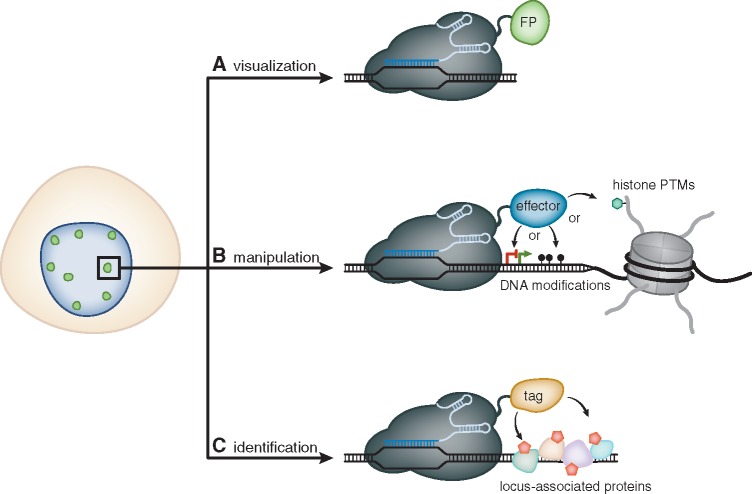
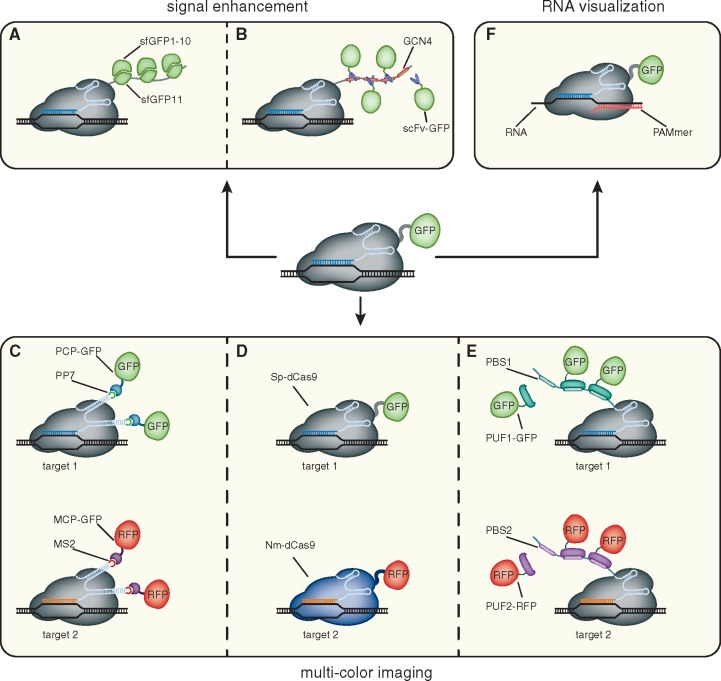
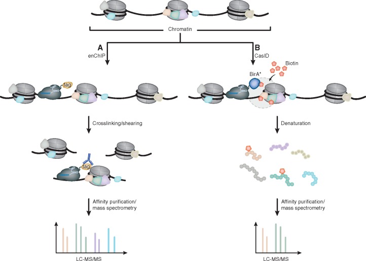
Since the discovery of the clustered regularly interspaced short palindromic repeats (CRISPR)/CRISPR-associated system (Cas) as a tool for gene editing a plethora of locus-specific as well as genome-wide approaches have been developed that allow efficient and reproducible manipulation of genomic sequences. However, the seemingly unbound potential of CRISPR/Cas does not stop with its utilization as a site-directed nuclease. Mutations in its catalytic centers render Cas9 (dCas9) a universal recruitment platform that can be utilized to control transcription, visualize DNA sequences, investigate in situ proteome compositions and manipulate epigenetic modifications at user-defined genomic loci. In this review, we give a comprehensive introduction and overview of the development, improvement and application of recent dCas9-based approaches.
Overview
The claims and statistics of this paper haven’t been checked yet.
Run the check — free, no account needed. It reads the open-access text and checks the sources it cites.
Integrity signals
Registry
Cites
Statistics
Figures
Verification
Claims the paper makes
The claims and statistics of this paper haven’t been checked yet.
Run the check — free, no account needed. It reads the open-access text and checks the sources it cites.
Claims other papers make about this one
Each is checked against this paper’s own text: does it say what the citing paper says it does?
In the citing paper
“Moreover, the CRISPR/dCas-9 can be also used to visualize and pinpoint where specifically the gene of interest is located inside the cell (subcellular localization) by fusing a marker such as Green Fluorescent Proteins (GFP) with dCas-9 enzyme.”
From Mechanism and Applications of CRISPR/Cas-9-Mediated Genome Editing (2021) · cites this paper as [51]
In the citing paper
“This enables site-specific labeling and imaging of endogenous loci in living cells for further utilization.51”
From Mechanism and Applications of CRISPR/Cas-9-Mediated Genome Editing (2021) · cites this paper as [51]
Self-check
Percentages, sums and reported differences re-computed from the paper’s own numbers.
The self-check needs the paper’s full text.
Citations
Cites
The mechanism of double-strand DNA break repair by the nonhomologous DNA end-joining pathway
Lieber, M. R. (2010) · Annual Review of Biochemistry · doi:10.1146/annurev.biochem.052308.093131 · Cited by 2,882
Genome-scale transcriptional activation by an engineered CRISPR-Cas9 complex
Konermann, S., Brigham, M. D., Trevino, A. E., Joung, J., Abudayyeh, O. O., Barcena, C., Hsu, P. D., Habib, N., Gootenberg, J. S., Nishimasu, H., et al. (2015) · Nature · doi:10.1038/nature14136 · Cited by 2,825
Genome-Scale CRISPR-Mediated Control of Gene Repression and Activation
Gilbert, L. A., Horlbeck, M. A., Adamson, B., Villalta, J. E., Chen, Y., Whitehead, E. H., Guimaraes, C., Panning, B., Ploegh, H. L., Bassik, M. C., et al. (2014) · Cell · doi:10.1016/j.cell.2014.09.029 · Cited by 2,728
Crystal structure of Cas9 in complex with guide RNA and target DNA
Nishimasu, H., Ran, F. A., Hsu, P. D., Konermann, S., Shehata, S. I., Dohmae, N., Ishitani, R., Zhang, F., & Nureki, O. (2014) · Cell · doi:10.1016/j.cell.2014.02.001 · Cited by 2,306
RNA targeting with CRISPR–Cas13
Abudayyeh, O. O., Gootenberg, J. S., Essletzbichler, P., Han, S., Joung, J., Belanto, J. J., Verdine, V., Cox, D. B. T., Kellner, M. J., Regev, A., et al. (2017) · Nature · doi:10.1038/nature24049 · Cited by 2,093
RNA-guided genetic silencing systems in bacteria and archaea
Wiedenheft, B., Sternberg, S. H., & Doudna, J. A. (2012) · Nature · doi:10.1038/nature10886 · Cited by 1,851
Mali, P., Aach, J., Stranges, P. B., Esvelt, K. M., Moosburner, M., Kosuri, S., Yang, L., & Church, G. M. (2013) · Nature Biotechnology · doi:10.1038/nbt.2675 · Cited by 1,752 · cited 1×
“Combining this modified sgRNA with a dCas9-VP64 fusion resulted in drastically increased expression levels of targeted genes and was shown to simultaneously upregulate 10 genes [114].”
BackgroundComparative Genomics of the Eukaryotes
Rubin, G. M., Yandell, M. D., Wortman, J. R., Gabor, G. L., Miklos, Nelson, C. R., Hariharan, I. K., Fortini, M. E., Li, P. W., Apweiler, R., et al. (2000) · Science · doi:10.1126/science.287.5461.2204 · Cited by 1,698
Structures of Cas9 endonucleases reveal RNA-mediated conformational activation
Jinek, M., Jiang, F., Taylor, D. W., Sternberg, S. H., Kaya, E., Ma, E., Anders, C., Hauer, M., Zhou, K., Lin, S., et al. (2014) · Science · doi:10.1126/science.1247997 · Cited by 1,230
Editing DNA Methylation in the Mammalian Genome
Liu, X. S., Wu, H., Ji, X., Stelzer, Y., Wu, X., Czauderna, S., Shu, J., Dadon, D., Young, R. A., & Jaenisch, R. (2016) · Cell · doi:10.1016/j.cell.2016.08.056 · Cited by 1,224
Research works printed but not linked: 103
- Samson, 2013, Revenge of the phages: defeating bacterial defences, Nat Rev Microbiol, 11, 675, 10.1038/nrmicro3096 · DOI
- Barrangou, 2007, CRISPR provides acquired resistance against viruses in prokaryotes, Science, 315, 1709., 10.1126/science.1138140 · DOI · Paper page
- Grissa, 2007, CRISPRFinder: a web tool to identify clustered regularly interspaced short palindromic repeats, Nucleic Acids Res, 35, W52, 10.1093/nar/gkm360 · DOI
- Ishino, 1987, Nucleotide sequence of the iap gene, responsible for alkaline phosphatase isozyme conversion in Escherichia coli, and identification of the gene product, J Bacteriol, 169, 5429, 10.1128/JB.169.12.5429-5433.1987 · DOI · Paper page
- Ratner, 2016, Overview of CRISPR–Cas9 biology, Cold Spring Harb Protoc, 2016, pdb.top088849., 10.1101/pdb.top088849 · DOI
- Carte, 2008, Cas6 is an endoribonuclease that generates guide RNAs for invader defense in prokaryotes, Genes Dev, 22, 3489, 10.1101/gad.1742908 · DOI
- Garside, 2012, Cas5d processes pre-crRNA and is a member of a larger family of CRISPR RNA endonucleases, RNA, 18, 2020, 10.1261/rna.033100.112 · DOI
- Nam, 2012, Cas5d protein processes pre-crRNA and assembles into a cascade-like interference complex in subtype I-C/Dvulg CRISPR-Cas system, Structure, 20, 1574, 10.1016/j.str.2012.06.016 · DOI
- Jore, 2011, Structural basis for CRISPR RNA-guided DNA recognition by Cascade, Nat Struct Mol Biol, 18, 529, 10.1038/nsmb.2019 · DOI
- Westra, 2012, CRISPR immunity relies on the consecutive binding and degradation of negatively supercoiled invader DNA by Cascade and Cas3, Mol. Cell, 46, 595, 10.1016/j.molcel.2012.03.018 · DOI
- Hrle, 2013, Structure and RNA-binding properties of the type III-A CRISPR-associated protein Csm3. RNA, Biol, 10, 1670
- Samai, 2015, Co-transcriptional DNA and RNA cleavage during Type III CRISPR-Cas immunity, Cell, 161, 1164, 10.1016/j.cell.2015.04.027 · DOI · Paper page
- Staals, 2013, Structure and activity of the RNA-targeting Type III-B CRISPR-Cas complex of Thermus thermophilus, Mol Cell, 52, 135, 10.1016/j.molcel.2013.09.013 · DOI
- Staals, 2014, RNA targeting by the type III-A CRISPR-Cas Csm complex of Thermus thermophilus, Mol Cell, 56, 518, 10.1016/j.molcel.2014.10.005 · DOI
- Dickinson, 2016, CRISPR-based methods for caenorhabditis elegans genome engineering, Genetics, 202, 885, 10.1534/genetics.115.182162 · DOI
- Henao-Mejia, 2016, Generation of genetically modified mice using the CRISPR-Cas9 genome-editing system, Cold Spring Harb Protoc, 2016, pdb.prot090704., 10.1101/pdb.prot090704 · DOI
- Housden, 2016, Cas9-mediated genome engineering in Drosophila melanogaster, Cold Spring Harb Protoc, 2016, pdb.top086843., 10.1101/pdb.top086843 · DOI
- Mali, 2013, RNA-guided human genome engineering via Cas9, Science, 339, 823, 10.1126/science.1232033 · DOI · Paper page
- Vejnar, 2016, Optimized CRISPR-Cas9 system for genome editing in zebrafish, Cold Spring Harb Protoc, 2016, pdb.prot086850., 10.1101/pdb.prot086850 · DOI
- McVey, 2008, MMEJ repair of double-strand breaks (director’s cut): deleted sequences and alternative endings, Trends Genet, 24, 529, 10.1016/j.tig.2008.08.007 · DOI
- Cong, 2013, Multiplex genome engineering using CRISPR/Cas systems, Science, 339, 819, 10.1126/science.1231143 · DOI · Paper page
- Jinek, 2012, A programmable dual-RNA-guided DNA endonuclease in adaptive bacterial immunity, Science, 337, 816, 10.1126/science.1225829 · DOI · Paper page
- Ariyoshi, 1994, Atomic structure of the RuvC resolvase: a holliday junction-specific endonuclease from E. coli, Cell, 78, 1063, 10.1016/0092-8674(94)90280-1 · DOI
- Gorecka, 2013, Crystal structure of RuvC resolvase in complex with Holliday junction substrate, Nucleic Acids, 10.1093/nar/gkt769 · DOI
- Biertümpfel, 2007, Crystal structure of T4 endonuclease VII resolving a Holliday junction, Nature, 449, 616, 10.1038/nature06152 · DOI
- Li, 2003, DNA binding and cleavage by the periplasmic nuclease Vvn: a novel structure with a known active site, EMBO J, 22, 4014, 10.1093/emboj/cdg377 · DOI
- Qi, 2013, Repurposing CRISPR as an RNA-guided platform for sequence-specific control of gene expression, Cell, 152, 1173, 10.1016/j.cell.2013.02.022 · DOI · Paper page
- Bolzer, 2005, Three-dimensional maps of all chromosomes in human male fibroblast nuclei and prometaphase rosettes, PLoS Biol, 3, e157, 10.1371/journal.pbio.0030157 · DOI
- Raj, 2009, Single-molecule approaches to stochastic gene expression, Annu Rev Biophys, 38, 255, 10.1146/annurev.biophys.37.032807.125928 · DOI
- Kanda, 1998, Histone-GFP fusion protein enables sensitive analysis of chromosome dynamics in living mammalian cells, Curr Biol, 8, 377, 10.1016/S0960-9822(98)70156-3 · DOI
- Manders, 1999, Direct imaging of DNA in living cells reveals the dynamics of chromosome formation, J Cell Biol, 144, 813, 10.1083/jcb.144.5.813 · DOI
- Martin, 2005, DNA labeling in living cells, Cytometry A, 67, 45, 10.1002/cyto.a.20172 · DOI
- Krawczyk, 2012, Chromatin mobility is increased at sites of DNA double-strand breaks, J Cell Sci, 125, 2127., 10.1242/jcs.089847 · DOI
- Shelby, 1996, Dynamic elastic behavior of alpha-satellite DNA domains visualized in situ in living human cells, J Cell Biol, 135, 545., 10.1083/jcb.135.3.545 · DOI
- Robinett, 1996, In vivo localization of DNA sequences and visualization of large-scale chromatin organization using lac operator/repressor recognition, J Cell Biol, 135, 1685, 10.1083/jcb.135.6.1685 · DOI
- Roukos, 2013, Spatial dynamics of chromosome translocations in living cells, Science, 341, 660., 10.1126/science.1237150 · DOI
- Gersbach, 2014, Activating human genes with zinc finger proteins, transcription activator-like effectors and CRISPR/Cas9 for gene therapy and regenerative medicine, Expert Opin Ther Targets, 18, 835, 10.1517/14728222.2014.913572 · DOI
- Laity, 2001, Zinc finger proteins: new insights into structural and functional diversity, Curr Opin Struct Biol, 11, 39, 10.1016/S0959-440X(00)00167-6 · DOI
- Miller, 1985, Repetitive zinc-binding domains in the protein transcription factor IIIA from Xenopus oocytes, EMBO J, 4, 1609, 10.1002/j.1460-2075.1985.tb03825.x · DOI
- Tupler, 2001, Expressing the human genome, Nature, 409, 832, 10.1038/35057011 · DOI
- Elrod-Erickson, 1996, Zif268 protein-DNA complex refined at 1.6 A: a model system for understanding zinc finger-DNA interactions, Structure, 4, 1171, 10.1016/S0969-2126(96)00125-6 · DOI · Paper page
- Pavletich, 1991, Zinc finger-DNA recognition: crystal structure of a Zif268-DNA complex at 2.1 A, Science, 252, 809, 10.1126/science.2028256 · DOI
- Liu, 1997, Design of polydactyl zinc-finger proteins for unique addressing within complex genomes, Proc Natl Acad Sci U S A, 94, 5525, 10.1073/pnas.94.11.5525 · DOI · Paper page
- Lindhout, 2007, Live cell imaging of repetitive DNA sequences via GFP-tagged polydactyl zinc finger proteins, Nucleic Acids Res, 35, e107., 10.1093/nar/gkm618 · DOI
- DeFrancesco, 2011, Move over ZFNs, Nat Biotechnol, 29, 681, 10.1038/nbt.1935 · DOI
- Ramirez, 2008, Unexpected failure rates for modular assembly of engineered zinc fingers, Nat Methods, 5, 374, 10.1038/nmeth0508-374 · DOI
- Segal, 1999, Toward controlling gene expression at will: selection and design of zinc finger domains recognizing each of the 5’-GNN-3' DNA target sequences, Proc Natl Acad Sci U S A, 96, 2758, 10.1073/pnas.96.6.2758 · DOI · Paper page
- Ma, 2013, Visualization of repetitive DNA sequences in human chromosomes with transcription activator-like effectors, Proc Natl Acad Sci U S A, 110, 21048, 10.1073/pnas.1319097110 · DOI
- Miyanari, 2013, Live visualization of chromatin dynamics with fluorescent TALEs, Nat Struct Mol Biol, 20, 1321, 10.1038/nsmb.2680 · DOI
- Ren, 2017, Visualization of aging-associated chromatin alterations with an engineered TALE system, Cell Res, 27, 483, 10.1038/cr.2017.18 · DOI
- Thanisch, 2014, Targeting and tracing of specific DNA sequences with dTALEs in living cells, Nucleic Acids Res, 42, e38., 10.1093/nar/gkt1348 · DOI
- Yuan, 2014, Illuminating DNA replication during Drosophila development using TALE-lights, Curr Biol, 24, R144, 10.1016/j.cub.2014.01.023 · DOI
- Boch, 2009, Breaking the code of DNA binding specificity of TAL-type III effectors, Science, 326, 1509, 10.1126/science.1178811 · DOI · Paper page
- Morbitzer, 2011, Assembly of custom TALE-type DNA binding domains by modular cloning, Nucleic Acids Res, 39, 5790, 10.1093/nar/gkr151 · DOI
- Reyon, 2012, FLASH assembly of TALENs for high-throughput genome editing, Nat Biotechnol, 30, 460, 10.1038/nbt.2170 · DOI
- Schmid-Burgk, 2013, A ligation-independent cloning technique for high-throughput assembly of transcription activator-like effector genes, Nat Biotechnol, 31, 76, 10.1038/nbt.2460 · DOI
- Anton, 2014, Visualization of specific DNA sequences in living mouse embryonic stem cells with a programmable fluorescent CRISPR/Cas system, Nucleus, 5, 163, 10.4161/nucl.28488 · DOI
- Chen, 2013, Dynamic imaging of genomic loci in living human cells by an optimized CRISPR/Cas system, Cell, 155, 1479, 10.1016/j.cell.2013.12.001 · DOI
- Köferle, 2016, CORALINA: a universal method for the generation of gRNA libraries for CRISPR-based screening, BMC Genomics, 17, 917, 10.1186/s12864-016-3268-z · DOI
- Lane, 2015, Enzymatically generated CRISPR libraries for genome labeling and screening, Dev Cell, 34, 373, 10.1016/j.devcel.2015.06.003 · DOI
- Tanenbaum, 2014, A protein-tagging system for signal amplification in gene expression and fluorescence imaging, Cell, 159, 635, 10.1016/j.cell.2014.09.039 · DOI
- Ye, 2017, Live cell imaging of genomic loci using dCas9-SunTag system and a bright fluorescent protein, Protein Cell, 8, 853, 10.1007/s13238-017-0460-0 · DOI
- Kamiyama, 2016, Versatile protein tagging in cells with split fluorescent protein, Nat Comms, 7, 11046, 10.1038/ncomms11046 · DOI
- Cabantous, 2005, Protein tagging and detection with engineered self-assembling fragments of green fluorescent protein, Nat Biotechnol, 23, 102, 10.1038/nbt1044 · DOI
- Ma, 2015, Multicolor CRISPR labeling of chromosomal loci in human cells, Proc Natl Acad Sci U S A, 112, 3002, 10.1073/pnas.1420024112 · DOI
- Esvelt, 2013, Orthogonal Cas9 proteins for RNA-guided gene regulation and editing, Nat Methods, 10, 1116, 10.1038/nmeth.2681 · DOI · Paper page
- Chen, 2016, Expanding the CRISPR imaging toolset with Staphylococcus aureus Cas9 for simultaneous imaging of multiple genomic loci, Nucleic Acids Res, 44, e75., 10.1093/nar/gkv1533 · DOI
- Fu, 2016, CRISPR-dCas9 and sgRNA scaffolds enable dual-colour live imaging of satellite sequences and repeat-enriched individual loci, Nat Comms, 7, 11707., 10.1038/ncomms11707 · DOI
- Qin, 2017, Live cell imaging of low- and non-repetitive chromosome loci using CRISPR-Cas9, Nat Comms, 8, 14725., 10.1038/ncomms14725 · DOI
- Wang, 2016, An RNA-aptamer-based two-color CRISPR labeling system, Sci Rep, 6, 26857., 10.1038/srep26857 · DOI
- Cheng, 2016, Casilio: a versatile CRISPR-Cas9-Pumilio hybrid for gene regulation and genomic labeling, Cell Res, 26, 254, 10.1038/cr.2016.3 · DOI
- Chen, 2013, Engineering RNA-binding proteins for biology, FEBS J, 280, 3734, 10.1111/febs.12375 · DOI
- Nelles, 2016, Programmable RNA tracking in live cells with CRISPR/Cas9, Cell, 165, 488, 10.1016/j.cell.2016.02.054 · DOI
- O’connell, 2014, Programmable RNA recognition and cleavage by CRISPR/Cas9, Nature, 516, 263, 10.1038/nature13769 · DOI
- Cox, 2017, RNA editing with CRISPR-Cas13, Science, 358, 1019, 10.1126/science.aaq0180 · DOI · Paper page
- Abudayyeh, 2016, C2c2 is a single-component programmable RNA-guided RNA-targeting CRISPR effector, Science, 353, aaf5573, 10.1126/science.aaf5573 · DOI · Paper page
- Bikard, 2013, Programmable repression and activation of bacterial gene expression using an engineered CRISPR-Cas system, Nucleic Acids Res, 41, 7429, 10.1093/nar/gkt520 · DOI · Paper page
- Gilbert, 2013, CRISPR-mediated modular RNA-guided regulation of transcription in eukaryotes, Cell, 154, 442, 10.1016/j.cell.2013.06.044 · DOI · Paper page
- Kearns, 2013, Cas9 effector-mediated regulation of transcription and differentiation in human pluripotent stem cells, Development, 141, 219, 10.1242/dev.103341 · DOI
- Konermann, 2013, Optical control of mammalian endogenous transcription and epigenetic states, Nature, 500, 472, 10.1038/nature12466 · DOI · Paper page
- Cheng, 2013, Multiplexed activation of endogenous genes by CRISPR-on, an RNA-guided transcriptional activator system, Cell Res, 23, 1163, 10.1038/cr.2013.122 · DOI
- Maeder, 2013, CRISPR RNA-guided activation of endogenous human genes, Nat Methods, 10, 977, 10.1038/nmeth.2598 · DOI · Paper page
- Perez-Pinera, 2013, RNA-guided gene activation by CRISPR-Cas9-based transcription factors, Nat Methods, 10, 973, 10.1038/nmeth.2600 · DOI · Paper page
- Chavez, 2015, Highly efficient Cas9-mediated transcriptional programming, Nat Methods, 12, 326, 10.1038/nmeth.3312 · DOI · Paper page
- Hilton, 2015, Epigenome editing by a CRISPR-Cas9-based acetyltransferase activates genes from promoters and enhancers, Nat Biotechnol, 33, 510, 10.1038/nbt.3199 · DOI
- Choudhury, 2016, CRISPR-dCas9 mediated TET1 targeting for selective DNA demethylation at BRCA1 promoter, Oncotarget, 7, 46545, 10.18632/oncotarget.10234 · DOI
- Xu, 2016, A CRISPR-based approach for targeted DNA demethylation, Cell Discov, 2, 16009., 10.1038/celldisc.2016.9 · DOI
- Kearns, 2015, Functional annotation of native enhancers with a Cas9-histone demethylase fusion, Nat Methods, 12, 401, 10.1038/nmeth.3325 · DOI · Paper page
- McDonald, 2016, Reprogrammable CRISPR/Cas9-based system for inducing site-specific DNA methylation, Biol Open, 5, 866., 10.1242/bio.019067 · DOI
- Vojta, 2016, Repurposing the CRISPR-Cas9 system for targeted DNA methylation, Nucleic Acids Res, 44, 5615, 10.1093/nar/gkw159 · DOI
- Huang, 2017, DNA epigenome editing using CRISPR-Cas SunTag-directed DNMT3A, Genome Biol, 18, 176., 10.1186/s13059-017-1306-z · DOI
- Morita, 2016, Targeted DNA demethylation in vivo using dCas9-peptide repeat and scFv-TET1 catalytic domain fusions, Nat Biotechnol, 34, 1060, 10.1038/nbt.3658 · DOI
- Anton, 2017, Site-specific recruitment of epigenetic factors with a modular CRISPR/Cas system, Nucleus, 8, 279, 10.1080/19491034.2017.1292194 · DOI
- Braun, 2017, Rapid and reversible epigenome editing by endogenous chromatin regulators, Nat Commun, 8, 560., 10.1038/s41467-017-00644-y · DOI
- Keung, 2015, Chromatin regulation at the frontier of synthetic biology, Nat Rev Genet, 16, 159, 10.1038/nrg3900 · DOI
- Thakore, 2016, Editing the epigenome: technologies for programmable transcription and epigenetic modulation, Nat Methods, 13, 127, 10.1038/nmeth.3733 · DOI · Paper page
- Amabile, 2016, Inheritable silencing of endogenous genes by hit-and-run targeted epigenetic editing, Cell, 167, 219, 10.1016/j.cell.2016.09.006 · DOI · Paper page
- Collas, 2010, The current state of chromatin immunoprecipitation, Mol Biotechnol, 45, 87, 10.1007/s12033-009-9239-8 · DOI
- Fujita, 2013, Efficient isolation of specific genomic regions and identification of associated proteins by engineered DNA-binding molecule-mediated chromatin immunoprecipitation (enChIP) using CRISPR, Biochem Biophys Res Commun, 439, 132, 10.1016/j.bbrc.2013.08.013 · DOI
- Roux, 2012, A promiscuous biotin ligase fusion protein identifies proximal and interacting proteins in mammalian cells, J Cell Biol, 196, 801., 10.1083/jcb.201112098 · DOI · Paper page
- Schmidtmann, 2016, Determination of local chromatin composition by CasID, Nucleus, 7, 476, 10.1080/19491034.2016.1239000 · DOI
- Liu, 2017, In situ capture of chromatin interactions by biotinylated dCas9, Cell, 170, 1028, 10.1016/j.cell.2017.08.003 · DOI
- Konermann, 2014, Genome-scale transcriptional activation by an engineered CRISPR-Cas9 complex, Nature, 517, 583, 10.1038/nature14136 · DOI · Paper page
Cited by
Sorted by citations
Mechanism and Applications of CRISPR/Cas-9-Mediated Genome Editing
Mengstie, M. A., & Wondimu, B. Z. (2021) · Biologics: Targets and Therapy · doi:10.2147/btt.s326422 · Cited by 478
Metadata
- Authors
- Anton, T., Karg, E., & Bultmann, S.
- Affiliations
- Department of Biology II and Center for Integrated Protein Science Munich (CIPSM), LMU Munich, 82152 Martinsried, Germany
- ORCID
- Bultmann, Sebastian
- Venue
- Biology Methods and Protocols
- Publisher
- Oxford University Press (OUP)
- Published
- 2018-01-01
- Volume
- 3
- Issue
- 1
- ISSN
- 2396-8923
- PMC
- PMC6994046
- Cited by
- 32
- References
- 10
- Language
- en
- Funders
- Deutsche Forschungsgemeinschaft (SPP1623)
About these checks
Registry, retraction, Cites and statistics results are computed, not judged, with one exception: a printed reference that no index lists is read once by a model before it counts as a citation error. Claims are extracted by a model and kept only when their quote appears verbatim in the text; each cited source is then checked, with a model’s help, for whether it says what the claim relies on. Those verdicts can be wrong — read the claim’s quote and the cited source before relying on one.
Statistics and arithmetic are recomputed, not judged.
Confidence bands are shown only for fields calibrated on our annotated test set. None is calibrated yet, so no band is shown.
What counts as a claim ›How we measure confidence ›How we check this ›
Are you an author, or do you see a problem with this page? Request a review
How to cite
Cite CoChat for the checks on this page, and the paper for its own findings.
- This page
- CoChat. (2026). Citation check: Applications of the CRISPR/Cas system beyond gene editing. https://app.cochat.ai/papers/10.1093/biomethods/bpy002
- The paper
- Anton, T., Karg, E., & Bultmann, S. (2018). Applications of the CRISPR/Cas system beyond gene editing. Biology Methods and Protocols. https://doi.org/10.1093/biomethods/bpy002
