

The DOI matches its Crossref record; 60 of the 60 references it cites were found in a registry or an index; 22 of the 47 sources its claims cite were checked for support.
Toward the human cellular microRNAome
Open accessPublished version of a preprint
Abstract
MicroRNAs are short RNAs that serve as regulators of gene expression and are essential components of normal development as well as modulators of disease. MicroRNAs generally act cell-autonomously, and thus their localization to specific cell types is needed to guide our understanding of microRNA activity. Current tissue-level data have caused considerable confusion, and comprehensive cell-level data do not yet exist. Here, we establish the landscape of human cell-specific microRNA expression. This project evaluated 8 billion small RNA-seq reads from 46 primary cell types, 42 cancer or immortalized cell lines, and 26 tissues. It identified both specific and ubiquitous patterns of expression that strongly correlate with adjacent superenhancer activity. Analysis of unaligned RNA reads uncovered 207 unknown minor strand (passenger) microRNAs of known microRNA loci and 495 novel putative microRNA loci. Although cancer cell lines generally recapitulated the expression patterns of matched primary cells, their isomiR sequence families exhibited increased disorder, suggesting DROSHA- and DICER1-dependent microRNA processing variability. Cell-specific patterns of microRNA expression were used to de-convolute variable cellular composition of colon and adipose tissue samples, highlighting one use of these cell-specific microRNA expression data. Characterization of cellular microRNA expression across a wide variety of cell types provides a new understanding of this critical regulatory RNA species.
Overview
AI snapshot
Written from the paper’s full text.
TL;DR
This study establishes a human cell-specific microRNA expression landscape by analyzing small RNA-seq across primary cells, cancer/immortalized cell lines, and tissues. It identifies cell-restricted and ubiquitous microRNAs, links expression to superenhancers, reports 495 high-confidence putative novel microRNAs, and shows substantial isomiR diversity that differs between primary and cancer cells.
Key findings
- Across 46 primary cell types, 42 cancer/immortalized cell lines, and 26 tissues, 2319 of 2546 known microRNAs (miRBase v21) were detected at ≥1 read per million microRNA reads in at least one sample; 320 microRNAs reached ≥1000 RPM in at least one of 46 normal cell types, and 94 (29%) were present in only a single cell class. Six microRNAs were present in all 46 cell types at ≥1000 RPM: miR-107, miR-103a-3p, miR-103b, miR-191-5p, miR-21-5p, and miR-92a-3p.
- MicroRNA expression strongly correlated with adjacent superenhancer activity: expression was significantly higher when a superenhancer was within 40 kb of the microRNA locus (Wilcoxon rank-sum test, P < 2.2−16), and cell-type-restricted microRNAs matched cell-specific superenhancer activity.
- Novel microRNA discovery from 1.2 billion unmapped reads yielded 21,338 miRDeep2 loci, which were refined to 495 highest-confidence putative novel microRNAs; 207 unassigned passenger microRNAs from known loci were also found. Validation included miRanalyzer overlap, Argonaute CLIP-seq evidence, PhyloP conservation, DICER1-knockout dependence, and PCR of nine novel microRNAs.
- IsomiR analysis of 126 primary cell and 82 cancer/immortalized cell samples showed that the miRBase v21 canonical sequence was the most abundant isomiR in only 33% of primary-cell microRNAs, and 37% never had the canonical sequence as most abundant; cancer/immortalized cells had a ∼20% increase in disorder over primary cells (P < 0.005, Wilcoxon rank-sum test).
Methods
- The study combined new small RNA-seq from 39 primary cell samples with public Sequence Read Archive/Array Express data, processing 694 samples through miRge and retaining 530 for analysis: 161 primary cell datasets (46 main cell types), 100 cancer/immortalized cell line datasets (42 cell lines), and 269 tissue datasets (26 tissues).
- MicroRNA expression states were discretized with DEXUS; batch effects were addressed with RUV normalization and SVA; clustering used t-SNE. Cell-specific signatures were used with CIBERSORT to de-convolute adipose and colon tissue composition.
- Novel microRNA detection used miRDeep2 and miRanalyzer on unmapped reads, followed by novoMiRank, PhyloP conservation, Argonaute CLIP-seq, DICER1-knockout data, and PCR validation. IsomiR families were analyzed for templated length variants, 5′ start variation, and nontemplated 3′ nucleotide additions.
Limitations
- The paper states that much data came from public sources across different platforms and sequencing methods, creating technical variation; extensive normalization was used, but technical factors still drive some variation and clustering. Some cell types had few to no replicates, it is unknown how well cultured cells match in vivo cells, and many cell types (hepatocytes, neutrophils, pneumocytes) were unavailable.
- The paper states that the 495 putative novel microRNAs are not definitive, many are likely false positives, and in vivo proof was beyond scope; requiring multiple evidence sources may also have lost true positives.
- Evident from the text: superenhancer correlations are not proof of superenhancer activity on adjacent microRNAs, and the authors caution that the activity of these superenhancers on adjacent genes and microRNAs is generally unknown.
Model-written summary. Check it against the paper before relying on it.
Integrity signals
Registry
Cites
Statistics
Figures
Verification
Claims the paper makes
64 Own result · 26 Own interpretation · 14 Own method · 10 No full text of the sources · 9 Supported · 8 Partly supported · 4 Sources not found · 3 No basis given · 2 Couldn’t confirm · 1 Supported so far · 1 Not checked yet · accuracy not yet measured
The cited sources support only part of this claim — read each source’s verdict below.
In the paper
“While providing valuable insights into the relationship of microRNA expression and disease (Lu et al. 2005), these and subsequent studies (Cheng et al. 2015; Ludwig et al. 2016) have not unraveled cellular microRNA expression.”
§ Toward the human cellular microRNAome > Abstract
What the sources say
- Not matchedcites Cheng et al. 2015
Couldn’t be matched to a paper in Crossref, so it can’t be checked.
- Supportedcites Ludwig et al. 2016 source
The Introduction (page unavailable) says Ludwig et al. profiled miRNAs across 61 human tissues, and the Results report tissue-specific expression. The study therefore provides tissue-level patterns rather than resolving expression in individual cells. This verdict concerns Ludwig et al.; it does not assess the other cited studies. (LLM, conf=0.84)
plain- Asserted: passes — The authors put it forward as true.
- Checkable: passes — It can be checked against evidence.
- Load-bearing: passes — The paper’s conclusions depend on it.
- Based: passes — The paper gives a reason why it holds.
- Not matched
Every source the paper cites for this claim was checked and supports it.
In the paper
“Additionally, the anonymity of microRNA nomenclature, with sequential numerical naming, has not allowed an intrinsic understanding of which microRNAs are ubiquitous and which have cell-restricted patterns of expression (Witwer and Halushka 2016).”
§ Toward the human cellular microRNAome > Abstract
What the sources say
- Supportedcites Witwer and Halushka 2016 source
The paper says its numerical nomenclature does not intrinsically convey a microRNA’s localization. In “Areas of microRNA confusion and pitfalls to avoid” (page not supplied), it links the anonymity of microRNA names to difficulty identifying cellular expression, and its Figure 1 distinguishes ubiquitous from cell-type-restricted microRNAs. (LLM, conf=0.92)
plaindirect- Asserted: passes — The authors put it forward as true.
- Checkable: passes — It can be checked against evidence.
- Load-bearing: passes — The paper’s conclusions depend on it.
- Based: passes — The paper gives a reason why it holds.
- Supported
Every source the paper cites for this claim was checked and supports it.
In the paper
“We used surrogate variable analysis (SVA) and 26 loosely described “biologic clusters” of tissues and cells to clearly demonstrate that, after adjusting for surrogate variables, similar tissue and cell types clustered together across experiments (Supplemental Fig. S3; Leek and Storey 2007).”
§ Toward the human cellular microRNAome > Results > Generation of a cellular microRNAome
What the sources say
- Supportedcites Leek and Storey 2007 source
In Results > SVA Applied to Human Expression Studies (page unavailable), Leek and Storey describe applying surrogate variable analysis and adjusting for a surrogate variable, supporting the citation’s identification of the method. The clustering across experiments is presented as the citing authors’ own result, illustrated by their Supplemental Fig. S3, rather than as a finding attributed to Leek and Storey. (LLM, conf=0.86)
boostedcorrelationaln = 387- Asserted: passes — The authors put it forward as true.
- Checkable: passes — It can be checked against evidence.
- Load-bearing: passes — The paper’s conclusions depend on it.
- Based: passes — The paper gives a reason why it holds.
- Supported
Every source the paper cites for this claim was checked and supports it.
In the paper
“In comparison to the 3707 novel TJU microRNAs (Londin et al. 2015), only 257 (52%) were shared between both data sets.”
§ Toward the human cellular microRNAome > Results > Novel human microRNAs
What the sources say
- Supportedcites Londin et al. 2015 source
Table 1 lists 3,707 novel miRNAs, and the Results describe the same total; the paper’s examples use TJU identifiers. The citation identifies the comparison dataset, not the citing authors’ finding that 257 (52%) were shared. (LLM, conf=0.92)
plain- Asserted: passes — The authors put it forward as true.
- Checkable: passes — It can be checked against evidence.
- Load-bearing: unclear — It is unclear whether the paper’s conclusions depend on it.
- Based: passes — The paper gives a reason why it holds.
- Supported
The cited sources support only part of this claim — read each source’s verdict below.
In the paper
“Although technical factors may be responsible for some variation between cell types, the data clearly demonstrate a need to revise our understanding of the appropriate canonical microRNA sequences for better reproducibility and computational target prediction (Mestdagh et al. 2014; Agarwal et al. 2015).”
§ Toward the human cellular microRNAome > Results > Wide variation in the distribution of isomiRs
What the sources say
- Not checked — no full text of the sourcecites Mestdagh et al. 2014 source
- Partly supportedcites Agarwal et al. 2015 sourceExperimental — accuracy not yet measured
In Materials and methods (‘MicroRNA sets for TargetScan7’; page not specified), the paper identifies abundant microRNA isoforms with different seed sequences and annotates them for target prediction. This supports considering alternative sequences in computational predictions, but the paper does not establish a need to revise canonical microRNA sequences for reproducibility. (LLM, conf=0.82)
boosted- Asserted: passes — The authors put it forward as true.
- Checkable: unclear — It is unclear whether it could be checked against evidence.
- Load-bearing: passes — The paper’s conclusions depend on it.
- Based: passes — The paper gives a reason why it holds.
Every source the paper cites for this claim was checked and supports it.
In the paper
“We determined that the main drivers of this difference were the changing methods of the Illumina sequencing library kits acting upon a small subset of microRNAs (Supplemental Fig. S10A; Baran-Gale et al. 2015).”
§ Toward the human cellular microRNAome > Results > Wide variation in the distribution of isomiRs
What the sources say
- Supportedcites Baran-Gale et al. 2015 source
The Introduction reports that 50 highly expressed miRNAs were detected at levels more than tenfold different between Illumina v1.5 and TruSeq. The Discussion attributes the differential detection bias to the two library-preparation methods, supporting the cited explanation that a subset of miRNAs is particularly affected by the change in kits. (LLM, conf=0.88)
plaindirect- Asserted: passes — The authors put it forward as true.
- Checkable: passes — It can be checked against evidence.
- Load-bearing: passes — The paper’s conclusions depend on it.
- Based: passes — The paper gives a reason why it holds.
- Supported
Every source the paper cites for this claim was checked and supports it.
In the paper
“These data further support the cell-type restricted microRNAs seen above and indicate that analyses of global tissue microRNA expression require an understanding of the source of each microRNA of interest, lest misinterpretations of the data result in spurious disease associations (Kent et al. 2014).”
§ Toward the human cellular microRNAome > Results > Diverse microRNAs expression patterns
What the sources say
- Supportedcites Kent et al. 2014 source
The Introduction explains that tissue-level miRNA signals depend on cell-type composition and that understanding cellular expression is critical to interpreting them. The Conclusion warns that changes in cellular composition between diseased and non-diseased tissues can be mistaken for changes in miRNA expression within a cell type. Page numbers are not provided in our copy. (LLM, conf=0.93)
hedgeddirect- Asserted: passes — The authors put it forward as true.
- Checkable: passes — It can be checked against evidence.
- Load-bearing: passes — The paper’s conclusions depend on it.
- Based: passes — The paper gives a reason why it holds.
- Supported
The cited sources support only part of this claim — read each source’s verdict below.
In the paper
“yet, because blood is found in all tissues, they have been inappropriately assigned a variety of functions in epithelial and mesenchymal cells based on misinterpreted tissue-level data (Kent et al. 2014; Halushka 2016).”
§ Toward the human cellular microRNAome > Discussion
What the sources say
- Partly supportedcites Kent et al. 2014 sourceExperimental — accuracy not yet measured
The Introduction section on miR-451a (page not provided) says erythrocytes are present in every human tissue and that tissue-level miR-451a findings have been erroneously interpreted as expression in epithelial cells; it also discusses a liposarcoma example. It mentions miR-144 as part of the miR-144/451a cluster, but does not establish that miR-144 itself was inappropriately assigned those cellular functions. (LLM, conf=0.87)
- Not checked — no full text of the sourcecites Halushka 2016 source
plaindirect- Asserted: passes — The authors put it forward as true.
- Checkable: passes — It can be checked against evidence.
- Load-bearing: passes — The paper’s conclusions depend on it.
- Based: passes — The paper gives a reason why it holds.
- Partly supported
The cited sources support only part of this claim — read each source’s verdict below.
In the paper
“Namely, miR-451a and miR-144 are exclusively expressed in red blood cells, yet, because blood is found in all tissues, they have been inappropriately assigned a variety of functions in epithelial and mesenchymal cells based on misinterpreted tissue-level data (Kent et al. 2014; Halushka 2016).”
§ Toward the human cellular microRNAome > Discussion
What the sources say
- Partly supportedcites Kent et al. 2014 sourceExperimental — accuracy not yet measured
The Introduction section “miR-451a—an erythrocyte specific microRNA” says the authors’ primary-cell comparisons found miR-451a exclusively in red blood cells. It identifies miR-144 as part of the same cluster, but does not say miR-144 is exclusively expressed in red blood cells. That part of the claim is unsupported by our copy. (LLM, conf=0.88)
- Not checked — no full text of the sourcecites Halushka 2016 source
plain- Asserted: passes — The authors put it forward as true.
- Checkable: passes — It can be checked against evidence.
- Load-bearing: passes — The paper’s conclusions depend on it.
- Based: passes — The paper gives a reason why it holds.
- Partly supported
Every source the paper cites for this claim was checked and supports it.
In the paper
“This will improve the interpretation of tissue microRNA expression levels, which, to date, has significantly muddied our understanding of microRNA localization and biologically relevant function (Kent et al. 2014).”
§ Toward the human cellular microRNAome > Discussion
What the sources say
- Supportedcites Kent et al. 2014 source
The Introduction states that understanding cellular patterns of miRNA expression is critical to interpreting tissue-level data. The Conclusion says that failure to account for tissue composition complicates knowledge of miRNA function and that identifying the cellular source is needed to make biologically appropriate connections. (LLM, conf=0.93)
plaindirect- Asserted: passes — The authors put it forward as true.
- Checkable: passes — It can be checked against evidence.
- Load-bearing: passes — The paper’s conclusions depend on it.
- Based: passes — The paper gives a reason why it holds.
- Supported
- Probably not a substantial claim. 19
- It may state two separate claims as one. 32
The sources with a verdict support this claim; the rest weren’t checked yet, have no full text we could read, or their verdicts aren’t rated yet.
In the paper
“In this role, they control essential cell processes in health and disease (Ambros 2004; Mendell and Olson 2012).”
§ Toward the human cellular microRNAome > Abstract
What the sources say
- Not checked — no full text of the sourcecites Ambros 2004 source
- Supportedcites Mendell and Olson 2012 source
The Introduction describes miRNA functions in normal mammalian physiology and disease, including their influence on how developed tissues respond to stress. The MicroRNAs in Cancer section identifies cell proliferation and apoptosis as processes they regulate. Together, these passages support the broad paraphrase. (LLM, conf=0.88)
plaindirect- Asserted: passes — The authors put it forward as true.
- Checkable: passes — It can be checked against evidence.
- Load-bearing: fails — The paper’s conclusions may not depend on it.
- Based: passes — The paper gives a reason why it holds.
Probably not a substantial claim. The paper’s conclusions may not depend on it.
The cited sources support only part of this claim — read each source’s verdict below.
In the paper
“Superenhancers are dense genomic regions of transcription factor binding sites that have a multiplicative effect on increasing adjacent gene expression (Whyte et al. 2013; Suzuki et al. 2017).”
§ Toward the human cellular microRNAome > Results > Diverse microRNAs expression patterns
What the sources say
- Partly supportedcites Whyte et al. 2013 sourceExperimental — accuracy not yet measured
The Results sections on large genomic domains and functional attributes describe super-enhancers as clusters of transcription-factor-bound enhancers associated with higher expression of target genes. They report 3.8-fold higher luciferase activity for constituent segments than for typical enhancer segments, but do not establish a multiplicative effect on adjacent gene expression. The section on key ESC identity genes also notes that enhancer targets can be distant, not necessarily adjacent. (LLM, conf=0.86)
- Partly supportedcites Suzuki et al. 2017 sourceExperimental — accuracy not yet measured
The Introduction describes super-enhancers as regions with high densities of transcription factors that are closely associated with genes. The Results (CRISPR/Cas9 functional genomics of miRNA super-enhancers) find cooperative, rather than additive, effects of their constituents on specific miRNAs. The paper does not establish a general multiplicative effect on the expression of adjacent genes. (LLM, conf=0.85)
plaindirect- Asserted: passes — The authors put it forward as true.
- Checkable: passes — It can be checked against evidence.
- Load-bearing: passes — The paper’s conclusions depend on it.
- Based: passes — The paper gives a reason why it holds.
Probably not a substantial claim.
- Partly supported
The cited sources support only part of this claim — read each source’s verdict below.
In the paper
“This included miR-199b-3p (+1 shift), miR-181c-3p (+1 shift), and miR-302a-5p (+3 shift), all of which had highly abundant reads containing a completely different seed sequence than the one currently assigned, with strong implications for the targeting of genes (Tan et al. 2014).”
§ Toward the human cellular microRNAome > Results > Wide variation in the distribution of isomiRs
What the sources say
- Partly supportedcites Tan et al. 2014 sourceExperimental — accuracy not yet measured
The Results section (‘MiRNAs from the same precursor have different 5′ and 3′ lengths’; page unavailable) reports a common 5′ isomiR of miR-302a and substantial differences in its predicted targets, supporting the general targeting implication. Our copy does not identify it as miR-302a-5p with a +3 shift, or report the claimed abundant reads and seed changes for miR-199b-3p and miR-181c-3p (searched by those names and shift terms). (LLM, conf=0.84)
plain- Asserted: passes — The authors put it forward as true.
- Checkable: passes — It can be checked against evidence.
- Load-bearing: passes — The paper’s conclusions depend on it.
- Based: passes — The paper gives a reason why it holds.
It may state two separate claims as one.
- Partly supported
The cited sources that could be checked couldn’t confirm or dispute this claim.
In the paper
“DICER1 knockouts are known to significantly repress 3p driver microRNA levels while allowing for longer 5p microRNAs that are loaded into Ago and trimmed back from the 3p end (Hill et al. 2009).”
§ Toward the human cellular microRNAome > Results > Novel human microRNAs
What the sources say
- Could not verifycites Hill et al. 2009 sourceExperimental — accuracy not yet measured
The section “Germline DICER1 Mutations in Familial Pleuropulmonary Blastoma” identifies DICER1 as involved in generating small RNAs and describes loss-of-function mutations, but does not address 3p versus 5p microRNA levels, Ago loading, or trimming. Searches for those strand-specific processing details found no passage supporting the attributed claim; page numbers are unavailable in our copy. (LLM, conf=0.76)
boosteddirect- Asserted: passes — The authors put it forward as true.
- Checkable: passes — It can be checked against evidence.
- Load-bearing: passes — The paper’s conclusions depend on it.
- Based: passes — The paper gives a reason why it holds.
Probably not a substantial claim. It may state two separate claims as one.
- Could not verify
The cited sources support only part of this claim — read each source’s verdict below.
In the paper
“This number of new microRNAs seems impossibly high and likely mostly false positives and/or nonfunctional transcripts (Mullokandov et al. 2012).”
§ Toward the human cellular microRNAome > Results > Novel human microRNAs
What the sources say
- Partly supportedcites Mullokandov et al. 2012 sourceExperimental — accuracy not yet measured
The Discussion (page unavailable in our copy) reports that almost 60% of miRNAs detected in the cells studied had no discernible suppressive activity, supporting concern that many detected miRNAs may have little biological activity. It does not assess the number of *new* miRNAs or conclude that most are false-positive identifications. The Results’ reference to possible false positives concerns a small set of sensor results, not the miRNA transcripts themselves. (LLM, conf=0.87)
hedgedn = 474- Asserted: passes — The authors put it forward as true.
- Checkable: passes — It can be checked against evidence.
- Load-bearing: passes — The paper’s conclusions depend on it.
- Based: passes — The paper gives a reason why it holds.
It may state two separate claims as one.
- Partly supported
The cited sources support only part of this claim — read each source’s verdict below.
In the paper
“Our data matched certain “ground truths” about specific microRNA expression in certain cell types, including miR-192 in epithelial cells, miR-126 in endothelial cells and platelets, and miR-9 in brain (McCall et al. 2011a; Haider et al. 2014; Kent et al. 2014).”
§ Toward the human cellular microRNAome > Discussion
What the sources say
- Not matchedcites McCall et al. 2011a
Couldn’t be matched to a paper in Crossref, so it can’t be checked.
- Partly supportedcites Haider et al. 2014 sourceExperimental — accuracy not yet measured
The Results section, “MicroRNA biomarker plausibility across studies” (page unavailable), states that miR-126 is highly expressed in endothelial cells. Our copy does not establish the other listed pairings—miR-126 in platelets, miR-192 in epithelial cells, or miR-9 in brain—so it supports only part of what this citation is offered for. The citing authors’ statement that their own data matched these patterns is their conclusion, not a finding attributed to Haider et al. (LLM, conf=0.86)
- Partly supportedcites Kent et al. 2014 sourceExperimental — accuracy not yet measured
The Introduction section “miR-126—an endothelial miRNA” identifies miR-126 as highly expressed in endothelial cells. The passages from our copy do not establish the other cited associations: miR-126 in platelets, miR-192 in epithelial cells, or miR-9 in brain. Page numbers were not available in our copy. (LLM, conf=0.87)
plaincorrelational- Asserted: passes — The authors put it forward as true.
- Checkable: passes — It can be checked against evidence.
- Load-bearing: passes — The paper’s conclusions depend on it.
- Based: passes — The paper gives a reason why it holds.
It may state two separate claims as one.
- Not matched
Every source the paper cites for this claim was checked and supports it.
In the paper
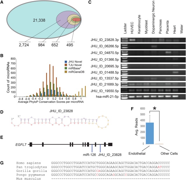
“The PhyloP conservation data (Fig. 3B) show that, beyond the most ideal microRNAs identified in miRGeneDB, there is a lack of genetic conservation, with overlap in a large segment of miRBase microRNAs and the novel microRNAs reported here and in a prior large study (Londin et al. 2015).”
§ Toward the human cellular microRNAome > Discussion
What the sources say
- Supportedcites Londin et al. 2015 source
Split into 2 atoms — 2 confirmed, 0 partially supported, 0 disputed, 0 misattributed, 0 unverifiable. Expand the claim to see per-atom verdicts.
plaincorrelational- Asserted: passes — The authors put it forward as true.
- Checkable: passes — It can be checked against evidence.
- Load-bearing: passes — The paper’s conclusions depend on it.
- Based: passes — The paper gives a reason why it holds.
It may state two separate claims as one.
- Supported
The cited sources that could be checked couldn’t confirm or dispute this claim.
In the paper
“As an example of potentially interesting novel microRNAs, JHU_ID_23828-3p was validated by PCR and is located in intron 8 of the EGFL7 gene, approximately 900 bp from mir-126 and within the same pri-miRNA transcript (Fig. 3D,E; Chang et al. 2015).”
§ Toward the human cellular microRNAome > Results > Novel human microRNAs
What the sources say
- Could not verifycites Chang et al. 2015 sourceExperimental — accuracy not yet measured
The Results passages describe PCR validation of other pri-miRNA transcripts, but do not identify JHU_ID_23828-3p. Searches for that identifier, EGFL7, miR-126, and the approximately 900-bp distance found no passage addressing the claimed location or shared transcript (page numbers unavailable). (LLM, conf=0.76)
plain- Asserted: passes — The authors put it forward as true.
- Checkable: passes — It can be checked against evidence.
- Load-bearing: unclear — It is unclear whether the paper’s conclusions depend on it.
- Based: passes — The paper gives a reason why it holds.
It may state two separate claims as one.
- Could not verify
Every source the paper cites for this claim was checked and supports it.
In the paper
“As well, another recent paper investigating microRNA RNA-seq across multiple laboratories found relative quantification to be “remarkably accurate and reproducible” (Giraldez et al. 2017), consistent with our work.”
§ Toward the human cellular microRNAome > Discussion
What the sources say
- Supportedcites Giraldez et al. 2017 source
The Accuracy and cross-protocol concordance section reports that all evaluated protocols were remarkably accurate at relative quantification of synthetic miRNAs. The Discussion reports that relative-quantification results were strongly correlated across institutions, even when they used different protocols. Together, these passages support the citing sentence’s characterization. (LLM, conf=0.94)
boosted- Asserted: passes — The authors put it forward as true.
- Checkable: passes — It can be checked against evidence.
- Load-bearing: fails — The paper’s conclusions may not depend on it.
- Based: passes — The paper gives a reason why it holds.
Probably not a substantial claim. The paper’s conclusions may not depend on it. It may state two separate claims as one.
- Supported
Every source the paper cites for this claim was checked and supports it.
In the paper
“The remaining miRBase microRNAs had modest conservation and were similarly distributed to both the putative novel microRNAs reported here and the 3707 novel microRNAs reported as TJU microRNAs (Fig. 3B; Londin et al. 2015).”
§ Toward the human cellular microRNAome > Results > Novel human microRNAs
What the sources say
- Supportedcites Londin et al. 2015 source
The citation identifies the comparison set as Londin et al.’s 3,707 novel TJU microRNAs. Table 1 lists 3,707 novel miRNAs, and Fig. 6 uses TJU identifiers for examples. The statement about their similar conservation distribution is presented as the citing paper’s Fig. 3B finding, not as a finding attributed to Londin et al. (LLM, conf=0.91)
plain- Asserted: passes — The authors put it forward as true.
- Checkable: passes — It can be checked against evidence.
- Load-bearing: passes — The paper’s conclusions depend on it.
- Based: passes — The paper gives a reason why it holds.
It may state two separate claims as one.
- Supported
A result the paper reports from its own data — it cites no other source for it, so there is none to check. The quote shows it is in the paper.
In the paper
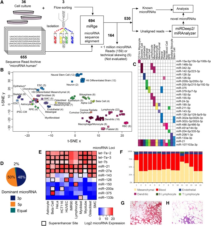
“miR-451a was abundantly present in 20 of 26 tissues but is from only one cell class (red blood cells).”
§ Toward the human cellular microRNAome > Results > Diverse microRNAs expression patterns
plain- Asserted: passes — The authors put it forward as true.
- Checkable: passes — It can be checked against evidence.
- Load-bearing: unclear — It is unclear whether the paper’s conclusions depend on it.
- Based: passes — The paper gives a reason why it holds.
It may state two separate claims as one.
A result the paper reports from its own data — it cites no other source for it, so there is none to check. The quote shows it is in the paper.
In the paper
“Strong clustering by biological group was observed for all samples, overcoming most technical concerns.”
§ Toward the human cellular microRNAome > Results > Generation of a cellular microRNAome
boostedcorrelational- Asserted: passes — The authors put it forward as true.
- Checkable: passes — It can be checked against evidence.
- Load-bearing: passes — The paper’s conclusions depend on it.
- Based: passes — The paper gives a reason why it holds.
It may state two separate claims as one.
A result the paper reports from its own data — it cites no other source for it, so there is none to check. The quote shows it is in the paper.
In the paper
“and miR-150-5p, abundant in lymphocytes, was present in 11 tissues.”
§ Toward the human cellular microRNAome > Results > Diverse microRNAs expression patterns
plain- Asserted: passes — The authors put it forward as true.
- Checkable: passes — It can be checked against evidence.
- Load-bearing: unclear — It is unclear whether the paper’s conclusions depend on it.
- Based: passes — The paper gives a reason why it holds.
It may state two separate claims as one.
In the paper
“We found the average z-score was right-shifted to 1.07 (indicating less similarity) for a subset of the 21,338 loci, while for the 495 highest confidence loci it was 0.90, indicating more consistency with miRBase microRNAs (Supplemental Fig. S8A,B; Backes et al. 2016).”
§ Toward the human cellular microRNAome > Results > Novel human microRNAs
What the sources say
- Not matchedcites Backes et al. 2016
Couldn’t be matched to a paper in Crossref, so it can’t be checked.
plain- Asserted: passes — The authors put it forward as true.
- Checkable: passes — It can be checked against evidence.
- Load-bearing: passes — The paper’s conclusions depend on it.
- Based: passes — The paper gives a reason why it holds.
It may state two separate claims as one.
- Not matched
A result the paper reports from its own data — it cites no other source for it, so there is none to check. The quote shows it is in the paper.
In the paper
“Eight primary T lymphocyte samples and 14 T lymphocyte malignancy samples also revealed moderate to strong correlation but with separate primary and cancer-derived cell clustering (Supplemental Fig. S6C).”
§ Toward the human cellular microRNAome > Results > Variable expression in cancer cell lines
plaincorrelationaln = 14- Asserted: passes — The authors put it forward as true.
- Checkable: passes — It can be checked against evidence.
- Load-bearing: passes — The paper’s conclusions depend on it.
- Based: passes — The paper gives a reason why it holds.
It may state two separate claims as one.
A result the paper reports from its own data — it cites no other source for it, so there is none to check. The quote shows it is in the paper.
In the paper
“Thirty-four were also adjacent to a known microRNA.”
§ Toward the human cellular microRNAome > Results > Novel human microRNAs
plaincorrelational- Asserted: passes — The authors put it forward as true.
- Checkable: passes — It can be checked against evidence.
- Load-bearing: fails — The paper’s conclusions may not depend on it.
- Based: passes — The paper gives a reason why it holds.
Probably not a substantial claim. The paper’s conclusions may not depend on it.
A result the paper reports from its own data — it cites no other source for it, so there is none to check. The quote shows it is in the paper.
In the paper
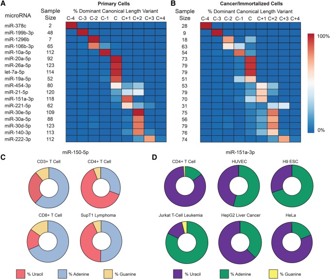
“but could vary widely between both primary and cancer/immortalized cells as a reflection of either biological differences or technical factors (Fig. 4C,D).”
§ Toward the human cellular microRNAome > Results > Wide variation in the distribution of isomiRs
hedgedconditionaln = 82- Asserted: passes — The authors put it forward as true.
- Checkable: passes — It can be checked against evidence.
- Load-bearing: fails — The paper’s conclusions may not depend on it.
- Based: passes — The paper gives a reason why it holds.
Probably not a substantial claim. The paper’s conclusions may not depend on it.
A result the paper reports from its own data — it cites no other source for it, so there is none to check. The quote shows it is in the paper.
In the paper
“Certain aspects of the data also helped with normalization, including 98% of sequencing runs being performed on an Illumina Genome Analyzer or HiSeq system and >74% of libraries being generated using an Illumina sequencing kit.”
§ Toward the human cellular microRNAome > Discussion
plain- Asserted: passes — The authors put it forward as true.
- Checkable: passes — It can be checked against evidence.
- Load-bearing: passes — The paper’s conclusions depend on it.
- Based: passes — The paper gives a reason why it holds.
It may state two separate claims as one.
A result the paper reports from its own data — it cites no other source for it, so there is none to check. The quote shows it is in the paper.
In the paper
“and 15 were orthologous to a different species’ microRNA (primarily primate) (Supplemental Tables S8, S9).”
§ Toward the human cellular microRNAome > Results > Novel human microRNAs
plain- Asserted: passes — The authors put it forward as true.
- Checkable: passes — It can be checked against evidence.
- Load-bearing: fails — The paper’s conclusions may not depend on it.
- Based: passes — The paper gives a reason why it holds.
Probably not a substantial claim. The paper’s conclusions may not depend on it.
A result the paper reports from its own data — it cites no other source for it, so there is none to check. The quote shows it is in the paper.
In the paper
“Additionally, while miR-126 is highly conserved throughout chordates, JHU_ID_ 23828 is only conserved among primates (Fig. 3G).”
§ Toward the human cellular microRNAome > Results > Novel human microRNAs
plain- Asserted: passes — The authors put it forward as true.
- Checkable: passes — It can be checked against evidence.
- Load-bearing: unclear — It is unclear whether the paper’s conclusions depend on it.
- Based: passes — The paper gives a reason why it holds.
It may state two separate claims as one.
A result the paper reports from its own data — it cites no other source for it, so there is none to check. The quote shows it is in the paper.
In the paper
“A test for the difference in proportions between wild-type and DICER1 knockout samples revealed the proportion in wild-type dropped from 0.151 to 0.047 in the DICER1 samples, representing a significant decrease (P < 2.2−16).”
§ Toward the human cellular microRNAome > Results > Novel human microRNAs
plaincorrelational- Asserted: passes — The authors put it forward as true.
- Checkable: passes — It can be checked against evidence.
- Load-bearing: passes — The paper’s conclusions depend on it.
- Based: passes — The paper gives a reason why it holds.
It may state two separate claims as one.
A result the paper reports from its own data — it cites no other source for it, so there is none to check. The quote shows it is in the paper.
In the paper
“The most abundant isomiR was widely variable between cells and often incongruent with the expected sequence.”
§ Toward the human cellular microRNAome > Results > Wide variation in the distribution of isomiRs
plain- Asserted: passes — The authors put it forward as true.
- Checkable: passes — It can be checked against evidence.
- Load-bearing: passes — The paper’s conclusions depend on it.
- Based: passes — The paper gives a reason why it holds.
It may state two separate claims as one.
In the paper
“This was quite variable and rarely >90%, suggesting methods that fail to acquire isomiRs significantly underestimate the presence of microRNAs in a variable fashion (Mestdagh et al. 2014).”
§ Toward the human cellular microRNAome > Results > Wide variation in the distribution of isomiRs
What the sources say
- Not checked — no full text of the sourcecites Mestdagh et al. 2014 source
hedgeddirect- Asserted: passes — The authors put it forward as true.
- Checkable: passes — It can be checked against evidence.
- Load-bearing: passes — The paper’s conclusions depend on it.
- Based: passes — The paper gives a reason why it holds.
It may state two separate claims as one.
A result the paper reports from its own data — it cites no other source for it, so there is none to check. The quote shows it is in the paper.
In the paper
“An analysis of 12 primary and three immortalized fibroblast cell line microRNA signatures identified overall strong correlation (RUV-corrected, log2-normalized, pairwise R > 0.80–0.99), with the immortalized lines being slightly less correlated (Supplemental Fig. S6A).”
§ Toward the human cellular microRNAome > Results > Variable expression in cancer cell lines
plaincorrelationaln = 3- Asserted: passes — The authors put it forward as true.
- Checkable: passes — It can be checked against evidence.
- Load-bearing: passes — The paper’s conclusions depend on it.
- Based: passes — The paper gives a reason why it holds.
It may state two separate claims as one.
In the paper
“There were more microRNAs that differed between primary and cancer-derived cells (Supplemental Fig. S6D), including miR-150-5p which was 64-fold higher in the primary T cells, as has been reported (He et al. 2014).”
§ Toward the human cellular microRNAome > Results > Variable expression in cancer cell lines
What the sources say
- Not matchedcites He et al. 2014
Couldn’t be matched to a paper in Crossref, so it can’t be checked.
plaincorrelationaln = 8- Asserted: passes — The authors put it forward as true.
- Checkable: passes — It can be checked against evidence.
- Load-bearing: passes — The paper’s conclusions depend on it.
- Based: passes — The paper gives a reason why it holds.
It may state two separate claims as one.
- Not matched
A result the paper reports from its own data — it cites no other source for it, so there is none to check. The quote shows it is in the paper.
In the paper
“Using a number of validation steps, we reduced this number down to 495 highest confidence miRNAs, which is roughly one new microRNA per sample evaluated.”
§ Toward the human cellular microRNAome > Discussion
plaindirect- Asserted: passes — The authors put it forward as true.
- Checkable: passes — It can be checked against evidence.
- Load-bearing: passes — The paper’s conclusions depend on it.
- Based: passes — The paper gives a reason why it holds.
It may state two separate claims as one.
A result the paper reports from its own data — it cites no other source for it, so there is none to check. The quote shows it is in the paper.
In the paper
“Our data also demonstrate general consistencies, but not without some concerning differences, between primary cells and immortalized/cancer cell lines.”
§ Toward the human cellular microRNAome > Discussion
boostedcorrelational- Asserted: passes — The authors put it forward as true.
- Checkable: passes — It can be checked against evidence.
- Load-bearing: passes — The paper’s conclusions depend on it.
- Based: passes — The paper gives a reason why it holds.
It may state two separate claims as one.
In the paper
“MicroRNAs are an established class of small regulatory RNAs that, within the RISC complex, bind mRNAs and repress protein production (Valencia-Sanchez et al. 2006).”
§ Toward the human cellular microRNAome > Abstract
What the sources say
- Not checked — no full text of the sourcecites Valencia-Sanchez et al. 2006 source
boosteddirect- Asserted: passes — The authors put it forward as true.
- Checkable: passes — It can be checked against evidence.
- Load-bearing: fails — The paper’s conclusions may not depend on it.
- Based: passes — The paper gives a reason why it holds.
Probably not a substantial claim. The paper’s conclusions may not depend on it.
A conclusion the paper draws from its own results — it cites no other source for it, so there is none to check. The quote shows it is in the paper.
In the paper
“Because all tissues are composed of multiple, unique cell types, it is essential to understand from which cell the microRNA signal is obtained.”
§ Toward the human cellular microRNAome > Abstract
plaindirect- Asserted: passes — The authors put it forward as true.
- Checkable: fails — It may be too vague to check against evidence.
- Load-bearing: passes — The paper’s conclusions depend on it.
- Based: passes — The paper gives a reason why it holds.
Probably not a substantial claim. It may be too vague to check against evidence.
In the paper
“The most thorough of these microRNA localization projects performed small RNA library sequencing (RNA-seq) on over 250 libraries from 26 organ systems. However, this nascent effort sequenced only ∼1200 reads per library (Landgraf et al. 2007).”
§ Toward the human cellular microRNAome > Abstract
What the sources say
- Not checked — no full text of the sourcecites Landgraf et al. 2007 source
plain- Asserted: passes — The authors put it forward as true.
- Checkable: passes — It can be checked against evidence.
- Load-bearing: passes — The paper’s conclusions depend on it.
- Based: passes — The paper gives a reason why it holds.
It may state two separate claims as one.
In the paper
“IsomiRs are related sequences with mostly 5′ and 3′ nucleotide modifications that collectively make up the totality of a given microRNA (Neilsen et al. 2012).”
§ Toward the human cellular microRNAome > Abstract
What the sources say
- Not checked — no full text of the sourcecites Neilsen et al. 2012 source
plain- Asserted: passes — The authors put it forward as true.
- Checkable: passes — It can be checked against evidence.
- Load-bearing: passes — The paper’s conclusions depend on it.
- Based: passes — The paper gives a reason why it holds.
Probably not a substantial claim.
None of the sources cited for this claim have been checked yet.
In the paper
“MicroRNA expression has been predominantly characterized in tissues, with no comprehensive cellular studies.”
§ Toward the human cellular microRNAome > Abstract
What the sources say
- Not matchedcites Lagos-Quintana et al. 2002
Couldn’t be matched to a paper in Crossref, so it can’t be checked.
- Not matchedcites Barad et al. 2004
Couldn’t be matched to a paper in Crossref, so it can’t be checked.
- Not checked — no full text of the sourcecites Liu et al. 2004 source
plain- Asserted: passes — The authors put it forward as true.
- Checkable: passes — It can be checked against evidence.
- Load-bearing: passes — The paper’s conclusions depend on it.
- Based: passes — The paper gives a reason why it holds.
It may state two separate claims as one.
- Not matched
A claim is checked only against the cited paper’s full text, never its abstract, and none is available for its sources.
In the paper
“Due to the inexact cutting of DICER1 and DROSHA and nucleotide additions/modifications, a collection of mostly similar sequences (with most diversity on the 3′ end) make up the isomiR family of a microRNA (Morin et al. 2008; Neilsen et al. 2012).”
§ Toward the human cellular microRNAome > Results > Wide variation in the distribution of isomiRs
What the sources say
plaindirect- Asserted: passes — The authors put it forward as true.
- Checkable: passes — It can be checked against evidence.
- Load-bearing: fails — The paper’s conclusions may not depend on it.
- Based: passes — The paper gives a reason why it holds.
Probably not a substantial claim. The paper’s conclusions may not depend on it.
A conclusion the paper draws from its own results — it cites no other source for it, so there is none to check. The quote shows it is in the paper.
In the paper
“This technical driver of isomiR type will challenge those groups working toward a standard isomiR nomenclature.”
§ Toward the human cellular microRNAome > Discussion
plaindirect- Asserted: passes — The authors put it forward as true.
- Checkable: fails — It may be too vague to check against evidence.
- Load-bearing: fails — The paper’s conclusions may not depend on it.
- Based: passes — The paper gives a reason why it holds.
Probably not a substantial claim. It may be too vague to check against evidence. The paper’s conclusions may not depend on it.
A conclusion the paper draws from its own results — it cites no other source for it, so there is none to check. The quote shows it is in the paper.
In the paper
“Either this suggests that much of miRBase is inaccurate or that, while conservation provides evidence for a microRNA to be authentic, it is not a requirement.”
§ Toward the human cellular microRNAome > Discussion
hedged- Asserted: passes — The authors put it forward as true.
- Checkable: passes — It can be checked against evidence.
- Load-bearing: passes — The paper’s conclusions depend on it.
- Based: passes — The paper gives a reason why it holds.
It may state two separate claims as one.
A conclusion the paper draws from its own results — it cites no other source for it, so there is none to check. The quote shows it is in the paper.
In the paper
“Altogether, these data demonstrate the importance and feasibility of solving for the cellular content of tissues to better understand the composition of the analyzed tissue.”
§ Toward the human cellular microRNAome > Results > Diverse microRNAs expression patterns
boostedn = 15- Asserted: passes — The authors put it forward as true.
- Checkable: passes — It can be checked against evidence.
- Load-bearing: passes — The paper’s conclusions depend on it.
- Based: passes — The paper gives a reason why it holds.
It may state two separate claims as one.
In the paper
“The cell line H1264, which is a lung carcinoma cell line, has been reported as being cross-contaminated with H157, which is a separate human lung carcinoma cell line (ICLAC.org).”
§ Toward the human cellular microRNAome > Methods > Publicly available RNA-seq data
What the sources say
- Not matchedcites ICLAC.org
Couldn’t be matched to a paper in Crossref, so it can’t be checked.
plaincorrelational- Asserted: passes — The authors put it forward as true.
- Checkable: passes — It can be checked against evidence.
- Load-bearing: fails — The paper’s conclusions may not depend on it.
- Based: passes — The paper gives a reason why it holds.
Probably not a substantial claim. The paper’s conclusions may not depend on it.
- Not matched
What the paper did — it cites no source for this, so there is none to check. The quote shows it is in the paper.
In the paper
“A consensus adapter sequence could not be determined for 45 samples, and the sequences were trimmed to 21 bp using the CutAdapt –u command (e.g., $ cutadapt <FILE>.fastq -u -14 -o <FILE>_cut.fastq for a 35-bp read length). These samples were excluded from isomiR analyses.”
§ Toward the human cellular microRNAome > Methods > microRNA annotation via miRge
plainn = 45- Asserted: passes — The authors put it forward as true.
- Checkable: passes — It can be checked against evidence.
- Load-bearing: fails — The paper’s conclusions may not depend on it.
- Based: passes — The paper gives a reason why it holds.
Probably not a substantial claim. The paper’s conclusions may not depend on it. It may state two separate claims as one.
What the paper did — it cites no source for this, so there is none to check. The quote shows it is in the paper.
In the paper
“The microRNAs designated as .5p/.3p were named as such, as each of these microRNA loci was identified in more than one sample, with two different pre-miRNA structures designating the sequence to the 5p or 3p arm. There were no “passenger” reads to distinguish the correct structure, so either location remains plausible.”
§ Toward the human cellular microRNAome > Methods > Comments on novel microRNA localization
plain- Asserted: passes — The authors put it forward as true.
- Checkable: passes — It can be checked against evidence.
- Load-bearing: unclear — It is unclear whether the paper’s conclusions depend on it.
- Based: passes — The paper gives a reason why it holds.
It may state two separate claims as one.
What the paper did — it cites no source for this, so there is none to check. The quote shows it is in the paper.
In the paper
“We verified the ability of the five estimated latent factors to capture and adjust for batch effects by examining biological clusters comprised of multiple experiments. The RUV-normalized data clustered by biological cluster and not experimental batch.”
§ Toward the human cellular microRNAome > Methods > Remove unwanted variation normalization of microRNA samples
plaindirect- Asserted: passes — The authors put it forward as true.
- Checkable: passes — It can be checked against evidence.
- Load-bearing: passes — The paper’s conclusions depend on it.
- Based: passes — The paper gives a reason why it holds.
It may state two separate claims as one.
A conclusion the paper draws from its own results — it cites no other source for it, so there is none to check. The quote shows it is in the paper.
In the paper
“These microRNA expression patterns from 42 cell types are the first step toward a complete understanding of microRNA expression across all cells and establishing a human cell atlas.”
§ Toward the human cellular microRNAome > Discussion
plain- Asserted: passes — The authors put it forward as true.
- Checkable: passes — It can be checked against evidence.
- Load-bearing: passes — The paper’s conclusions depend on it.
- Based: passes — The paper gives a reason why it holds.
It may state two separate claims as one.
- Rests on an assumption: There are more microRNAs to be discovered in rare cell types and in developmental stages that have not been assessed.
A conclusion the paper draws from its own results — it cites no other source for it, so there is none to check. The quote shows it is in the paper.
In the paper
“Finally, we are only moving toward a complete cellular microRNAome, as many cell types (hepatocytes, neutrophils, pneumocytes) were not available for this study.”
§ Toward the human cellular microRNAome > Discussion
plain- Asserted: passes — The authors put it forward as true.
- Checkable: passes — It can be checked against evidence.
- Load-bearing: passes — The paper’s conclusions depend on it.
- Based: passes — The paper gives a reason why it holds.
Probably not a substantial claim.
What the paper did — it cites no source for this, so there is none to check. The quote shows it is in the paper.
In the paper
“Of note, distinguishing linker sequences in this data set was not necessarily feasible, and 54% of reads were adjusted by CutAdapt to only 21- or 22-bp lengths using the command described above, likely resulting in an overidentification of Ago-bound microRNAs.”
§ Toward the human cellular microRNAome > Methods > Novel microRNA discovery—miRDeep2
hedgedconditional- Asserted: passes — The authors put it forward as true.
- Checkable: passes — It can be checked against evidence.
- Load-bearing: unclear — It is unclear whether the paper’s conclusions depend on it.
- Based: passes — The paper gives a reason why it holds.
It may state two separate claims as one.
A conclusion the paper draws from its own results — it cites no other source for it, so there is none to check. The quote shows it is in the paper.
In the paper
“We caution that the activity of these superenhancers on adjacent genes and microRNAs is generally unknown, and these reported correlations are not proof of activity of the superenhancer on the microRNA.”
§ Toward the human cellular microRNAome > Methods > Superenhancer analysis
plain- Asserted: passes — The authors put it forward as true.
- Checkable: passes — It can be checked against evidence.
- Load-bearing: fails — The paper’s conclusions may not depend on it.
- Based: passes — The paper gives a reason why it holds.
Probably not a substantial claim. The paper’s conclusions may not depend on it.
What the paper did — it cites no source for this, so there is none to check. The quote shows it is in the paper.
In the paper
“The 159 samples that had less than 1 million microRNA reads were excluded and are not represented in the data.”
§ Toward the human cellular microRNAome > Methods > microRNA annotation via miRge
plainn = 159- Asserted: passes — The authors put it forward as true.
- Checkable: passes — It can be checked against evidence.
- Load-bearing: fails — The paper’s conclusions may not depend on it.
- Based: passes — The paper gives a reason why it holds.
Probably not a substantial claim. The paper’s conclusions may not depend on it. It may state two separate claims as one.
In the paper
“This type of expression discretization has been shown to greatly reduce batch effects when combining data across studies and technologies (McCall et al. 2011b, 2014).”
§ Toward the human cellular microRNAome > Methods > DEXUS analysis
What the sources say
- Not matchedcites McCall et al. 2011b, 2014
Couldn’t be matched to a paper in Crossref, so it can’t be checked.
boosteddirect- Asserted: passes — The authors put it forward as true.
- Checkable: passes — It can be checked against evidence.
- Load-bearing: fails — The paper’s conclusions may not depend on it.
- Based: passes — The paper gives a reason why it holds.
Probably not a substantial claim. The paper’s conclusions may not depend on it.
- Not matched
What the paper did — it cites no source for this, so there is none to check. The quote shows it is in the paper.
In the paper
“We also removed five tissue samples (SRR1635903-8) with extreme technical skewing of microRNA reads (>60% of all reads were microRNA let-7b-5p).”
§ Toward the human cellular microRNAome > Methods > microRNA annotation via miRge
plainn = 5- Asserted: passes — The authors put it forward as true.
- Checkable: passes — It can be checked against evidence.
- Load-bearing: fails — The paper’s conclusions may not depend on it.
- Based: passes — The paper gives a reason why it holds.
Probably not a substantial claim. The paper’s conclusions may not depend on it.
The paper relies on this without support.
MicroRNAs generally act cell-autonomously.
“MicroRNAs generally act cell-autonomously, and thus their localization to specific cell types is needed to guide our understanding of microRNA activity.”
The paper relies on this without support.
Current tissue-level microRNA data have caused considerable confusion.
“Current tissue-level data have caused considerable confusion, and comprehensive cell-level data do not yet exist.”
The paper relies on this without support.
Small RNA-seq has become a robust method to fully characterize known microRNAs, capture complete isomiR families, and identify novel microRNAs.
“Small RNA-seq has become a robust method to fully characterize known microRNAs, capture complete isomiR families, and identify novel microRNAs.”
The paper relies on this without support.
The microRNA community has been forthright in depositing RNA-seq data into central public repositories.
“The microRNA community has been forthright in depositing RNA-seq data into central public repositories.”
The paper relies on this without support.
Immortalized and cancer-derived cell lines are frequent surrogates for primary cells in understanding biologic pathways.
“Immortalized and cancer-derived cell lines are frequent surrogates for primary cells in understanding biologic pathways.”
The paper relies on this without support.
The extent of differences in microRNA expression between immortalized/cancer cell lines and primary cells is unknown.
“However, the extent of differences in microRNA expression between these cell lines and primary cells is unknown.”
The paper relies on this without support.
The cross-contamination of H1264 with H157 is of no consequence for the way data from this cell line were used.
“However, in the context of the way data from this cell line were used, that distinction is of no consequence here.”
The paper relies on this without support.
There are more microRNAs to be discovered in rare cell types and in developmental stages that have not been assessed.
“It is likely that there are more microRNAs to be discovered in rare cell types and in developmental stages that have not been assessed.”
Claims other papers make about this one
Each is checked against this paper’s own text: does it say what the citing paper says it does?
The source backs part of the claim, not all of it — or backs it for a different population, condition or comparison.
In the citing paper
“Unfortunately, many miRNAs, such as the let-7 family, miR-10a, miR-22, and others are also both commonly identified as disease biomarkers and common to many different cell types [79].”
From miR-21: a non‐specific biomarker of all maladies (2021) · cites this paper as [79]
Experimental — accuracy not yet measuredThe source backs part of the claim, not all of it — or backs it for a different population, condition or comparison.
In the citing paper
“the AGO-CLIP, while often used as a “gold standard” of miRNA validation (24)”
From A mouse tissue atlas of small noncoding RNA (2020) · cites this paper as [24]
Experimental — accuracy not yet measuredThe cited source says what the claim attributes to it.
In the citing paper
“for the majority of these identified RNA we could not call the cell type of origin based in the data generated within previous studies offering cell type resolution (24, 25).”
From A mouse tissue atlas of small noncoding RNA (2020) · cites this paper as [24]
The cited source says what the claim attributes to it.
In the citing paper
“Recent efforts in profiling of the most studied types of small ncRNA, miRNA, across cells and tissues demonstrated the existence of tissue- and cell type-specific short noncoding transcripts (16, 24, 25, 33).”
From A mouse tissue atlas of small noncoding RNA (2020) · cites this paper as [24]
The cited source says what the claim attributes to it.
In the citing paper
“Previously, we described a cellular microRNAome based upon 46 primary cell types from 161 samples [12].”
From A curated human cellular microRNAome based on 196 primary cell types (2022) · cites this paper as [12]
The cited source says what the claim attributes to it.
In the citing paper
“It generally replicated miRNA expression estimates from prior studies [11, 12, 23], while significantly adding to the number of samples upon which those estimates are based.”
From A curated human cellular microRNAome based on 196 primary cell types (2022) · cites this paper as [12]
The cited source says what the claim attributes to it.
In the citing paper
“In our current collection of 196 cell types, 30 were not part of previously published large cellular microRNAomes projects (McCall et al. [12] and de Rie et al. [11]).”
From A curated human cellular microRNAome based on 196 primary cell types (2022) · cites this paper as [12]
The check wasn’t confident either way, so it gives no verdict — never disputed.
In the citing paper
“Each of these three miRNAs are also implicated in a range of neoplastic and non-neoplastic disorders.”
From miR-21: a non‐specific biomarker of all maladies (2021) · cites this paper as [79]
Experimental — accuracy not yet measuredThe check wasn’t confident either way, so it gives no verdict — never disputed.
In the citing paper
“we could identify that muscle-specific mir-133a-2 is expressed in cardiomyocytes, while mir-148a is expressed in hepatocytes and duct cells (Fig. 6 C and D) and mir-194–2 comes from the enterocytes in the gut (24, 25).”
From A mouse tissue atlas of small noncoding RNA (2020) · cites this paper as [24]
Experimental — accuracy not yet measuredThe check wasn’t confident either way, so it gives no verdict — never disputed.
In the citing paper
“We identified both well-described brain-specific miRNAs, such as mir-9, mir-124, mir-219, mir-338 (16, 24, 33), as well as those which are missing from existing catalogs, such as mir-666, mir-878, mir-433, etc.”
From A mouse tissue atlas of small noncoding RNA (2020) · cites this paper as [24]
Experimental — accuracy not yet measuredIn the citing paper
“Of note, each tissue or cell type might contain different isomiR composition (35), and therefore, we would recommend to ensure that the designed oligo covers all nucleotides at the 5′ end of the target sequence.”
From Depletion of erythropoietic miR-486-5p and miR-451a improves detectability of rare microRNAs in peripheral blood-derived small RNA sequencing libraries (2020) · cites this paper as [35]
Self-check
Percentages, sums and reported differences re-computed from the paper’s own numbers.
Each contradiction between two passages listed here was confirmed by an independent check.
Rated minor: it would not touch a result — a value no result rests on, a p-value that stays on the same side of the significance level, or a conflict that is clerical, resolved elsewhere in the paper, or outside its own findings.
In the paper
§ Toward the human cellular microRNAome > Results > Novel human microRNAs
“eight among 29 nonendothelial cell types”
§ Toward the human cellular microRNAome > Results > Novel human microRNAs
“four endothelial cell lines (avg. 3673 reads) than 30 other cell types (avg. eight reads; P = 0.001, Mann-Whitney U test)”
Rated minor: it would not touch a result — a value no result rests on, a p-value that stays on the same side of the significance level, or a conflict that is clerical, resolved elsewhere in the paper, or outside its own findings.
In the paper
“A small percent (394, 0.7%) were identified in more than 50 samples.”
§ Toward the human cellular microRNAome > Results > Novel human microRNAs
Reported 0.7 · recomputed 1.562376
Rated minor: it would not touch a result — a value no result rests on, a p-value that stays on the same side of the significance level, or a conflict that is clerical, resolved elsewhere in the paper, or outside its own findings.
In the paper
“miRDeep2 identified, and we assigned names (JHU_ID_ xxx) to, 25,218 putative “driver” (thermodynamically stable) and “passenger” (thermodynamically unstable and degraded) microRNAs from 21,338 loci, with the majority (18,480, 65%) being from individual samples”
§ Toward the human cellular microRNAome > Results > Novel human microRNAs
Reported 65 · recomputed 73.28099
Rated minor: it would not touch a result — a value no result rests on, a p-value that stays on the same side of the significance level, or a conflict that is clerical, resolved elsewhere in the paper, or outside its own findings.
In the paper
§ Toward the human cellular microRNAome > Methods > Novel microRNA discovery—miRDeep2
“identifying 105 reads from 19 tissues and cells”
§ Toward the human cellular microRNAome > Methods > Novel microRNA discovery—miRDeep2
“Both the 479 standard small microRNA RNA-seq unmapped samples and the 108 Ago reads were processed in miRDeep2 for novel microRNA detection”
Rated minor: it would not touch a result — a value no result rests on, a p-value that stays on the same side of the significance level, or a conflict that is clerical, resolved elsewhere in the paper, or outside its own findings.
In the paper
§ Toward the human cellular microRNAome > Results > Generation of a cellular microRNAome
“The 161 primary cell RNA-seq data sets encompassed 46 main cell types”
§ Toward the human cellular microRNAome > Methods > Determining the ubiquity of microRNAs
“All 162 primary cell runs were collapsed into their 46 unique cell types, keeping the maximum RPM value for each microRNA.”
Rated minor: it would not touch a result — a value no result rests on, a p-value that stays on the same side of the significance level, or a conflict that is clerical, resolved elsewhere in the paper, or outside its own findings.
In the paper
§ Toward the human cellular microRNAome > Discussion
“These microRNA expression patterns from 42 cell types are the first step toward a complete understanding of microRNA expression across all cells”
§ Toward the human cellular microRNAome > Results > Generation of a cellular microRNAome
“The 161 primary cell RNA-seq data sets encompassed 46 main cell types”
Citations
Cites
Ultrafast and memory-efficient alignment of short DNA sequences to the human genome.
Langmead, B., Trapnell, C., Pop, M., & Salzberg, S. L. (2009) · Genome Biology · doi:10.1186/gb-2009-10-3-r25 · Cited by 21,849
Robust enumeration of cell subsets from tissue expression profiles
Newman, A. M., Liu, C. L., Green, M. R., Gentles, A. J., Feng, W., Xu, Y., Hoang, C. D., Diehn, M., & Alizadeh, A. A. (2015) · Nature Methods · doi:10.1038/nmeth.3337 · Cited by 13,224
The functions of animal microRNAs
Ambros, V. (2004) · Nature · doi:10.1038/nature02871 · Cited by 10,728
MicroRNA expression profiles classify human cancers
Lü, J., Getz, G. A., Miska, E. A., Alvarez-Saavedra, E., Lamb, J., Peck, D., Sweet-Cordero, A., Ebert, B. L., Mak, R. H., Ferrando, A. A., et al. (2005) · Nature · doi:10.1038/nature03702 · Cited by 9,637
Circulating microRNAs as stable blood-based markers for cancer detection
Mitchell, P. S., Parkin, R. K., Kroh, E. M., Fritz, B. R., Wyman, S. K., Pogosova-Agadjanyan, E. L., Peterson, A., Noteboom, J., O'Briant, K. C., Allen, A., et al. (2008) · Proceedings of the National Academy of Sciences · doi:10.1073/pnas.0804549105 · Cited by 7,969
Predicting effective microRNA target sites in mammalian mRNAs
Agarwal, V., Bell, G. W., Nam, J.-W., & Bartel, D. P. (2015) · eLife · doi:10.7554/elife.05005 · Cited by 7,318
Master Transcription Factors and Mediator Establish Super-Enhancers at Key Cell Identity Genes
Whyte, W. A., Orlando, D. A., Hnisz, D., Abraham, B. J., Lin, C. Y., Kagey, M. H., Rahl, P. B., Lee, T. I., & Young, R. A. (2013) · Cell · doi:10.1016/j.cell.2013.03.035 · Cited by 4,143
A Mammalian microRNA Expression Atlas Based on Small RNA Library Sequencing
Landgraf, P., Rusu, M., Sheridan, R., Sewer, A., Iovino, N., Aravin, A., Pfeffer, S., Rice, A., Kamphorst, A. O., Landthaler, M., et al. (2007) · Cell · doi:10.1016/j.cell.2007.04.040 · Cited by 3,626
Non-coding RNAs and cancer: microRNAs and beyond
Raia, R. M., & Călin, G. A. (2011) · Journal of Nucleic Acids Investigation · doi:10.4081/jnai.2011.e5 · Cited by 3,394
Identification of tissue-specific microRNAs from mouse
Lagos-Quintana, M., Rauhut, R., Yalcin, A., Meyer, J., Lendeckel, W., & Tuschl, T. (2002) · Current Biology · doi:10.1016/s0960-9822(02)00809-6 · Cited by 3,355
miRDeep2 accurately identifies known and hundreds of novel microRNA genes in seven animal clades
Friedländer, M. R., Mackowiak, S. D., Li, N., Chen, W., & Rajewsky, N. (2012) · Nucleic Acids Research · doi:10.1093/nar/gkr688 · Cited by 3,207
Detection of nonneutral substitution rates on mammalian phylogenies
Pollard, K. S., Hubisz, M. J., Rosenbloom, K. R., & Siepel, A. (2010) · Genome Research · doi:10.1101/gr.097857.109 · Cited by 2,353
Treatment of HCV Infection by Targeting MicroRNA
Janssen, H. L. A., Reesink, H. W., Lawitz, E., Zeuzem, S., Rodriguez‐Torres, M., Patel, K., van der Meer, A. J., Patick, A. K., Chen, A. P., Zhou, Y., et al. (2013) · New England Journal of Medicine · doi:10.1056/nejmoa1209026 · Cited by 2,262
Phenotypic characterization of human colorectal cancer stem cells
Dalerba, P. D., Dylla, S. J., Park, I.-K., Liu, R., Wang, X., Cho, R. W., Hoey, T., Gurney, A. L., Huang, E. H., Simeone, D. M., et al. (2007) · Proceedings of the National Academy of Sciences · doi:10.1073/pnas.0703478104 · Cited by 2,237
Control of translation and mRNA degradation by miRNAs and siRNAs: Table 1.
Valencia-Sanchez, M. A., Liu, J., Hannon, G. J., & Parker, R. (2006) · Genes & Development · doi:10.1101/gad.1399806 · Cited by 2,060
Normalization of RNA-seq data using factor analysis of control genes or samples
Risso, D., Ngai, J., Speed, T. P., & Dudoit, S. (2014) · Nature Biotechnology · doi:10.1038/nbt.2931 · Cited by 2,052
Kriks, S., Shim, J., Piao, J., Ganat, Y. M., Wakeman, D. R., Xie, Z., Carrillo‐Reid, L., Auyeung, G., Antonacci, C. L., Buch, A., et al. (2011) · Nature · doi:10.1038/nature10648 · Cited by 1,951
Capturing Heterogeneity in Gene Expression Studies by Surrogate Variable Analysis
Leek, J. T., & Storey, J. D. (2007) · PLoS Genetics · doi:10.1371/journal.pgen.0030161 · Cited by 1,930
MicroRNAs in Stress Signaling and Human Disease
Mendell, J. T., & Olson, E. N. (2012) · Cell · doi:10.1016/j.cell.2012.02.005 · Cited by 1,673
BASKERVILLE, S., & BARTEL, D. P. (2005) · RNA · doi:10.1261/rna.7240905 · Cited by 1,508
Morin, R. D., O’Connor, M. D., Griffith, M., Kuchenbauer, F., Delaney, A., Prabhu, A.-L., Zhao, Y., McDonald, H., Zeng, T., Hirst, M., et al. (2008) · Genome Research · doi:10.1101/gr.7179508 · Cited by 1,110
Distribution of miRNA expression across human tissues
Ludwig, N., Leidinger, P., Becker, K., Backes, C., Fehlmann, T., Pallasch, C., Rheinheimer, S., Meder, B., Stähler, C., Meese, E., et al. (2016) · Nucleic Acids Research · doi:10.1093/nar/gkw116 · Cited by 968
An oligonucleotide microchip for genome-wide microRNA profiling in human and mouse tissues
Liu, C.-G., Calin, G. A., Meloon, B., Gamliel, N., Sevignani, C., Ferracin, M., Dumitru, C. D., Shimizu, M., Zupo, S., Dono, M., et al. (2004) · Proceedings of the National Academy of Sciences · doi:10.1073/pnas.0403293101 · Cited by 948
DICER1 Mutations in Familial Pleuropulmonary Blastoma
Hill, D. A., Ivanovich, J., Priest, J. R., Gurnett, C. A., Dehner, L. P., Desruisseau, D., Jarzembowski, J. A., Wikenheiser-Brokamp, K. A., Suarez, B. K., Whelan, A. J., et al. (2009) · Science · doi:10.1126/science.1174334 · Cited by 751
Evaluation of quantitative miRNA expression platforms in the microRNA quality control (miRQC) study
Mestdagh, P., Hartmann, N., Baeriswyl, L., Andreasen, D., Bernard, N., Chen, C., Cheo, D., D'Andrade, P., DeMayo, M., Dennis, L., et al. (2014) · Nature Methods · doi:10.1038/nmeth.3014 · Cited by 660
Barad, O., Meiri, E., Avniel, A., Aharonov, R., Barzilai, A., Bentwich, I., Einav, U., Gilad, S., Hurban, P., Karov, Y., et al. (2004) · Genome Research · doi:10.1101/gr.2845604 · Cited by 582
Fromm, B., Billipp, T., Peck, L. E., Johansen, M., Tarver, J. E., King, B. L., Newcomb, J. M., Sempere, L. F., Flatmark, K., Hovig, E. J., et al. (2015) · Annual Review of Genetics · doi:10.1146/annurev-genet-120213-092023 · Cited by 540
IsomiRs – the overlooked repertoire in the dynamic microRNAome
Neilsen, C. T., Goodall, G. J., & Bracken, C. P. (2012) · Trends in Genetics · doi:10.1016/j.tig.2012.07.005 · Cited by 499
PHAST and RPHAST: phylogenetic analysis with space/time models
Hubisz, M. J., Pollard, K. S., & Siepel, A. (2011) · Briefings in Bioinformatics · doi:10.1093/bib/bbq072 · Cited by 490
Re-evaluation of the roles of DROSHA , Exportin 5 , and DICER in microRNA biogenesis
Kim, Y. K., Kim, B., & Kim, V. N. (2016) · Proceedings of the National Academy of Sciences · doi:10.1073/pnas.1602532113 · Cited by 441
Londin, E., Loher, P., Telonis, A. G., Quann, K., Clark, P., Jing, Y., Hatzimichael, E., Kirino, Y., Honda, S., Lally, M., et al. (2015) · Proceedings of the National Academy of Sciences · doi:10.1073/pnas.1420955112 · Cited by 404
Mullokandov, G., Baccarini, A., Ruzo, A., Jayaprakash, A. D., Tung, N., Israelow, B., Evans, M. J., Sachidanandam, R., & Brown, B. D. (2012) · Nature Methods · doi:10.1038/nmeth.2078 · Cited by 394
dbSUPER: a database of super-enhancers in mouse and human genome
Khan, A., & Zhang, X. (2015) · Nucleic Acids Research · doi:10.1093/nar/gkv1002 · Cited by 392
Tyner, C., Barber, G. P., Casper, J., Clawson, H., Diekhans, M., Eisenhart, C., Fischer, C. M., Gibson, D., Gonzalez, J. N., Guruvadoo, L., et al. (2016) · Nucleic Acids Research · doi:10.1093/nar/gkw1134 · Cited by 299
Super-Enhancer-Mediated RNA Processing Revealed by Integrative MicroRNA Network Analysis
Suzuki, H. I., Young, R. A., & Sharp, P. A. (2017) · Cell · doi:10.1016/j.cell.2017.02.015 · Cited by 284
Hackenberg, M., Rodríguez‐Ezpeleta, N., & Aransay, A. M. (2011) · Nucleic Acids Research · doi:10.1093/nar/gkr247 · Cited by 272
5′ isomiR variation is of functional and evolutionary importance
Tan, G. C., Chan, E., Molnar, A., Sarkar, R., Alexieva, D., Isa, I. M., Robinson, S., Zhang, S., Ellis, P., Langford, C. F., et al. (2014) · Nucleic Acids Research · doi:10.1093/nar/gku656 · Cited by 255
Software for pre-processing Illumina next-generation sequencing short read sequences
Chen, C., Khaleel, S. S., Huang, H., & Wu, C. H. (2014) · Source Code for Biology and Medicine · doi:10.1186/1751-0473-9-8 · Cited by 225
Lessons from miR-143/145: the importance of cell-type localization of miRNAs
Kent, O. A., McCall, M. N., Cornish, T. C., & Halushka, M. K. (2014) · Nucleic Acids Research · doi:10.1093/nar/gku461 · Cited by 200
Self-citationMcCall, M. N., Uppal, K., Jaffee, H. A., Zilliox, M. J., & Irizarry, R. A. (2010) · Nucleic Acids Research · doi:10.1093/nar/gkq1259 · Cited by 177
Self-citationA Critical Evaluation of microRNA Biomarkers in Non-Neoplastic Disease
Haider, B. A., Baras, A. S., McCall, M. N., Hertel, J. A., Cornish, T. C., & Halushka, M. K. (2014) · PLoS ONE · doi:10.1371/journal.pone.0089565 · Cited by 157
Self-citationToward the promise of microRNAs - Enhancing reproducibility and rigor in microRNA research
Witwer, K. W., & Halushka, M. K. (2016) · RNA Biology · doi:10.1080/15476286.2016.1236172 · Cited by 151
Self-citationThe role of miR-150 in normal and malignant hematopoiesis
He, Y., Jiang, X., & Chen, J. (2013) · Oncogene · doi:10.1038/onc.2013.346 · Cited by 125
Baran-Gale, J., Kurtz, C. L., Erdos, M. R., Sison, C., Young, A., Fannin, E. E., Chines, P. S., & Sethupathy, P. (2015) · Frontiers in Genetics · doi:10.3389/fgene.2015.00352 · Cited by 120
Genome-wide annotation of microRNA primary transcript structures reveals novel regulatory mechanisms
Chang, T.-C., Pertea, M., Lee, S., Salzberg, S. L., & Mendell, J. T. (2015) · Genome Research · doi:10.1101/gr.193607.115 · Cited by 103
MicroRNA profiling of diverse endothelial cell types
McCall, M. N., Kent, O. A., Yu, J., Fox-Talbot, K., Zaiman, A., & Halushka, M. K. (2011) · BMC Medical Genomics · doi:10.1186/1755-8794-4-78 · Cited by 100
Self-citationXu, J.-C., Fan, J., Wang, X., Eacker, S. M., Kam, T., Chen, L., Yin, X., Zhu, J., Chi, Z., Jiang, H., et al. (2016) · Science Translational Medicine · doi:10.1126/scitranslmed.aad0623 · Cited by 94
Self-citationLeucci, E., Zriwil, A., Gregersen, L. H., Jensen, K. T., Obad, S., Bellan, C., Leoncini, L., Kauppinen, S., & Lund, A. H. (2012) · Oncogene · doi:10.1038/onc.2012.15 · Cited by 89
Genetic regulation of human adipose microRNA expression and its consequences for metabolic traits
Civelek, M., Hagopian, R., Pan, C., Che, N., Yang, W.-P., Kayne, P. S., Saleem, N. K., Cederberg, H., Kuusisto, J., Gargalovic, P. S., et al. (2013) · Human Molecular Genetics · doi:10.1093/hmg/ddt159 · Cited by 89
miRge - A Multiplexed Method of Processing Small RNA-Seq Data to Determine MicroRNA Entropy
Baras, A. S., Mitchell, C. J., Myers, J. R., Gupta, S., Weng, L.-C., Ashton, J. M., Cornish, T. C., Pandey, A., & Halushka, M. K. (2015) · PLoS ONE · doi:10.1371/journal.pone.0143066 · Cited by 84
Self-citationThe Gene Expression Barcode 3.0: improved data processing and mining tools
McCall, M. N., Jaffee, H. A., Zelisko, S., Sinha, N., Hooiveld, G. J. E. J., Irizarry, R. A., & Zilliox, M. J. (2013) · Nucleic Acids Research · doi:10.1093/nar/gkt1204 · Cited by 70
Self-citationComplex Sources of Variation in Tissue Expression Data: Analysis of the GTEx Lung Transcriptome
McCall, M. N., Illei, P. B., & Halushka, M. K. (2016) · The American Journal of Human Genetics · doi:10.1016/j.ajhg.2016.07.007 · Cited by 68
Self-citationPrioritizing and selecting likely novel miRNAs from NGS data
Backes, C., Meder, B., Hart, M., Ludwig, N., Leidinger, P., Vogel, B., Galata, V., Roth, P., Menegatti, J., Grässer, F. A., et al. (2015) · Nucleic Acids Research · doi:10.1093/nar/gkv1335 · Cited by 55
YM500v2: a small RNA sequencing (smRNA-seq) database for human cancer miRNome research
Cheng, W., Chung, I., Tsai, C.-F., Huang, T.-S., Chen, C., Wang, S., Chang, T., Sun, H.-J., Chao, J. Y. C., Cheng, C., et al. (2014) · Nucleic Acids Research · doi:10.1093/nar/gku1156 · Cited by 48
DEXUS: identifying differential expression in RNA-Seq studies with unknown conditions
Klambauer, G., Unterthiner, T., & Hochreiter, S. (2013) · Nucleic Acids Research · doi:10.1093/nar/gkt834 · Cited by 31
Guo, L., Zhao, Y., Yang, S., Zhang, H., & Chen, F. (2014) · Molecular Biology Reports · doi:10.1007/s11033-014-3548-0 · Cited by 23
Lin, J., Zhang, X., Zhao, Z., Welker, N. C., Li, Y. H., Liu, Y., & Bronner, M. P. (2017) · MicroRNA · doi:10.2174/2211536605666161117113031 · Cited by 11
MicroRNA-144 is unlikely to play a role in bronchiolitis obliterans syndrome
Halushka, M. K. (2016) · The Journal of Heart and Lung Transplantation · doi:10.1016/j.healun.2016.01.008 · Cited by 7
Self-citationGiraldez, M., Spengler, R., Etheridge, A., Godoy, P., Barczak, A., Srinivasan, S., De Hoff, P., Tanriverdi, K., Courtright, A., Lu, S., et al. (2017) · doi:10.1101/113050 · Cited by 5
MicroRNA-144 is unlikely to play a role in bronchiolitis obliterans syndromeTo the Editor:
Xu, Z., Ramachandran, S., Gunasekaran, M., Zhou, F., Trulock, E. P., Kreisel, D., Hachem, R. R., & Mohanakumar, T. (2016) · The Journal of Heart and Lung Transplantation · doi:10.1016/j.healun.2016.01.022 · Cited by 4
Research works printed but not linked: 1
- 2008, Visualizing data using t-SNE, Mach Learn Res, 9, 2579
Cited by
Sorted by citations
MirGeneDB 2.0: the metazoan microRNA complement
Fromm, B., Domanska, D., Høye, E., Ovchinnikov, V., Kang, W., Aparicio-Puerta, E., Johansen, M., Flatmark, K., Mathelier, A., Hovig, E., et al. (2020) · Nucleic Acids Research · doi:10.1093/nar/gkz885 · Cited by 261
miR-21: a non‐specific biomarker of all maladies
Jenike, A. E., & Halushka, M. K. (2021) · Biomarker Research · doi:10.1186/s40364-021-00272-1 · Cited by 248
Self-citationStructural Basis for Target-Directed MicroRNA Degradation
Sheu-Gruttadauria, J., Pawlica, P., Klum, S. M., Wang, S., Yario, T. A., Schirle Oakdale, N. T., Steitz, J. A., & MacRae, I. J. (2019) · Molecular Cell · doi:10.1016/j.molcel.2019.06.019 · Cited by 230
MicroRNAs as regulators of brain function and targets for treatment of epilepsy
Brennan, G. P., & Henshall, D. C. (2020) · Nature Reviews Neurology · doi:10.1038/s41582-020-0369-8 · Cited by 169
Sarcopenia: Etiology, Nutritional Approaches, and miRNAs
Cannataro, R., Carbone, L., Petro, J. L., Cione, E., Vargas-Molina, S., Romero, H. M. A., Forero, D. A., Odriozola-Martínez, A., Kreider, R. B., & Bonilla, D. A. (2021) · International Journal of Molecular Sciences · doi:10.3390/ijms22189724 · Cited by 179
Metadata
- Authors
- McCall, M. N., Kim, M.-S., Adil, M., Patil, A. H., Lu, Y., Mitchell, C. J., Leal-Rojas, P., Xu, J., Kumar, M., Dawson, V. L., Dawson, T. M., Baras, A. S., Rosenberg, A. Z., Arking, D. E., Burns, K. H., Pandey, A., & Halushka, M. K.
- Affiliations
- University of Rochester Medicine (US)
- Johns Hopkins University (US)
- B.S. Abdur Rahman Crescent Institute of Science & Technology (IN)
- Ginkgo Bioworks, Inc. (United States) (US)
- ORCID
- Venue
- Genome Research
- Publisher
- Cold Spring Harbor Laboratory
- Published
- 2017-09-06
- Volume
- 27
- Issue
- 10
- Pages
- 1769-1781
- ISSN
- 1088-9051 (ISSN-L)
- 1549-5469
- PMC
- PMC5630040
- OpenAlex
- W2951861059
- Semantic Scholar
- ac6a9702a5d5503965da7115ba3331e833f71007
- Cited by
- 151
- References
- 60
- Citations by year
- 2026: 9 · 2025: 13 · 2024: 8 · 2023: 17 · 2022: 17 · 2021: 19 · 2020: 19 · 2019: 20 · 2018: 26 · 2017: 3
- Type
- posted content
- Language
- en
- Open access
- bronze
- doi.org
- Locations
- Genome Research (publishedVersion)
- PubMed Central (publishedVersion, cc-by-nc)
- PubMed (publishedVersion)
- HighWire Press Open Archive (submittedVersion)
- Omeka S (Universidad de La Frontera) (submittedVersion, cc-by-nc-nd)
- Funders
- American Heart Association (13GRNT16420015)
- National Institutes of Health
- Cancer Center Support (P30CA016087)
- Laura and Isaac Perlmutter Cancer Center
- NCI's Clinical Proteomic Tumor Analysis Consortium
- NIH (U24CA160036, U24CA210985)
- National Fund for Scientific and Technological Development, FONDECYT (1151008, MSCRFII-0429, MSCRFII-0125, 2013-MSCRF-0054, 2014-MSCRF-0665)
- NIH/NINDS (NS67525, NS37388)
- Topics
- Life Sciences › Biochemistry, Genetics and Molecular Biology › Cancer Research › MicroRNA in disease regulation
- Life Sciences › Agricultural and Biological Sciences › Plant Science › Plant Molecular Biology Research
- Life Sciences › Biochemistry, Genetics and Molecular Biology › Cancer Research › Cancer-related molecular mechanisms research
- Keywords
- microRNA, human cell types, cancer cell lines, super-enhancers, Drosha, DICER1, small RNA sequencing, colon tissue, adipose tissue, gene expression regulation
- MeSH terms
- Adult, Cell Line, Transformed, Humans, Male, Organ Specificity, RNA Processing, Post-Transcriptional, MicroRNAs, Cell Line, Tumor
About these checks
Registry, retraction, Cites and statistics results are computed, not judged, with one exception: a printed reference that no index lists is read once by a model before it counts as a citation error. Claims are extracted by a model and kept only when their quote appears verbatim in the text; each cited source is then checked, with a model’s help, for whether it says what the claim relies on. Those verdicts can be wrong — read the claim’s quote and the cited source before relying on one.
A language model drew the claims from the paper on 2026-11-06, under version 3 of the claim definition (model deepseek-v4.1-flash, prompt extract-v3).
A second model labelled each claim on 2026-11-06 (model jev-1.13-20260917, definition revision gold-guideline-v0.2).
Cited sources are checked with a language model’s help (gpt-6-sol).
Statistics and arithmetic are recomputed, not judged.
Confidence bands are shown only for fields calibrated on our annotated test set. None is calibrated yet, so no band is shown.
What counts as a claim ›How we measure confidence ›How we check this ›
Are you an author, or do you see a problem with this page? Request a review
