

The DOI matches its Crossref record; 197 of the 199 references it cites were found in a registry or an index.
Ceramides in Alzheimer’s Disease: Key Mediators of Neuronal Apoptosis Induced by Oxidative Stress and A β Accumulation
Open accessAbstract
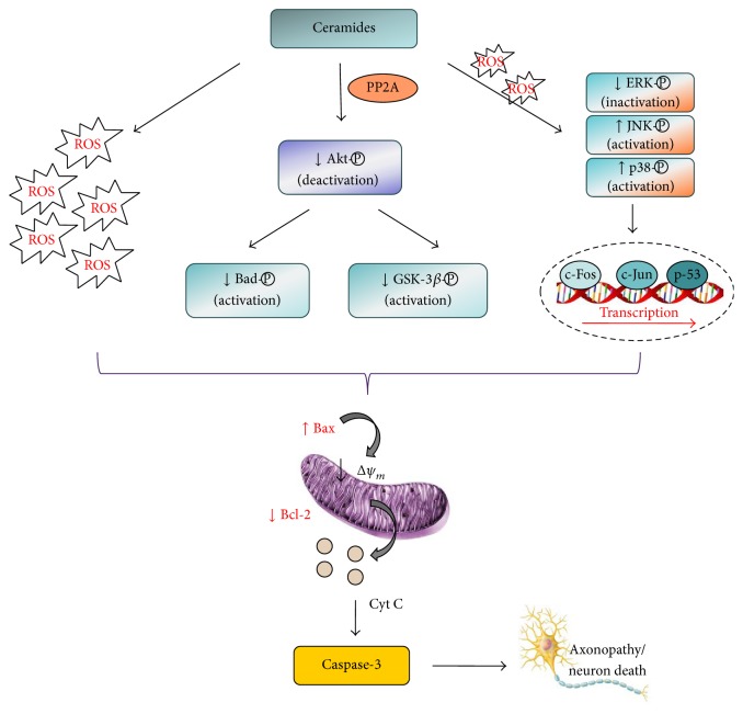
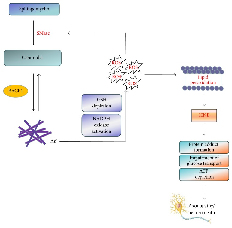
Alzheimer’s disease (AD), the most common chronic and progressive neurodegenerative disorder, is characterized by extracellular deposits of amyloid β -peptides (A β ) and intracellular deposits of hyperphosphorylated tau (phospho-tau) protein. Ceramides, the major molecules of sphingolipid metabolism and lipid second messengers, have been associated with AD progression and pathology via A β generation. Enhanced levels of ceramides directly increase A β through stabilization of β -secretase, the key enzyme in the amyloidogenic processing of A β precursor protein (APP). As a positive feedback loop, the generated oligomeric and fibrillar A β induces a further increase in ceramide levels by activating sphingomyelinases that catalyze the catabolic breakdown of sphingomyelin to ceramide. Evidence also supports important role of ceramides in neuronal apoptosis. Ceramides may initiate a cascade of biochemical alterations, which ultimately leads to neuronal death by diverse mechanisms, including depolarization and permeabilization of mitochondria, increased production of reactive oxygen species (ROS), cytochrome c release, Bcl-2 depletion, and caspase-3 activation, mainly by modulating intracellular signalling, particularly along the pathways related to Akt/PKB kinase and mitogen-activated protein kinases (MAPKs). This review summarizes recent findings related to the role of ceramides in oxidative stress-driven neuronal apoptosis and interplay with A β in the cascade of events ending in neuronal degeneration.
Overview
The claims and statistics of this paper haven’t been checked yet.
Run the check — free, no account needed. It reads the open-access text and checks the sources it cites.
Integrity signals
Registry
Cites
Statistics
Figures
Verification
Claims the paper makes
The claims and statistics of this paper haven’t been checked yet.
Run the check — free, no account needed. It reads the open-access text and checks the sources it cites.
Claims other papers make about this one
Each is checked against this paper’s own text: does it say what the citing paper says it does?
The source backs part of the claim, not all of it — or backs it for a different population, condition or comparison.
In the citing paper
“Elevated Cer levels have been associated with AD pathology, including promoting Aβ accumulation, mitochondrial dysfunction, and inflammation. 24 , 25”
From Protective mechanisms against Alzheimer's disease in APOE3‐Christchurch homozygous astrocytes (2025) · cites this paper as [25]
Experimental — accuracy not yet measuredThe cited source says what the claim attributes to it.
In the citing paper
“Ceramides are also known to initiate some of the pathological changes that drive AD progression. 24 , 25”
From Protective mechanisms against Alzheimer's disease in APOE3‐Christchurch homozygous astrocytes (2025) · cites this paper as [25]
Self-check
Percentages, sums and reported differences re-computed from the paper’s own numbers.
The self-check needs the paper’s full text.
Citations
Cites
1 retracted
Puglielli, L., Ellis, B. C., Saunders, A. J., & Kovacs, D. M. (2003) · Journal of Biological Chemistry · doi:10.1074/jbc.m300466200 · Cited by 265
RetractedLi, P., Nijhawan, D., Budihardjo, I., Srinivasula, S. M., Ahmad, M., Alnemri, E. S., & Wang, X. (1997) · Cell · doi:10.1016/s0092-8674(00)80434-1 · Cited by 7,289
Apolipoprotein E and Alzheimer disease: risk, mechanisms and therapy
Liu, C.-C., Kanekiyo, T., Xu, H., & Bu, G. (2013) · Nature Reviews Neurology · doi:10.1038/nrneurol.2012.263 · Cited by 3,459
Principles of bioactive lipid signalling: lessons from sphingolipids
Hannun, Y. A., & Obeid, L. M. (2008) · Nature Reviews Molecular Cell Biology · doi:10.1038/nrm2329 · Cited by 3,411
Uttara, B., Singh, A., Zamboni, P., & Mahajan, R. (2009) · Current Neuropharmacology · doi:10.2174/157015909787602823 · Cited by 3,378
Reactive oxygen species, cellular redox systems, and apoptosis
Circu, M. L., & Aw, T. Y. (2010) · Free Radical Biology and Medicine · doi:10.1016/j.freeradbiomed.2009.12.022 · Cited by 3,340
Blennow, K., de Leon, M. J., & Zetterberg, H. (2006) · The Lancet · doi:10.1016/s0140-6736(06)69113-7 · Cited by 3,134
Copper toxicity, oxidative stress, and antioxidant nutrients
Gaetke, L. M. (2003) · Toxicology · doi:10.1016/s0300-483x(03)00159-8 · Cited by 2,223
Copper, iron and zinc in Alzheimer's disease senile plaques
Lovell, M. A., Robertson, J. D., Teesdale, W. J., Campbell, J. L., & Markesbery, W. R. (1998) · Journal of the Neurological Sciences · doi:10.1016/s0022-510x(98)00092-6 · Cited by 2,120
Janssens, V., & Goris, J. (2001) · Biochemical Journal · doi:10.1042/0264-6021:3530417 · Cited by 1,927
Suppression of ceramide-mediated programmed cell death by sphingosine-1-phosphate
Cuvillier, O., Pirianov, G., Kleuser, B., Vanek, P. G., Coso, O. A., Gutkind, J. S., & Spiegel, S. (1996) · Nature · doi:10.1038/381800a0 · Cited by 1,532
Life and Death of Neurons in the Aging Brain
Morrison, J. H., & Hof, P. R. (1997) · Science · doi:10.1126/science.278.5337.412 · Cited by 1,344
Self-citationAmyloid Precursor Protein Trafficking, Processing, and Function
Thinakaran, G., & Koo, E. H. (2008) · Journal of Biological Chemistry · doi:10.1074/jbc.r800019200 · Cited by 1,141
Consumption of Fish and n-3 Fatty Acids and Risk of Incident Alzheimer Disease
Morris, M. C., Evans, D. A., Bienias, J. L., Tangney, C. C., Bennett, D. A., Wilson, R. S., Aggarwal, N., & Schneider, J. (2003) · Archives of Neurology · doi:10.1001/archneur.60.7.940 · Cited by 1,135
Neurodegenerative diseases and oxidative stress
Emerit, J., Edeas, M. A., & Bricaire, F. (2003) · Biomedicine & Pharmacotherapy · doi:10.1016/j.biopha.2003.11.004 · Cited by 1,057
Cutler, R. G., Kelly, J., Storie, K., Pedersen, W. A., Tammara, A., Hatanpaa, K., Troncoso, J. C., & Mattson, M. P. (2004) · Proceedings of the National Academy of Sciences · doi:10.1073/pnas.0305799101 · Cited by 1,008
Tau phosphorylation: the therapeutic challenge for neurodegenerative disease
Hanger, D. P., Anderton, B. H., & Noble, W. (2009) · Trends in Molecular Medicine · doi:10.1016/j.molmed.2009.01.003 · Cited by 965
Fassbender, K., Simons, M., Bergmann, C., Stroick, M., Lütjohann, D., Keller, P., Runz, H., Kühl, S., Bertsch, T., von Bergmann, K., et al. (2001) · Proceedings of the National Academy of Sciences · doi:10.1073/pnas.081620098 · Cited by 860
Oxidative Stress and the Pathogenesis of Alzheimer's Disease
Zhao, Y., & Zhao, B. (2013) · Oxidative Medicine and Cellular Longevity · doi:10.1155/2013/316523 · Cited by 855
The Genetics of Alzheimer Disease: Back to the Future
Bertram, L., Lill, C. M., & Tanzi, R. E. (2010) · Neuron · doi:10.1016/j.neuron.2010.10.013 · Cited by 834
Oxidative Alterations in Alzheimer's Disease
Markesbery, W. R., & Carney, J. M. (1999) · Brain Pathology · doi:10.1111/j.1750-3639.1999.tb00215.x · Cited by 789
Does neuroinflammation fan the flame in neurodegenerative diseases?
Frank-Cannon, T. C., Alto, L. T., McAlpine, F. E., & Tansey, M. G. (2009) · Molecular Neurodegeneration · doi:10.1186/1750-1326-4-47 · Cited by 751
Allanbutterfield, D., Castegna, A., Lauderback, C. M., & Drake, J. (2002) · Neurobiology of Aging · doi:10.1016/s0197-4580(01)00340-2 · Cited by 715
Chen, Z., & Zhong, C. (2013) · Progress in Neurobiology · doi:10.1016/j.pneurobio.2013.06.004 · Cited by 698
Lim, G. P., Calon, F., Morihara, T., Yang, F., Teter, B., Ubeda, O. J., Salem, N., Frautschy, S. A., & Cole, G. M. (2005) · Journal of Neuroscience · doi:10.1523/jneurosci.4225-04.2005 · Cited by 697
Ceramide activates heterotrimeric protein phosphatase 2A
Dobrowsky, R. T., Kamibayashi, C., Mumby, M. C., & Hannun, Y. A. (1993) · Journal of Biological Chemistry · doi:10.1016/s0021-9258(18)82288-8 · Cited by 666
Han, X., M. Holtzman, D., W. McKeel, D., Kelley, J., & Morris, J. C. (2002) · Journal of Neurochemistry · doi:10.1046/j.1471-4159.2002.00997.x · Cited by 626
Lipid Rafts in the Maintenance of Synapses, Dendritic Spines, and Surface AMPA Receptor Stability
Hering, H., Lin, C.-C., & Sheng, M. (2003) · Journal of Neuroscience · doi:10.1523/jneurosci.23-08-03262.2003 · Cited by 622
The redox chemistry of the Alzheimer's disease amyloid β peptide
Smith, D. G., Cappai, R., & Barnham, K. J. (2007) · Biochimica et Biophysica Acta (BBA) - Biomembranes · doi:10.1016/j.bbamem.2007.02.002 · Cited by 608
Cell death and diseases related to oxidative stress: 4-hydroxynonenal (HNE) in the balance
Dalleau, S., Baradat, M., Guéraud, F., & Huc, L. (2013) · Cell Death & Differentiation · doi:10.1038/cdd.2013.138 · Cited by 580
Chemistry and biochemistry of lipid peroxidation products
Guéraud, F., Atalay, M., Bresgen, N., Gašparović, A. Č., Eckl, P., Laurence Therese, H. U. C., Jouanin, I., Siems, W., & Uchida, K. (2010) · Free Radical Research · doi:10.3109/10715762.2010.498477 · Cited by 570
Mark, R. J., Pang, Z., Geddes, J. W., Uchida, K., & Mattson, M. P. (1997) · The Journal of Neuroscience · doi:10.1523/jneurosci.17-03-01046.1997 · Cited by 551
Butterfield, D. A., Swomley, A. M., & Sultana, R. (2013) · Antioxidants & Redox Signaling · doi:10.1089/ars.2012.5027 · Cited by 526
Deregulation of sphingolipid metabolism in Alzheimer’s disease
He, X., Huang, Y., Li, B., Gong, C.-X., & Schuchman, E. H. (2010) · Neurobiology of Aging · doi:10.1016/j.neurobiolaging.2008.05.010 · Cited by 516
The Alzheimer's Aβ peptide induces neurodegeneration and apoptotic cell death in transgenic mice
LaFerla, F. M., Tinkle, B. T., Bieberich, C. J., Haudenschild, C. C., & Jay, G. (1995) · Nature Genetics · doi:10.1038/ng0195-21 · Cited by 513
Evidence for Apoptotic Cell Death in Alzheimer's Disease
Smale, G., Nichols, N. R., Brady, D. R., Finch, C. E., & Horton, W. E. (1995) · Experimental Neurology · doi:10.1006/exnr.1995.1025 · Cited by 495
Dinkins, M. B., Dasgupta, S., Wang, G., Zhu, G., & Bieberich, E. (2014) · Neurobiology of Aging · doi:10.1016/j.neurobiolaging.2014.02.012 · Cited by 494
Oxidative stress in Alzheimer disease: A possibility for prevention
Bonda, D., Wang, X., Perry, G. L., Nunomura, A., Tabaton, M., Zhu, X., & Smith, M. A. (2010) · Neuropharmacology · doi:10.1016/j.neuropharm.2010.04.005 · Cited by 487
MORISHIMA, Y., Gotoh, Y., Zieg, J., Barrett, T., Takano, H., Flavell, R. A., Davis, R. J., Shirasaki, Y., & Greenberg, M. E. (2001) · Journal of Neuroscience · doi:10.1523/jneurosci.21-19-07551.2001 · Cited by 486
Sultana, R., Perluigi, M., & Butterfield, D. A. (2013) · Free Radical Biology and Medicine · doi:10.1016/j.freeradbiomed.2012.09.027 · Cited by 478
Griffin, R. J., Moloney, A., Kelliher, M., Johnston, J. A., Ravid, R., Dockery, P., O'Connor, R., & O'Neill, C. (2005) · Journal of Neurochemistry · doi:10.1111/j.1471-4159.2004.02949.x · Cited by 459
Sparks, D. L., & Schreurs, B. G. (2003) · Proceedings of the National Academy of Sciences · doi:10.1073/pnas.1832769100 · Cited by 455
Regulation of cholesterol and sphingomyelin metabolism by amyloid-β and presenilin
Grimm, M. O. W., Grimm, H. S., Pätzold, A. J., Zinser, E. G., Halonen, R., Duering, M., Tschäpe, J., Strooper, B. D., Müller, U., Shen, J., et al. (2005) · Nature Cell Biology · doi:10.1038/ncb1313 · Cited by 454
Keller, J. N., Pang, Z., Geddes, J. W., Begley, J. G., Germeyer, A., Waeg, G., & Mattson, M. P. (1997) · Journal of Neurochemistry · doi:10.1046/j.1471-4159.1997.69010273.x · Cited by 439
Liver cell death and anemia in Wilson disease involve acid sphingomyelinase and ceramide
Lang, P. A., Schenck, M., Nicolay, J. P., Becker, J. U., Kempe, D. S., Lupescu, A., Koka, S., Eisele, K., Klarl, B. A., Rubben, H., et al. (2007) · Nature Medicine · doi:10.1038/nm1539 · Cited by 438
Parkin prevents mitochondrial swelling and cytochrome c release in mitochondria-dependent cell death
Darios, F. D. (2003) · Human Molecular Genetics · doi:10.1093/hmg/ddg044 · Cited by 407
Ceramide Channels Increase the Permeability of the Mitochondrial Outer Membrane to Small Proteins
Siskind, L. J., Kolesnick, R., & Colombini, M. (2002) · Journal of Biological Chemistry · doi:10.1074/jbc.m200754200 · Cited by 390
Masliah, E., Sisk, A., Mallory, M. E., Mucke, L., Schenk, D., & Games, D. (1996) · Journal of Neuroscience · doi:10.1523/jneurosci.16-18-05795.1996 · Cited by 389
Weber, J. T., Slemmer, J. E., Shacka, J. J., & Sweeney, M. I. (2008) · Current Medicinal Chemistry · doi:10.2174/092986708783497337 · Cited by 386
FAN, a Novel WD-Repeat Protein, Couples the p55 TNF-Receptor to Neutral Sphingomyelinase
Adam‐Klages, S., Adam, D., Wiegmann, K., Struve, S., Kolanus, W., Schneider‐Mergener, J., & Krönke, M. (1996) · Cell · doi:10.1016/s0092-8674(00)80169-5 · Cited by 381
Farooqui, A. A., Ong, W., & Horrocks, L. A. (2006) · Pharmacological Reviews · doi:10.1124/pr.58.3.7 · Cited by 378
Intraneuronal β-amyloid accumulation and synapse pathology in Alzheimer’s disease
Gouras, G. K., Tampellini, D., Takahashi, R. H., & Capetillo-Zarate, E. (2010) · Acta Neuropathologica · doi:10.1007/s00401-010-0679-9 · Cited by 372
Kuo, Y.-C., Huang, K.-Y., Yang, C.-H., Yang, Y.-S., Lee, W.-Y., & Chiang, C.-W. (2008) · Journal of Biological Chemistry · doi:10.1074/jbc.m709585200 · Cited by 367
Zhou, H., Summers, S. A., Birnbaum, M. J., & Pittman, R. N. (1998) · Journal of Biological Chemistry · doi:10.1074/jbc.273.26.16568 · Cited by 356
Soluble Aβ oligomer production and toxicity
Larson, M. E., & Lesné, S. E. (2012) · Journal of Neurochemistry · doi:10.1111/j.1471-4159.2011.07478.x · Cited by 356
Cordy, J. M., Hussain, I., Dingwall, C., Hooper, N. M., & Turner, A. J. (2003) · Proceedings of the National Academy of Sciences · doi:10.1073/pnas.1635130100 · Cited by 356
Acid and neutral sphingomyelinases: roles and mechanisms of regulation
Marchesini, N., & Hannun, Y. A. (2004) · Biochemistry and Cell Biology · doi:10.1139/o03-091 · Cited by 342
Fibrillar Amyloid-β Peptides Activate Microglia via TLR2: Implications for Alzheimer’s Disease
Jana, M., Palencia, C. A., & Pahan, K. (2008) · The Journal of Immunology · doi:10.4049/jimmunol.181.10.7254 · Cited by 338
Ceramide Induces Bcl2 Dephosphorylation via a Mechanism Involving Mitochondrial PP2A
Ruvolo, P. P., Deng, X., Ito, T., Carr, B. K., & May, W. S. (1999) · Journal of Biological Chemistry · doi:10.1074/jbc.274.29.20296 · Cited by 338
Inhibition of the Neutral Magnesium-dependent Sphingomyelinase by Glutathione
Liu, B., & Hannun, Y. A. (1997) · Journal of Biological Chemistry · doi:10.1074/jbc.272.26.16281 · Cited by 304
The many “faces” of copper in medicine and treatment
Hordyjewska, A., Popiołek, Ł. P., & Kocot, J. (2014) · BioMetals · doi:10.1007/s10534-014-9736-5 · Cited by 301
Morrison, C. D., Pistell, P. J., Ingram, D. K., Johnson, W. D., Liu, Y., Fernandez‐Kim, S. O., White, C. L., Purpera, M. N., Uranga, R. M., Bruce‐Keller, A. J., et al. (2010) · Journal of Neurochemistry · doi:10.1111/j.1471-4159.2010.06865.x · Cited by 300
Long Chain Ceramides Activate Protein Phosphatase-1 and Protein Phosphatase-2A
Chalfant, C. E., Kishikawa, K., Mumby, M. C., Kamibayashi, C., Bielawska, A. E., & Hannun, Y. A. (1999) · Journal of Biological Chemistry · doi:10.1074/jbc.274.29.20313 · Cited by 300
Poon, H. F., Calabrese, V., Scapagnini, G., & Butterfield, D. A. (2004) · Clinics in Geriatric Medicine · doi:10.1016/j.cger.2004.02.005 · Cited by 300
Choi, Y., Kim, H., Shin, K. Y., Kim, E., Kim, M., Kim, H.-S., Park, C. H., Jeong, Y. H., Yoo, J., Lee, J., et al. (2007) · Neuropsychopharmacology · doi:10.1038/sj.npp.1301377 · Cited by 296
Roles and regulation of secretory and lysosomal acid sphingomyelinase
Jenkins, R. W., Canals, D., & Hannun, Y. A. (2009) · Cellular Signalling · doi:10.1016/j.cellsig.2009.01.026 · Cited by 286
Lee, J.-T., Xu, J., Lee, J.-M., Ku, G., Han, X., Yang, D.-I., Chen, S., & Hsu, C. Y. (2004) · The Journal of Cell Biology · doi:10.1083/jcb.200307017 · Cited by 280
Ceramide function in the brain: when a slight tilt is enough
Mencarelli, C., & Martínez‐Martínez, P. (2012) · Cellular and Molecular Life Sciences · doi:10.1007/s00018-012-1038-x · Cited by 278
Roles for dysfunctional sphingolipid metabolism in Alzheimer's disease neuropathogenesis
Haughey, N. J., Bandaru, V. V. R., Bae, M. H., & Mattson, M. P. (2010) · Biochimica et Biophysica Acta (BBA) - Molecular and Cell Biology of Lipids · doi:10.1016/j.bbalip.2010.05.003 · Cited by 277
Giraldo, E., Lloret, A., Fuchsberger, T., & Viña, J. (2014) · Redox Biology · doi:10.1016/j.redox.2014.03.002 · Cited by 268
Ceramide Induces Cytochrome c Release from Isolated Mitochondria
Ghafourifar, P., Klein, S. D., Schucht, O., Schenk, U., Pruschy, M. N., Rocha, S., & Richter, C. (1999) · Journal of Biological Chemistry · doi:10.1074/jbc.274.10.6080 · Cited by 266
Increased ceramide in brains with Alzheimer's and other neurodegenerative diseases
Filippov, V., Song, M. A., Zhang, K., Vinters, H. V., Tung, S., Kirsch, W. M., Yang, J., & Duerksen-Hughes, P. J. (2012) · Journal of Alzheimer s Disease · doi:10.3233/jad-2011-111202 · Cited by 264
Apoptosis in Alzheimer's disease—an update
Shimohama, S. (2000) · APOPTOSIS · doi:10.1023/a:1009625323388 · Cited by 255
Katsel, P., Li, C., & Haroutunian, V. H. (2007) · Neurochemical Research · doi:10.1007/s11064-007-9297-x · Cited by 244
Rushworth, J. V., & Hooper, N. M. (2010) · International Journal of Alzheimer s Disease · doi:10.4061/2011/603052 · Cited by 234
Jana, A., Hogan, E. L., & Pahan, K. (2009) · Journal of the Neurological Sciences · doi:10.1016/j.jns.2008.12.010 · Cited by 233
Yao, M., Nguyen, T. V., & Pike, C. J. (2005) · Journal of Neuroscience · doi:10.1523/jneurosci.4736-04.2005 · Cited by 233
4-Hydroxynonenal in the Pathogenesis and Progression of Human Diseases
Shoeb, M., Ansari, N., Srivastava, S., & Ramana, K. (2013) · Current Medicinal Chemistry · doi:10.2174/09298673113209990181 · Cited by 230
Dietary fat composition and dementia risk
Morris, M. C., & Tangney, C. (2014) · Neurobiology of Aging · doi:10.1016/j.neurobiolaging.2014.03.038 · Cited by 228
Serum ceramides increase the risk of Alzheimer disease
Mielke, M. M., Bandaru, V. V. R., Haughey, N. J., Xia, J., Fried, L. P., Yaşar, S., Albert, M., Varma, V. R., Harris, G., Schneider, E. B., et al. (2012) · Neurology · doi:10.1212/wnl.0b013e318264e380 · Cited by 227
Spatial Segregation of γ-Secretase and Substrates in DistinctMembraneDomains
Vetrivel, K. S., Cheng, H., Kim, S.-H., Chen, Y., Barnes, N. Y., Parent, A., Sisodia, S. S., & Thinakaran, G. (2005) · Journal of Biological Chemistry · doi:10.1074/jbc.m503570200 · Cited by 225
Malaplate, C., Florent‐Béchard, S., Youssef, I., Koziel, V., Sponne, I., Kriem, B., Leininger‐Muller, B., Olivier, J. L., Oster, T., & Pillot, T. (2006) · Neurobiology of Disease · doi:10.1016/j.nbd.2006.02.010 · Cited by 224
Alzheimer´s Disease and Oxidative Stress: A Review
Pohanka, M. (2013) · Current Medicinal Chemistry · doi:10.2174/09298673113206660258 · Cited by 219
Ferreiro, E. B., Oliveira, C. R., & Pereira, C. (2008) · Neurobiology of Disease · doi:10.1016/j.nbd.2008.02.003 · Cited by 218
Mukhopadhyay, A., Saddoughi, S. A., Song, P., Sultan, I. Y., Ponnusamy, S., Senkal, C. E., Snook, C. F., Arnold, H., Sears, R. C., Hanniui, Y. A., et al. (2008) · The FASEB Journal · doi:10.1096/fj.08-120550 · Cited by 217
Decreased thioredoxin and increased thioredoxin reductase levels in alzheimer’s disease brain
Lovell, M. A., Xie, C., Gabbita, S. P., & Markesbery, W. R. (2000) · Free Radical Biology and Medicine · doi:10.1016/s0891-5849(99)00258-0 · Cited by 206
JNK pathway: diseases and therapeutic potential
Cui, J., Zhang, M., Zhang, Y., & Xu, Z. (2007) · Acta Pharmacologica Sinica · doi:10.1111/j.1745-7254.2007.00579.x · Cited by 202
LEVENSON, R. W., Sturm, V. E., & Haase, C. M. (2014) · Annual Review of Clinical Psychology · doi:10.1146/annurev-clinpsy-032813-153653 · Cited by 200
Salinas, M., López‐Valdaliso, R., Martı́n, D., Alvarez, A. M., & Cuadrado, A. (2000) · Molecular and Cellular Neuroscience · doi:10.1006/mcne.1999.0813 · Cited by 199
Sphingolipid Metabolic Pathway: An Overview of Major Roles Played in Human Diseases
Rao, R. P., Vaidyanathan, N., Rengasamy, M., Oommen, A. M., Somaiya, N., & Jagannath, M. R. (2013) · Journal of Lipids · doi:10.1155/2013/178910 · Cited by 199
The role of inflammasome in Alzheimer's disease
Liu, L., & Chan, C. (2014) · Ageing Research Reviews · doi:10.1016/j.arr.2013.12.007 · Cited by 196
Cholesterol Level and Statin Use in Alzheimer Disease
Shepardson, N. E. (2011) · Archives of Neurology · doi:10.1001/archneurol.2011.242 · Cited by 190
Serum sphingomyelins and ceramides are early predictors of memory impairment
Mielke, M. M., Bandaru, V. V. R., Haughey, N. J., Rabins, P. V., Lyketsos, C. G., & Carlson, M. C. (2008) · Neurobiology of Aging · doi:10.1016/j.neurobiolaging.2008.03.011 · Cited by 188
Amyloid Beta: Multiple Mechanisms of Toxicity and Only Some Protective Effects?
Carrillo‐Mora, P., Luna, R., & Colín-Barenque, L. (2014) · Oxidative Medicine and Cellular Longevity · doi:10.1155/2014/795375 · Cited by 187
Jana, A., & Pahan, K. (2004) · Journal of Biological Chemistry · doi:10.1074/jbc.m404635200 · Cited by 186
France‐Lanord, V., Brugg, B., Michel, P. P., Agid, Y., & Ruberg, M. (1997) · Journal of Neurochemistry · doi:10.1046/j.1471-4159.1997.69041612.x · Cited by 182
DHA Improves Cognition and Prevents Dysfunction of Entorhinal Cortex Neurons in 3xTg-AD Mice
Arsenault, D., Julien, C., Tremblay, C., & Calon, F. (2011) · PLoS ONE · doi:10.1371/journal.pone.0017397 · Cited by 177
Ferrer, I. (2009) · Journal of Bioenergetics and Biomembranes · doi:10.1007/s10863-009-9243-5 · Cited by 172
Fabelo, N., Martín, M. V., Marín, R., Moreno, D., Ferrer, I., & Díaz, M. L. (2014) · Neurobiology of Aging · doi:10.1016/j.neurobiolaging.2014.02.005 · Cited by 169
Martı́n, D., Salinas, M., Fujita, N., Tsuruo, T., & Cuadrado, A. (2002) · Journal of Biological Chemistry · doi:10.1074/jbc.m201070200 · Cited by 168
Sphingomyelin synthase-related protein SMSr controls ceramide homeostasis in the ER
Vacaru, A. M., Tafesse, F., Ternes, P., Kondylis, V., Hermansson, M., Brouwers, J. F. H. M., Somerharju, P. J., Rabouille, C., & Holthuis, J. C. M. (2009) · The Journal of Cell Biology · doi:10.1083/jcb.200903152 · Cited by 168
Kitazawa, M., Cheng, D., & LaFerla, F. M. (2009) · Journal of Neurochemistry · doi:10.1111/j.1471-4159.2009.05901.x · Cited by 165
Ceramide and the mitochondrial respiratory chain
Kogot‐Levin, A., & Saada, A. (2013) · Biochimie · doi:10.1016/j.biochi.2013.07.027 · Cited by 162
Stoica, B. A., Movsesyan, V. A., Lea, P. M., & Faden, A. I. (2003) · Molecular and Cellular Neuroscience · doi:10.1016/s1044-7431(02)00028-3 · Cited by 160
Oxidative Stress and Cell Membranes in the Pathogenesis of Alzheimer's Disease
Axelsen, P. H., Komatsu, H., & Murray, I. V. J. (2011) · Physiology · doi:10.1152/physiol.00024.2010 · Cited by 159
Plasma Sphingomyelins are Associated with Cognitive Progression in Alzheimer's Disease
Mielke, M. M., Haughey, N. J., Bandaru, V. V. R., Weinberg, D. D., Darby, E. J., Zaidi, N., Pavlik, V., Doody, R. S., & Lyketsos, C. G. (2011) · Journal of Alzheimer s Disease · doi:10.3233/jad-2011-110405 · Cited by 158
4-Hydroxy-2-Nonenal Pyrrole Adducts in Human Neurodegenerative Disease
Montine, K. S., KIM, P. J., Olson, S. J., Markesbery, W. R., & Montine, T. J. (1997) · Journal of Neuropathology & Experimental Neurology · doi:10.1097/00005072-199708000-00004 · Cited by 158
Oxidative Stress and Metabolic Syndrome: Cause or Consequence of Alzheimer's Disease?
Contreras, D. L., Carvajal, K., Toral-Ríos, D., Franco-Bocanegra, D., & Campos‐Peña, V. (2014) · Oxidative Medicine and Cellular Longevity · doi:10.1155/2014/497802 · Cited by 155
Lyn‐Cook, L. E., Lawton, M., Tong, M., Silbermann, E., Longato, L., Jiao, P., Mark, P., Wands, J. R., Xu, H., & de la Monte, S. M. (2009) · Journal of Alzheimer s Disease · doi:10.3233/jad-2009-0984 · Cited by 154
A TrkA-to-p75NTR molecular switch activates amyloid β-peptide generation during aging
Costantini, C., Weindruch, R. H., Valle, G. D., & Puglielli, L. (2005) · Biochemical Journal · doi:10.1042/bj20050700 · Cited by 149
Personality changes in Alzheimer's disease: a systematic review
Wahlin, T. R., & Byrne, G. J. (2010) · International Journal of Geriatric Psychiatry · doi:10.1002/gps.2655 · Cited by 147
Lin, C., Chen, C., Chiang, C.-W., Jan, M., Huang, W., & Lin, Y.-S. (2007) · Journal of Cell Science · doi:10.1242/jcs.03473 · Cited by 144
Mielke, M. M., & Lyketsos, C. G. (2010) · NeuroMolecular Medicine · doi:10.1007/s12017-010-8121-y · Cited by 143
The structural requirements for ceramide activation of serine-threonine protein phosphatases
Chalfant, C. E., Szulc, Z. M., Roddy, P. L., Bielawska, A. E., & Hannun, Y. A. (2004) · Journal of Lipid Research · doi:10.1194/jlr.m300347-jlr200 · Cited by 141
Lipid Integration in Neurodegeneration: An Overview of Alzheimer’s Disease
Yadav, R. S., & Tiwari, N. K. (2014) · Molecular Neurobiology · doi:10.1007/s12035-014-8661-5 · Cited by 140
Ceramide channels and their role in mitochondria-mediated apoptosis
Colombini, M. (2010) · Biochimica et Biophysica Acta (BBA) - Bioenergetics · doi:10.1016/j.bbabio.2010.01.021 · Cited by 139
Dietary fatty acids intake: possible role in cognitive decline and dementia
Solfrizzi, V., D’Introno, A., Colacicco, A. M., Capurso, C. A., DelParigi, A., Capurso, S. A., Gadaleta, A., Capurso, A., & Panza, F. (2005) · Experimental Gerontology · doi:10.1016/j.exger.2005.01.001 · Cited by 136
Patil, S. P., Melrose, J., & Chan, C. (2007) · European Journal of Neuroscience · doi:10.1111/j.1460-9568.2007.05797.x · Cited by 129
The Role of the Ceramide Acyl Chain Length in Neurodegeneration: Involvement of Ceramide Synthases
Ben‐David, O., & Futerman, A. H. (2010) · NeuroMolecular Medicine · doi:10.1007/s12017-010-8114-x · Cited by 129
Gong, C., Liu, F., Grundke-Iqbal, I., & Iqbal, K. (2006) · Journal of Alzheimer's Disease · doi:10.3233/jad-2006-9101 · Cited by 128
Phosphoprotein Phosphatase 2A: A Novel Druggable Target for Alzheimer’s Disease
Voronkov, M. V., Braithwaite, S. P., & Stock, J. B. (2011) · Future Medicinal Chemistry · doi:10.4155/fmc.11.47 · Cited by 127
Willaime‐Morawek, S., Vanhoutte, P., Caboche, J., Lemaigre‐Dubreuil, Y., Mariani, J., & Brugg, B. (2001) · European Journal of Neuroscience · doi:10.1046/j.0953-816x.2001.01581.x · Cited by 127
Sphingosine-1-phosphate: the Swiss army knife of sphingolipid signaling
Maceyka, M. W., Milstien, S., & Spiegel, S. (2008) · Journal of Lipid Research · doi:10.1194/jlr.r800065-jlr200 · Cited by 124
The case for soluble Aβ oligomers as a drug target in Alzheimer's disease
Hefti, F. F., Goure, W. F., Jerecìć, J., Iverson, K. S., Walicke, P. A., & Krafft, G. A. (2013) · Trends in Pharmacological Sciences · doi:10.1016/j.tips.2013.03.002 · Cited by 122
Papaioannou, N. G., Tooten, P. C. J., van Ederen, A. M., Böhl, J., Rofina, J., Tsangaris, T., & Gruys, E. (2001) · Amyloid · doi:10.3109/13506120108993810 · Cited by 121
Montine, T. J., Markesbery, W. R., Zackert, W. E., Sanchez, S. C., Roberts, L. J., & Morrow, J. D. (1999) · American Journal Of Pathology · doi:10.1016/s0002-9440(10)65185-1 · Cited by 116
Liu, B., Hassler, D. F., Smith, G. K., Weaver, K. L., & Hannun, Y. A. (1998) · Journal of Biological Chemistry · doi:10.1074/jbc.273.51.34472 · Cited by 115
Arnal, N., Cristalli, D. O., de Alaniz, M. J. T., & Marra, C. A. (2009) · Brain Research · doi:10.1016/j.brainres.2009.11.085 · Cited by 113
Tamboli, I. Y., Prager, K., Barth, E., Heneka, M., Sandhoff, K., & Walter, J. (2005) · Journal of Biological Chemistry · doi:10.1074/jbc.m414525200 · Cited by 110
Ceramidase Regulates Synaptic Vesicle Exocytosis and Trafficking
Rohrbough, J. C., Rushton, E., Musselman, L. P., Woodruff, E. A., Matthies, H. J. G., Acharya, U. R., Acharya, J., & Broadie, K. S. (2004) · Journal of Neuroscience · doi:10.1523/jneurosci.1146-04.2004 · Cited by 104
Elevated cerebrospinal fluid sphingomyelin levels in prodromal Alzheimer's disease
Košiček, M., Zetterberg, H., Andreasen, N., Peter‐Katalinić, J., & Hecimovic, S. K. (2012) · Neuroscience Letters · doi:10.1016/j.neulet.2012.04.019 · Cited by 102
Stranahan, A. M., Cutler, R. G., Button, C., Telljohann, R., & Mattson, M. P. (2011) · Journal of Neurochemistry · doi:10.1111/j.1471-4159.2011.07351.x · Cited by 100
Ceramide induces neuronal apoptosis through the caspase-9/caspase-3 pathway
Movsesyan, V. A., Yakovlev, A. G., Dabaghyan, E. A., Stoica, B. A., & Faden, A. I. (2002) · Biochemical and Biophysical Research Communications · doi:10.1016/s0006-291x(02)02593-7 · Cited by 99
Asiatic acid protects primary neurons against C2-ceramide-induced apoptosis
Zhang, X., Wu, J., Dou, Y., Xia, B., Rong, W., Rimbach, G., & Lou, Y. (2012) · European Journal of Pharmacology · doi:10.1016/j.ejphar.2012.01.006 · Cited by 99
Olivieri, G., Baysang, G., Meier, F., Müller-Spahn, F., Stähelin, H. B., Brockhaus, M., & Brack, C. (2001) · Journal of Neurochemistry · doi:10.1046/j.1471-4159.2001.00090.x · Cited by 98
Stoica, B. A., Movsesyan, V. A., Knoblach, S. M., & Faden, A. I. (2005) · Molecular and Cellular Neuroscience · doi:10.1016/j.mcn.2005.02.009 · Cited by 98
Protective effects of S-nitrosoglutathione against amyloid β-peptide neurotoxicity
Ju, T., Chen, S., Liu, C.-C., & Yang, D. (2005) · Free Radical Biology and Medicine · doi:10.1016/j.freeradbiomed.2004.12.019 · Cited by 96
Patil, S. P., Sheng, L., Masserang, A., & Chan, C. (2006) · Neuroscience Letters · doi:10.1016/j.neulet.2006.07.015 · Cited by 95
Falluel‐Morel, A., Aubert, N., Vaudry, D., Basille, M., Fontaine, M., Fournier, A., Vaudry, H., & Gonzalez, B. J. (2004) · Journal of Neurochemistry · doi:10.1111/j.1471-4159.2004.02810.x · Cited by 95
Ceramide in the Molecular Mechanisms of Neuronal Cell Death. The Role of Sphingosine-1-Phosphate
Czubowicz, K., & Strosznajder, R. P. (2014) · Molecular Neurobiology · doi:10.1007/s12035-013-8606-4 · Cited by 91
Expression of apoptosis related proteins in brains of patients with Alzheimer's disease
Engidawork, E., Gulesserian, T., Seidl, R., Cairns, N., & Lubec, G. (2001) · Neuroscience Letters · doi:10.1016/s0304-3940(01)01618-4 · Cited by 91
Intersection between metabolic dysfunction, high fat diet consumption, and brain aging
Uranga, R. M., Bruce‐Keller, A. J., Morrison, C. D., Fernandez‐Kim, S. O., Ebenezer, P. J., Zhang, L., Dasuri, K., & Keller, J. N. (2010) · Journal of Neurochemistry · doi:10.1111/j.1471-4159.2010.06803.x · Cited by 90
Dietary fats, cerebrovasculature integrity and Alzheimer’s disease risk
Takechi, R., Galloway, S., Pallebage‐Gamarallage, M. M., Lam, V., & Mamo, J. C. L. (2009) · Progress in Lipid Research · doi:10.1016/j.plipres.2009.10.004 · Cited by 89
Anderson, A. J., Stoltzner, S., Lai, F., Su, J. H., & Nixon, R. A. (2000) · Neurobiology of Aging · doi:10.1016/s0197-4580(00)00126-3 · Cited by 88
Alessenko, A. V., Бугрова, А. Е., & Дудник, Л. Б. (2004) · Biochemical Society Transactions · doi:10.1042/bst0320144 · Cited by 88
Zeng, C., Lee, J. T., Chen, H., Chen, S., Hsu, C. Y., & Xu, J. (2005) · Journal of Neurochemistry · doi:10.1111/j.1471-4159.2005.03217.x · Cited by 80
Neutral sphingomyelinases and nSMase2: Bridging the gaps
Clarke, C. J., & Hannun, Y. A. (2006) · Biochimica et Biophysica Acta (BBA) - Biomembranes · doi:10.1016/j.bbamem.2006.06.025 · Cited by 80
Cross-talk of membrane lipids and Alzheimer-related proteins
Walter, J., & van Echten‐Deckert, G. (2013) · Molecular Neurodegeneration · doi:10.1186/1750-1326-8-34 · Cited by 79
Sphingolipid and Ceramide Homeostasis: Potential Therapeutic Targets
Young, S. A., Mina, J. G., Denny, P. W., & Smith, T. (2012) · Biochemistry Research International · doi:10.1155/2012/248135 · Cited by 77
Nutrient intake and plasma β-amyloid
Gu, Y., Schupf, N., Cosentino, S., Luchsinger, J. A., & Scarmeas, N. (2012) · Neurology · doi:10.1212/wnl.0b013e318258f7c2 · Cited by 77
Díaz, M. L., Fabelo, N., Martín, M. V., Ferrer, I., Gómez, T., & Marín, R. (2014) · Journal of Alzheimer s Disease · doi:10.3233/jad-141146 · Cited by 75
Liu, L., Martin, R., & Chan, C. (2012) · Neurobiology of Aging · doi:10.1016/j.neurobiolaging.2012.05.017 · Cited by 75
Aβ1–42 modulation of Akt phosphorylation via α7 nAChR and NMDA receptors
Abbott, J. J., Howlett, D. R., Francis, P. T., & Williams, R. J. (2007) · Neurobiology of Aging · doi:10.1016/j.neurobiolaging.2007.01.003 · Cited by 71
C-jun N-terminal kinases/c-Jun and p38 pathways cooperate in ceramide-induced neuronal apoptosis
Willaime‐Morawek, S., Brami‐Cherrier, K., Mariani, J., Caboche, J., & Brugg, B. (2003) · Neuroscience · doi:10.1016/s0306-4522(02)00996-x · Cited by 71
Toman, R. E., Movsesyan, V. A., Murthy, S. K., Milstien, S., Spiegel, S., & Faden, A. I. (2002) · Journal of Neuroscience Research · doi:10.1002/jnr.10190 · Cited by 70
Ceramides and sphingomyelinases in senile plaques
Panchal, M., Gaudin, M., Lazar, A. N., Salvati, E., Rivals, I., Ayciriex, S., Dauphinot, L., Dargère, D., Auzeil, N., Masserini, M., et al. (2014) · Neurobiology of Disease · doi:10.1016/j.nbd.2014.01.010 · Cited by 69
Dinkins, M. B., Dasgupta, S., Wang, G., Zhu, G., He, Q., Kong, J. N., & Bieberich, E. (2015) · Journal of Alzheimer s Disease · doi:10.3233/jad-150088 · Cited by 68
Müller, W. E. G., Romero, F. J., Perovic, S., Pergande, G., & Pialoglou, P. (1997) · Journal of Neurochemistry · doi:10.1046/j.1471-4159.1997.68062371.x · Cited by 67
Darios, F. D., Lambeng, N., Troadec, J., Michel, P. P., & Ruberg, M. (2003) · Journal of Neurochemistry · doi:10.1046/j.1471-4159.2003.01590.x · Cited by 65
Qin, X., Cheng, Y., & Yu, L. (2010) · Neuroscience Letters · doi:10.1016/j.neulet.2010.05.062 · Cited by 64
Involvement of sphingomyelinases in TNF signaling pathways
Krönke, M. (1999) · Chemistry and Physics of Lipids · doi:10.1016/s0009-3084(99)00084-5 · Cited by 64
Chakrabarti, S., Sinha, M., Thakurta, I. G., Banerjee, P., & Chattopadhyay, M. (2013) · Current Medicinal Chemistry · doi:10.2174/09298673113209990152 · Cited by 63
Ceramide and mitochondria in ischemic brain injury.
Novgorodov, S. A., & Gudz, T. I. (2011) · Europe PMC (PubMed Central) · Cited by 62
Rosales‐Corral, S., Acuña-Castroviejo, D., Tan, D. X., López-Armas, G. D. C., Cruz-Ramos, J. A., Munoz, R., Melnikov, V., Manchester, L. C., & Reiter, R. J. (2012) · Oxidative Medicine and Cellular Longevity · doi:10.1155/2012/843649 · Cited by 61
Ito, A., Uehara, T., Tokumitsu, A., Okuma, Y., & Nomura, Y. (1999) · Biochimica et Biophysica Acta (BBA) - Molecular Cell Research · doi:10.1016/s0167-4889(99)00131-7 · Cited by 61
Gomez‐Brouchet, A., Pchejetski, D., Brizuela, L., Garcia, V., Altié, M.-F., Maddelein, M., Delisle, M., & Cuvillier, O. (2007) · Molecular Pharmacology · doi:10.1124/mol.106.033738 · Cited by 60
Protective effects of S-nitrosoglutathione against amyloid beta-peptide neurotoxicity
Jü, Chert, Sd, Liu, Yang, Y., & Di (2006) · 中国生物学文摘 · Cited by 60
Chen, S., Yin, J., Hwang, C., Tang, C.-M., & Yang, D. (2012) · Free Radical Research · doi:10.3109/10715762.2012.674640 · Cited by 59
Woltjer, R. L., Nghiem, W., Maezawa, I., Milatović, D., Vaisar, T., Montine, K. S., & Montine, T. J. (2005) · Journal of Neurochemistry · doi:10.1111/j.1471-4159.2005.03109.x · Cited by 58
Chen, C., Lin, C., Chiang, C.-W., Jan, M., & Lin, Y.-S. (2006) · Molecular Pharmacology · doi:10.1124/mol.106.024059 · Cited by 58
Could plasma sphingolipids be diagnostic or prognostic biomarkers for Alzheimer’s disease?
Mielke, M. M., & Haughey, N. J. (2012) · Clinical Lipidology · doi:10.2217/clp.12.59 · Cited by 56
Kuljiš, R. O., & Šalković‐Petrišić, M. (2011) · Journal of Alzheimer s Disease · doi:10.3233/jad-2011-101392 · Cited by 55
Barth, B. M., Gustafson, S. J., & Kuhn, T. B. (2011) · Journal of Neuroscience Research · doi:10.1002/jnr.22748 · Cited by 53
Geekiyanage, H., Upadhye, A., & Chan, C. (2013) · Neurobiology of Aging · doi:10.1016/j.neurobiolaging.2013.02.001 · Cited by 52
Inhibition of sphingomyelinase activity helps to prevent neuron death caused by ischemic stress
SOEDA, S. (2004) · Neurochemistry International · doi:10.1016/j.neuint.2004.04.001 · Cited by 48
Rapid cognitive decline in Alzheimer's disease: a literature review
Sona, A., Ellis, K. A., & Ames, D. J. (2013) · International Review of Psychiatry · doi:10.3109/09540261.2013.859128 · Cited by 46
Sortino, M. A., Condorelli, F., Vancheri, C., & Canonico, P. L. (1999) · Endocrinology · doi:10.1210/endo.140.10.7062 · Cited by 45
Erythropoietin prevents PC12 cells from beta-amyloid-induced apoptosis via PI3K⁄Akt pathway
Sun, Z., Yang, H., Wang, Z., Jing, P., Zhen, H., & Sheng-Di, C. (2012) · Translational Neurodegeneration · doi:10.1186/2047-9158-1-7 · Cited by 41
Lambeng, N., Michel, P. P., Brugg, B., Agid, Y., & Ruberg, M. (1999) · Brain Research · doi:10.1016/s0006-8993(99)01061-6 · Cited by 40
Crivello, N. A., Rosenberg, I. H., Dallal, G. E., Bielinski, D. F., & Joseph, J. A. (2005) · Neurochemistry International · doi:10.1016/j.neuint.2005.06.011 · Cited by 40
Kim, N. H., Kim, K., Park, W. S., Son, H. S., & Bae, Y. M. (2007) · Journal of Cellular Biochemistry · doi:10.1002/jcb.21344 · Cited by 39
Xu, Y., Hou, X., Liu, Y. U., & Zong, Y. (2008) · Journal of Neuroscience Research · doi:10.1002/jnr.21909 · Cited by 39
Synthetic ceramide analogues increase amyloid-β 42 production by modulating γ-secretase activity
Takasugi, N., Sasaki, T., Shinohara, M., Iwatsubo, T., & Tomita, T. (2014) · Biochemical and Biophysical Research Communications · doi:10.1016/j.bbrc.2014.12.087 · Cited by 37
Dong, H., Mao, S., Wei, J., Liu, B., Zhang, Z., Zhang, Q., & Yan, M. (2012) · Molecular Biology Reports · doi:10.1007/s11033-012-1477-3 · Cited by 35
Chen, S., Lee, J., Zeng, C., Chen, H., Hsu, C. Y., & Xu, J. (2006) · Journal of Neurochemistry · doi:10.1111/j.1471-4159.2006.03774.x · Cited by 34
Jembrek, M. J., Vlainić, J., Radovanović, V., Erhardt, J., & Oršolić, N. (2014) · BioMetals · doi:10.1007/s10534-014-9792-x · Cited by 32
Prasad, V. V. T. S., Nithipatikom, K., & Harder, D. R. (2008) · Neurochemistry International · doi:10.1016/j.neuint.2008.07.002 · Cited by 30
Mitochondria morphology and DNA content upon sublethal exposure to beta-amyloid(1-42) peptide.
Diana, A., Šimić, G., Sinforiani, E., Orrù, N., Pichiri, G., & Bono, G. G. (2008) · PubMed · Cited by 28
Self-citationAction of the neurotrophins on calcium uptake
Jiang, H., & Guroff, G. (1997) · Journal of Neuroscience Research · doi:10.1002/(sici)1097-4547(19971101)50:3 · Cited by 27
Jembrek, M. J., Leko, M. B., Pivac, N., Hof, P. R., & Šimić, G. (2013) · Translational Neuroscience · doi:10.2478/s13380-013-0144-z · Cited by 25
Self-citationDotson, P. P., Karakashian, A., & Nikolova‐Karakashian, M. (2014) · Biochemical Journal · doi:10.1042/bj20140665 · Cited by 23
PTEN: A molecular target for neurodegenerative disorders
Ismail, A., Ning, K., Al‐Hayani, A., Sharrack, B., & Azzouz, M. (2012) · Translational Neuroscience · doi:10.2478/s13380-012-0018-9 · Cited by 21
Modulation of amyloid precursor protein processing by synthetic ceramide analogues
Li, H., Kim, W. S., Guillemin, G. J., Hill, A. F., Evin, G. M., & Garner, B. (2010) · Biochimica et Biophysica Acta (BBA) - Molecular and Cell Biology of Lipids · doi:10.1016/j.bbalip.2010.05.012 · Cited by 20
Cytoskeletal changes as an alternative view on pathogenesis of Alzheimer's disease
Šimić, G., Gnjidić, M., & Kostović, I. (1998) · Periodicum Biologorum · Cited by 19
Self-citationTang, C.-M., Hwang, C., Chen, S., & Yang, D. (2010) · Free Radical Biology and Medicine · doi:10.1016/j.freeradbiomed.2010.12.024 · Cited by 16
?imi?, G., Kostovi?, I., Winblad, B., & Bogdanovi?, N. (1997) · The Journal of Comparative Neurology · doi:10.1002/(sici)1096-9861(19970324)379:4
Self-citation
Research works printed but not linked: 8
- 1998, Periodicum Biologorum, 100, 165
- 10.1002/(sici)1096-9861(19970324)379:4lt;482::aid-cne2>3.0.co;2-z · DOI
- 2011, International Journal of Biochemistry and Molecular Biology, 2, 347
- 10.1016/j.freeradbiomed.2004.12.019 · DOI
- 2008, Collegium Antropologicum, 32, 51
- 2014, The Biochemical Journal, 465, 371
- 2009, Molecular Neurodegeneration, 4, article 47
- 10.1002/(sici)1097-4547(19971101)50:360;466::aid-jnr1362;3.0.co;2-4 · DOI
Cited by
Sorted by citations
Regulation of apoptosis in health and disease: the balancing act of BCL-2 family proteins
Singh, R., Letai, A., & Sarosiek, K. (2019) · Nature Reviews Molecular Cell Biology · doi:10.1038/s41580-018-0089-8 · Cited by 2,111
Park, M. W., Cha, H. W., Kim, J., Kim, J. H., Yang, H., Yoon, S., Boonpraman, N., Yi, S. S., Yoo, I. D., & Moon, J.-S. (2021) · Redox Biology · doi:10.1016/j.redox.2021.101947 · Cited by 683
Activation of microglia and astrocytes: a roadway to neuroinflammation and Alzheimer’s disease
Kaur, D., Sharma, V., & Deshmukh, R. R. (2019) · Inflammopharmacology · doi:10.1007/s10787-019-00580-x · Cited by 503
Lipids and Alzheimer’s Disease
Kao, Y.-C., Ho, P.-C., Tu, Y.-K., Jou, I.-M., & Tsai, K.-J. (2020) · International Journal of Molecular Sciences · doi:10.3390/ijms21041505 · Cited by 485
Varma, V. R., Oommen, A. M., Varma, S., Casanova, R., An, Y., Andrews, R. M., O’Brien, R., Pletniková, O., Troncoso, J. C., Toledo, J. B., et al. (2018) · PLoS Medicine · doi:10.1371/journal.pmed.1002482 · Cited by 469
Metadata
- Authors
- Jazvinšćak Jembrek, M., Hof, P. R., & Šimić, G.
- Affiliations
- Laboratory for Molecular Neuropharmacology, Division of Molecular Medicine, Rudjer Bošković Institute, 10000 Zagreb, Croatia
- Icahn School of Medicine at Mount Sinai (US)
- University of Zagreb (HR)
- Venue
- Oxidative Medicine and Cellular Longevity
- Publisher
- Wiley
- Published
- 2015
- Volume
- 2015
- Pages
- 1-17
- ISSN
- 1942-0994 (ISSN-L)
- 1942-0900
- PMC
- PMC4458271
- OpenAlex
- W1484835368
- Cited by
- 244
- References
- 199
- Citations by year
- 2026: 13 · 2025: 22 · 2024: 26 · 2023: 23 · 2022: 27 · 2021: 29 · 2020: 21 · 2019: 29 · 2018: 20 · 2017: 21 · 2016: 10 · 2015: 3
- FWCI
- 1.74
- Type
- journal article
- Language
- en
- Open access
- hybrid
- doi.org
- Locations
- Oxidative Medicine and Cellular Longevity (publishedVersion, cc-by)
- Europe PMC (PubMed Central) (publishedVersion, cc-by)
- PubMed Central (publishedVersion, cc-by)
- PubMed (publishedVersion)
- Repository of the Medical Faculty of the University of Zagreb (University of Zagreb) (acceptedVersion)
- DOAJ (DOAJ: Directory of Open Access Journals) (submittedVersion)
- Institutional Repository of the Ruđer Bošković Institute (Ruđer Bošković Institute) (submittedVersion, cc-by)
- Institutional Repository of the Ruđer Bošković Institute (Ruđer Bošković Institute) (submittedVersion, other-oa)
- Funders
- Croatian Ministry of Science, Education and Sport (098-0000000-2448, 09/16, AG005138)
- Croatian Science Foundation (098-0000000-2448, 09/16, AG005138)
- National Institutes of Health (098-0000000-2448, 09/16, AG005138)
- Topics
- Life Sciences › Biochemistry, Genetics and Molecular Biology › Molecular Biology › Sphingolipid Metabolism and Signaling
- Life Sciences › Neuroscience › Cellular and Molecular Neuroscience › Alzheimer's disease research and treatments
- Health Sciences › Medicine › Genetics (clinical) › Lysosomal Storage Disorders Research
- Keywords
- ceramide, Alzheimer's disease, amyloid-beta, oxidative stress, neuronal apoptosis, sphingolipid metabolism, beta-secretase, sphingomyelinase, tau hyperphosphorylation, mitochondrial membrane permeabilization, reactive oxygen species, caspase-3, Akt signaling, MAPK signaling, neurodegeneration
- MeSH terms
- Alzheimer Disease, Animals, Ceramides, Humans, Neurons, Amyloid beta-Peptides, Apoptosis, Oxidative Stress
About these checks
Registry, retraction, Cites and statistics results are computed, not judged, with one exception: a printed reference that no index lists is read once by a model before it counts as a citation error. Claims are extracted by a model and kept only when their quote appears verbatim in the text; each cited source is then checked, with a model’s help, for whether it says what the claim relies on. Those verdicts can be wrong — read the claim’s quote and the cited source before relying on one.
Statistics and arithmetic are recomputed, not judged.
Confidence bands are shown only for fields calibrated on our annotated test set. None is calibrated yet, so no band is shown.
What counts as a claim ›How we measure confidence ›How we check this ›
Are you an author, or do you see a problem with this page? Request a review
