

The DOI matches its Crossref record; 129 of the 129 references it cites were found in a registry or an index.
Aberrant repair and fibrosis development in skeletal muscle
Open accessAbstract
The repair process of damaged tissue involves the coordinated activities of several cell types in response to local and systemic signals. Following acute tissue injury, infiltrating inflammatory cells and resident stem cells orchestrate their activities to restore tissue homeostasis. However, during chronic tissue damage, such as in muscular dystrophies, the inflammatory-cell infiltration and fibroblast activation persists, while the reparative capacity of stem cells (satellite cells) is attenuated. Abnormal dystrophic muscle repair and its end stage, fibrosis, represent the final common pathway of virtually all chronic neurodegenerative muscular diseases. As our understanding of the pathogenesis of muscle fibrosis has progressed, it has become evident that the muscle provides a useful model for the regulation of tissue repair by the local microenvironment, showing interplay among muscle-specific stem cells, inflammatory cells, fibroblasts and extracellular matrix components of the mammalian wound-healing response. This article reviews the emerging findings of the mechanisms that underlie normal versus aberrant muscle-tissue repair.
Overview
The claims and statistics of this paper haven’t been checked yet.
Run the check — free, no account needed. It reads the open-access text and checks the sources it cites.
Integrity signals
Registry
Cites
Statistics
Figures
Verification
Claims the paper makes
The claims and statistics of this paper haven’t been checked yet.
Run the check — free, no account needed. It reads the open-access text and checks the sources it cites.
Claims other papers make about this one
Each is checked against this paper’s own text: does it say what the citing paper says it does?
In the citing paper
“Previous work has shown that the accumulation of inflammatory macrophages and age-related declines in interferon-gamma signaling delay muscle repair following injury”
From Skeletal muscle aging, cellular senescence, and senotherapeutics: current knowledge and future directions (2021) · cites this paper as Mann et al., 2011
Self-check
Percentages, sums and reported differences re-computed from the paper’s own numbers.
The self-check needs the paper’s full text.
Citations
Cites
1 retracted
RETRACTED ARTICLE: The role of nitric oxide during healing of trauma to the skeletal muscle
Filippin, L. I., Cuevas, M. J., Lima‐Cabello, E., Marroni, N. P., González‐Gallego, J., & Xavier, R. M. (2010) · Inflammation Research · doi:10.1007/s00011-010-0277-2 · Cited by 34 · cited 4×
“In addition, NO, which is released by local and invading cells during injury, can act as a pro-inflammatory molecule, and is able to activate MMP-2, which in turn releases HGF. Importantly, blockers of NOS function, such as nitro- Larginine methyl ester (L-NAME), have been shown to decrease the acute inflammatory reac…”
MethodInfluentialRetractedThe chemokine system in diverse forms of macrophage activation and polarization
MANTOVANI, A., SICA, A., SOZZANI, S., ALLAVENA, P., VECCHI, A., & LOCATI, M. (2004) · Trends in Immunology · doi:10.1016/j.it.2004.09.015 · Cited by 6,534 · cited 2×
“A nomenclature for polarized macrophages has been proposed [24,25] and they are now referred to as classically and alternatively activated macrophages, or M1 and M2 macrophages, respectively (Figure 2, Figure 3).”
BackgroundMechanisms of TGF-β Signaling from Cell Membrane to the Nucleus
Shi, Y., & Massagué, J. (2003) · Cell · doi:10.1016/s0092-8674(03)00432-x · Cited by 5,860
Visse, R., & Nagase, H. (2003) · Circulation Research · doi:10.1161/01.res.0000070112.80711.3d · Cited by 4,575 · cited 1×
“Activation requires proteolytic cleavage of the inactive precursor by either membrane-type (MT)1MMP [105] or plasmin, and cleavage of their corresponding inhibitors [106].”
Cellular and molecular mechanisms of fibrosis
Wynn, T. (2008) · The Journal of Pathology · doi:10.1002/path.2277 · Cited by 4,495
SATELLITE CELL OF SKELETAL MUSCLE FIBERS
Mauro, A. (1961) · The Journal of Cell Biology · doi:10.1083/jcb.9.2.493 · Cited by 3,683
Hinz, B., Phan, S. H., Thannickal, V. J., Galli, A., Bochaton-Piallat, M.-L., & Gabbiani, G. (2007) · The American Journal of Pathology · doi:10.2353/ajpath.2007.070112 · Cited by 2,068 · cited 1×
“These cells are associated with tissue repair and fibrosis in many tissues and organs, including muscle, skin, liver, lung, bone and cartilage [56].”
BackgroundArnold, L., Henry, A., Poron, F., Baba-Amer, Y., van Rooijen, N., Plonquet, A., Gherardi, R. K., & Chazaud, B. (2007) · The Journal of Experimental Medicine · doi:10.1084/jem.20070075 · Cited by 2,002 · cited 3×
“An important development in our understanding of muscle repair and fibrosis was the demonstration that a heterogeneous population of macrophages exists in regenerating muscle after injury, exhibiting opposing activities (either pro-inflammatory or anti-inflammatory) and different kinetics [23].”
BackgroundInfluentialFibrotic disease and the TH1/TH2 paradigm
Wynn, T. A. (2004) · Nature Reviews Immunology · doi:10.1038/nri1412 · Cited by 1,765
Muscle injury activates resident fibro/adipogenic progenitors that facilitate myogenesis
Joe, A. W. B., Yi, L., Natarajan, A., Le Grand, F., So, L., Wang, J., Rudnicki, M. A., & Rossi, F. M. V. (2010) · Nature Cell Biology · doi:10.1038/ncb2015 · Cited by 1,690 · cited 7×
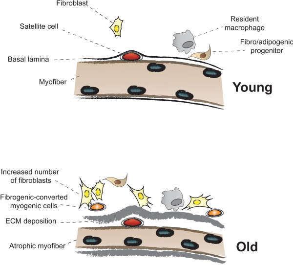
“Whether FAP conversion also occurs during aging, and whether Basal lamina ECM deposition Satellite cell Fibro/adipogenic progenitor Resident macrophage Fibroblast Increased number of fibroblasts Fibrogenic-converted myogenic cells Myofiber Atrophic myofiber Young Old Figure 5 Myofiber growth and extracellular matrix (…”
BackgroundInfluential
Cited by
Sorted by citations
Items 201–300 of 854
From muscle to food: oxidative challenges and developmental anomalies in poultry breast muscle
Lilburn, M. S., Griffin, J. R., & Wick, M. P. (2018) · Poultry Science · doi:10.3382/ps/pey409 · Cited by 48
Long, H., Lichtnekert, J., Andrassy, J., Schraml, B. U., Romagnani, P., & Anders, H.-J. (2023) · Frontiers in Immunology · doi:10.3389/fimmu.2023.1194988 · Cited by 46
Fuqua, J. D., Lawrence, M. M., Hettinger, Z. R., Borowik, A., Brecheen, P., Szczygiel, M. M., Abbott, C. B., Peelor, F. F., Confides, A. L., Kinter, M. T., et al. (2023) · Journal of Cachexia Sarcopenia and Muscle · doi:10.1002/jcsm.13285 · Cited by 50
Coaxing stem cells for skeletal muscle repair
McCullagh, K. J. A., & Perlingeiro, R. C. R. (2014) · Advanced Drug Delivery Reviews · doi:10.1016/j.addr.2014.07.007 · Cited by 49
“Muscular dystrophies and aging are both associated with large amounts of fibrosis caused by an accumulation of ECM components particularly collagen [37,38].”
BackgroundPincu, Y., Linden, M. A., Zou, K., Baynard, T., & Boppart, M. D. (2015) · Cytokine · doi:10.1016/j.cyto.2015.01.013 · Cited by 47
“Increased accumulation of collagen in skeletal muscle has other unfavorable consequences [9].”
BackgroundMehuron, T., Kumar, A., Duarte, L., Yamauchi, J., Accorsi, A., & Girgenrath, M. (2014) · Skeletal Muscle · doi:10.1186/2044-5040-4-14 · Cited by 47
Jensen, A. R., Kelley, B. V., Mosich, G. M., Ariniello, A., Eliasberg, C. D., Vu, B. K., Shah, P., Devana, S. K., Murray, I. R., Péault, B., et al. (2018) · Journal of Shoulder and Elbow Surgery · doi:10.1016/j.jse.2018.02.040 · Cited by 47
“Deposition of fibrotic extracellular matrix differs between acute and chronic skeletal muscle injuries.(32) The regeneration of myofibers, as well as the removal of dead myofibers and collagen, is tightly coordinated with a short inflammatory phase following acute injury.”
BackgroundA Mathematical Model of Idiopathic Pulmonary Fibrosis
Hao, W., Marsh, C. B., & Friedman, A. (2015) · PLoS ONE · doi:10.1371/journal.pone.0135097 · Cited by 47
“Excessive accumulation of ECM components (particularly collagen) associated with tissue injury and inflammation, results in permanent scar formation [37].”
BackgroundTrypsin, Tryptase, and Thrombin Polarize Macrophages towards a Pro-Fibrotic M2a Phenotype
White, M. J. V., & Gomer, R. H. (2015) · PLoS ONE · doi:10.1371/journal.pone.0138748 · Cited by 48
The histone deacetylase SIRT6 blocks myostatin expression and development of muscle atrophy
Samant, S. A., Kanwal, A., Pillai, V. B., Bao, R., & Gupta, M. P. (2017) · Scientific Reports · doi:10.1038/s41598-017-10838-5 · Cited by 46
Piñol‐Jurado, P., Suárez‐Calvet, X., Fernández‐Simón, E., Gallardo, E., de la Oliva, N., Martínez‐Muriana, A., Gómez‐Gálvez, P., Escudero, L. M., Pérez-Peiró, M., Wollin, L., et al. (2018) · Cell Death and Disease · doi:10.1038/s41419-018-0792-6 · Cited by 46
Contreras, O., Rossi, F. M. V., & Brandan, E. (2019) · Matrix Biology Plus · doi:10.1016/j.mbplus.2019.04.003 · Cited by 46
Changes in Respiratory Muscle Thickness during Mechanical Ventilation: Focus on Expiratory Muscles
Shi, Z., de Vries, H. J., de Grooth, H., Jonkman, A. H., Zhang, Y., Haaksma, M. E., van de Ven, P. M., de Man, A., Girbes, A. R. J., Tuinman, P. R., et al. (2021) · Anesthesiology · doi:10.1097/aln.0000000000003736 · Cited by 46
Baumert, P., Temple, S., Stanley, J. M., Cocks, M. S., Strauss, J. A., Shepherd, S. O., Drust, B., Lake, M., Stewart, C. E., & Erskine, R. M. (2021) · Scientific Reports · doi:10.1038/s41598-021-87195-x · Cited by 46
A Stromal Progenitor and ILC2 Niche Promotes Eosinophilia in Skeletal Muscle
Kastenschmidt, J. M., Coulis, G., Farahat, P. K., Pham, P., Rios, R., Cristal, T. T., Mannaa, A. H., Ayer, R. E., Yahia, R., Deshpande, A. A., et al. (2020) · Cell Reports · doi:10.1016/j.celrep.2021.108997 · Cited by 47
“Given that transcriptional programs associated with fibrosis can impede muscle regeneration (Gardner et al., 2020; Mann et al., 2011), the increased proportion of small regenerating myofibers when ILC2s are depleted in mdx mice may reflect the beginning phases of an improved muscle regenerative…”
Cardiac MRI biomarkers for Duchenne muscular dystrophy
Magrath, P., Maforo, N. G., Renella, P., Nelson, S. F., Halnon, N. J., & Ennis, D. B. (2018) · Biomarkers in Medicine · doi:10.2217/bmm-2018-0125 · Cited by 45
“DMD is a form of chronic muscle injury wherein chronic inflammation results in excessive accumulation of extracellular matrix components, which inhibit myogenic repair and lead to muscle being replaced by fibrotic scar [18].”
BackgroundShibaguchi, T., Sugiura, T., Fujitsu, T., Nomura, T., Yoshihara, T., Naito, H., Yoshioka, T., Ogura, A., & Ohira, Y. (2016) · Journal of Physiological Sciences · doi:10.1007/s12576-015-0433-0 · Cited by 41
“TGF-b is one of the most powerful fibrotic cytokines and plays a central role in the development of fibrosis in various tissues including skeletal muscle [1, 39].”
InfluentialEffect of Resistance Training on Extracellular Matrix Adaptations in Skeletal Muscle of Older Rats
Guzzoni, V., Ribeiro, M. B. T., Lopes, G. N., Marqueti, R. D. C., de Andrade, R. V., Selistre-de-Araújo, H. S., & Durigan, J. L. Q. (2018) · Frontiers in Physiology · doi:10.3389/fphys.2018.00374 · Cited by 46
“RT blunted the ageinduced accumulation of connective tissue concomitant to the upregulation of genes related to synthesis (COL-1A1/COL-3A1; TGFβ and CTGF) and degradation (MMP-2/MMP-9; TIMP1/TIMP-2) of the ECM network in the SOL and GAS muscles of older rats.”
BackgroundInfluentialShared and distinct mechanisms of skeletal muscle atrophy: A narrative review
Wilburn, D. T., Ismaeel, A., Machek, S. B., Fletcher, E., & Koutakis, P. (2021) · Ageing Research Reviews · doi:10.1016/j.arr.2021.101463 · Cited by 47
Interactions between p-Akt and Smad3 in injured muscles initiate myogenesis or fibrogenesis
Dong, Y., Lakhia, R., Thomas, S. S., Dong, Y., Wang, X. H., Silva, K. A. S., & Zhang, L. (2013) · American Journal of Physiology-Endocrinology and Metabolism · doi:10.1152/ajpendo.00644.2012 · Cited by 43
“In contrast, suppression of TGF- 1 signaling inhibits fibrosis and improves muscle regeneration (23).”
BackgroundKosmac, K., Gonzalez-Freire, M., McDermott, M. M., White, S. H., Walton, R. G., Sufit, R., Tian, L., Li, L., Kibbe, M. R., Criqui, M., et al. (2020) · Journal of the American Heart Association : Cardiovascular and Cerebrovascular Disease · doi:10.1161/jaha.118.015929 · Cited by 35
Bioengineered human skeletal muscle capable of functional regeneration
Fleming, J. W., Capel, A. J., Rimington, R. P., Wheeler, P. C., Leonard, A. N., BISHOP, N. C., Davies, O. G., & Lewis, M. P. (2020) · BMC Biology · doi:10.1186/s12915-020-00884-3 · Cited by 45
Alves, A. N., Ribeiro, B., Fernandes, K., Souza, N., Rocha, L. A., Nunes, F., Bussadori, S., & Mesquita-Ferrari, R. (2016) · Lasers in Medical Science · doi:10.1007/s10103-016-1908-9 · Cited by 36
Platelet-rich plasma in the treatment of skeletal muscle injuries
Andı́a, I., & Abate, M. (2015) · Expert Opinion on Biological Therapy · doi:10.1517/14712598.2015.1038234 · Cited by 42
“3 Fibrosis Muscle fibroblasts also play an important role in muscle repair by secreting collagen (type I and III), fibronectin, elastin, proteoglycans laminin and growth factors [37].”
BackgroundFornaro, M., Hinken, A. C., Needle, S., Hu, E., Trendelenburg, A., Mayer, A., Rosenstiel, A., Chang, C. K. W., Meier, V., Billin, A. N., et al. (2013) · American Journal of Physiology-Endocrinology and Metabolism · doi:10.1152/ajpendo.00408.2013 · Cited by 42
“Poor recovery from muscle damage is a prominent feature of several neuromuscular disorders, including muscular dystrophy and the frailty associated with old age, leading to excessive muscle fibrosis and reduced function (16).”
BackgroundRowlands, D., Nelson, A. R., Raymond, F., Métairon, S., Mansourian, R., Clarke, J., Stellingwerff, T., & Phillips, S. M. (2016) · Physiological Genomics · doi:10.1152/physiolgenomics.00068.2015 · Cited by 44
“Other literature evidence supports a role for postexercise protein feeding in ECM turnover, for example, expression of matrix metallopeptidase and inhibitor genes (increased MMP9, MMP13, MMP19; decreased TIMP1, TIMP2) (57), which control basement membrane degradation and ECM remodeling facilitating the recruitment of…”
BackgroundBarreto, G. N. L. S., de Oliveira, S. S., dos Anjos, I. V., Chalkidis, H. D. M., Mourão, R. H. V., Moura-da-Silva, A. M., Sano-Martins, I. S., & Gonçalves, L. R. C. (2017) · PLoS neglected tropical diseases · doi:10.1371/journal.pntd.0005458 · Cited by 42
Recruitment and therapeutic application of macrophages in skeletal muscles after hind limb ischemia
Hsieh, P.-L., Rybalko, V. Y., Baker, A., Suggs, L., & Farrar, R. (2017) · Journal of Vascular Surgery · doi:10.1016/j.jvs.2017.04.070 · Cited by 35
“It is well established that residual inflammation in the course of tissue regeneration may adversely affect tissue repair by facilitating excessive connective tissue deposition, also known as fibrosis.(27) We evaluated the extent of fibrotic tissue deposition in the recovering muscle by performing Masson trichrome sta…”
Available In Vitro Models for Human Satellite Cells from Skeletal Muscle
Romagnoli, C., Iantomasi, T., & Brandi, M. L. (2021) · International Journal of Molecular Sciences · doi:10.3390/ijms222413221 · Cited by 44
Exploring the Role of Oxidative Stress in Skeletal Muscle Atrophy: Mechanisms and Implications
Agrawal, S., Chakole, S. V., Shetty, N., Prasad, R., Lohakare, T., & Wanjari, M. B. (2023) · Cureus · doi:10.7759/cureus.42178 · Cited by 48
Andrés-Mateos, E., Mejías, R., Soleimani, A., Lin, B., Burks, T. N., Marx, R., Lin, B., Zellars, R., Zhang, Y.-G., Huso, D., et al. (2012) · PLoS ONE · doi:10.1371/journal.pone.0048884 · Cited by 40
“Therefore, multiple studies have focused on identifying ECM proteins, cytokines, growth factors and the downstream signaling pathways involved in aberrant muscle regeneration and fibrotic tissue formation [14,15,16].”
BackgroundRole of proteoglycans in the regulation of the skeletal muscle fibrotic response
Brandan, E., & Gutiérrez, J. (2013) · FEBS Journal · doi:10.1111/febs.12278 · Cited by 41
Seasonal and post-trauma remodeling in cone-dominant ground squirrel retina
Merriman, D. K., Sajdak, B. S., Li, W., & Jones, B. W. (2016) · Experimental Eye Research · doi:10.1016/j.exer.2016.01.011 · Cited by 42
Kozakowska, M., Pietraszek‐Gremplewicz, K., Cieśla, M., Seczyńska, M., Bronisz-Budzyńska, I., Podkalicka, P., Bukowska-Straková, K., Łoboda, A., Józkowicz, A., & Dulak, J. (2017) · American Journal Of Pathology · doi:10.1016/j.ajpath.2017.10.017 · Cited by 43
Hoffmann, C., Höckele, S., Kappler, L., de Angelis, M. H., Häring, H., & Weigert, C. (2018) · Scientific Reports · doi:10.1038/s41598-017-18658-3 · Cited by 43
Morgoulis, D., Berenstein, P., Cazacu, S., Kazimirsky, G., Dori, A., Barnea, E. R., & Brodie, C. (2019) · Cell Death and Disease · doi:10.1038/s41419-019-1307-9 · Cited by 42
Reconstruction of Muscle Fascicle‐Like Tissues by Anisotropic 3D Patterning
Jin, Y., Jeon, E. J., Jeong, S., Min, S., Choi, Y. S., Ys, K., Lee, J. S., Shin, J., Yu, J. H., Ahn, D., et al. (2021) · Advanced Functional Materials · doi:10.1002/adfm.202006227 · Cited by 41
The genetic pleiotropy of musculoskeletal aging
Karasik, D., & Cohen, M. W. (2012) · Frontiers in Physiology · doi:10.3389/fphys.2012.00303 · Cited by 41
“Some form of inflammatory response is necessary to repair damaged tissues effectively; however, chronic inflammatory responses drive unrestrained wound healing and fibrosis especially in aging (Mann et al., 2011).”
BackgroundInfluentialGumpenberger, M., Wessner, B., Graf, A. C., Narici, M. V., Fink, C., Braun, S., Hoser, C., Blazevich, A. J., & Csapo, R. (2020) · International Journal of Molecular Sciences · doi:10.3390/ijms21197089 · Cited by 40
Goetsch, K. P., Myburgh, K. H., & Niesler, C. U. (2013) · Journal of Muscle Research and Cell Motility · doi:10.1007/s10974-013-9364-7 · Cited by 41
Sugihara, H., Teramoto, N., Yamanouchi, K., Matsuwaki, T., & Nishihara, M. (2018) · Aging · doi:10.18632/aging.101425 · Cited by 41
“In sarcopenia, muscle frequently exhibits adipose- and fibrous-tissue infiltration [14], and this impairs skeletal muscle function: whereas intramuscular adipose tissue (IMAT) causes poor physical performance [15], fibrous tissue reduces motile and contractile functions [16].”
BackgroundThrown for a loop: fibro-adipogenic progenitors in skeletal muscle fibrosis
Loomis, T., & Smith, L. (2023) · American Journal of Physiology-Cell Physiology · doi:10.1152/ajpcell.00245.2023 · Cited by 39
Vidal, B., Ardite, E., Suelves, M., Ruiz‐Bonilla, V., Janué, A., Flick, M. J., Degen, J. L., Serrano, A. L., & Muñoz‐Cánoves, P. (2012) · Human Molecular Genetics · doi:10.1093/hmg/dds012 · Cited by 39
A homogenization model of the Voigt type for skeletal muscle
Spyrou, L. A., Agoras, M., & Danas, K. (2016) · Journal of Theoretical Biology · doi:10.1016/j.jtbi.2016.11.018 · Cited by 39
“…1995), b) it has a major contribution to myo-fascial force transmission between neighboring fibers and fascicles (Huijing, 1999), c) it is implicated in muscle diseases (e.g. muscular dystrophy (Emery et al., 2002; Mann et al., 2011), spasticity (Lieber et al., 2003)), and ∗Corresponding author.”
BackgroundOkerblom, J., Schwarz, F., Olson, J., Fletes, W., Ali, S. R., Martin, P. T., Glass, C. K., Nizet, V., & Varki, A. P. (2017) · The Journal of Immunology · doi:10.4049/jimmunol.1601471 · Cited by 39
Baudequin, T., & Tabrizian, M. (2017) · Advanced Healthcare Materials · doi:10.1002/adhm.201700734 · Cited by 40
Circulating low density lipoprotein (LDL)
Khosravi, M., Hosseini‐Fard, S. R., & Najafi, M. (2018) · Hormone Molecular Biology and Clinical Investigation · doi:10.1515/hmbci-2018-0024 · Cited by 41
Tissue cross talks governing limb muscle development and regeneration
Helmbacher, F., & Stricker, S. (2020) · Seminars in Cell and Developmental Biology · doi:10.1016/j.semcdb.2020.05.005 · Cited by 42
“Muscle disease or large scale muscle trauma is often associated with replacement of muscle tissue with fibrotic tissue as well as with adipose tissue infiltration [187].”
BackgroundYao, L., Tichy, E. D., Zhong, L., Mohanty, S., Wang, L., Ai, E., Yang, S., Mourkioti, F., & Qin, L. (2020) · Journal of Bone and Mineral Research · doi:10.1002/jbmr.4265 · Cited by 39
“Muscle regeneration associates with a transient ectopic adipocyte accumulation((3,4,35)) as well as fibrosis deposition, especially under disease conditions.((48)) Although the fibrogenic effect of FAPs has been studied extensively both in vitro and in vivo,((1,8,10,37,47,49,50)) their role in adipogenic regulation in…”
BackgroundEffects on Contralateral Muscles after Unilateral Electrical Muscle Stimulation and Exercise
Song, Y.-F., Forsgren, S., Yu, J.-G., Lorentzon, R., & Stål, P. (2012) · PLoS ONE · doi:10.1371/journal.pone.0052230 · Cited by 39
“Fibrosis is essentially an excessive accumulation of extracellular matrix components, particularly collagen, which is the end result of a cascade of events proceeding from tissue damage via inflammation [57].”
BackgroundMukund, K., Mathewson, M. A., Minamoto, V. B., Ward, S. R., Subramaniam, S., & Lieber, R. L. (2014) · Muscle & Nerve · doi:10.1002/mus.24211 · Cited by 39
Yaden, B. C., Wang, Y. X., Wilson, J. M., Culver, A. E., Milner, A., Datta‐Mannan, A., Shetler, P. K., Croy, J. E., Dai, G., & Krishnan, V. (2014) · American Journal Of Pathology · doi:10.1016/j.ajpath.2013.12.029 · Cited by 39
Cholok, D. J., Lee, E., Lisiecki, J. L., Agarwal, S., Loder, S. J., Ranganathan, K., Qureshi, A. T., Davis, T. A., & Lévi, B. (2016) · The Journal of Trauma: Injury, Infection, and Critical Care · doi:10.1097/ta.0000000000001290 · Cited by 38
Galectin‐1 promotes an M2 macrophage response to polydioxanone scaffolds
Abebayehu, D., Spence, A., Boyan, B. D., Schwartz, Z., Ryan, J. J., & McClure, M. J. (2017) · Journal of Biomedical Materials Research Part A · doi:10.1002/jbm.a.36113 · Cited by 38
Kim, S.-Y., Ahn, G., Kim, H., Je, J.-G., Kim, K., & Jeon, Y. (2020) · Marine Drugs · doi:10.3390/md18110529 · Cited by 38
Giha, H. A., Sater, M. S., & Alamin, O. A. O. (2022) · Acta Diabetologica · doi:10.1007/s00592-022-01860-9 · Cited by 39
“This uncontrolled and irreversible ECM expansion is accompanied by an accumulation of ECM due to inhibited degradation, which results in a fibrotic phenotype within the muscle [48].”
BackgroundEvidence of induced muscle regeneration persists for years in the mouse
Meyer, G. A. (2018) · Muscle & Nerve · doi:10.1002/mus.26329 · Cited by 37
Recovery from Volumetric Muscle Loss Injury: A Comparison Between Young and Aged Rats
Kim, J., Kasukonis, B. M., Brown, L., Washington, T. A., & Wolchok, J. (2016) · Experimental Gerontology · doi:10.1016/j.exger.2016.07.008 · Cited by 37
Venter, C., & Niesler, C. U. (2018) · BioTechniques · doi:10.2144/btn-2017-0100 · Cited by 40
Glycerol-induced injury as a new model of muscle regeneration
Mahdy, M. A. A. (2018) · Cell and Tissue Research · doi:10.1007/s00441-018-2846-6 · Cited by 37
“acts as a scaffold to support the newly formed myofibers (Mann et al. 2011; Serrano et al. 2011).”
BackgroundChronic infection stunts macrophage heterogeneity and disrupts immune-mediated myogenesis
Jin, R. M., Warunek, J., & Wohlfert, E. A. (2018) · JCI Insight · doi:10.1172/jci.insight.121549 · Cited by 37
Role of Metabolic Stress and Exercise in Regulating Fibro/Adipogenic Progenitors
Collao, N., Farup, J., & Lisio, M. D. (2020) · Frontiers in Cell and Developmental Biology · doi:10.3389/fcell.2020.00009 · Cited by 39
“This chronic, low-grade inflammation is associated impaired function of MuSCs, immune cells, and FAPs, leading to fibrosis, and poor skeletal muscle regeneration (Rostasy et al., 2008; Mann et al., 2011; D’Souza et al., 2015; Wang et al., 2015).”
BackgroundLee, E. M., Kim, A.-Y., Lee, E. H., Park, J., Lee, M., Hwang, M., Kim, C.-Y., Kim, S., & Jeong, K. (2014) · Cell Transplantation · doi:10.3727/096368914x678599 · Cited by 36
“The frequent cycle of necrosis and regeneration induces chronic inflammation and fibrosis, progressively replacing the muscle tissue (27).”
BackgroundHeitman, K., Alexander, M. S., & Faul, C. (2024) · International Journal of Molecular Sciences · doi:10.3390/ijms25105117 · Cited by 39
Zheng, H., Yang, Z., Zhou, L., Zhang, B., Cheng, R., & Zhang, Q. (2024) · Small · doi:10.1002/smll.202310483 · Cited by 36
Hypermethylation: Causes and Consequences in Skeletal Muscle Myopathy
Majumder, A., Behera, J., Jeremić, N., & Tyagi, S. C. (2017) · Journal of Cellular Biochemistry · doi:10.1002/jcb.25841 · Cited by 34
“…of the media and extracellular matrix (ECM) remodeling [Vermeulen et al., 2001; Mujumdar et al., 2002; Kundu et al., 2009; Tyagi et al., 2009; Mann et al., 2011; Steed and Tyagi, 2011; Lee et al., 2012a; Dick, 2013; Veeranki et al., 2015; Winchester et al., 2015], however, the mechanism of…”
BackgroundChiu, C., Chang, T.-H., Chang, S., Chang, G., Chen, A. C., Cheng, C., Chen, S., Fu, J., Wen, C.-J., & Chan, Y. (2020) · The American Journal of Sports Medicine · doi:10.1177/0363546520905853 · Cited by 38
“The parallel occurrence of myofiber regeneration and the replacement of muscle tissue with fibrotic tissue in the injured muscle leads to proliferative competition between both systems during the recovery process.(32) Fibrosis prevents patients from returning to baseline function, in part by inhibiting satellite cell…”
BackgroundRole of sphingosine 1-phosphate signalling in tissue fibrosis
Donati, C., Cencetti, F., Bernacchioni, C., Vannuzzi, V., & Bruni, P. (2020) · Cellular Signalling · doi:10.1016/j.cellsig.2020.109861 · Cited by 36
Muscle fibrosis and maladaptation occur progressively in CKD and are rescued by dialysis
Brightwell, C. R., Kulkarni, A. S., Paredes, W. M., Zhang, K., Perkins, J. B., Gatlin, K. J., Custodio, M. J., Farooq, H., Zaidi, B., Pai, R. N., et al. (2021) · JCI Insight · doi:10.1172/jci.insight.150112 · Cited by 37
“JCI Insight 2021;6(24):e150112 https://doi.”
Alcalde-Estévez, E., Asenjo-Bueno, A., Sosa, P., Olmos, G., Plaza, P., Caballero-Mora, M. Á., Rodríguez-Puyol, D., Ruíz-Torres, M. P., & López-Ongil, S. (2020) · Aging · doi:10.18632/aging.103450 · Cited by 35
“Muscular fibrosis has been related to the activation of resident fibroblasts [10] or may also be caused by a faulty muscle-repair process initiated by the activation of muscle satellite cells [11].”
BackgroundBarbe, M. F., Harris, M. Y., Cruz, G. E., Amin, M., Billett, N. M., Dorotan, J. T., Day, E. P., Kim, S. Y., & Bove, G. M. (2021) · BMC Musculoskeletal Disorders · doi:10.1186/s12891-021-04270-0 · Cited by 35
Ribeiro, B. G., Alves, A. N., Andreo, L., Cantero, T. M., Fernandes, K. P. S., Dias, D. D. S., Bernardes, N., Angelis, K. D., & Mesquita‐Ferrari, R. A. (2016) · PLoS ONE · doi:10.1371/journal.pone.0153618 · Cited by 35
“After an injury, the muscle repair process begins and is divided into interdependent phases: degeneration and inflammation, regeneration, fibrosis/scar formation and remodeling [3].”
BackgroundEpicatechin elicits MyoD-dependent myoblast differentiation and myogenic conversion of fibroblasts
Lee, S.-J., Leem, Y.-E., Go, G.-Y., Choi, Y., Song, Y. J., Kim, I., Kim, D. Y., Kim, Y. K., Seo, D. W., Kang, J., et al. (2017) · PLoS ONE · doi:10.1371/journal.pone.0175271 · Cited by 34
Plasticity of the Muscle Stem Cell Microenvironment
Dinulović, I., Furrer, R., & Handschin, C. (2017) · Advances in experimental medicine and biology · doi:10.1007/978-3-319-69194-7_8 · Cited by 37
“which in chronic diseases such as muscular dystrophies result in fibrotic tissue formation at the expense of functional muscle tissue (119).”
Sorensen, J. R., Kaluhiokalani, J. P., Hafen, P. S., Deyhle, M. R., Parcell, A. C., & Hyldahl, R. D. (2019) · The FASEB Journal · doi:10.1096/fj.201900519r · Cited by 37
“are characteristics observed in aged muscle (18).”
Ollewagen, T., Myburgh, K. H., van de Vyver, M., & Smith, C. (2021) · Journal of Biomedical Science · doi:10.1186/s12929-021-00714-w · Cited by 37
“Analysis of blood samples from patients with active RA highlighted differences in cell–cell interactions, altered EMT, and increased TGF-β proteins suggestive of increased de novo ECM synthesis and fibrosis [89].”
BackgroundInfluentialThe effect of low‐level laser therapy (LLLT) applied prior to muscle injury
Ribeiro, B., Alves, A. N., dos Santos, L. A. D., Fernandes, K., Cantero, T. M., Gomes, M. T., França, C., da Silva, D. D. F. T., Bussadori, S., & Mesquita-Ferrari, R. (2015) · Lasers in Surgery and Medicine · doi:10.1002/lsm.22381 · Cited by 31
CCR2 deficiency does not provide sustained improvement of muscular dystrophy in mdx 5cv mice
Zhao, W., Wang, X., Ransohoff, R. M., & Zhou, L. (2016) · The FASEB Journal · doi:10.1096/fj.201600619r · Cited by 33
Cabello‐Verrugio, C., Rivera, J. C. A., & García, D. (2017) · Current Opinion in Clinical Nutrition & Metabolic Care · doi:10.1097/mco.0000000000000361 · Cited by 34
Nederveen, J. P., Joanisse, S., Thomas, A. C. Q., Snijders, T., Manta, K., Bell, K. E., Phillips, S. M., Kumbhare, D. A., & Parise, G. (2020) · The FASEB Journal · doi:10.1096/fj.201900787r · Cited by 35
Farsijani, S., Santanasto, A. J., Miljkovic, I., Boudreau, R. M., Goodpaster, B. H., Kritchevsky, S. B., & Newman, A. B. (2020) · The Journals of Gerontology Series A · doi:10.1093/gerona/glaa161 · Cited by 33
“The increased intermuscular fat accompanied by muscle wasting is an aberrant adipogenic reparative response that is observed in degenerative conditions (eg, muscular dystrophies) (50), where adipose tissue and fibrosis “replace” the attenuated muscle.”
BackgroundMuscle PGC-1α modulates satellite cell number and proliferation by remodeling the stem cell niche
Dinulović, I., Furrer, R., Beer, M., Ferry, A., Cardel, B., & Handschin, C. (2016) · Skeletal Muscle · doi:10.1186/s13395-016-0111-9 · Cited by 35
“, the fibrosis and BL thickening upon chronic elevation of FN in the uninjured state [7] or the elevated resting SC numbers and reduced SC proliferative potential on fibers ex vivo caused by the increased basal expression of FN in likeacetylglucosaminyltransferase myodystrophy (LARGE) mice [6].”
BackgroundGiovarelli, M., Arnaboldi, F., Zecchini, S., Cornaghi, L., Nava, A., Sommariva, M., Clementi, E., & Gagliano, N. (2022) · International Journal of Molecular Sciences · doi:10.3390/ijms23158735 · Cited by 32
Bernier, L., Hefendehl, J. K., Scott, R. W., Tung, L. W., Lewis, C. B., Soliman, H., Simm, S., Dissing‐Olesen, L., Hofmann, J. C., Guo, D., et al. (2025) · Nature Neuroscience · doi:10.1038/s41593-025-01872-y · Cited by 38
Enzyme-Based Anti-Inflammatory Therapeutics for Inflammatory Diseases
Narayanan, K. B. (2025) · Pharmaceutics · doi:10.3390/pharmaceutics17050606 · Cited by 41
“The enzyme is also involved in breaking down excess collagen associated with scarring or fibrosis, thereby reducing fibrosis in anti-fibrotic therapies [92].”
Jensen, S. M., Bechshøft, C. J., Heisterberg, M. F., Schjerling, P., Andersen, J., Kjaer, M., & Mackey, A. (2020) · Frontiers in Physiology · doi:10.3389/fphys.2020.00811 · Cited by 26
“, 2019) which has been associated with the development of fibrosis (Mann et al., 2011) although it is possible that macrophage content increases to combat fibrosis rather than contributing to it.”
BackgroundPhotoreactive Hydrogel Stiffness Influences Volumetric Muscle Loss Repair
Basurto, I. M., Passipieri, J. A., Gardner, G. M., Smith, K. K., Amacher, A. R., Hansrisuk, A., Christ, G. J., & Caliari, S. R. (2021) · Tissue Engineering Part A · doi:10.1089/ten.tea.2021.0137 · Cited by 35
Selective modulation through the glucocorticoid receptor ameliorates muscle pathology in mdx mice
Huynh, T., Uaesoontrachoon, K., Quinn, J. L., Tatem, K. S., Heier, C. R., van der Meulen, J. H., Yu, Q., Harris, M. A., Nolan, C. J., Haegeman, G., et al. (2013) · The Journal of Pathology · doi:10.1002/path.4231 · Cited by 33
“In damaged muscle, the macrophage population response transitions from Th-1-responsive promoting satellite cell activation and proliferation, while inhibiting differentiation, to Th-2-responsive for differentiation, growth and repair [47–49].”
BackgroundMuscle weakness after critical illness: unravelling biological mechanisms and clinical hurdles
Pierre, A., Favory, R., Bourel, C., Howsam, M., Romien, R., Lancel, S., & Préau, S. (2025) · Critical Care · doi:10.1186/s13054-025-05462-z · Cited by 39
Hindi, S. M., Sato, S., Choi, Y., & Kumar, A. (2013) · Human Molecular Genetics · doi:10.1093/hmg/ddt536 · Cited by 31
“Fibrosis is a pathogenic factor characterized by chronic inflammation with persistent production of profibrotic cytokines and excessive deposition of ECM proteins, including collagens and fibronectin which can impair tissue function (50).”
BackgroundSwiderski, K., Todorov, M., Gehrig, S. M., Naim, T., Chee, A., Stapleton, D., Koopman, R., & Lynch, G. S. (2014) · Fibrogenesis & Tissue Repair · doi:10.1186/1755-1536-7-1 · Cited by 32
“This environment limits muscle regenerative capacity and there is concomitant replacement of formerly functional muscle fibres with adipose and fibrotic material (reviewed in [1]).”
BackgroundSamaan, M. C., Missiuna, P. C., Peterson, D. C., & Thabane, L. (2016) · BMJ Open · doi:10.1136/bmjopen-2016-011812 · Cited by 31
HIF prolyl hydroxylase inhibition protects skeletal muscle from eccentric contraction-induced injury
Billin, A., Honeycutt, S. E., McDougal, A., Kerr, J. P., Chen, Z., Freudenberg, J., Rajpal, D., Luo, G., Kramer, H. F., Geske, R., et al. (2018) · Skeletal Muscle · doi:10.1186/s13395-018-0179-5 · Cited by 17
Neuromuscular disease modeling on a chip
Santoso, J. W., & McCain, M. L. (2020) · Disease Models & Mechanisms · doi:10.1242/dmm.044867 · Cited by 36
Dodd, T., Simon, L., LeCapitaine, N. J., Zabaleta, J., Mussell, J., Berner, P., Ford, S. M., Dufour, J., Bagby, G. J., Nelson, S., et al. (2014) · Alcoholism Clinical and Experimental Research · doi:10.1111/acer.12545 · Cited by 32
“The systemic and timely process of breakdown and replacement of the ECM, aided by MMPs, is critical for ensuring full and rapid repair, as well as for avoiding fibrosis (Mann et al., 2011).”
BackgroundAll for One and One for All: Regenerating Skeletal MusclePreprint
Hindi, S. M., & Millay, D. P. (2021) · Cold Spring Harbor Perspectives in Biology · doi:10.1101/cshperspect.a040824 · Cited by 29
Demonbreun, A. R., Allen, M. V., Warner, J. L., Barefield, D. Y., Krishnan, S., Swanson, K. E., Earley, J. U., & McNally, E. M. (2016) · American Journal Of Pathology · doi:10.1016/j.ajpath.2016.02.005 · Cited by 31
Xu, P., Werner, J.-U., Milerski, S., Hamp, C. M., Kuzenko, T., Jähnert, M., Gottmann, P., de Roy, L., Warnecke, D., Abaei, A., et al. (2018) · Frontiers in Physiology · doi:10.3389/fphys.2018.00674 · Cited by 34
“cells and clearance of tissue debris becomes problematic, thus leading to a hampered response in obese individuals (Mann et al., 2011; Akhmedov and Berdeaux, 2013; Urciuolo et al., 2013; Garg et al., 2015; Sziksz et al., 2015; Darby et al., 2016; Lin et al., 2016).”
Hayward, K. L., Kouthouridis, S., & Zhang, B. (2020) · ACS Biomaterials Science & Engineering · doi:10.1021/acsbiomaterials.0c01089 · Cited by 33
Kobak, K. A., Lawrence, M. M., Pharaoh, G. A., Borowik, A., Peelor, F. F., Shipman, P. D., Griffin, T. M., Remmen, H. V., & Miller, B. F. (2021) · Journal of Cachexia Sarcopenia and Muscle · doi:10.1002/jcsm.12772 · Cited by 31
Metadata
- Authors
- Mann, C. J., Perdiguero, E., Kharraz, Y., Aguilar, S., Pessina, P., Serrano, A. L., & Muñoz-Cánoves, P.
- Affiliations
- Universitat Pompeu Fabra (ES)
- Venue
- Skeletal Muscle
- Published
- 2011-05-04
- Volume
- 1
- Issue
- 1
- Pages
- 21-21
- ISSN
- 2044-5040 (ISSN-L)
- PMC
- PMC3156644
- OpenAlex
- W2076787014
- Cited by
- 853
- References
- 129
- Citations by year
- 2026: 37 · 2025: 59 · 2024: 55 · 2023: 53 · 2022: 59 · 2021: 71 · 2020: 81 · 2019: 85 · 2018: 76 · 2017: 51 · 2016: 68 · 2015: 48 · 2014: 49 · 2013: 33 · 2012: 27
- FWCI
- 14.30
- Type
- journal article
- Language
- en
- Open access
- gold
- doi.org
- Licence
- cc-by
- Locations
- Skeletal Muscle (publishedVersion, cc-by)
- Europe PMC (PubMed Central) (publishedVersion, cc-by)
- PubMed Central (publishedVersion, cc-by)
- PubMed (publishedVersion)
- RECERCAT (Consorci de Serveis Universitaris de Catalunya) (publishedVersion, other-oa)
- DOAJ (DOAJ: Directory of Open Access Journals) (submittedVersion, cc-by-sa)
- Repositori digital de la UPF (Universitat Pompeu Fabra) (submittedVersion, cc-by)
- Funders
- Ministerio de Ciencia e Innovación (PLE2009)
- Centro de Investigación Biomédica en Red sobre Enfermedades Neurodegenerativas
- Topics
- Life Sciences › Biochemistry, Genetics and Molecular Biology › Molecular Biology › Muscle Physiology and Disorders
- Health Sciences › Medicine › Cardiology and Cardiovascular Medicine › Cardiac Fibrosis and Remodeling
- Health Sciences › Medicine › Surgery › Wound Healing and Treatments
- Keywords
- muscle fibrosis, muscle repair, muscular dystrophy, satellite cells, muscle stem cells, fibroblast activation, inflammatory cell infiltration, extracellular matrix, wound healing, microenvironment
About these checks
Registry, retraction, Cites and statistics results are computed, not judged, with one exception: a printed reference that no index lists is read once by a model before it counts as a citation error. Claims are extracted by a model and kept only when their quote appears verbatim in the text; each cited source is then checked, with a model’s help, for whether it says what the claim relies on. Those verdicts can be wrong — read the claim’s quote and the cited source before relying on one.
Statistics and arithmetic are recomputed, not judged.
Confidence bands are shown only for fields calibrated on our annotated test set. None is calibrated yet, so no band is shown.
What counts as a claim ›How we measure confidence ›How we check this ›
Are you an author, or do you see a problem with this page? Request a review
