

The DOI matches its Crossref record; 129 of the 129 references it cites were found in a registry or an index.
Aberrant repair and fibrosis development in skeletal muscle
Open accessAbstract
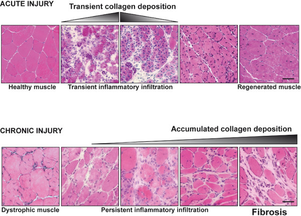
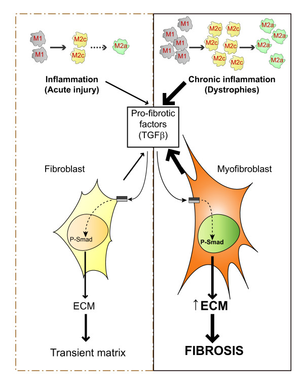
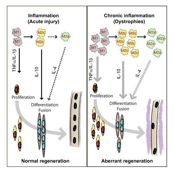
The repair process of damaged tissue involves the coordinated activities of several cell types in response to local and systemic signals. Following acute tissue injury, infiltrating inflammatory cells and resident stem cells orchestrate their activities to restore tissue homeostasis. However, during chronic tissue damage, such as in muscular dystrophies, the inflammatory-cell infiltration and fibroblast activation persists, while the reparative capacity of stem cells (satellite cells) is attenuated. Abnormal dystrophic muscle repair and its end stage, fibrosis, represent the final common pathway of virtually all chronic neurodegenerative muscular diseases. As our understanding of the pathogenesis of muscle fibrosis has progressed, it has become evident that the muscle provides a useful model for the regulation of tissue repair by the local microenvironment, showing interplay among muscle-specific stem cells, inflammatory cells, fibroblasts and extracellular matrix components of the mammalian wound-healing response. This article reviews the emerging findings of the mechanisms that underlie normal versus aberrant muscle-tissue repair.
Overview
The claims and statistics of this paper haven’t been checked yet.
Run the check — free, no account needed. It reads the open-access text and checks the sources it cites.
Integrity signals
Registry
Cites
Statistics
Figures
Verification
Claims the paper makes
The claims and statistics of this paper haven’t been checked yet.
Run the check — free, no account needed. It reads the open-access text and checks the sources it cites.
Claims other papers make about this one
Each is checked against this paper’s own text: does it say what the citing paper says it does?
In the citing paper
“Previous work has shown that the accumulation of inflammatory macrophages and age-related declines in interferon-gamma signaling delay muscle repair following injury”
From Skeletal muscle aging, cellular senescence, and senotherapeutics: current knowledge and future directions (2021) · cites this paper as Mann et al., 2011
Self-check
Percentages, sums and reported differences re-computed from the paper’s own numbers.
The self-check needs the paper’s full text.
Citations
Cites
1 retracted
RETRACTED ARTICLE: The role of nitric oxide during healing of trauma to the skeletal muscle
Filippin, L. I., Cuevas, M. J., Lima‐Cabello, E., Marroni, N. P., González‐Gallego, J., & Xavier, R. M. (2010) · Inflammation Research · doi:10.1007/s00011-010-0277-2 · Cited by 34 · cited 4×
“In addition, NO, which is released by local and invading cells during injury, can act as a pro-inflammatory molecule, and is able to activate MMP-2, which in turn releases HGF. Importantly, blockers of NOS function, such as nitro- Larginine methyl ester (L-NAME), have been shown to decrease the acute inflammatory reac…”
MethodInfluentialRetractedThe chemokine system in diverse forms of macrophage activation and polarization
MANTOVANI, A., SICA, A., SOZZANI, S., ALLAVENA, P., VECCHI, A., & LOCATI, M. (2004) · Trends in Immunology · doi:10.1016/j.it.2004.09.015 · Cited by 6,534 · cited 2×
“A nomenclature for polarized macrophages has been proposed [24,25] and they are now referred to as classically and alternatively activated macrophages, or M1 and M2 macrophages, respectively (Figure 2, Figure 3).”
BackgroundMechanisms of TGF-β Signaling from Cell Membrane to the Nucleus
Shi, Y., & Massagué, J. (2003) · Cell · doi:10.1016/s0092-8674(03)00432-x · Cited by 5,860
Visse, R., & Nagase, H. (2003) · Circulation Research · doi:10.1161/01.res.0000070112.80711.3d · Cited by 4,575 · cited 1×
“Activation requires proteolytic cleavage of the inactive precursor by either membrane-type (MT)1MMP [105] or plasmin, and cleavage of their corresponding inhibitors [106].”
Cellular and molecular mechanisms of fibrosis
Wynn, T. (2008) · The Journal of Pathology · doi:10.1002/path.2277 · Cited by 4,495
SATELLITE CELL OF SKELETAL MUSCLE FIBERS
Mauro, A. (1961) · The Journal of Cell Biology · doi:10.1083/jcb.9.2.493 · Cited by 3,683
Hinz, B., Phan, S. H., Thannickal, V. J., Galli, A., Bochaton-Piallat, M.-L., & Gabbiani, G. (2007) · The American Journal of Pathology · doi:10.2353/ajpath.2007.070112 · Cited by 2,068 · cited 1×
“These cells are associated with tissue repair and fibrosis in many tissues and organs, including muscle, skin, liver, lung, bone and cartilage [56].”
BackgroundArnold, L., Henry, A., Poron, F., Baba-Amer, Y., van Rooijen, N., Plonquet, A., Gherardi, R. K., & Chazaud, B. (2007) · The Journal of Experimental Medicine · doi:10.1084/jem.20070075 · Cited by 2,002 · cited 3×
“An important development in our understanding of muscle repair and fibrosis was the demonstration that a heterogeneous population of macrophages exists in regenerating muscle after injury, exhibiting opposing activities (either pro-inflammatory or anti-inflammatory) and different kinetics [23].”
BackgroundInfluentialFibrotic disease and the TH1/TH2 paradigm
Wynn, T. A. (2004) · Nature Reviews Immunology · doi:10.1038/nri1412 · Cited by 1,765
Muscle injury activates resident fibro/adipogenic progenitors that facilitate myogenesis
Joe, A. W. B., Yi, L., Natarajan, A., Le Grand, F., So, L., Wang, J., Rudnicki, M. A., & Rossi, F. M. V. (2010) · Nature Cell Biology · doi:10.1038/ncb2015 · Cited by 1,690 · cited 7×
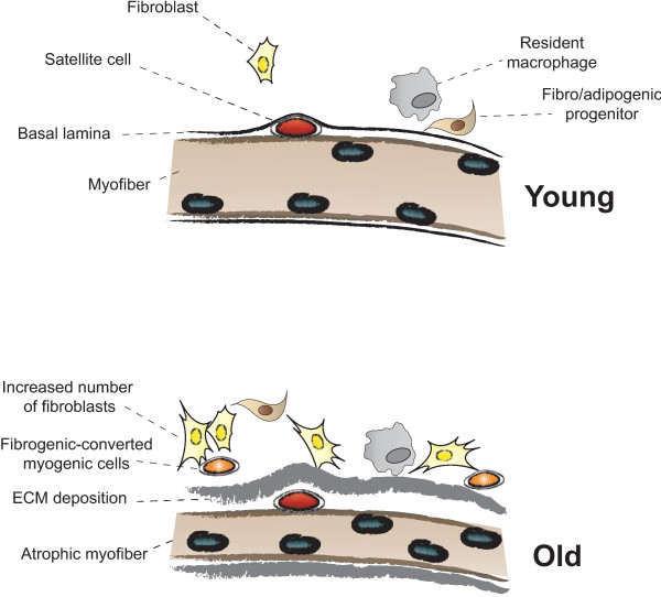
“Whether FAP conversion also occurs during aging, and whether Basal lamina ECM deposition Satellite cell Fibro/adipogenic progenitor Resident macrophage Fibroblast Increased number of fibroblasts Fibrogenic-converted myogenic cells Myofiber Atrophic myofiber Young Old Figure 5 Myofiber growth and extracellular matrix (…”
BackgroundInfluential
Cited by
Sorted by citations
Items 601–700 of 854
Tazawa, R., Uchida, K., Kenmoku, T., Nakawaki, M., Muneshige, K., Ishii, D., Inoue, G., & Takaso, M. (2021) · Journal of Orthopaedic Surgery and Research · doi:10.1186/s13018-021-02675-0 · Cited by 4
Stretch-Induced Healing of Injured Muscles Is Associated With Myogenesis and Decreased Fibrosis
Hu, C., Chen, C. P.-C., Tsui, P., Chen, C., & Hsu, C. (2022) · The American Journal of Sports Medicine · doi:10.1177/03635465221083995 · Cited by 4
“Although muscle is noted for its high capacity for regeneration and repair of myofibers,(16) muscle fibrosis impairs muscle regeneration and enhances muscle susceptibility to further injury.(19,20) In current management after muscle injury, therapeutic stretching exercise has been applied in rehabilitation settings to…”
BackgroundJiang, X., Zhang, H., Ren, Y., Li, Y., Zhong, L., Guo, J., & Zhang, X. (2023) · Journal of Orthopaedic Surgery and Research · doi:10.1186/s13018-023-04069-w · Cited by 4
New Strategies for Muscular Repair and Regeneration
Cianforlini, M., Coppa, V., Grassi, M., & Gigante, A. P. (2017) · Springer eBooks · doi:10.1007/978-3-662-54184-5_14 · Cited by 5
Cilia Control Fat Deposition during Tissue Repair
Perdiguero, E., Serrano, A. L., & Muñoz‐Cánoves, P. (2017) · Developmental Cell · doi:10.1016/j.devcel.2017.06.023 · Cited by 4
Fibrous myopathy induced by intramuscular injections of cyclizine
Tinker, R. J., Wiltshire, R., du Plessis, D., & Lilleker, J. B. (2020) · BMJ Case Reports · doi:10.1136/bcr-2019-233544 · Cited by 4
Collagen I and III Ratios and Tenacity of Rats’ Muscle Injured and Treated with Platelet-Rich Plasma
Garcia, T. A., Ozaki, G. A. T., Castoldi, R. C., Job, A. E., Camargo, R. C. T., Filho, J. C. S. C., & Belangero, W. D. (2020) · International Journal of Morphology · doi:10.4067/s0717-95022020000501392 · Cited by 4
“Although the importance of the extracellular matrix during muscle regeneration is already recognized (Mann et al., 2011), it is not known exactly, what role type I and III collagens play at each stage of inflammation.”
BackgroundWalsh, C. J., Batt, J. A. E., Herridge, M. S., Mathur, S., Bader, G. D., Hu, P., Khatri, P., & dos Santos, C. C. (2022) · Scientific Reports · doi:10.1038/s41598-022-15003-1 · Cited by 3
Effects of hydrogen sulfide in hyperhomocysteinemia-mediated skeletal muscle myopathy.
Majumder, A. (2018) · ThinkIR: The University of Louisville's Institutional Repository (University of Louisville) · doi:10.18297/etd/3114 · Cited by 4
“The basic unit of the muscle is the muscle fiber, so any structural or morphological changes in muscle fiber can play havoc with it functional capabilities (368).”
BackgroundA Novel Minimally Invasive Surgically Induced Skeletal Muscle Injury Model in Sheep
Vidal, L., Vila, I., Venegas, V., Sacristán, A., Contreras-Muñoz, P., López-Garzón, M., Giné, C., Rodas, G., & Marotta, M. (2024) · International Journal of Molecular Sciences · doi:10.3390/ijms25115612 · Cited by 3
“Additionally, pro-fibrotic cytokines, such as TGFs, induce the migration of fibroblasts into the wound to produce and remodel the ECM [41].”
BackgroundZuntini, A. C. S., Damico, M. V., Gil, C. D., Godinho, R. O., Pacini, E. S. A., Fortes-Dias, C. L., & Moreira, V. (2023) · Toxicology and Applied Pharmacology · doi:10.1016/j.taap.2023.116384 · Cited by 3
Tuset, L., Cano, M. L., Fortuny, G., López, J. M., Herrero, J., & Puigjaner, D. (2024) · Scientific Reports · doi:10.1038/s41598-024-54119-4 · Cited by 3
“On the other hand, the regrown tissue in the SW might become scarred 9,10 and exceedingly stiff 11 .”
BackgroundKim, T. K., & Gil, H. Y. (2024) · Journal of Clinical Medicine · doi:10.3390/jcm13092607 · Cited by 3
“Impairment of these stages can result in unsuccessful muscle regeneration, usually characterized by persistent degeneration, inflammation, and fibrosis of muscle fibers [40].”
BackgroundFennelly, C., Wang, Z., Criswell, T. L., & Söker, S. (2016) · Stem Cell Reviews and Reports · doi:10.1007/s12015-016-9687-z · Cited by 3
“Other strategies have been able to stimulate regeneration by altering the tissue microenvironment through the use of antiinflammatory and anti-fibrotic drugs [9].”
BackgroundFeasibility study for inducing the skeletal muscle fibrosis via irradiation using two mouse strains
Murakoshi, Y., Yoshikawa, R., Mori, T., & Iwasaki, R. (2021) · Japanese Journal of Radiology · doi:10.1007/s11604-021-01219-0 · Cited by 3
“When a muscle is damaged, inflammatory cells such as monocytes and macrophages infiltrate the damaged site [40].”
BackgroundAge‐Related ECM Stiffness Mediates TRAIL Activation in Muscle Stem Cell Differentiation
Alakhdar, A. A., Sivakumar, S., Kopchak, R., Hunter, A. N., Ambrosio, F., & Washburn, N. R. (2024) · Advanced Biology · doi:10.1002/adbi.202400334 · Cited by 3
Tambaro, F., Gigante, A., Gallicchio, C., Pellicano, C., Ramaccini, C., Belli, R., Gasperini-Zacco, M. L., Rosato, E., & Muscaritoli, M. (2025) · European Journal of Internal Medicine · doi:10.1016/j.ejim.2025.03.034 · Cited by 5
Emerging Biological Approaches to Muscle Injuries
van der Made, A. D., Reurink, G., Tol, J. L., Marotta, M., Rodas, G., & Kerkhoffs, G. M. M. J. (2017) · Springer eBooks · doi:10.1007/978-3-662-54181-4_19 · Cited by 3
Targeting Cell Contractile Forces: A Novel Minimally Invasive Treatment Strategy for Fibrosis
Atluri, K., Chinnathambi, S., Mendenhall, A., Martin, J. A., Sander, E. A., & Salem, A. K. (2020) · Annals of Biomedical Engineering · doi:10.1007/s10439-020-02497-x · Cited by 3
Influencing the secretion of myogenic factors from mesenchymal stem cells
Smith, L. (2014) · Stem Cell Research & Therapy · doi:10.1186/scrt485 · Cited by 3
Alameddine, H. S. (2017) · Springer eBooks · doi:10.1007/978-981-10-2513-6_3 · Cited by 3
Basic Muscle Physiology in Relation to Hamstring Injury and Repair
Bayer, M. L., & Järvinen, T. A. H. (2020) · Springer eBooks · doi:10.1007/978-3-030-31638-9_2 · Cited by 5
Evaluation by fractal dimension of muscle regeneration after photobiomodulation
de Oliveira, B. M., Moschini, G. A. D. L., Dias, R. D. J. D., Tenório, P. R., Pacagnelli, F. L., & de Freitas, C. E. A. (2020) · Fisioterapia em Movimento · doi:10.1590/1980-5918.033.ao39 · Cited by 3
“They are commonly caused by injuries or strains [1, 2].”
BackgroundEdmundsson, D., Toolanen, G., & Stål, P. (2017) · Muscle & Nerve · doi:10.1002/mus.25715 · Cited by 3
“These changes are the end stage of a cascade of events proceeding from tissue damage via inflammation.(39) In diabetic patients with CECS, low oxygenation, tissue damage, and inflammation probably provide an environment for intramuscular fibrosis and fat infiltration.”
BackgroundMouse Models of Muscle Fibrosis
Serrano, A. L., & Muñoz‐Cánoves, P. (2021) · Methods in molecular biology · doi:10.1007/978-1-0716-1382-5_24 · Cited by 3
Zfp697 is an RNA-binding protein that regulates skeletal muscle inflammation and regenerationPreprint
Correia, J. C., Jannig, P. R., Gosztyla, M. L., Červenka, I., Ducommun, S., Præstholm, S. M., Dumont, K. D., Liu, Z., Liang, Q., Edsgärd, D., et al. (2023) · bioRxiv (Cold Spring Harbor Laboratory) · doi:10.1101/2023.06.12.544338 · Cited by 3
Cockrell, C., Yoram Vodovotz, B., Zamora, R., & An, G. C. (2024) · bioRxiv (Cold Spring Harbor Laboratory) · doi:10.1101/2024.06.04.595972 · Cited by 3
Mitra, A., Chaudhury, D., Rai, A. B., Das, U. N., Prasad, T. S. K., Bose, B., & Sudheer Shenoy, P. (2025) · The FASEB Journal · doi:10.1096/fj.202502321rr · Cited by 2
Hunt, M., Vincent, A. E., McNiff, M. M., Ettridge, G., Sabin, C. A., Winston, A., & Payne, B. A. I. (2025) · npj Aging · doi:10.1038/s41514-025-00273-6 · Cited by 2
“This, in turn, is associated with a decline in contractability and muscle strength 10 .”
InfluentialZhang, H., Li, W., Zhang, H., Lu, M. M., Zhao, X., Li, J., Hu, R., & Zhang, Y. (2025) · Journal of Orthopaedic Surgery and Research · doi:10.1186/s13018-025-06400-z · Cited by 2
“…tissue growth factor (CTGF) [30], which amplifies the pro-fibrotic response; the imbalance between matrix metalloproteinases (MMPs) and their inhibitors (TIMPs) [31], which governs ECM remodeling; and key inflammatory cytokines (e.g., IL-6 [32], TNF-α [31]) that initiate the fibrotic cascade.”
Zhang, W., Zuo, Y., Xu, D., Lin, Y., Zang, Y., Chen, R., Chen, M., & Su, Z. (2025) · Peptides · doi:10.1016/j.peptides.2025.171456 · Cited by 2
Long, F., Pan, X., He, A., Wang, X., Wei, Z., & Gao, S. Y. (2026) · Experimental Biology and Medicine · doi:10.3389/ebm.2026.10835 · Cited by 2
“TGF-β signaling is a central inhibitor of regeneration, promoting FAP differentiation into fibrotic and adipogenic lineages; its blockade with agents such as losartan or TGF-β –neutralizing antibodies reduces fibrosis and enhances muscle regeneration in injury models [76, 77].”
Barbosa, S. C. A. (2019) · Anais do VIII Seminário de Integração Científica da Universidade do Estado do Pará · doi:10.31792/21759766.viiisic.2019.476-480 · Cited by 2
Muscle injury induces an increase in total and non-rapid eye movement sleep time
Vanneau, T., Quiquempoix, M., Erkel, M.-C., Drogou, C., Trignol, A., Sauvet, F., Léger, D., Gomez-Mérino, D., & Chennaoui, M. (2023) · SLEEP · doi:10.1093/sleep/zsad012 · Cited by 2
Optimized Biomanufacturing for Treatment of Volumetric Muscle Loss Enables Physiomimetic Recovery
Bour, R. K., Garner, G. T., Peirce, S. M., & Christ, G. J. (2024) · Tissue Engineering Part A · doi:10.1089/ten.tea.2023.0315 · Cited by 2
C60 Fullerene Reduces the Development of Post-Traumatic Dysfunction in Rat Soleus Muscle
Prylutskyy, Y. І., Nozdrenko, D. M., Motuziuk, O., Prylutska, S., Bogutska, K. I., Abramchuk, O., Morenko, A., Franskevych, D., Scharff, P., & Ritter, U. (2024) · International Journal of Molecular Sciences · doi:10.3390/ijms252212206 · Cited by 2
“At the same time, during an intense inflammatory process, fibroblasts remain active, whereas the reparative capacity of stem cells decreases [10].”
Morsy, M. M., Salah, B., Hulail, M. A. E., Abdo, W., Mahfouz, H., Hakami, Z. H., Alsulimani, A., Alnasser, S. M., Alhegaili, A. S., Abouzed, T. K., et al. (2024) · Tissue and Cell · doi:10.1016/j.tice.2024.102680 · Cited by 2
Hyperspectral Imaging of Uterine Fibroids
Therien, A. M., Majumder, J. A., Joasil, A. S., Fodera, D. M., Myers, K. M., Chen, X., & Hendon, C. P. (2025) · Journal of Biophotonics · doi:10.1002/jbio.202400499 · Cited by 2
“…is visible across all optical systems shown in Figure 2, but is most clear in Figure 2A. Additionally, the accumulation of ECM components, a main characteristic of fibroids and fibrosis, displaces smooth muscle cells [46] that contain hemodynamic proteins with robust fingerprint absorption spectra.”
Nguyen‐Tran, D., & Ruohola‐Baker, H. (2015) · Stem cell biology and regenerative medicine · doi:10.1007/978-1-4939-3228-3_11 · Cited by 2
(2020) · Nanosistemi Nanomateriali Nanotehnologii · doi:10.15407/nnn.18.03.449 · Cited by 2
Muscle Satellite Cells and Duchenne Muscular Dystrophy
Miyagoe-Suzuki, Y., Fukada, S.-I., & Takeda, S. (2012) · InTech eBooks · doi:10.5772/30769 · Cited by 2
Koike, T. E., Camargo, R. C. T., Ozaki, G. A. T., Castoldi, R. C., Seraphim, P. M., Oikawa, S. M., & Filho, J. C. S. C. (2016) · International Journal of Morphology · doi:10.4067/s0717-95022016000300042 · Cited by 2
“The main purpose of physical therapy is to act in the repair process, reducing the inflammatory process, accelerating tissue repair and inhibiting the occurrence of sequelae such as the formation of fibrosis (Mann et al., 2011).”
BackgroundWolf, M. T. (2014) · D-Scholarship@Pitt (University of Pittsburgh) · Cited by 2
Low Intensity Training Provokes Adaptations on Muscle Fibrosis of a Muscular Dystrophy Model
Pinto, P. A. F., Machado, A. S. D., Libório, L. R., Santos, A. P., Oliveira, M. X., & Gaiad, T. P. (2018) · International Journal of Morphology · doi:10.4067/s0717-95022018000200471 · Cited by 2
“The funders had no role in study design, data collection and analysis, decision to publish or preparati n of the manuscript. fragmented sarcolemma (Briguet et al., 2004; Mann et al., 2011; Nakamura & Takeda).”
BackgroundPriono, B. H., Rasyid, H. N., Ismiarto, Y. D., & Ramdan, A. (2019) · Pharmaceutical Sciences and Research · doi:10.7454/psr.v6i1.4256 · Cited by 2
“Although TGF-β blocking substances have already been found and known for its effectiveness in fibrotic tissue problems, such as Decorin, Suramin, and Relaxin (Mann et al., 2011; Terada et al., 2013), yet these drugs are not yet openly distributed in Indonesia, which makes it quite expensive and mostly requires injecti…”
BackgroundCellular regulators of myoblast migration and myogenesis
Dimchev, G. A. (2026) · e-space (Manchester Metropolitan University) · doi:10.83056/mmu.32434632 · Cited by 2
The Physiological and Genetic Factors Underpinning the Response to Muscle Damaging Exercise
Baumert, P. (2019) · Liverpool John Moores University · doi:10.24377/ljmu.t.00010325 · Cited by 2
Contreras, O., Soliman, H., Théret, M., Rossi, F. M. V., & Brandan, E. (2020) · bioRxiv (Cold Spring Harbor Laboratory) · doi:10.1101/2020.01.05.895334 · Cited by 3
Hettinger, Z. R., Hu, S., Mamiya, H., Sahu, A., Iijima, H., Wang, K., Gilmer, G. G., Miller, A. K., Nasello, G., D’Amore, A., et al. (2023) · bioRxiv (Cold Spring Harbor Laboratory) · doi:10.1101/2023.02.24.529950 · Cited by 2
“Aged muscle is typically characterized by abnormal ECM deposition and remodeling, contributing 129 to increased tissue stiffness (10-14).”
BackgroundmiR-1, miR-133a, and miR-29b and Skeletal Muscle Fibrosis in Chronic Limb-Threatening Ischaemia.Preprint
Keane, A. J., Sanz‐Nogués, C., Jayasooriya, D., Creane, M., Chen, X., Lyons, C. J., Sikri, I., Goljanek‐Whysall, K., & O’Brien, T. (2024) · Research Square · doi:10.21203/rs.3.rs-3960935/v1 · Cited by 2
Meng, J. H., Kang, Y., Lai, A., Feyerabend, M., Inoue, W., Martínez-Trujillo, J., Rudy, B., & Wang, X. (2024) · bioRxiv (Cold Spring Harbor Laboratory) · doi:10.1101/2024.12.05.627057 · Cited by 2
Antxr2-mediated fine-tuning of Collagen VI ensures skeletal muscle functionPreprint
Metti, S., Bracq, L. J., Signor, M., Facchinello, N., Gambarotto, L., Nogara, L., Pigato, G., Abrami, L., Kunz, B., Duc, J., et al. (2025) · bioRxiv (Cold Spring Harbor Laboratory) · doi:10.1101/2025.09.11.675515 · Cited by 2
Zazula, M. F., Marca, A. P., Oliveira, M. D. C., Barbosa, A., Bonfleur, M. L., Ribeiro, L. D. F. C., & Torrejais, M. M. (2024) · Muscles Ligaments and Tendons Journal · doi:10.32098/mltj.01.2024.02 · Cited by 2
Walluks, K., Praetorius, J., Arnold, D., & Figge, M. T. (2024) · Scientific Reports · doi:10.1038/s41598-024-62805-6 · Cited by 1
Hou, T., Zhang, L., Zhao, L., Gao, F., & Xing, T. (2025) · Poultry Science · doi:10.1016/j.psj.2025.105576 · Cited by 2
“Mutryn et al. (2015) indicated that genes associated with ECM deposition and remodeling are upregulated in the PM muscle of WB-affected broilers.”
New genotype–phenotype correlations and transcriptomic findings in limb-girdle muscular dystrophy R9
Yuan, Q., Xie, Z., Lu, Y., Liu, C., Lu, Y., Han, X., Li, Z., Zhang, W., Wang, Z., Yuan, Y., et al. (2025) · Journal of Neurology · doi:10.1007/s00415-025-13252-4 · Cited by 1
Isolated Neck Extensor Myopathy Associated with Sarcopenia
Fujiki, S., Matsubayashi, T., Sakurai, Y., Ichijo, M., & Kamata, T. (2025) · Internal Medicine · doi:10.2169/internalmedicine.5305-25 · Cited by 1
“Furthermore, the muscle regeneration and remodeling processes associated with sarcopenia cause persistent muscle inflammation (9), which may exacerbate the load on muscles with reduced tissue elasticity.”
Pauletto, P. A., Eduardo, C. H., da Silva, F. F. A., Luckner, M. E., Ribeiro, L. D. F. C., Torrejais, M. M., & Bertolini, G. R. F. (2025) · Photochemistry and Photobiology · doi:10.1111/php.70034 · Cited by 1
“39,40 In this study, only the combined A-PRF + PBM treatment resulted in an increase in type III collagen, whereas the individual treatments showed no significant differences between groups.”
Biofabrication of a 3D human skeletal muscle microenvironment to study the early steps of fibrosis
Francescato, R., Ishmaku, M., Talò, G., Francese, M., Cascione, L., Martini, V., Uguccioni, M., Moretti, M., & Bersini, S. (2025) · Materials Today Bio · doi:10.1016/j.mtbio.2025.102386 · Cited by 1
“Most importantly, ECs can undergo phenotypic changes through an endothelial-mesenchymal transition (EndoMT), hence acquiring a myofibroblastic phenotype (e.g. alpha-smooth muscle actin - α SMA, N-cadherin expression) [8,9].”
Carpenter, M. L., Cruz, E., Hirpara, A., Sidrak, J. P., Dayton, M. R., & Hogan, C. A. (2025) · Orthopedics · doi:10.3928/01477447-20250909-01 · Cited by 1
Doxorubicin Impairs Skeletal Muscle Regeneration by Dysregulating the Early Inflammatory Response
Kondo, S., Ishizaka, T., Yamada, M., Yonehara, S., Sasaki, H., Isaji, M., Nakagawa, T., & Horiuchi, K. (2025) · FASEB BioAdvances · doi:10.1096/fba.2025-00165 · Cited by 1
“Although skeletal muscle has a robust regenerative capacity, this can be compromised under various pathological conditions, often resulting in irreversible changes such as intramuscular fibrosis and fatty infiltration [15–17].”
Odria, R., Mercado-Amarilla, A., Soler‐Botija, C., Borràs, D., Ohana, J., Maestre-Mora, P., Bigot, A., Suárez‐Calvet, X., Gallardo, E., Nogales‐Gadea, G., et al. (2025) · Life Sciences · doi:10.1016/j.lfs.2025.124150 · Cited by 1
Naralan, M. E., & Hatipoğlu, Ö. (2026) · Oral Radiology · doi:10.1007/s11282-026-00914-w · Cited by 2
Awad, K. R., Aguirre, J., Adegbite, M., Kumar, A. S., Yacoub, A. S., Xia, M., & Brotto, M. A. P. (2026) · Frontiers in Physiology · doi:10.3389/fphys.2026.1789642 · Cited by 3
Lysophosphatidic acid mediates skeletal muscle fibrosis in denervation via activation of YAP/TAZ
Cruz-Soca, M., Córdova-Casanova, A., Faundez-Contreras, J., Martínez, N. W., Vaccaro-Rivera, F., Bazaes-Astorga, S., Gutiérrez‐Rojas, C., Gallardo, F. S., Rebolledo, D. L., Court, F. A., et al. (2026) · JCI Insight · doi:10.1172/jci.insight.198388 · Cited by 1
“Fibrosis is closely related to other pathological features in skeletal muscle, including degeneration, inflammation, and atrophy, ultimately exacerbating disease progression (19, 20).”
Shrestha, S., Politeski, A., & Dick, S. A. (2026) · Journal of Leukocyte Biology · doi:10.1093/jleuko/qiag054 · Cited by 1
“9 13 Concurrently, FAPs expand and provide structural and paracrine support to promote MuSC activation and differentiation.”
Different effects of 3‐week disuse on phenotype and gene expression in calf and thigh muscles
Orlova, M. A., Kurochkina, N. S., Zhedyaev, R. Y., Vavilov, N. E., Zgoda, V. G., Puchkova, А. A., Borzykh, A. A., Vepkhvadze, T. F., Lednev, E. M., Makhnovskii, P. A., et al. (2026) · The Journal of Physiology · doi:10.1113/jp290722 · Cited by 1
Macrophage regulation of extracellular matrix remodeling in aging skeletal muscle
Cui, C., Nazario, J. C., Stephen, I. A., Baranzini, M., Zhang, Y., De, S., Sen, P., Ferrucci, L., & Gorospe, M. (2026) · Ageing Research Reviews · doi:10.1016/j.arr.2026.103208 · Cited by 1
Preserved satellite cell pool in a laminin-α2-deficient model of congenital muscular dystrophy
Coelho, L. A., Ribeiro, A. F., Nogueira, L., de Vasconcelos, F. T. G. R., Bezerra, J. C., & Vainzof, M. (2026) · Disease Models & Mechanisms · doi:10.1242/dmm.052782 · Cited by 1
“Fibrosis is defined by the excessive accumulation of ECM proteins (Mann et al., 2011).”
Effect of green tea on aged rat skeletal muscle: a light and electron microscopic study
Elgamal, D. A., & Ahmed, S. (2012) · The Egyptian Journal of Histology · doi:10.1097/01.ehx.0000414274.75968.92 · Cited by 3
¿Podemos Mejorar La Cicatrización de un Desgarro Muscular Masivo?
Burucker, A. V., Gonçalves, M. S., Proboste, I. A., Mena, R. C., Poblete, D. F., Ángulo, M. G., Rodríguez, R. C., & Molina, P. C. (2018) · Revista Chilena de Ortopedia y Traumatología · doi:10.1055/s-0038-1641564 · Cited by 1
Immune System Regulation of Muscle Injury and Disease
Kastenschmidt, J. M., Mannaa, A. H., Muñoz, K. J., & Villalta, S. A. (2019) · Springer eBooks · doi:10.1007/978-3-030-03095-7_7 · Cited by 1
Soares, I. D. S. (2019) · Anais do VIII Seminário de Integração Científica da Universidade do Estado do Pará · doi:10.31792/21759766.viiisic.2019.322-326 · Cited by 1
MITOS ASSURINI DO TROCARÁ: FONTES DE SABERES INTERDISCIPLINARES
Assurini, V., Assurini, W., Assurini, M., & Assurini, T. (2019) · Anais do VIII Seminário de Integração Científica da Universidade do Estado do Pará · doi:10.31792/21759766.viiisic.2019.161 · Cited by 1
Skeletal Muscle-Resident Pericyte Responses to Conditions of Use and Disuse
Munroe, M., Dvoretskiy, S., & Boppart, M. D. (2021) · Stem cell biology and regenerative medicine · doi:10.1007/978-3-030-62129-2_9 · Cited by 1
Bani, A., Putera, I., Susanto, E., & Nora, R. L. D. (2022) · Current Eye Research · doi:10.1080/02713683.2022.2153871 · Cited by 1
“may potentially alter muscle regeneration as M2 macrophage affect myoblast activity.(6,8,16,31) Compared to M1, M2 macrophages have more anti-inflammatory properties.”
BackgroundCirculating TGF-β1 Levels: Linking Muscle and Trauma
La, L., & Tuan, R. S. (2023) · Biomarkers in disease · doi:10.1007/978-3-031-07395-3_37 · Cited by 1
Maldonado, L., Orozco-Aguilar, J., Valero‐Breton, M., Tacchi, F., Cifuentes-Silva, E., & Cabello‐Verrugio, C. (2023) · Advances in experimental medicine and biology · doi:10.1007/978-3-031-26163-3_12 · Cited by 1
Targeting Hub Genes Involved in Muscle Injury Induced by Jumping Load Based on Transcriptomics
Shi, X.-L., Wang, Y., Liu, H., & Han, R. (2023) · DNA and Cell Biology · doi:10.1089/dna.2022.0285 · Cited by 1
Cathepsin B Levels Correlate with the Severity of Canine Myositis
Pasquale, V. D., Vaccaro, E., Rossin, F., Ciampa, M., Scarcella, M., Paciello, O., & Tafuri, S. (2025) · Biomolecules · doi:10.3390/biom15050743 · Cited by 1
“Interestingly, elevated expression levels of cathepsin S were found in muscle biopsies from IM-affected individuals and in human myoblasts cultured in the presence of IFN-gamma In IM, advanced stages are characterized by the replacement of muscle by fibrous tissue [26,27].”
Orthobiologic Augmentation to Improve Rotator Cuff Repair Outcomes: Current and Future Strategies
Jabara, J. T., Kiani, S., Youn, A., & Feeley, B. T. (2025) · Journal of the American Academy of Orthopaedic Surgeons · doi:10.5435/jaaos-d-25-00069 · Cited by 1
Fibrosis-Inducing Strategies in Regenerating Dystrophic and Normal Skeletal Muscle
Pessina, P., & Muñoz‐Cánoves, P. (2016) · Methods in molecular biology · doi:10.1007/978-1-4939-3810-0_7 · Cited by 1
Fausto, D. A., Ferraz, A. L. J., Delgado, E. F., Andrade, S. C. S., Coutinho, L. L., & Feijó, G. L. D. (2018) · animal · doi:10.1017/s1751731118001490 · Cited by 1
The Bioengineering of Exogenic Organs and/or Cells for Use in Regenerative Medicine
Aravalli, R. N., Shiao, M. L., Lu, W.-C., Xie, H. M., Pearce, C., Toman, N. G., Danczyk, G., Sipe, C. J., Miller, Z. D., Crane, A. T., et al. (2018) · Elsevier eBooks · doi:10.1016/b978-0-12-813068-1.00015-4 · Cited by 1
TUDO O QUE MEU AMOR DESEJAR: UM ESTUDO SOBRE OS TRABALHOS DE MAGIA NOS TERREIROS EM BELÉM DO PARÁ
Sena, F. O. D. (2019) · Anais do VIII Seminário de Integração Científica da Universidade do Estado do Pará · doi:10.31792/21759766.viiisic.2019.374-377 · Cited by 1
O BELO RELIGIOSO CONVENIENTE DE ARNOLD SCHÖNBERG
da Silva, S. W. D. (2019) · Anais do VIII Seminário de Integração Científica da Universidade do Estado do Pará · doi:10.31792/21759766.viiisic.2019.411-414 · Cited by 1
Behavior of skeletal muscle after severe burn injury
Quintana, H. T., & Gomes, D. M. C. F. (2020) · World Journal of Advanced Research and Reviews · doi:10.30574/wjarr.2020.5.2.0042
“These elements serve to stabilize the tissue, and act as a scaffold for the new fibers [49].”
BackgroundBMP1.3 protein as potential target in treatment of fibrosis
Bordukalo-Nikšić, T., & Kufner, V. (2021) · Rad Hrvatske akademije znanosti i umjetnosti Medicinske znanosti · doi:10.21857/y7v64t0oxy · Cited by 1
“Chronic inflammatory processes in dystrophic muscle result in excessive accumulation of ECM components, leading to the replacement of muscle with fibrotic tissue (84).”
BackgroundBone Metastases: Systemic Regulation and Impact on Host
Suresh, S., & Guise, T. A. (2022) · Springer eBooks · doi:10.1007/978-3-031-09518-4_3 · Cited by 1
Research Progress of Skeletal Muscle Injury Repair and Treatment Strategies
(2023) · International Journal of Health and Pharmaceutical Medicine · doi:10.38007/ijhpm.2023.040113 · Cited by 1
The effect of antiretrovirals on myoblast proliferation : migration and differentation.
Sibanda, W. N. (2013) · ResearchSpace (University of KwaZulu-Natal) · Cited by 1
Benfotiamine reduces pathology and improves muscle function in mdx micePreprint
Woodman, K., Coles, C., Toulson, S., Gibbs, E. M., Knight, M., Mcdonagh, M., Crosbie-Watson, R. H., Lamandé, S., & White, J. (2018) · bioRxiv · doi:10.1101/288621 · Cited by 1
Lemoine, G. G., Scott‐Boyer, M., Ambroise, B., Périn, O., & Droit, A. (2020) · Research Square · doi:10.21203/rs.3.rs-134425/v1 · Cited by 2
Palmieri, L., Pili, L., Jaber, A., Hong, A. V., Marcello, M., El‐Khoury, R., Brochier, G., Bigot, A., Israeli, D., Richard, I., et al. (2023) · bioRxiv (Cold Spring Harbor Laboratory) · doi:10.1101/2023.07.26.550063 · Cited by 2
yao, H., qian, J., Bian, X., Tang, Y., Xiao, Y., he, G., Tang, H., Liu, X., zhang, A., xie, Q., et al. (2024) · Research Square · doi:10.21203/rs.3.rs-3975799/v1 · Cited by 1
Dapper, H., Waltenberger, M., Pigorsch, S. U., Combs, S. E., Bauermeister, K., & Bauermeister, W. (2024) · Research Square · doi:10.21203/rs.3.rs-4516893/v1 · Cited by 1
“Essentially, �brosis results from a misdirected wound healing process Mann et al (2011).”
BackgroundShahin‐Shamsabadi, A., & Cappuccitti, J. (2024) · bioRxiv (Cold Spring Harbor Laboratory) · doi:10.1101/2024.11.16.623949 · Cited by 1
Pathophysiological remodeling of the skeletal muscle microenvironment in patients with lung cancerPreprint
Battey, E., Irazoki, A., Wang, J., Sørensen, J., Modvig, J. L., Themsen, S. S. B., Voldstedlund, C. T., Knos, C., Andersen, N. R., Frank, E., et al. (2025) · bioRxiv (Cold Spring Harbor Laboratory) · doi:10.1101/2025.11.19.689342 · Cited by 4
Leucine-Protein Supplemented Recovery and Exercise
Nelson, A. R., Karagounis, L. G., & ROWLANDS, D. S. (2014) · Springer eBooks · doi:10.1007/978-1-4939-1914-7_2
Metadata
- Authors
- Mann, C. J., Perdiguero, E., Kharraz, Y., Aguilar, S., Pessina, P., Serrano, A. L., & Muñoz-Cánoves, P.
- Affiliations
- Universitat Pompeu Fabra (ES)
- Venue
- Skeletal Muscle
- Published
- 2011-05-04
- Volume
- 1
- Issue
- 1
- Pages
- 21-21
- ISSN
- 2044-5040 (ISSN-L)
- PMC
- PMC3156644
- OpenAlex
- W2076787014
- Cited by
- 853
- References
- 129
- Citations by year
- 2026: 37 · 2025: 59 · 2024: 55 · 2023: 53 · 2022: 59 · 2021: 71 · 2020: 81 · 2019: 85 · 2018: 76 · 2017: 51 · 2016: 68 · 2015: 48 · 2014: 49 · 2013: 33 · 2012: 27
- FWCI
- 14.30
- Type
- journal article
- Language
- en
- Open access
- gold
- doi.org
- Licence
- cc-by
- Locations
- Skeletal Muscle (publishedVersion, cc-by)
- Europe PMC (PubMed Central) (publishedVersion, cc-by)
- PubMed Central (publishedVersion, cc-by)
- PubMed (publishedVersion)
- RECERCAT (Consorci de Serveis Universitaris de Catalunya) (publishedVersion, other-oa)
- DOAJ (DOAJ: Directory of Open Access Journals) (submittedVersion, cc-by-sa)
- Repositori digital de la UPF (Universitat Pompeu Fabra) (submittedVersion, cc-by)
- Funders
- Ministerio de Ciencia e Innovación (PLE2009)
- Centro de Investigación Biomédica en Red sobre Enfermedades Neurodegenerativas
- Topics
- Life Sciences › Biochemistry, Genetics and Molecular Biology › Molecular Biology › Muscle Physiology and Disorders
- Health Sciences › Medicine › Cardiology and Cardiovascular Medicine › Cardiac Fibrosis and Remodeling
- Health Sciences › Medicine › Surgery › Wound Healing and Treatments
- Keywords
- muscle fibrosis, muscle repair, muscular dystrophy, satellite cells, muscle stem cells, fibroblast activation, inflammatory cell infiltration, extracellular matrix, wound healing, microenvironment
About these checks
Registry, retraction, Cites and statistics results are computed, not judged, with one exception: a printed reference that no index lists is read once by a model before it counts as a citation error. Claims are extracted by a model and kept only when their quote appears verbatim in the text; each cited source is then checked, with a model’s help, for whether it says what the claim relies on. Those verdicts can be wrong — read the claim’s quote and the cited source before relying on one.
Statistics and arithmetic are recomputed, not judged.
Confidence bands are shown only for fields calibrated on our annotated test set. None is calibrated yet, so no band is shown.
What counts as a claim ›How we measure confidence ›How we check this ›
Are you an author, or do you see a problem with this page? Request a review
