

The DOI matches its Crossref record; 129 of the 129 references it cites were found in a registry or an index.
Aberrant repair and fibrosis development in skeletal muscle
Open accessAbstract
The repair process of damaged tissue involves the coordinated activities of several cell types in response to local and systemic signals. Following acute tissue injury, infiltrating inflammatory cells and resident stem cells orchestrate their activities to restore tissue homeostasis. However, during chronic tissue damage, such as in muscular dystrophies, the inflammatory-cell infiltration and fibroblast activation persists, while the reparative capacity of stem cells (satellite cells) is attenuated. Abnormal dystrophic muscle repair and its end stage, fibrosis, represent the final common pathway of virtually all chronic neurodegenerative muscular diseases. As our understanding of the pathogenesis of muscle fibrosis has progressed, it has become evident that the muscle provides a useful model for the regulation of tissue repair by the local microenvironment, showing interplay among muscle-specific stem cells, inflammatory cells, fibroblasts and extracellular matrix components of the mammalian wound-healing response. This article reviews the emerging findings of the mechanisms that underlie normal versus aberrant muscle-tissue repair.
Overview
The claims and statistics of this paper haven’t been checked yet.
Run the check — free, no account needed. It reads the open-access text and checks the sources it cites.
Integrity signals
Registry
Cites
Statistics
Figures
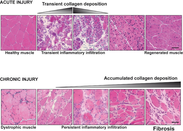
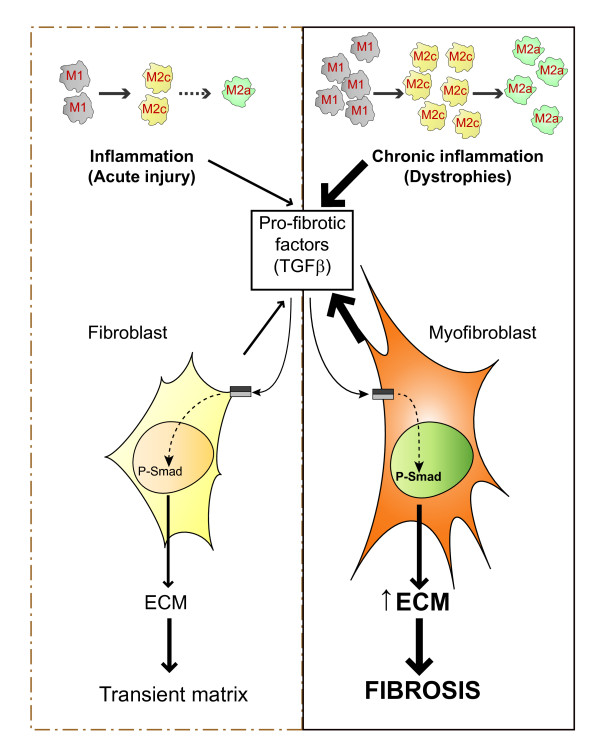
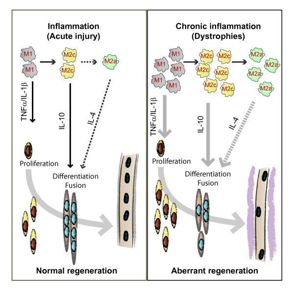
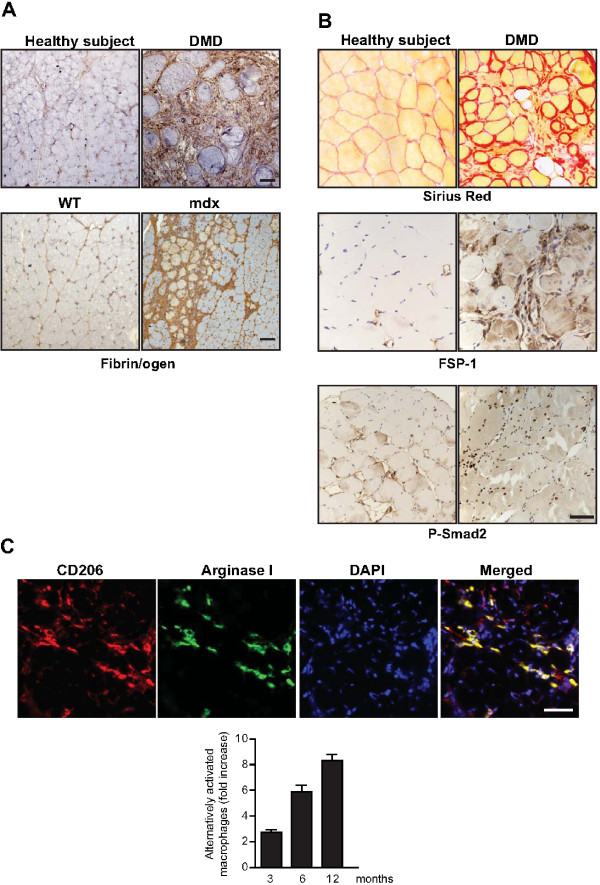
Verification
Claims the paper makes
The claims and statistics of this paper haven’t been checked yet.
Run the check — free, no account needed. It reads the open-access text and checks the sources it cites.
Claims other papers make about this one
Each is checked against this paper’s own text: does it say what the citing paper says it does?
In the citing paper
“Previous work has shown that the accumulation of inflammatory macrophages and age-related declines in interferon-gamma signaling delay muscle repair following injury”
From Skeletal muscle aging, cellular senescence, and senotherapeutics: current knowledge and future directions (2021) · cites this paper as Mann et al., 2011
Self-check
Percentages, sums and reported differences re-computed from the paper’s own numbers.
The self-check needs the paper’s full text.
Citations
Cites
1 retracted
RETRACTED ARTICLE: The role of nitric oxide during healing of trauma to the skeletal muscle
Filippin, L. I., Cuevas, M. J., Lima‐Cabello, E., Marroni, N. P., González‐Gallego, J., & Xavier, R. M. (2010) · Inflammation Research · doi:10.1007/s00011-010-0277-2 · Cited by 34 · cited 4×
“In addition, NO, which is released by local and invading cells during injury, can act as a pro-inflammatory molecule, and is able to activate MMP-2, which in turn releases HGF. Importantly, blockers of NOS function, such as nitro- Larginine methyl ester (L-NAME), have been shown to decrease the acute inflammatory reac…”
MethodInfluentialRetractedThe chemokine system in diverse forms of macrophage activation and polarization
MANTOVANI, A., SICA, A., SOZZANI, S., ALLAVENA, P., VECCHI, A., & LOCATI, M. (2004) · Trends in Immunology · doi:10.1016/j.it.2004.09.015 · Cited by 6,534 · cited 2×
“A nomenclature for polarized macrophages has been proposed [24,25] and they are now referred to as classically and alternatively activated macrophages, or M1 and M2 macrophages, respectively (Figure 2, Figure 3).”
BackgroundMechanisms of TGF-β Signaling from Cell Membrane to the Nucleus
Shi, Y., & Massagué, J. (2003) · Cell · doi:10.1016/s0092-8674(03)00432-x · Cited by 5,860
Visse, R., & Nagase, H. (2003) · Circulation Research · doi:10.1161/01.res.0000070112.80711.3d · Cited by 4,575 · cited 1×
“Activation requires proteolytic cleavage of the inactive precursor by either membrane-type (MT)1MMP [105] or plasmin, and cleavage of their corresponding inhibitors [106].”
Cellular and molecular mechanisms of fibrosis
Wynn, T. (2008) · The Journal of Pathology · doi:10.1002/path.2277 · Cited by 4,495
SATELLITE CELL OF SKELETAL MUSCLE FIBERS
Mauro, A. (1961) · The Journal of Cell Biology · doi:10.1083/jcb.9.2.493 · Cited by 3,683
Hinz, B., Phan, S. H., Thannickal, V. J., Galli, A., Bochaton-Piallat, M.-L., & Gabbiani, G. (2007) · The American Journal of Pathology · doi:10.2353/ajpath.2007.070112 · Cited by 2,068 · cited 1×
“These cells are associated with tissue repair and fibrosis in many tissues and organs, including muscle, skin, liver, lung, bone and cartilage [56].”
BackgroundArnold, L., Henry, A., Poron, F., Baba-Amer, Y., van Rooijen, N., Plonquet, A., Gherardi, R. K., & Chazaud, B. (2007) · The Journal of Experimental Medicine · doi:10.1084/jem.20070075 · Cited by 2,002 · cited 3×
“An important development in our understanding of muscle repair and fibrosis was the demonstration that a heterogeneous population of macrophages exists in regenerating muscle after injury, exhibiting opposing activities (either pro-inflammatory or anti-inflammatory) and different kinetics [23].”
BackgroundInfluentialFibrotic disease and the TH1/TH2 paradigm
Wynn, T. A. (2004) · Nature Reviews Immunology · doi:10.1038/nri1412 · Cited by 1,765
Muscle injury activates resident fibro/adipogenic progenitors that facilitate myogenesis
Joe, A. W. B., Yi, L., Natarajan, A., Le Grand, F., So, L., Wang, J., Rudnicki, M. A., & Rossi, F. M. V. (2010) · Nature Cell Biology · doi:10.1038/ncb2015 · Cited by 1,690 · cited 7×
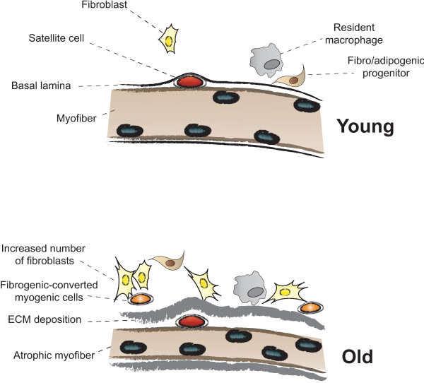
“Whether FAP conversion also occurs during aging, and whether Basal lamina ECM deposition Satellite cell Fibro/adipogenic progenitor Resident macrophage Fibroblast Increased number of fibroblasts Fibrogenic-converted myogenic cells Myofiber Atrophic myofiber Young Old Figure 5 Myofiber growth and extracellular matrix (…”
BackgroundInfluential
Cited by
Sorted by citations
Items 401–500 of 854
Mosich, G. M., Husman, R., Shah, P., Sharma, A. K., Ressadeh, K., Aderibigbe, T., Hu, V. J., McClintick, D. J., Wu, G., Gatto, J. D., et al. (2019) · JCI Insight · doi:10.1172/jci.insight.125334 · Cited by 18
“For instance, the myogenicity of satellite cells may be compromised in the fibrotic muscle (34).”
BackgroundMuscle Diversity, Heterogeneity, and Gradients: Learning from Sarcoglycanopathies
Riera, C. S., Lozanoska‐Ochser, B., Testa, S., Fornetti, E., Bouché, M., & Madaro, L. (2021) · International Journal of Molecular Sciences · doi:10.3390/ijms22052502 · Cited by 18
“Nonetheless, after acute muscle injury, transient extracellular matrix (ECM) remodeling is essential for normal muscle repair [55].”
BackgroundPryce, B. R., Al‐Zahrani, K. N., Dufresne, S. S., Belkina, N., Labrèche, C., Patiño‐López, G., Frenette, J., Shaw, S., & Sabourin, L. A. (2017) · Skeletal Muscle · doi:10.1186/s13395-016-0119-1 · Cited by 18
“Consistent damage to myofibers and decreased force generation is often associated with muscle wasting and fibrosis, as is the case in animal models of muscular dystrophy [52].”
BackgroundLaurino, A., Spinelli, V., Gencarelli, M., Balducci, V., Dini, L., Diolaiuti, L., Ghionzoli, M., Messineo, A., Mugelli, A., Cerbai, E., et al. (2019) · International Journal of Molecular Sciences · doi:10.3390/ijms20194912 · Cited by 18
Altered Gene Expression of Muscle Satellite Cells Contributes to Agerelated Sarcopenia in Mice
Budai, Z., Balogh, L., & Sarang, Z. (2018) · Current Aging Science · doi:10.2174/1874609811666180925104241 · Cited by 17
“Inflammation induces ECM production in activated fibroblasts, which normally take place during muscle regeneration, however, this can lead to muscle fibrosis when the inflammation does not resolve properly [27].”
Young, B. A., Koppenhaver, S. L., Timo-Dondoyano, R. M., Baumann, K., Scheirer, V., Wolff, A., Sutlive, T. G., & Elliott, J. M. (2020) · Journal of Electromyography and Kinesiology · doi:10.1016/j.jelekin.2020.102488 · Cited by 20
Inflammation, fibrosis and skeletal muscle regeneration in LGMDR9 are orchestrated by macrophages
Kölbel, H., Preuße, C., Brand, L., von Moers, A., Marina, A. D., Schuelke, M., Roos, A., Goebel, H., Schara, U., & Stenzel, W. (2021) · Neuropathology and Applied Neurobiology · doi:10.1111/nan.12730 · Cited by 18
Mobilizing Endogenous Repair Through Understanding Immune Reaction With Biomaterials
Karkanitsa, M. L., Fathi, P., Ngo, T. B., & Sadtler, K. (2021) · Frontiers in Bioengineering and Biotechnology · doi:10.3389/fbioe.2021.730938 · Cited by 20
Dreher, S. I., Höckele, S., Huypens, P., Irmler, M., Hoffmann, C., Jeske, T., Hastreiter, M., Moller, A., Birkenfeld, A. L., Häring, H., et al. (2021) · Cells · doi:10.3390/cells10123443 · Cited by 17
Laghi, V., Ricci, V., Santa, F. D., & Torcinaro, A. (2022) · Diagnostics · doi:10.3390/diagnostics12030561 · Cited by 17
Zazula, M. F., Rodriguez, D. F. S., Theodoro, J. L., Maciel, M., Sepulveda, E. V. D. S., de Andrade, B. Z., Boaretto, M. L., Maciel, J. I. H. N., Bronczek, G. A., Soares, G. M., et al. (2023) · International Journal of Molecular Sciences · doi:10.3390/ijms24054730 · Cited by 17
Skeletal muscle omics signatures in cancer cachexia: perspectives and opportunities
Gilmore, L. A., Parry, T. L., Thomas, G. A., & Khamoui, A. V. (2023) · JNCI Monographs · doi:10.1093/jncimonographs/lgad006 · Cited by 19
Identification and functional characterization of TRPA1 in human myoblasts
Osterloh, M., Boehm, M., Kalbe, B., Osterloh, S., & Hatt, H. H. (2015) · Pflügers Archiv - European Journal of Physiology · doi:10.1007/s00424-015-1729-x · Cited by 17
“Muscle injury is always accompanied by local inflammation and the invasion of inflammatory cells which is essential for the repair process [41, 53].”
BackgroundLong non coding RNA H19: An emerging therapeutic target in fibrosing diseases
Li, J., Cao, L., Liu, H., Yin, X., & Wang, J. (2019) · Autoimmunity · doi:10.1080/08916934.2019.1681983 · Cited by 17
“Morgoulis et al, showed that muscle cells derived from DMD patients exhibited decreased expression of lncRNA H19 and miR-675 compared to healthy donor-derived cell.”
BackgroundInfluentialNon-thermal plasma accelerates the healing process of peripheral nerve crush injury in rats
Lee, H.-G., Choi, J., Jang, Y.-S., Kim, U., Kim, G., & Hwang, D. (2020) · International Journal of Medical Sciences · doi:10.7150/ijms.44041 · Cited by 17
“When the muscles are restored normally, the amount of collagen decreases, but in chronic muscle wounds, the amount of collagen remains higher [36].”
BackgroundCeafalan, L., Dobre, M., Milanesi, E., Niculae, A., Manole, E., Gherghiceanu, M., & Hinescu, M. (2020) · Journal of Cellular and Molecular Medicine · doi:10.1111/jcmm.15624 · Cited by 16
Carraro, U., & Yablonka‐Reuveni, Z. (2021) · European Journal of Translational Myology · doi:10.4081/ejtm.2021.9743 · Cited by 18
Miyake, K., Miyagawa, S., Harada, A., & Sawa, Y. (2022) · Molecular Therapy · doi:10.1016/j.ymthe.2022.01.008 · Cited by 17
“as a scaffold for muscle fiber regeneration.(20,42) By balancing the temporary ECM formation and degradation, ECM remodeling occurs, which is essential for angiogenesis and muscle regeneration; this balance is regulated by both proinflammatory and anti-inflammatory macrophages.”
BackgroundInfluentialMautner, K., & Blazuk, J. (2015) · PM&R · doi:10.1016/j.pmrj.2014.12.012 · Cited by 17
“In dysfunctional muscle repair, such as in muscular dystrophies, inflammatory fibroblast activation persists and the reparative capacity of stem cells (satellite cells) is attenuated, resulting in muscular fibrosis and the laying down of excessive type I collagen [34].”
BackgroundPhelps, M., Stuelsatz, P., & Yablonka‐Reuveni, Z. (2016) · FEBS Journal · doi:10.1111/febs.13682 · Cited by 17
Exercise and tissue fibrosis: recent advances in therapeutic potential and molecular mechanisms
Zhao, Z., Zhu, Y., & Wan, D. (2025) · Frontiers in Endocrinology · doi:10.3389/fendo.2025.1557797 · Cited by 19
Kharraz, Y., Lukesova, V., Serrano, A. L., Davison, A., & Muñoz‐Cánoves, P. (2022) · Cytometry Part A · doi:10.1002/cyto.a.24568 · Cited by 16
“During regeneration, skeletal muscle undergoes a series of tightly regulated steps that require the coordination of stem cells with several muscle-resident and blood-derived cell populations to restore tissue homeostasis [9].”
BackgroundAlheib, O., da Silva, L. D., Kwon, I., Reis, R., & Correlo, V. (2022) · Trends in Biotechnology · doi:10.1016/j.tibtech.2022.09.010 · Cited by 16
Che, S., Pham, P. H., Barbut, S., Bienzle, D., & Susta, L. (2024) · Animals · doi:10.3390/ani14020176 · Cited by 16
Nie, L., He, K., Qiu, C., Li, Q., Xiong, B., Gao, C., Zhang, X., Jing, M., Wu, W., Liu, J., et al. (2024) · Biomedicine & Pharmacotherapy · doi:10.1016/j.biopha.2024.116415 · Cited by 19
Abreu, P., Pinheiro, C. H. D. J., Vitzel, K. F., de Vasconcelos, D. A. A., Torres, R. P., Fortes, M. A. S., Marzuca-Nassr, G. N., Mancini‐Filho, J., Hirabara, S. M., & Curi, R. (2016) · Experimental Physiology · doi:10.1113/ep085899 · Cited by 18
“…expected, these findings suggest partial recovery of skeletal muscle contractile function and an increase in fibrous tissue area (collagen deposition) after 28 days of severe injury, similar to what was reported by others (Wynn, 2008; Gillies & Lieber, 2011; Mann et al. 2011; Meyer & Lieber, 2012).”
BackgroundResultInfluentialEffect of Anabolic Steroids on the Cardiac and Skeletal Muscles of Adult Male Rats
Elgendy, H. A. (2018) · International Journal of Clinical and Developmental Anatomy · doi:10.11648/j.ijcda.20180401.11 · Cited by 16
“It has been documented that the increase of intrafasicular connective tissue usually represent a response to a loss of myofibers and attributed that to cytokine imbalance [14, 41].”
BackgroundThooyamani, A. S., & Mukhopadhyay, A. (2021) · Scientific Reports · doi:10.1038/s41598-020-79771-4 · Cited by 17
Lahmann, I., Griger, J., Chen, J.-S., Zhang, Y., Schuelke, M., & Birchmeier, C. (2021) · eLife · doi:10.7554/elife.57356 · Cited by 17
The Involvement of Collagen Triple Helix Repeat Containing 1 in Muscular Dystrophies
Spector, I., Zilberstein, Y., Lavy, A., Genin, O., Barzilai-Tutsch, H., Bodanovsky, A., Halevy, O., & Pines, M. (2012) · American Journal Of Pathology · doi:10.1016/j.ajpath.2012.11.004 · Cited by 16
Angiogenic Properties of Mesenchymal Stem Cells in a Mouse Model of Limb Ischemia
Martins, L., Martin, P. K. M., & Han, S. W. (2014) · Methods in molecular biology · doi:10.1007/978-1-4939-1453-1_13 · Cited by 16
Glycerol induces early fibrosis in regenerating rat skeletal muscle
Mahdy, M. A. A., Warita, K., & Hosaka, Y. Z. (2018) · Journal of Veterinary Medical Science · doi:10.1292/jvms.18-0328 · Cited by 16
“Excessive deposition of collagen fibers hinders normal muscle repair following injury [17].”
BackgroundMechanical induction of osteoanabolic Wnt1 promotes osteoblast differentiation via Plat
Ahmad, M., Haffner‐Luntzer, M., Schoppa, A., Najafova, Z., Lukic, T., Yorgan, T. A., Amling, M., Schinke, T., & Ignatius, A. A. (2024) · The FASEB Journal · doi:10.1096/fj.202301424rr · Cited by 15
“64,65 They help remove damaged ECM components and allow new ECM synthesis for tissue regeneration.”
BackgroundRoig-Soriano, J., Edo, Á., Verdés, S., Martín-Alonso, C., Sánchez‐de‐Diego, C., Rodríguez-Estévez, L., Serrano, A. L., Abraham, C. R., Bosch, A., Ventura, F., et al. (2025) · Molecular Therapy · doi:10.1016/j.ymthe.2025.02.030 · Cited by 16
Bioelectric Properties of Myogenic Progenitor Cells
Fennelly, C., & Söker, S. (2018) · Bioelectricity · doi:10.1089/bioe.2018.0002 · Cited by 14
“188,189”
Nguyen, T. T., Dang-Van, P., Dang-Ngoc, P., Kim, W. B., Le-Minh, T., & Nguyễn, V. Q. H. (2022) · International Journal of Reproductive BioMedicine (IJRM) · doi:10.18502/ijrm.v20i7.11559 · Cited by 15
“Heat stress increases the excessive accumulation of extracellular components, in which collagen plays an important role, and this process often occurs after trauma; inflammation leads to increased fibrosis, resulting in reduced function and causing chronic diseases in many tissues and organs (18).”
BackgroundFeng, X., Wang, A. H., Juan, A. H., Ko, K. D., Jiang, K., Riparini, G., Ciuffoli, V., Kaba, A., Lopez, C., Naz, F., et al. (2023) · Developmental Cell · doi:10.1016/j.devcel.2023.04.005 · Cited by 16
Raghavan, K., Dedeepiya, V. D., Srinivasan, S., Pushkala, S., Bharatidasan, S. S., Ikewaki, N., Iwasaki, M., Senthilkumar, R., Preethy, S., & Abraham, S. J. (2023) · IBRO Neuroscience Reports · doi:10.1016/j.ibneur.2023.06.007 · Cited by 15
“While IL-6 is an acute inflammatory Non-ambulatory Test couldn ’ t be performed biomarker (Mann et al., 2011), IL-13 is a pro-fibrotic biomarker (Mann et al., 2011) and was significantly decreased (Fig.”
BackgroundNieves‐Rodriguez, S., Barthélémy, F., Woods, J. D., Douine, E. D., Wang, R. T., Scripture-Adams, D. D., Chesmore, K. N., Galasso, F. I., Miceli, M. C., & Nelson, S. F. (2023) · Frontiers in Genetics · doi:10.3389/fgene.2023.1216066 · Cited by 15
“Most of the genetic modifiers (10 of 17) had expression mainly within a nonmyofiber cell type, consistent with the known role of nonmyofiber lineage cells in orchestrating muscle remodeling during regeneration and fibrosis (Mann et al., 2011).”
BackgroundFactors, mechanisms and improvement methods of muscle strength loss
Wang, K., Wang, X., & Wang, Y. (2024) · Frontiers in Cell and Developmental Biology · doi:10.3389/fcell.2024.1509519 · Cited by 18
Mahdy, M. A. A., Warita, K., & Hosaka, Y. (2017) · Animal science journal = Nihon chikusan Gakkaiho · doi:10.1111/asj.12845 · Cited by 13
Formicola, L., Pannérec, A., Correra, R. M., Gayraud-Morel, B., Ollitrault, D., Besson, V., Tajbakhsh, S., Lachey, J. L., Seehra, J. S., Marazzi, G., et al. (2018) · Frontiers in Physiology · doi:10.3389/fphys.2018.00515 · Cited by 17
“During the early phase of DMD, muscle undergoes continuous rounds of degeneration/regeneration but eventually regenerative competence declines accompanied by a decrease in satellite cell number (Wallace and McNally, 2009; Mann et al., 2011; Tabebordbar et al., 2013).”
BackgroundPorcari, P., Hall, M. G., Clark, C. A., Greally, E., Straub, V. W., & Blamire, A. M. (2020) · NMR in Biomedicine · doi:10.1002/nbm.4276 · Cited by 18
“Subsequently, muscle becomes fibrotic and is eventually replaced by fat.(10,11) Skeletal and cardiac muscle function is gradually lost over time leading to respiratory and cardiac failure and premature death.”
BackgroundAddinsall, A. B., Forgan, L. G., McRae, N. L., Kelly, R. W., McDonald, P., McNeil, B., McCulloch, D. R., & Stupka, N. (2020) · Biomolecules · doi:10.3390/biom10030416 · Cited by 15
“Muscle degeneration was assessed by manually circling regions of fibrosis and mononuclear infiltrate, which is composed of muscle progenitor cells, inflammatory cells and fibroblasts [43] and expressing these regions as a percentage of the total muscle cross section [37].”
MethodPhotopolymerizable Hydrogel-Encapsulated Fibromodulin-Reprogrammed Cells for Muscle Regeneration
Yang, P., Li, C., Lee, M., Marzvanyan, A., Zhao, Z., Ting, K., Soo, C. B., & Zheng, Z. S. (2020) · Tissue Engineering Part A · doi:10.1089/ten.tea.2020.0026 · Cited by 15
Strategies and opportunities to control breast myopathies: An opinion paper
Bailey, R. A. (2023) · Frontiers in Physiology · doi:10.3389/fphys.2023.1173564 · Cited by 14
“During the first week post hatch these cells are most active and their population increases rapidly (Mann et al., 2011; Daughtry et al., 2017; Halevy, 2020).”
BackgroundCernisova, V., Lu‐Nguyen, N., Trundle, J., Herath, S., Malerba, A., & Popplewell, L. J. (2023) · International Journal of Molecular Sciences · doi:10.3390/ijms24098174 · Cited by 14
Ortved, K. F., Alward, L., Cowles, B., Linardi, R. L., Barot, D., Usimaki, A., Fedie, J. R., Amodie, D., & Goodrich, L. R. (2024) · Frontiers in Veterinary Science · doi:10.3389/fvets.2024.1335972 · Cited by 14
Yao, H., Qian, J., Bian, X., Tang, Y., ye, X., He, G., Tang, H., liu, X., Zhang, A., xie, Q., et al. (2024) · Journal of Orthopaedic Surgery and Research · doi:10.1186/s13018-024-04733-9 · Cited by 15
“Recurrent damage and inadequate treatment of underlying conditions can lead to the progression of acute injury to chronic injury, resulting in delayed clearance of resident cells, disrupted inter-cellular coordination, and ultimately pathological repair [13].”
BackgroundSynergistic effects of conductive hydrogels and electrical stimulation in volumetric muscle loss
Park, S., Le, T. P., Byun, H. J., Lee, S., Lee, M. K., Huh, K. M., & Lee, J. Y. (2025) · Chemical Engineering Journal · doi:10.1016/j.cej.2025.162362 · Cited by 14
Kakutani, N., Takada, S., Nambu, H., Matsumoto, J., Furihata, T., Yokota, T., Fukushima, A., & Kinugawa, S. (2020) · Skeletal Muscle · doi:10.1186/s13395-020-00230-9 · Cited by 15
M-Ras is Muscle-Ras, Moderate-Ras, Mineral-Ras, Migration-Ras, and Many More-Ras
Endo, T. (2020) · Experimental Cell Research · doi:10.1016/j.yexcr.2020.112342 · Cited by 14
Li, N., Bai, R., Li, C., Dang, L.-H., Du, Q.-X., Jin, Q., Cao, J., Wang, Y., & Sun, J. (2021) · Bioscience Reports · doi:10.1042/bsr20203699 · Cited by 14
“responses(17), and coordination between inflammation and regeneration.”
Could we also be regenerative superheroes, like salamanders?
Dall’Agnese, A., & Puri, P. (2016) · BioEssays · doi:10.1002/bies.201600015 · Cited by 14
“Failed or aberrant regeneration often leads to maladaptive repair of tissues and organs by scarring (fibrosis) [1, 2] or uncontrolled cell”
BackgroundStem cells in skeletal muscle growth and regeneration in amniotes and teleosts: Emerging themes
Ruparelia, A. A., Ratnayake, D., & Currie, P. D. (2019) · Wiley Interdisciplinary Reviews Developmental Biology · doi:10.1002/wdev.365 · Cited by 15
“…M2 macrophages led to muscle fibrosis in aged muscle (Y. Wang, Wehling-Henricks, Samengo, & Tidball, 2015) and in conditions of chronic muscle damage presenting with prolonged nonresolute inflammatory responses (Lemos et al., 2015; Mann et al., 2011; Villalta, Nguyen, Deng, Gotoh, & Tidball, 2008).”
BackgroundMiyake, K., Kikuchi, S., Tatsukawa, T., Uchida, D., Koya, A., Sawa, Y., & Azuma, N. (2020) · Annals of Vascular Surgery · doi:10.1016/j.avsg.2020.07.033 · Cited by 14
Regulation of Myogenic Activity by Substrate and Electrical Stimulation In Vitro
Patel, A., Vendrell, S., Haas, G. J., Marcinczyk, M., Ziemkiewicz, N., Talovic, M., Fisher, J. S., & Garg, K. (2019) · BioResearch open access · doi:10.1089/biores.2019.0016 · Cited by 12
Yang, Y., Liu, M., Liu, X., Zhang, Y.-C., Hu, Y., Gao, R.-S., Pang, E., Hou, L., Wang, J., & Fei, W. (2022) · Nanomedicine · doi:10.2217/nnm-2022-0173 · Cited by 14
Muscle regeneration and muscle stem cells in metabolic disease
Chung, J. D., Porrello, E. R., & Lynch, G. S. (2024) · Free Radical Biology and Medicine · doi:10.1016/j.freeradbiomed.2024.11.041 · Cited by 16
Jaimes, N. G. O., Ginja, M., Regueiro-Purriños, M. M., Oliveira, P. A., Gama, A., Rodríguez-Altónaga, J. A., & Gonzalo-Orden, J. M. (2013) · Journal of Small Animal Practice · doi:10.1111/jsap.12080 · Cited by 13
“However, in severe muscular dystrophies satellite cells may become exhausted over time or lose the capacity to mediate repair and the muscle tissue is replaced by adipose and fibrous tissue (Mann et al. 2011).”
BackgroundSevere murine limb‐girdle muscular dystrophy type 2C pathology is diminished by FTY720 treatment
Heydemann, A. (2016) · Muscle & Nerve · doi:10.1002/mus.25503 · Cited by 13
“In muscular dystrophy (MD), the satellite cells are impeded by chronic inflammation (2), excessive fibrosis, and excessive ECM (3), and the muscles are incapable of remodeling the scar tissue at the required pace.”
The pathophysiology and treatment of musculoskeletal fibrosis
Sheets, K., Overbey, J., Ksajikian, A., Bovid, K. M., Kenter, K., & Li, Y. (2022) · Journal of Cellular Biochemistry · doi:10.1002/jcb.30217 · Cited by 14
“However, excess deposition of ECM and collagen becomes problematic in the formation of scar tissue.(5) Studies from molecular and cellular biology have shown multiple factors influence pro‐fibrotic changes in musculoskeletal tissues.”
BackgroundSugihara, H., MIYAJI, K., Yamanouchi, K., Matsuwaki, T., & Nishihara, M. (2017) · Journal of Veterinary Medical Science · doi:10.1292/jvms.17-0638 · Cited by 12
“Although M2 macrophages contribute to tissue repair, excess number of M2 macrophages is known to lead to hyper-activation of fibroblasts, resulting in the fibrosis of skelatal muscle [15, 23].”
BackgroundWound healing, cellular regeneration and plasticity: the elegans way
Vibert, L., Daulny, A., & Jarriault, S. (2018) · The International Journal of Developmental Biology · doi:10.1387/ijdb.180123sj · Cited by 16
“In humans, epithelia, including skin, intestine and lung, can heal and regrow, and a few organs, such as the kidney, endometrium and liver exhibit some regenerative abilities (Barker, 2014; Chou et al., 2014; Gargett et al., 2012; Kotton and Morrisey, 2014; Mann et al., 2011; Michalopoulos, 2007).”
BackgroundPericytes in Muscular Dystrophies
Moyle, L. A., Tedesco, F. S., & Benedetti, S. (2019) · Advances in experimental medicine and biology · doi:10.1007/978-3-030-16908-4_15 · Cited by 15
“4c. Pericytes and fibrosis Increased fibrosis is a typical feature of aged and dystrophic muscles, which ultimately results in muscle weakness, atrophy and reduction of its regenerative potential (Kragstrup et al., 2011; Mann et al., 2011; Ryall et al., 2008; Thompson, 2009; Walston, 2012).”
BackgroundDistant regulatory effects of genetic variation in multiple human tissuesPreprint
Jo, B., He, Y. M., Strober, B. J., Parsana, P., Aguet, F., Brown, A. A., Castel, S. E., Gamazon, E. R., Gewirtz, A. D., Gliner, G., et al. (2016) · bioRxiv (Cold Spring Harbor Laboratory) · doi:10.1101/074419 · Cited by 14
Tang, Y., Shi, C., & Zhang, X. (2025) · Food Hydrocolloids · doi:10.1016/j.foodhyd.2025.111482 · Cited by 12
Organotypic cultures as aging associated disease models
Sanchez, M. M., Bagdasarian, I. A., Darch, W., & Morgan, J. T. (2022) · Aging · doi:10.18632/aging.204361 · Cited by 12
Freeze-Dried Porous Collagen Scaffolds for the Repair of Volumetric Muscle Loss Injuries
Basurto, I. M., Bandara, G. C., Boudreau, R. D., Shriver, S. B., Muhammad, S. A., Christ, G. J., & Caliari, S. R. (2025) · ACS Biomaterials Science & Engineering · doi:10.1021/acsbiomaterials.4c01601 · Cited by 13
“33,34 Interestingly, the quantity of CD68 + /CD163 − macrophages within nonrepaired muscles was most similar to native muscle tissues, suggesting that other inflammatory cell types may be the primary drivers of chronic inflammation characteristic of VML. − 37 3.7.”
InfluentialCuijpers, I., Sthijns, M. M. J. P. E., van den Bogart, V. A. R., Katsburg, J., Leenders, C. F. M., & Troost, F. J. (2025) · International Journal of Molecular Sciences · doi:10.3390/ijms26115151 · Cited by 12
“The secretion of ECM proteins by activated myofibroblasts results in structural remodeling of the ECM, which is crucial for both normal skeletal muscle regeneration and the development of skeletal muscle fibrosis [12].”
Role of Sphingosine 1-Phosphate in Skeletal Muscle Cell Biology
Bruni, P., & Donati, C. (2013) · Handbook of experimental pharmacology · doi:10.1007/978-3-7091-1511-4_23 · Cited by 12
Neonatal Satellite Cells Form Small Myotubes In Vitro
Monroy, P. L. C., Grefte, S., Kuijpers‐Jagtman, A. M., den Hoff, J. W. V., & Wagener, F. A. D. T. G. (2016) · Journal of Dental Research · doi:10.1177/0022034516679136 · Cited by 12
“This could explain the decreased myogenic potential of BrHM SCs during healing (Mann et al. 2011).”
BackgroundErythropoietin Alleviates Burn-induced Muscle Wasting
Wu, S., Lu, I., Tai, M., Chai, C., Kwan, A., & Huang, S. (2020) · International Journal of Medical Sciences · doi:10.7150/ijms.38590 · Cited by 12
“Transforming growth factor beta 1 (TGF-β1) is a crucial factor to regulate ECM remodeling [32, 33] and drives tissue to fibrosis in chronic inflammatory diseases.”
BackgroundChaiyasing, R., Sugiura, A., Ishikawa, T., Ojima, K., Warita, K., & Hosaka, Y. Z. (2021) · Journal of Veterinary Medical Science · doi:10.1292/jvms.21-0495 · Cited by 13
“However, the muscle injury caused by mechanical loading or chemical injection activates satellite cells from a quiescent state to a state of proliferation, leading to the myoregeneration process [5, 28, 39, 42, 45].”
BackgroundMo, Y., Kang, S., Bang, J., Kim, Y., Jeong, J., Jeong, E. M., Kim, H. Y., Cho, S., & Kang, H. (2022) · Molecules and Cells · doi:10.14348/molcells.2022.0038 · Cited by 12
“M2c macrophages have been reported to drive pulmonary fibrosis (Lu et al., 2013; Mann et al., 2011).”
BackgroundXynos, A., Neguembor, M. V., Caccia, R., Licastro, D., Nonis, A., Serio, C. D., Stupka, E., & Gabellini, D. (2013) · Journal of Cell Science · doi:10.1242/jcs.121533 · Cited by 11
Pai, M., Lei, C.-S., Su, S.-T., Yeh, S., & Hou, Y.-C. (2019) · European Journal of Nutrition · doi:10.1007/s00394-019-01951-4 · Cited by 13
Venter, C., Myburgh, K., & Niesler, C. (2021) · Physiological Reports · doi:10.14814/phy2.14704 · Cited by 10
“Intercellular communication between the different macrophage and fibroblast phenotypes has knock-on effects regulating myogenesis; a dysregulated communication leads to aberrant muscle wound repair (Bosurgi et al., 2011; Mann et al., 2011).”
BackgroundInfluentialIqbal, A., May, U., Prince, S., Järvinen, T. A. H., & Heydemann, A. (2021) · Pharmaceutics · doi:10.3390/pharmaceutics13091506 · Cited by 12
Effects of Vitamin C on Cytotherapy-Mediated Muscle Regeneration
Kim, A.-Y., Lee, E. M., Lee, E. H., Min, C., Kang, K., Park, J., Hong, I., Ishigami, A., Tremblay, J. P., & Jeong, K. (2013) · Cell Transplantation · doi:10.3727/096368912x657846 · Cited by 12
“In addition, it also inhibits satellite cell proliferation (28) and induces transdifferentiation of myogenic progenitors to myofibroblasts (18,21).”
BackgroundPawlikowski, B. T., Betta, N. D., Antwine, T., & Olwin, B. B. (2019) · bioRxiv (Cold Spring Harbor Laboratory) · doi:10.1101/627851 · Cited by 13
“Muscle regeneration is diminished in aged mice, where repaired myofibers are smaller, generate less force, and muscle tissue is more fibrotic (Brack et al., 2007; I M Conboy et al., 2005; Cosgrove et al., 2014; Hall, Banks, Chamberlain, & Olwin, 2010; Mann et al., 2011; Zhang et al., 2016).”
BackgroundZwetsloot, K. A., Nedergaard, A., Gilpin, L. T., Childs, T. E., & Booth, F. W. (2012) · Biogerontology · doi:10.1007/s10522-012-9382-7 · Cited by 11
Huang, X., Kui, X., Ma, J., Chen, J., Huang, Y., & He, B. (2025) · Mechanisms of Ageing and Development · doi:10.1016/j.mad.2025.112067 · Cited by 11
“In C2C12 cells, D-gal treat-ment decreased proliferation, increased apoptosis, and upregulated aging-associated markers p53/p16, confirming successful aging model establishment.”
Melatonin ameliorates age-related sarcopenia by inhibiting fibrogenic conversion of satellite cell
Zhu, G.-Z., Zhao, K., Li, H.-Z., Wu, D.-Z., Chen, Y.-B., Han, D., Gao, J.-W., Chen, X., Yu, Y.-P., Huang, Z., et al. (2024) · Molecular Medicine · doi:10.1186/s10020-024-00998-2 · Cited by 15
Li, N., Lu, X., Zhang, J., Dang, L.-H., Liu, J.-F., Wu, F.-Y., Cao, X., Liang, X., & Sun, J. (2025) · Experimental Cell Research · doi:10.1016/j.yexcr.2025.114677 · Cited by 11
Mendes, S., Leal, D. V., Baker, L. A., Ferreira, A., Smith, A. C., & Viana, J. L. (2023) · International Journal of Molecular Sciences · doi:10.3390/ijms24076017 · Cited by 12
Low-intensity training provokes adaptive extracellular matrix turnover of a muscular dystrophy model
Gaiad, T. P., Oliveira, M. X., Lobo-Jr, A. R., Libório, L. R., Pinto, P. A. F., Fernandes, D. C., Santos, A. P., Ambrósio, C. E., & Machado, A. S. D. (2017) · Journal of Exercise Rehabilitation · doi:10.12965/jer.1735094.547 · Cited by 11
“…formation of a permanent fibrotic tissue by producing an accumulation of fibrotic interstitial ECM components such as hyaluronic acid, fibronectin, proteoglycans and interstitial collagens (Gillies et al., 2017; Serrano and MuñozCánoves, 2010), such as collagen types I and III (Mann et al., 2011).”
BackgroundInfluentialPlasma MMP-9, TIMP-1, and TGF-β1 Responses to Exercise-Induced Muscle Injury
Kim, J., & Lee, J. (2020) · International Journal of Environmental Research and Public Health · doi:10.3390/ijerph17020566 · Cited by 11
Harish, P., Forrest, L., Herath, S., Dickson, G., Malerba, A., & Popplewell, L. J. (2020) · Frontiers in Physiology · doi:10.3389/fphys.2020.00184 · Cited by 11
Lipin1 plays complementary roles in myofibre stability and regeneration in dystrophic muscles
Jama, A., Alshudukhi, A. A., Burke, S., Dong, L., Kamau, J. K., Voss, A. A., & Ren, H. (2023) · The Journal of Physiology · doi:10.1113/jp284085 · Cited by 11
“Excessive deposition of collagen and extracellular matrix elements progressively remodels and destroy normal skeletal muscle architecture in mdx mice (Mahdy 2019; Mann et al., 2011).”
BackgroundZazula, M. F., de Andrade, B. Z., Boaro, C. D. T., Kirsch, C. B., Reginato, A., Peretti, A. L., Brancalhão, R. M. C., Bertolini, G. R. F., Naliwaiko, K., Guimarães, A. T. B., et al. (2022) · Acta Histochemica · doi:10.1016/j.acthis.2022.151892 · Cited by 14
Ragozzino, E., Bortolani, S., Pietro, L. D., Papait, A., Parolini, O., Monforte, M., Tasca, G. A., & Ricci, E. (2023) · Acta Neuropathologica Communications · doi:10.1186/s40478-023-01660-4 · Cited by 10
“In the light of different studies supporting the causative role of inflammation in driving fibrosis in skeletal muscle [28, 36, 37], we evaluated a possible correlation among the immune cells found in FSHD muscles and the amount of fibrosis.”
BackgroundAssessment of gait mechanics and muscle strength in hypermobile Ehlers Danlos Syndrome
Ball, L. N., Jacobs, M. V., McLouth, C. J., Clasey, J. L., Francomano, C. A., Sheppard, M. B., & Samaan, M. A. (2024) · Clinical Biomechanics · doi:10.1016/j.clinbiomech.2024.106210 · Cited by 10
Duong, V. T., Dang, T. T., Le, V. P., Le, T. H., Nguyen, C. T., Phan, H. L., Seo, J.-M., Lin, C., Back, S. H., & Koo, K.-I. (2024) · Biomaterials · doi:10.1016/j.biomaterials.2024.122840 · Cited by 10
Shahin‐Shamsabadi, A., & Cappuccitti, J. (2024) · Advanced Healthcare Materials · doi:10.1002/adhm.202404465 · Cited by 10
Gurel, V., Lins, J., Lambert, K., Lazauski, J., Spaulding, J., & McMichael, J. C. (2015) · Dose-Response · doi:10.1177/1559325815616351 · Cited by 10
“Changes in this environment can speed up muscle repair, enhance muscle strength, and reduce inflammation and fibrosis, leading to the amelioration of symptoms.(21,22) Therefore, molecules that can modify these complex interactions between muscle cells and their environment can be powerful therapeutics for the treatmen…”
BackgroundLeiva‐Cepas, F., Jimena, I., Ruz‐Caracuel, I., Luque, E., Villalba, R., & Peña, J. (2020) · PubMed · doi:10.14670/hh-18-163 · Cited by 10
Eager, J. M., Warrender, W. J., Deusenbery, C. B., Jamgochian, G. C., Singh, A. M., Abboud, J. A., & Spiller, K. L. (2021) · The American Journal of Sports Medicine · doi:10.1177/03635465211023212 · Cited by 10
“Deviations from these processes, either temporally or in potency, result in improper healing.(20,25,33) While macrophage phenotype has not been thoroughly assessed in rotator cuff healing, especially in humans, the M1-to-M2a transition has been observed in a rat model of tendon healing.”
BackgroundSrikuea, R., & Hirunsai, M. (2023) · Scientific Reports · doi:10.1038/s41598-023-40978-w · Cited by 10
“Dysregulation of the TGF-β/Smad signalling cascade through increased phosphorylation of Smad2/3 proteins induces accumulation of ECM components and subsequently forms fibrosis in dystrophic muscle that is susceptible to progressive degeneration and regeneration 6,28 .”
Repairing Volumetric Muscle Loss with Commercially Available Hydrogels in an Ovine Model
Su, E. Y., Kennedy, C. S., Vega-Soto, E. E., Pallas, B. D., Lukpat, S. N., Hwang, D., Bosek, D. W., Forester, C. E., Loebel, C., & Larkin, L. M. (2023) · Tissue Engineering. Part A · doi:10.1089/ten.tea.2023.0240 · Cited by 10
“17,18 Because excessive fibrotic infiltration of the repair site is one of the biggest obstacles for regeneration of healthy skeletal muscle tissue, 19,20 various regenerative medicine strategies, such as hydrogels (HGs), are being developed to decrease the acute inflammation and fibrosis that occur…”
Metadata
- Authors
- Mann, C. J., Perdiguero, E., Kharraz, Y., Aguilar, S., Pessina, P., Serrano, A. L., & Muñoz-Cánoves, P.
- Affiliations
- Universitat Pompeu Fabra (ES)
- Venue
- Skeletal Muscle
- Published
- 2011-05-04
- Volume
- 1
- Issue
- 1
- Pages
- 21-21
- ISSN
- 2044-5040 (ISSN-L)
- PMC
- PMC3156644
- OpenAlex
- W2076787014
- Cited by
- 853
- References
- 129
- Citations by year
- 2026: 37 · 2025: 59 · 2024: 55 · 2023: 53 · 2022: 59 · 2021: 71 · 2020: 81 · 2019: 85 · 2018: 76 · 2017: 51 · 2016: 68 · 2015: 48 · 2014: 49 · 2013: 33 · 2012: 27
- FWCI
- 14.30
- Type
- journal article
- Language
- en
- Open access
- gold
- doi.org
- Licence
- cc-by
- Locations
- Skeletal Muscle (publishedVersion, cc-by)
- Europe PMC (PubMed Central) (publishedVersion, cc-by)
- PubMed Central (publishedVersion, cc-by)
- PubMed (publishedVersion)
- RECERCAT (Consorci de Serveis Universitaris de Catalunya) (publishedVersion, other-oa)
- DOAJ (DOAJ: Directory of Open Access Journals) (submittedVersion, cc-by-sa)
- Repositori digital de la UPF (Universitat Pompeu Fabra) (submittedVersion, cc-by)
- Funders
- Ministerio de Ciencia e Innovación (PLE2009)
- Centro de Investigación Biomédica en Red sobre Enfermedades Neurodegenerativas
- Topics
- Life Sciences › Biochemistry, Genetics and Molecular Biology › Molecular Biology › Muscle Physiology and Disorders
- Health Sciences › Medicine › Cardiology and Cardiovascular Medicine › Cardiac Fibrosis and Remodeling
- Health Sciences › Medicine › Surgery › Wound Healing and Treatments
- Keywords
- muscle fibrosis, muscle repair, muscular dystrophy, satellite cells, muscle stem cells, fibroblast activation, inflammatory cell infiltration, extracellular matrix, wound healing, microenvironment
About these checks
Registry, retraction, Cites and statistics results are computed, not judged, with one exception: a printed reference that no index lists is read once by a model before it counts as a citation error. Claims are extracted by a model and kept only when their quote appears verbatim in the text; each cited source is then checked, with a model’s help, for whether it says what the claim relies on. Those verdicts can be wrong — read the claim’s quote and the cited source before relying on one.
Statistics and arithmetic are recomputed, not judged.
Confidence bands are shown only for fields calibrated on our annotated test set. None is calibrated yet, so no band is shown.
What counts as a claim ›How we measure confidence ›How we check this ›
Are you an author, or do you see a problem with this page? Request a review
