

The DOI matches its Crossref record; 129 of the 129 references it cites were found in a registry or an index.
Aberrant repair and fibrosis development in skeletal muscle
Open accessAbstract
The repair process of damaged tissue involves the coordinated activities of several cell types in response to local and systemic signals. Following acute tissue injury, infiltrating inflammatory cells and resident stem cells orchestrate their activities to restore tissue homeostasis. However, during chronic tissue damage, such as in muscular dystrophies, the inflammatory-cell infiltration and fibroblast activation persists, while the reparative capacity of stem cells (satellite cells) is attenuated. Abnormal dystrophic muscle repair and its end stage, fibrosis, represent the final common pathway of virtually all chronic neurodegenerative muscular diseases. As our understanding of the pathogenesis of muscle fibrosis has progressed, it has become evident that the muscle provides a useful model for the regulation of tissue repair by the local microenvironment, showing interplay among muscle-specific stem cells, inflammatory cells, fibroblasts and extracellular matrix components of the mammalian wound-healing response. This article reviews the emerging findings of the mechanisms that underlie normal versus aberrant muscle-tissue repair.
Overview
The claims and statistics of this paper haven’t been checked yet.
Run the check — free, no account needed. It reads the open-access text and checks the sources it cites.
Integrity signals
Registry
Cites
Statistics
Figures
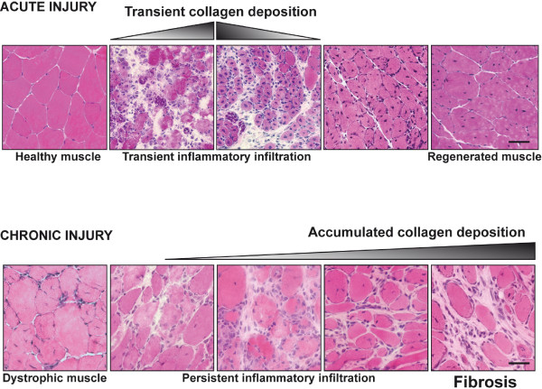
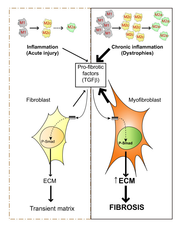
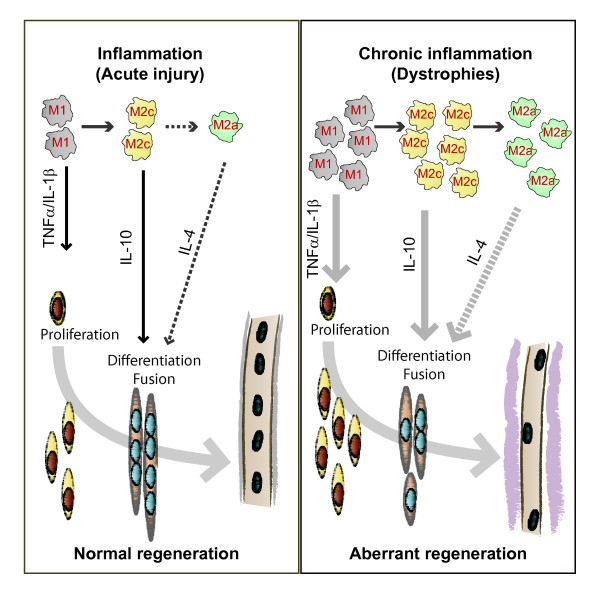
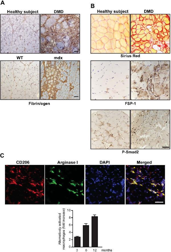
Verification
Claims the paper makes
The claims and statistics of this paper haven’t been checked yet.
Run the check — free, no account needed. It reads the open-access text and checks the sources it cites.
Claims other papers make about this one
Each is checked against this paper’s own text: does it say what the citing paper says it does?
In the citing paper
“Previous work has shown that the accumulation of inflammatory macrophages and age-related declines in interferon-gamma signaling delay muscle repair following injury”
From Skeletal muscle aging, cellular senescence, and senotherapeutics: current knowledge and future directions (2021) · cites this paper as Mann et al., 2011
Self-check
Percentages, sums and reported differences re-computed from the paper’s own numbers.
The self-check needs the paper’s full text.
Citations
Cites
1 retracted
RETRACTED ARTICLE: The role of nitric oxide during healing of trauma to the skeletal muscle
Filippin, L. I., Cuevas, M. J., Lima‐Cabello, E., Marroni, N. P., González‐Gallego, J., & Xavier, R. M. (2010) · Inflammation Research · doi:10.1007/s00011-010-0277-2 · Cited by 34 · cited 4×
“In addition, NO, which is released by local and invading cells during injury, can act as a pro-inflammatory molecule, and is able to activate MMP-2, which in turn releases HGF. Importantly, blockers of NOS function, such as nitro- Larginine methyl ester (L-NAME), have been shown to decrease the acute inflammatory reac…”
MethodInfluentialRetractedThe chemokine system in diverse forms of macrophage activation and polarization
MANTOVANI, A., SICA, A., SOZZANI, S., ALLAVENA, P., VECCHI, A., & LOCATI, M. (2004) · Trends in Immunology · doi:10.1016/j.it.2004.09.015 · Cited by 6,534 · cited 2×
“A nomenclature for polarized macrophages has been proposed [24,25] and they are now referred to as classically and alternatively activated macrophages, or M1 and M2 macrophages, respectively (Figure 2, Figure 3).”
BackgroundMechanisms of TGF-β Signaling from Cell Membrane to the Nucleus
Shi, Y., & Massagué, J. (2003) · Cell · doi:10.1016/s0092-8674(03)00432-x · Cited by 5,860
Visse, R., & Nagase, H. (2003) · Circulation Research · doi:10.1161/01.res.0000070112.80711.3d · Cited by 4,575 · cited 1×
“Activation requires proteolytic cleavage of the inactive precursor by either membrane-type (MT)1MMP [105] or plasmin, and cleavage of their corresponding inhibitors [106].”
Cellular and molecular mechanisms of fibrosis
Wynn, T. (2008) · The Journal of Pathology · doi:10.1002/path.2277 · Cited by 4,495
SATELLITE CELL OF SKELETAL MUSCLE FIBERS
Mauro, A. (1961) · The Journal of Cell Biology · doi:10.1083/jcb.9.2.493 · Cited by 3,683
Hinz, B., Phan, S. H., Thannickal, V. J., Galli, A., Bochaton-Piallat, M.-L., & Gabbiani, G. (2007) · The American Journal of Pathology · doi:10.2353/ajpath.2007.070112 · Cited by 2,068 · cited 1×
“These cells are associated with tissue repair and fibrosis in many tissues and organs, including muscle, skin, liver, lung, bone and cartilage [56].”
BackgroundArnold, L., Henry, A., Poron, F., Baba-Amer, Y., van Rooijen, N., Plonquet, A., Gherardi, R. K., & Chazaud, B. (2007) · The Journal of Experimental Medicine · doi:10.1084/jem.20070075 · Cited by 2,002 · cited 3×
“An important development in our understanding of muscle repair and fibrosis was the demonstration that a heterogeneous population of macrophages exists in regenerating muscle after injury, exhibiting opposing activities (either pro-inflammatory or anti-inflammatory) and different kinetics [23].”
BackgroundInfluentialFibrotic disease and the TH1/TH2 paradigm
Wynn, T. A. (2004) · Nature Reviews Immunology · doi:10.1038/nri1412 · Cited by 1,765
Muscle injury activates resident fibro/adipogenic progenitors that facilitate myogenesis
Joe, A. W. B., Yi, L., Natarajan, A., Le Grand, F., So, L., Wang, J., Rudnicki, M. A., & Rossi, F. M. V. (2010) · Nature Cell Biology · doi:10.1038/ncb2015 · Cited by 1,690 · cited 7×
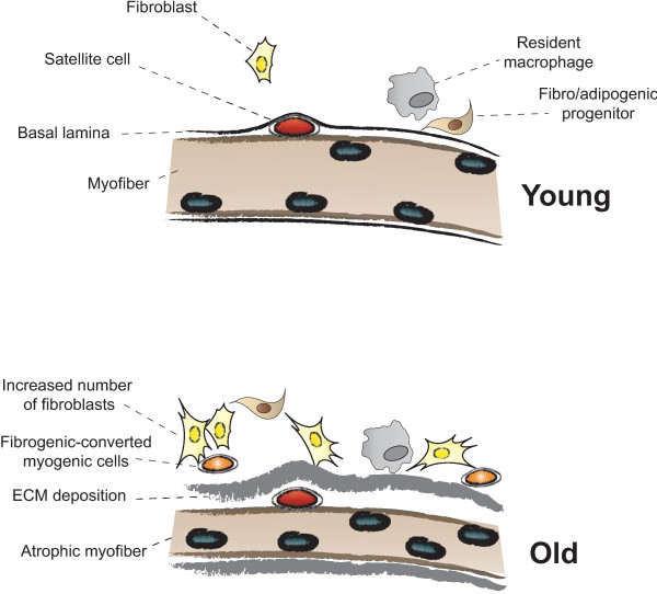
“Whether FAP conversion also occurs during aging, and whether Basal lamina ECM deposition Satellite cell Fibro/adipogenic progenitor Resident macrophage Fibroblast Increased number of fibroblasts Fibrogenic-converted myogenic cells Myofiber Atrophic myofiber Young Old Figure 5 Myofiber growth and extracellular matrix (…”
BackgroundInfluential
Cited by
Sorted by citations
Items 501–600 of 854
Pinheiro-Dardis, C. M., & Russo, T. L. (2016) · American Journal of Physical Medicine & Rehabilitation · doi:10.1097/phm.0000000000000601 · Cited by 10
Naughton, N. M., & Georgiadis, J. G. (2019) · Magnetic Resonance in Medicine · doi:10.1002/mrm.28014 · Cited by 10
“14 In complex tissues such as muscle, the apparent diffusion tensor is anisotropic and composed of three distinct eigenvalues (λ1, λ2, and λ3), which correspond to the princi(1) M(x, t)”
BackgroundTrends in Mesenchymal Stem Cells' Applications for Skeletal Muscle Repair and Regeneration
Caseiro, A. R., Pereira, T., Bártolo, P., Santos, J. D., Luís, A. L., & Maurício, A. C. (2015) · InTech eBooks · doi:10.5772/60919 · Cited by 10
Prenatal myonuclei play a crucial role in skeletal muscle hypertrophy in rodents
Kawano, F., Ono, Y., Fujita, R., Watanabe, A., Masuzawa, R., Shibata, K., Hasegawa, S., Nakata, K., & Nakai, N. (2016) · American Journal of Physiology-Cell Physiology · doi:10.1152/ajpcell.00151.2016 · Cited by 10
“However, the connective tissues were enlarged in the interstitial space of regenerated muscles, presumably because of the activation of fibroblasts during muscle regeneration (21, 25).”
BackgroundUnderstanding muscle-immune interactions in adolescent idiopathic scoliosis: a feasibility study
Rudrapatna, S., Peterson, D. C., Missiuna, P. C., Aditya, I., Drew, B. M., Sahar, N., Thabane, L., & Samaan, M. C. (2017) · Pilot and Feasibility Studies · doi:10.1186/s40814-017-0193-0 · Cited by 10
Venter, C., & Niesler, C. U. (2018) · Experimental Cell Research · doi:10.1016/j.yexcr.2018.07.019 · Cited by 10
DQF-MT MRI of connective tissues: application to tendon and muscle
Kuśmia, S., Eliav, U., Navon, G., & Guillot, G. (2012) · Magnetic Resonance Materials in Physics Biology and Medicine · doi:10.1007/s10334-012-0346-3 · Cited by 9
Skeletal muscle cell aging and stem cells
Rafi, S. T. M., Sambandam, Y., Sittadjody, S., Pathak, S., Ramachandran, I., & Kumaran, R. (2021) · Elsevier eBooks · doi:10.1016/b978-0-12-820071-1.00009-8 · Cited by 9
Iezzi, S., Purslow, P. P., Sara, C., Lanusse, C., & Lifschitz, A. (2017) · Food and Chemical Toxicology · doi:10.1016/j.fct.2017.05.005 · Cited by 9
Jang, S.-H., Lim, J., & Lee, O. (2017) · Microscopy Research and Technique · doi:10.1002/jemt.22920 · Cited by 9
Gautam, J., & Yao, Y. (2019) · Advances in experimental medicine and biology · doi:10.1007/978-3-030-11093-2_4 · Cited by 10
Progression of inflammation during immunodeficient mouse skeletal muscle regeneration
Grabowska, I., Mazur, M., Kowalski, K., Helinska, A., Moraczewski, J., Stremińska, W., Hoser, G., Kawiak, J., Ciemerych, M., & Brzóska, E. (2015) · Journal of Muscle Research and Cell Motility · doi:10.1007/s10974-015-9433-1 · Cited by 8
“ECM is mainly produced by muscle resident fibroblasts that migrate to the site of injury immediately after muscle damage (Mann et al. 2011; Serrano et al. 2011).”
BackgroundMolecular signatures of age-associated chronic degeneration of shoulder muscles
Raz, Y., Henseler, J. F., Kolk, A., Tatum, Z., Groosjohan, N. K., Verwey, N. E., Arindrarto, W., Kiełbasa, S. M., Nagels, J., Hoen, P. A. C. ’., et al. (2016) · Oncotarget · doi:10.18632/oncotarget.7382 · Cited by 9
“In addition, efforts to reduce the extracellular region are developed in models for Duchenne muscular dystrophy [31], and those could be also applied for shoulder patients to order to regenerate myofibers.”
BackgroundThomas, K. A., Gibbons, M. C., Lane, J. G., Singh, A., Ward, S. R., & Engler, A. J. (2016) · Journal of Orthopaedic Research® · doi:10.1002/jor.23453 · Cited by 9
“Conversely, excessive fibrosis also limits SMP renewal through chronic inflammatory responses that block entry into muscle fibers.(39) Our examination of proteomic changes in muscles from torn versus untorn RCTs illustrates the increased ECM deposition and loss of cytoskeletal proteins seen in RCT tear muscles, which…”
BackgroundBechshøft, C. J. L., Schjerling, P., Kjær, M., & Mackey, A. L. (2019) · Journal of Applied Physiology · doi:10.1152/japplphysiol.00215.2019 · Cited by 9
Barbe, M. F., White, A. R., Hilliard, B. A., Salvadeo, D. M., Amin, M., Harris, M. Y., Cruz, G. E., Hobson, L. J., & Popoff, S. N. (2019) · Europe PMC (PubMed Central) · Cited by 9
Cavin4b/Murcb Is Required for Skeletal Muscle Development and Function in Zebrafish
Housley, M. P., Njaine, B., Ricciardi, F., Stone, O. A., Hölper, S., Krüger, M., Sawa Kostin, D., & Stainier, D. Y. R. (2016) · PLoS Genetics · doi:10.1371/journal.pgen.1006099 · Cited by 9
“Apart from a direct upstream effect of Cavin4/Murc on Erk signaling, one explanation for the observed increase in phosphorylated Erk in Cavin4/Murc deficient zebrafish skeletal muscle is chronic injury [41].”
BackgroundLow intensity training improves redox status and reduces collagen fibers on dystrophic muscle
Fernandes, D. C., Cardoso-Nascimento, J. J. A., Garcia, B. C. C., Costa, K. B., Rocha‐Vieira, E., Oliveira, M. X., Machado, A. S. D., Santos, A. P., & Gaiad, T. P. (2019) · Journal of Exercise Rehabilitation · doi:10.12965/jer.1938060.030 · Cited by 9
“In DMD, this microenvironment of regeneration without synchrony is exacerbated by inflammatory cytokines and by oxidative stress, frequently associated with the fibrogenesis process (Barbieri and Sestili, 2012; Chazaud, 2016; Mann et al., 2011; Zanotti et al., 2016).”
BackgroundFibro-Adipogenic Progenitors require autocrine IGF-I in homeostatic and regenerating skeletal musclePreprint
Luo, Y. E., Abe-Teh, Z., Alsaghir, T. Y., Kuo, L.-Y., Yu, F., Stoker, B. E., Appu, A. B., Zhou, Y., Yue, F., Kopinke, D., et al. (2025) · bioRxiv (Cold Spring Harbor Laboratory) · doi:10.1101/2025.04.11.648330 · Cited by 9
Ohashi, A., Terai, S., Furukawa, S., Yamamoto, S., Kashimoto, R., & Satoh, A. (2023) · Developmental Biology · doi:10.1016/j.ydbio.2023.09.012 · Cited by 8
Bencze, M., Periou, B., Punzón, I., Barthélémy, I., Taglietti, V., Hou, C., Zaidan, L. R., Kefi, K., Blot, S., Agbulut, O., et al. (2023) · Journal of Cachexia Sarcopenia and Muscle · doi:10.1002/jcsm.13265 · Cited by 8
“26 Accumulation of extracellular matrix deposition following myonecrosis is a central pathogenic process contributing to the loss of function of dystrophic muscles, by progressively replacing functional muscle tissue in both the myocardium and skeletal muscles.”
BackgroundZfp697 is an RNA-binding protein that regulates skeletal muscle inflammation and remodeling
Correia, J. C., Jannig, P. R., Gosztyla, M. L., Červenka, I., Ducommun, S., Præstholm, S. M., Dias, J. M. M., Dumont, K. D., Liu, Z., Liang, Q., et al. (2024) · Proceedings of the National Academy of Sciences · doi:10.1073/pnas.2319724121 · Cited by 11
Li, D., Hou, T., Du, X., Zhao, L., Zhang, L., Gao, F., & Xing, T. (2024) · International Journal of Biological Macromolecules · doi:10.1016/j.ijbiomac.2024.137990 · Cited by 8
Advancements in two-dimensional nanomaterials for regenerative medicine in skeletal muscle repair
Bai, H., Liu, L., Luo, Z., Wan, R., & Chen, J. (2025) · Materials Today Bio · doi:10.1016/j.mtbio.2025.101924 · Cited by 10
Li, Y., Liu, J., Cao, W., He, S., Zhang, J., Ding, J., & Liu, B. (2026) · Advanced Fiber Materials · doi:10.1007/s42765-026-00677-3 · Cited by 8
“Prolonged denervation and chronic inflammation activate fibroblasts and myofibroblasts, increasing collagen deposition and impairing regeneration and functional recovery [46].”
Pferdehirt, L., Guo, P., Lu, A., Huard, M., Guilak, F., & Huard, J. (2022) · Journal of Orthopaedic Research® · doi:10.1002/jor.25311 · Cited by 8
“A major challenge in muscle repair and regeneration after injury is the injury‐associated fibrotic microenvironment causing cell differentiation down a fibrotic lineage, further exacerbating fibrosis.(35,36) We developed an MDSC‐based smart cell using CRISPR‐Cas9 where the cells sense and respond to fibrotic stimuli b…”
BackgroundLee, D. Y., Choi, Y. H., Choi, J. S., Eom, M. R., & Kwon, S. K. (2022) · Biomaterials Research · doi:10.1186/s40824-022-00261-x · Cited by 8
“As impaired muscle repair characterized by uncontrolled ECM remodeling results in the formation of fibrotic tissue in skeletal muscle [43], restoration of muscle volume may result in decreased interstitial fibrosis.”
BackgroundRole of Stem Cells and Extracellular Matrix in the Regeneration of Skeletal Muscle
Dunn, A. J., Marcinczyk, M., Talovic, M., Patel, K., Haas, G. J., & Garg, K. (2018) · InTech eBooks · doi:10.5772/intechopen.75828 · Cited by 8
“While skeletal muscle can regenerate very well following minor injuries, severe penetrating trauma involving physical loss of muscle tissue is well beyond the innate capacity for repair [1, 2].”
BackgroundInfluentialPešl, M., Jelínková, Š., Caluori, G., Holická, M., Krejčí, J., Němec, P., Kohutová, A., Žampachová, V., Dvořák, P., & Rotrekl, V. (2020) · Orphanet Journal of Rare Diseases · doi:10.1186/s13023-019-1257-4 · Cited by 8
“Such behavior could originate in the presence of an activated myofibroblast subpopulation, due to a chronic state of myocardial injury and inflammation [32] with progressive fibrosis [33, 34].”
BackgroundGutiérrez, J., González, D., Escalona‐Rivano, R., Takahashi, C., & Brandan, E. (2021) · The FASEB Journal · doi:10.1096/fj.202001646rr · Cited by 8
Polarization gating enables sarcomere length measurements by laser diffraction in fibrotic muscle
Young, K. W., Dayanidhi, S., & Lieber, R. L. (2014) · Journal of Biomedical Optics · doi:10.1117/1.jbo.19.11.117009 · Cited by 8
“Some clinical and experimental examples that show connective tissue increases in muscle include: muscular dystrophies,(18) developmental disorders such as cerebral palsy,(10) immobilization,(19) and aging.(20) Detailed models of diffraction(21) and diffuse propagation of polarized light(14,22) suggest that they differ…”
BackgroundThe genetic association with exercise-induced muscle damage and muscle injury risk
Baumert, P., Hall, E. C. R., & Erskine, R. M. (2019) · Elsevier eBooks · doi:10.1016/b978-0-12-816193-7.00017-8 · Cited by 9
Herbelet, S., Paepe, B. D., & Bleecker, J. D. (2020) · International Journal of Molecular Sciences · doi:10.3390/ijms21239225 · Cited by 8
miR-1, miR-133a, miR-29b and skeletal muscle fibrosis in chronic limb-threatening ischaemia
Keane, A. J., Sanz‐Nogués, C., Jayasooriya, D., Creane, M., Chen, X., Lyons, C. J., Sikri, I., Goljanek‐Whysall, K., & O’Brien, T. (2024) · Scientific Reports · doi:10.1038/s41598-024-76415-9 · Cited by 7
“Fibrosis is a pathological process whereby there is excessive deposition of ECM (in particular, collagen), impairment in ECM degradation, or both of these processes 88,89 .”
Spatial genomics: Mapping the landscape of fibrosis
Velu, P. P., Abhari, R. E., & Henderson, N. C. (2025) · Science Translational Medicine · doi:10.1126/scitranslmed.adm6783 · Cited by 8
Nutritional strategies targeting age-related skeletal muscle fibrosis: underlying mechanisms
Cuijpers, I., Katsburg, J., van Loon, L. J. C., Troost, F. J., & Sthijns, M. M. J. P. E. (2025) · Critical Reviews in Food Science and Nutrition · doi:10.1080/10408398.2025.2498676 · Cited by 8
“In this way, activated myofibroblasts start secreting ECM components such as collagen type I, III, laminin, fibronectin, and proteoglycans to form a new and temporary ECM that protects the muscle tissue and facilitates healing (Mann et al. 2011).”
Donovan, J. M., Silverman, J. A., Barthel, B. L., DuVall, M., Madden, M. C., MacDougall, J. E., Kilburn, N. R., Bronson, A., Evanchik, M. J., Gordon, G., et al. (2025) · Muscle & Nerve · doi:10.1002/mus.28444 · Cited by 7
Function of epigenetic modifications in wound healing and potential therapies (Review)
Cheng, J., Qian, W., Chen, F., Liu, X., Fu, M., Cao, W., & Zhou, Y. (2025) · International Journal of Molecular Medicine · doi:10.3892/ijmm.2025.5631 · Cited by 11
Across the space: applications of spatial transcriptomic technology in healthy and diseased muscle
Virtanen, L., D’Ercole, C., & Giordani, L. (2025) · Frontiers in Cell and Developmental Biology · doi:10.3389/fcell.2025.1656918 · Cited by 8
“As multiple lines of evidence indicate, macrophage activity is deregulated in DMD (Dadgar et al., 2014; Villalta et al., 2009; Mann et al., 2011).”
Pattanshetty, R. B., & Rao, M. S. (2021) · Journal of Dr. YSR University of Health Sciences. · doi:10.4103/jdrntruhs.jdrntruhs_104_21 · Cited by 7
“Excessive calcium deposition may cause additional skeletal tissue injury [30-33].”
BackgroundÖzdemir, C., Akçay, D., Yöyen-Ermiş, D., Taşkıran, E. Z., Soylu-Kucharz, R., Esendağlı, G., & Kocaefe, Ç. (2023) · PLoS ONE · doi:10.1371/journal.pone.0288800 · Cited by 7
“deposition of extracellular matrix (ECM) components, and fatty infiltration [1].”
BackgroundInfluentialTarban, N., Papp, A. B., Deák, D., Szentesi, P., Halász, H., Patsalos, A., Csernoch, L. J., Sarang, Z., & Szondy, Z. (2023) · Cell Death and Disease · doi:10.1038/s41419-023-06228-7 · Cited by 7
Lewis, F., Cottle, B. J., Shone, V., Marazzi, G., Sassoon, D. A., Tseng, C. C. S., Dankers, P. Y. W., Chamuleau, S. A. J., Nadal‐Ginard, B., & Ellison, G. M. (2017) · JACC Basic to Translational Science · doi:10.1016/j.jacbts.2017.08.002 · Cited by 7
“This leads to decreased proliferation, delayed fusion, and reduced differentiation of muscle progenitors, as well as a gradual replacement of muscle fibers by fat and fibrosis with poor vascularization (4,5).”
BackgroundPerspectives on hiPSC-Derived Muscle Cells as Drug Discovery Models for Muscular Dystrophies
Abati, E., Sclarandi, E., Comi, G. P., Parente, V., & Corti, S. P. (2021) · International Journal of Molecular Sciences · doi:10.3390/ijms22179630 · Cited by 7
MAK, R. H. K., Querfeld, U., Gonzalez, A., Gunta, S. S., & Cheung, W. W. (2021) · Cells · doi:10.3390/cells10123382 · Cited by 7
Ritter, P. D., Nübler, S., Buttgereit, A., Smith, L., Mühlberg, A., Bauer, J., Michael, M., Kreiss, L. A., Haug, M., Barton, E. R., et al. (2022) · International Journal of Molecular Sciences · doi:10.3390/ijms231810841 · Cited by 7
Hirota, J., Hasegawa, T., Inui, A., Takeda, D., Amano, R., Yatagai, N., Saito, I., Arimoto, S., & Akashi, M. (2022) · International Wound Journal · doi:10.1111/iwj.13974 · Cited by 7
“One of the most potent fibrosis-promoting cytokines is TGF-β.(25) TGF-β is implicated in the pathogenesis of fibrosis in most organs, including skeletal muscle.”
BackgroundInfluentialSasaki, E., Furuhata, K., Mizukami, T., & Hamaguchi, I. (2022) · The Journal of Toxicological Sciences · doi:10.2131/jts.47.439 · Cited by 7
Jorda, A., Campos-Campos, J., Aldasoro, C., Campo-Palacio, I., Alvarez-Gamez, K., Blasco, M. C., Aldasoro, M., & Vallés, S. L. (2022) · PLoS ONE · doi:10.1371/journal.pone.0276634 · Cited by 10
“However, chronic inflammation does not achieve complete recovery, with macrophages and lymphocytes being the primary cells involved [8,9].”
BackgroundAdaptability to eccentric exercise training is diminished with age in female mice
Baumann, C. W., Deane, C. S., Etheridge, T., Szewczyk, N. J., Willis, C. R. G., & Lowe, D. A. (2023) · Journal of Applied Physiology · doi:10.1152/japplphysiol.00428.2023 · Cited by 8
“As reviewed by Mann et al. (53) remodeling of the extracellular matrix can result in fibrotic deposition in aged skeletal muscle due to aberrant repair and regeneration of damaged proteins and fibers.”
Türkel, İ., Tahtalıoğlu, S., Çelik, E., Yazgan, B., Kubat, G. B., Özerkliğ, B., & Koşar, Ş. N. (2024) · Molecular Biology Reports · doi:10.1007/s11033-024-09637-9 · Cited by 7
“Tgfβ1 signaling has been identified as a key regulator of skeletal muscle regeneration, ECM remodeling, and fibrosis [44].”
Kiseleva, E. V., Serbina, O., Karpukhina, A. A., Mouly, V., & Vassetzky, Y. S. (2022) · Journal of Cellular Physiology · doi:10.1002/jcp.30789 · Cited by 7
“MSCs can produce and secrete extracellular matrix proteins, such as collagen and fibronectin, thus participating in the accumulation of extracellular matrix components and in fibrosis, a process of replacement of muscle tissue with the connective one (Mann et al., 2011).”
BackgroundGliozzi, M., Scarano, F., Musolino, V., Carresi, C., Scarcella, A., Nucera, S., Scicchitano, M., Ruga, S., Bosco, F., Maiuolo, J., et al. (2020) · Journal of Cardiovascular Medicine · doi:10.2459/jcm.0000000000001046 · Cited by 7
Ergan, M., Keskіn, T., Candan, İ. A., Erzurumlu, Y., Aşçı, H., Çömlekçı, S., & Başkurt, F. (2025) · BMC Musculoskeletal Disorders · doi:10.1186/s12891-025-08442-0 · Cited by 6
“Although various growth factors such as VEGF and FGF released from neutrophils, macrophages, fibroblasts, and myogenic precursors increase fibrosis, the most profibrogenic growth factor identified in the literature is TGF-β1 [42, 43].”
Shaikh, S., Ahmad, K., Lim, J. H., Ahmad, S. S., Lee, E. J., & Choi, I. (2025) · Pharmaceuticals · doi:10.3390/ph18091407 · Cited by 7
“These factors work synergistically to induce tissue stiffness and promote fibrosis [132].”
Li, Y., Chen, J., Chen, S., Ding, X., Zhou, X., Jin, L., Zhu, T., Xu, Y. X., & He, F. (2025) · Acta Biomaterialia · doi:10.1016/j.actbio.2025.12.019 · Cited by 6
Molecular and Phenotypic Changes in FLExDUX4 Mice
Murphy, K. M., Zhang, A., Bittel, A. J., & Chen, Y. (2023) · Journal of Personalized Medicine · doi:10.3390/jpm13071040 · Cited by 6
Satellite cell-derived TRIM28 is pivotal for mechanical load- and injury-induced myogenesis
Lin, K., Hibbert, J. E., Flynn, C. G. K., Lemens, J. L., Torbey, M. M., Steinert, N. D., Flejsierowicz, P. M., Melka, K. M., Lindley, G. T., Lares, M., et al. (2024) · EMBO Reports · doi:10.1038/s44319-024-00227-1 · Cited by 6
“This was intriguing because excessive fi brosis has been widely associated with aberrant regeneration (Mann et al, 2011).”
Fibroadipogenic progenitors: a potential target for preventing breast muscle myopathies in broilers
Jung, U., Kim, M., & Voy, B. H. (2024) · Frontiers in Physiology · doi:10.3389/fphys.2024.1458151 · Cited by 7
Sex-specific response of intramuscular fat to diet-induced obesity in rats
Smith, H. E., Abughazaleh, N., Seerattan, R. A., Syed, F., Young, D. W., Dufour, A., Hart, D. A., Reimer, R. A., & Herzog, W. (2025) · Scientific Reports · doi:10.1038/s41598-024-85084-7 · Cited by 6
Tran, M. P., Tsutsumi, R., Erberich, J. M., Chen, K. D., Flores, M., & Cooper, K. L. (2019) · eLife · doi:10.7554/elife.50645 · Cited by 7
“…found that the surrounding cells, which correlate with the positions of fibroblasts in electron micrographs, express the intracellular pro-peptide of Collagen I (Figure 4I), the major component of tendon and other fibrous connective tissues and of fibrotic tissue after injury (Mann et al., 2011).”
BackgroundEnhancement of myogenic potential of muscle progenitor cells and muscle healing during pregnancy
Lu, A., Guo, P., Pan, H., Tseng, C., Sinha, K. M., Yang, F., Scibetta, A. C., Cui, Y., Huard, M., Zhong, L., et al. (2021) · The FASEB Journal · doi:10.1096/fj.202001914r · Cited by 6
Świerczek‐Lasek, B., Tolak, Ł., Bijoch, Ł., Stefaniuk, M., Szpak, P., Kalaszczyńska, I., Stremińska, W., Ciemerych, M. A., & Archacka, K. (2022) · Cells · doi:10.3390/cells11182843 · Cited by 7
Walluks, K., Hoffmann, B., Svensson, C., Förster, G., Müller, A., Jarvis, J. C., PERKINS, J. D., Figge, M. T., & Arnold, D. (2024) · Scientific Reports · doi:10.1038/s41598-024-60875-0 · Cited by 6
“If the problem persists, the inflammation becomes chronic, and non-functional muscle and fibrotic tissue are formed 48 , which can eventually cause the muscle to harden and lose its ability to contract 49 .”
BackgroundMuscle Stem Cells: A Model System for Adult Stem Cell Biology
Cornelison, D., & Perdiguero, E. (2017) · Methods in molecular biology · doi:10.1007/978-1-4939-6771-1_1 · Cited by 6
“muscle by fibrotic and fat tissue [137].”
Sahyouni, R., Haidar, Y. M., Moshtaghi, O., Wang, B. Y., Djalilian, H. R., Middlebrooks, J. C., & Lin, H. W. (2017) · Otology & Neurotology · doi:10.1097/mao.0000000000001545 · Cited by 6
“Complete facial paralysis results in atonic facial musculature that can atrophy and irreversibly convert to scar tissue over time (1).”
BackgroundRepairing Volumetric Muscle Loss in the Ovine Peroneus Tertius Following a 6-Month Recovery
Rodriguez, B. L., Novakova, S., Vega-Soto, E. E., Nutter, G., Macpherson, P., & Larkin, L. (2021) · Tissue Engineering. Part A · doi:10.1089/ten.tea.2021.0187 · Cited by 8
Zhang, W., Gao, X., Wang, X., Li, D., Zhao, Y., Zhang, T., Ne, J., Xu, B., Li, S., Jiang, Z., et al. (2021) · Frontiers in Cardiovascular Medicine · doi:10.3389/fcvm.2021.753664 · Cited by 6
Messner, F., Thurner, M., Müller, J., Blumer, M. J. F., Hofmann, J. D., Marksteiner, R., Couillard‐Després, S., Troppmair, J., Öfner, D., Schneeberger, S., et al. (2021) · Stem Cell Research & Therapy · doi:10.1186/s13287-021-02208-w · Cited by 6
Grounds, M. D. (2011) · Encyclopedia of Life Sciences · doi:10.1002/9780470015902.a0001106.pub2 · Cited by 6
Involvement of lysophosphatidic acid-LPA1-YAP signaling in healthy and pathological FAPs migration
Bock-Pereda, A., Cruz-Soca, M., Gallardo, F. S., Córdova-Casanova, A., Gutiérrez‐Rojas, C., Faundez-Contreras, J., Chun, J. J. M., Casar, J. C., & Brandan, E. (2024) · Matrix Biology · doi:10.1016/j.matbio.2024.08.005 · Cited by 8
Spaghetti meat and woody breast myopathies in broiler chickens: similarities and differences
Che, S., & Hall, P. (2024) · Frontiers in Physiology · doi:10.3389/fphys.2024.1453322 · Cited by 5
Repetitive Overuse Injury Causes Entheseal Damage and Palmar Muscle Fibrosis in Older Rats
Patel, P. R., Tamas, I. P., Bas, M. V. D., Kegg, A., Hilliard, B. A., Lambi, A. G., Popoff, S. N., & Barbe, M. F. (2024) · International Journal of Molecular Sciences · doi:10.3390/ijms252413546 · Cited by 5
Niu, Q.-J., Yang, J.-C., Huang, R., Xia, Z., Refaie, A., & Sun, L. (2025) · The Journal of Nutritional Biochemistry · doi:10.1016/j.jnutbio.2025.110009 · Cited by 5
Cellular senescence—from solid organs to vascularized composite allotransplants
Knoedler, L., Schroeter, A., Iske, J., Dean, J., Boroumand, S., Schaschinger, T., Niederegger, T., Knoedler, S. A., Panayi, A. C., Heiland, M., et al. (2025) · GeroScience · doi:10.1007/s11357-025-01788-2 · Cited by 6
“This fibrotic response alters the mechanical properties of the muscle, limiting its functional capacity and volume, reducing graft endurance over time [107, 108].”
Stage-specific requirement for METTL3-dependent m6A epitranscriptomic regulation during myogenesis
Tan, Y.-Y., Ou, Y.-W., Zuo, Q., Luo, Y., Chen, W., Chen, W., Zhi, X., Lin, P.-W., Lu, J.-X., Liu, P., et al. (2025) · Communications Biology · doi:10.1038/s42003-025-08759-5 · Cited by 6
“The resolution of in fl ammation gradually occurs concomitantly with the progression of skeletal muscle repair, leading to a gradual recovery in skeletal muscle morphology 16 .”
Yamada, A. K., Ferretti, R., Matsumura, C. Y., Antunes, L., Silva, C. A. D., & Pertille, A. (2022) · Brazilian Journal of Medical and Biological Research · doi:10.1590/1414-431x2021e11597 · Cited by 5
“However, post-injury excess connective tissue results in increased tissue scarring, which impairs muscle function (2).”
BackgroundRoberts, I. V., Donno, R., Galli, F., Leon‐Valdivieso, C. Y., Siani, A., Cossu, G., Tirella, A., & Tirelli, N. (2022) · Biomaterials Advances · doi:10.1016/j.msec.2022.112661 · Cited by 5
“However, contractures play an important role in pathologies such as muscular dystrophy: there, progressive muscle wastage is accompanied by its replacement with irreversibly contracted, fibrotic tissue, which hampers the contractile function of the residual healthy muscle [58,59].”
BackgroundPhotobiomodulation and trigger band technique on groin adductor strain in athletes
Abutaleb, E. E. M., Albalawi, H., Mostafa, M. S. E. M., Nahas, E. M. E., Said, D., Embaby, H., Ahmed, E. T., Mahmoud, S. M., & Eldesoky, M. T. (2022) · International Journal of Health Sciences · doi:10.53730/ijhs.v6n2.10937 · Cited by 4
“, 2013) through its capacity to minimize oxidative damage and minimize inflammatory responses (Mann et al., 2011).”
BackgroundGender differences in lateral pterygoid muscle in patients with anterior disk displacement
Wang, S., Chen, Y., Guo, W., She, D., Liao, Y., Xing, Z., Huang, N., Huang, H., & Cao, D. (2022) · Oral Diseases · doi:10.1111/odi.14391 · Cited by 5
Brown, A., Morris, B., Kamau, J. K., Alshudukhi, A. A., Jama, A., & Ren, H. (2023) · Archives of Microbiology & Immunology · doi:10.26502/ami.936500115 · Cited by 5
“The heightened inflammatory response observed in the dystrophic muscle eventually leads to excessive collagen deposition which causes muscle stiffness and functional deficit [12].”
BackgroundInfluentialLeveraging Biomaterial Platforms to Study Aging-Related Neural and Muscular Degeneration
Hidalgo‐Alvarez, V., & Madl, C. M. (2024) · Biomolecules · doi:10.3390/biom14010069 · Cited by 5
Single‐Nucleus RNA Sequencing Unravels Early Mechanisms of Human Becker Muscular Dystrophy
Xie, Z., Liu, C., Sun, C., Liu, Y., Peng, J., Meng, L., Deng, J., Wang, Z., Yang, C., Yuan, Y., et al. (2024) · Annals of Neurology · doi:10.1002/ana.27070 · Cited by 5
Ex-vivo Expansion of Muscle-Regenerative Cells for the Treatment of Muscle Disorders
Schaaf, G. (2012) · Journal of Stem Cell Research & Therapy · doi:10.4172/2157-7633.s11-003 · Cited by 5
Manipulating the mechanics of extracellular matrix to study effects on the nucleus and its structure
Xia, Y., Cho, S., Vashisth, M., Ivanovska, I. L., Dingal, P. C. D. P., & Discher, D. E. (2018) · Methods · doi:10.1016/j.ymeth.2018.12.009 · Cited by 5
“late stage liver cirrhosis) injury [54,76].”
Roubos, S., D’Souza, D. M., Hernández‐Saavedra, D., Xu, G. B., Collao, N., Emmons, R. S., Larkin, J., Lloyd, J. C., Vanhie, J. J., Pan, Y., et al. (2021) · Applied Physiology, Nutrition, and Metabolism · doi:10.1139/apnm-2020-0956 · Cited by 6
Zazula, M. F., Kirsch, C. B., Theodoro, J. L., Boaro, C. D. T., Saraiva, D. F., Oliveira, S., de Andrade, B. Z., Peretti, A. L., Naliwaiko, K., Bertolini, G. R. F., et al. (2021) · Muscles Ligaments and Tendons Journal · doi:10.32098/mltj.04.2021.07 · Cited by 5
Miyake, K., Azuma, N., Rinoie, C., Maeda, S., Harada, A., Li, L., Minami, I., Miyagawa, S., & Sawa, Y. (2022) · Circulation Journal · doi:10.1253/circj.cj-22-0257 · Cited by 5
Zou, J., Wang, W., Kratz, K. L., Xu, X., Nie, Y. G., Ma, N., & Lendlein, A. (2018) · Clinical Hemorheology and Microcirculation · doi:10.3233/ch-189322 · Cited by 5
Kim, G.-W., Song, N., Park, M.-R., Kim, T.-E., Kim, D.-S., Oh, Y., & Lee, D. (2021) · Nanomaterials · doi:10.3390/nano11102571 · Cited by 5
Raghavan, K., Dedeepiya, V. D., Srinivasan, S., Pushkala, S., Subramanian, S., Ikewaki, N., Iwasaki, M., Senthilkumar, R., Preethy, S., & Abraham, S. J. (2021) · medRxiv · doi:10.1101/2021.12.13.21267706 · Cited by 5
“14 biomarker [15] and was significantly decreased (Figure 3).”
BackgroundKim, J. H., & Baek, J.-H. (2025) · Exploration of Immunology · doi:10.37349/ei.2025.1003192 · Cited by 4
Ibarra, J., Rueckert, H. E., Ruderman, L. V., Leinroth, A. P., Tabarestani, T. Q., Levin, J. M., Cook, C. E., Klifto, C. S., Hilton, M. J., & Anakwenze, O. A. (2025) · Journal of Shoulder and Elbow Surgery · doi:10.1016/j.jse.2025.03.040 · Cited by 4
Targeting Skeletal Muscle in Duchenne Muscular Dystrophy
Dostal, C., Reiner, J., Goncalves, A. I. A., Sousa, L. S., van der Knapp, M., Fischlein, J., Marksteiner, J., Sauer, J., Oudit, G. Y., Wagner, A., et al. (2025) · American Journal Of Pathology · doi:10.1016/j.ajpath.2025.11.002 · Cited by 5
C 60 fullerene promotes post-traumatic recovery of the rat muscle gastrocnemius
Prylutskyy, Y. І., Nozdrenko, D. M., Motuziuk, O., Prylutska, S., Vareniuk, І., Nurishchenko, N., Franskevych, D., Soroca, V. M., Bogutska, K. I., & Ritter, U. (2025) · Nanomedicine · doi:10.1080/17435889.2025.2461988 · Cited by 4
“…first two tests (on the 5 th and 10 th day of the experiment) can be explained by the suppression of inflammatory processes by the antioxidant properties of C 60 fullerenes, on the 15 th day of the experiment inflammatory processes do not play a key role in the increase of muscle stiffness [11,41].”
Skeletal muscle injury treatment using the Silk Elastin® injection in a rat model
Nakata, K., Ishikawa, M., Kamei, N., Miyaki, S., Adachi, N., Inoue, K., & Kawabata, S. (2024) · Regenerative Therapy · doi:10.1016/j.reth.2024.05.012 · Cited by 4
“It is well-known that substantial scarring of regenerated muscle tissues occurs in the presence of fi ssure damage beyond the repair capacity [16].”
BackgroundGoličnik, A. (2012) · African Journal of Pharmacy and Pharmacology · doi:10.5897/ajpp11.615 · Cited by 4
“…et al., 2005; Nielsen and Pedersen, 2008), has already been demonstrated in the skeletal muscle making it an endocrine organ actively participating through this communication in the immunoregulatory, anti-inflammatory, regenerative and metabolic effects (Delaigle et al., 2004; Mann et al., 2011).”
BackgroundBlum, J., Gheller, B. J. F., Hwang, S., Bender, E. L., Gheller, M. E., & Thalacker‐Mercer, A. E. (2020) · Journal of Nutrition · doi:10.1093/jn/nxaa190 · Cited by 4
Lazzarin, M. C., Quintana, H. T., Baptista, V. I. D. A., & de Oliveira, F. (2020) · Motriz Revista de Educação Física · doi:10.1590/s1980-6574202000030228 · Cited by 4
SMART approaches for genome-wide analyses of skeletal muscle stem and niche cells
Blackburn, D. M., Lazure, F., & Soleimani, V. D. (2021) · Critical Reviews in Biochemistry and Molecular Biology · doi:10.1080/10409238.2021.1908950 · Cited by 4
Metadata
- Authors
- Mann, C. J., Perdiguero, E., Kharraz, Y., Aguilar, S., Pessina, P., Serrano, A. L., & Muñoz-Cánoves, P.
- Affiliations
- Universitat Pompeu Fabra (ES)
- Venue
- Skeletal Muscle
- Published
- 2011-05-04
- Volume
- 1
- Issue
- 1
- Pages
- 21-21
- ISSN
- 2044-5040 (ISSN-L)
- PMC
- PMC3156644
- OpenAlex
- W2076787014
- Cited by
- 853
- References
- 129
- Citations by year
- 2026: 37 · 2025: 59 · 2024: 55 · 2023: 53 · 2022: 59 · 2021: 71 · 2020: 81 · 2019: 85 · 2018: 76 · 2017: 51 · 2016: 68 · 2015: 48 · 2014: 49 · 2013: 33 · 2012: 27
- FWCI
- 14.30
- Type
- journal article
- Language
- en
- Open access
- gold
- doi.org
- Licence
- cc-by
- Locations
- Skeletal Muscle (publishedVersion, cc-by)
- Europe PMC (PubMed Central) (publishedVersion, cc-by)
- PubMed Central (publishedVersion, cc-by)
- PubMed (publishedVersion)
- RECERCAT (Consorci de Serveis Universitaris de Catalunya) (publishedVersion, other-oa)
- DOAJ (DOAJ: Directory of Open Access Journals) (submittedVersion, cc-by-sa)
- Repositori digital de la UPF (Universitat Pompeu Fabra) (submittedVersion, cc-by)
- Funders
- Ministerio de Ciencia e Innovación (PLE2009)
- Centro de Investigación Biomédica en Red sobre Enfermedades Neurodegenerativas
- Topics
- Life Sciences › Biochemistry, Genetics and Molecular Biology › Molecular Biology › Muscle Physiology and Disorders
- Health Sciences › Medicine › Cardiology and Cardiovascular Medicine › Cardiac Fibrosis and Remodeling
- Health Sciences › Medicine › Surgery › Wound Healing and Treatments
- Keywords
- muscle fibrosis, muscle repair, muscular dystrophy, satellite cells, muscle stem cells, fibroblast activation, inflammatory cell infiltration, extracellular matrix, wound healing, microenvironment
About these checks
Registry, retraction, Cites and statistics results are computed, not judged, with one exception: a printed reference that no index lists is read once by a model before it counts as a citation error. Claims are extracted by a model and kept only when their quote appears verbatim in the text; each cited source is then checked, with a model’s help, for whether it says what the claim relies on. Those verdicts can be wrong — read the claim’s quote and the cited source before relying on one.
Statistics and arithmetic are recomputed, not judged.
Confidence bands are shown only for fields calibrated on our annotated test set. None is calibrated yet, so no band is shown.
What counts as a claim ›How we measure confidence ›How we check this ›
Are you an author, or do you see a problem with this page? Request a review
