

The DOI matches its Crossref record; 129 of the 129 references it cites were found in a registry or an index.
Aberrant repair and fibrosis development in skeletal muscle
Open accessAbstract
The repair process of damaged tissue involves the coordinated activities of several cell types in response to local and systemic signals. Following acute tissue injury, infiltrating inflammatory cells and resident stem cells orchestrate their activities to restore tissue homeostasis. However, during chronic tissue damage, such as in muscular dystrophies, the inflammatory-cell infiltration and fibroblast activation persists, while the reparative capacity of stem cells (satellite cells) is attenuated. Abnormal dystrophic muscle repair and its end stage, fibrosis, represent the final common pathway of virtually all chronic neurodegenerative muscular diseases. As our understanding of the pathogenesis of muscle fibrosis has progressed, it has become evident that the muscle provides a useful model for the regulation of tissue repair by the local microenvironment, showing interplay among muscle-specific stem cells, inflammatory cells, fibroblasts and extracellular matrix components of the mammalian wound-healing response. This article reviews the emerging findings of the mechanisms that underlie normal versus aberrant muscle-tissue repair.
Overview
The claims and statistics of this paper haven’t been checked yet.
Run the check — free, no account needed. It reads the open-access text and checks the sources it cites.
Integrity signals
Registry
Cites
Statistics
Figures
Verification
Claims the paper makes
The claims and statistics of this paper haven’t been checked yet.
Run the check — free, no account needed. It reads the open-access text and checks the sources it cites.
Claims other papers make about this one
Each is checked against this paper’s own text: does it say what the citing paper says it does?
In the citing paper
“Previous work has shown that the accumulation of inflammatory macrophages and age-related declines in interferon-gamma signaling delay muscle repair following injury”
From Skeletal muscle aging, cellular senescence, and senotherapeutics: current knowledge and future directions (2021) · cites this paper as Mann et al., 2011
Self-check
Percentages, sums and reported differences re-computed from the paper’s own numbers.
The self-check needs the paper’s full text.
Citations
Cites
1 retracted
RETRACTED ARTICLE: The role of nitric oxide during healing of trauma to the skeletal muscle
Filippin, L. I., Cuevas, M. J., Lima‐Cabello, E., Marroni, N. P., González‐Gallego, J., & Xavier, R. M. (2010) · Inflammation Research · doi:10.1007/s00011-010-0277-2 · Cited by 34 · cited 4×
“In addition, NO, which is released by local and invading cells during injury, can act as a pro-inflammatory molecule, and is able to activate MMP-2, which in turn releases HGF. Importantly, blockers of NOS function, such as nitro- Larginine methyl ester (L-NAME), have been shown to decrease the acute inflammatory reac…”
MethodInfluentialRetractedThe chemokine system in diverse forms of macrophage activation and polarization
MANTOVANI, A., SICA, A., SOZZANI, S., ALLAVENA, P., VECCHI, A., & LOCATI, M. (2004) · Trends in Immunology · doi:10.1016/j.it.2004.09.015 · Cited by 6,534 · cited 2×
“A nomenclature for polarized macrophages has been proposed [24,25] and they are now referred to as classically and alternatively activated macrophages, or M1 and M2 macrophages, respectively (Figure 2, Figure 3).”
BackgroundMechanisms of TGF-β Signaling from Cell Membrane to the Nucleus
Shi, Y., & Massagué, J. (2003) · Cell · doi:10.1016/s0092-8674(03)00432-x · Cited by 5,860
Visse, R., & Nagase, H. (2003) · Circulation Research · doi:10.1161/01.res.0000070112.80711.3d · Cited by 4,575 · cited 1×
“Activation requires proteolytic cleavage of the inactive precursor by either membrane-type (MT)1MMP [105] or plasmin, and cleavage of their corresponding inhibitors [106].”
Cellular and molecular mechanisms of fibrosis
Wynn, T. (2008) · The Journal of Pathology · doi:10.1002/path.2277 · Cited by 4,495
SATELLITE CELL OF SKELETAL MUSCLE FIBERS
Mauro, A. (1961) · The Journal of Cell Biology · doi:10.1083/jcb.9.2.493 · Cited by 3,683
Hinz, B., Phan, S. H., Thannickal, V. J., Galli, A., Bochaton-Piallat, M.-L., & Gabbiani, G. (2007) · The American Journal of Pathology · doi:10.2353/ajpath.2007.070112 · Cited by 2,068 · cited 1×
“These cells are associated with tissue repair and fibrosis in many tissues and organs, including muscle, skin, liver, lung, bone and cartilage [56].”
BackgroundArnold, L., Henry, A., Poron, F., Baba-Amer, Y., van Rooijen, N., Plonquet, A., Gherardi, R. K., & Chazaud, B. (2007) · The Journal of Experimental Medicine · doi:10.1084/jem.20070075 · Cited by 2,002 · cited 3×
“An important development in our understanding of muscle repair and fibrosis was the demonstration that a heterogeneous population of macrophages exists in regenerating muscle after injury, exhibiting opposing activities (either pro-inflammatory or anti-inflammatory) and different kinetics [23].”
BackgroundInfluentialFibrotic disease and the TH1/TH2 paradigm
Wynn, T. A. (2004) · Nature Reviews Immunology · doi:10.1038/nri1412 · Cited by 1,765
Muscle injury activates resident fibro/adipogenic progenitors that facilitate myogenesis
Joe, A. W. B., Yi, L., Natarajan, A., Le Grand, F., So, L., Wang, J., Rudnicki, M. A., & Rossi, F. M. V. (2010) · Nature Cell Biology · doi:10.1038/ncb2015 · Cited by 1,690 · cited 7×
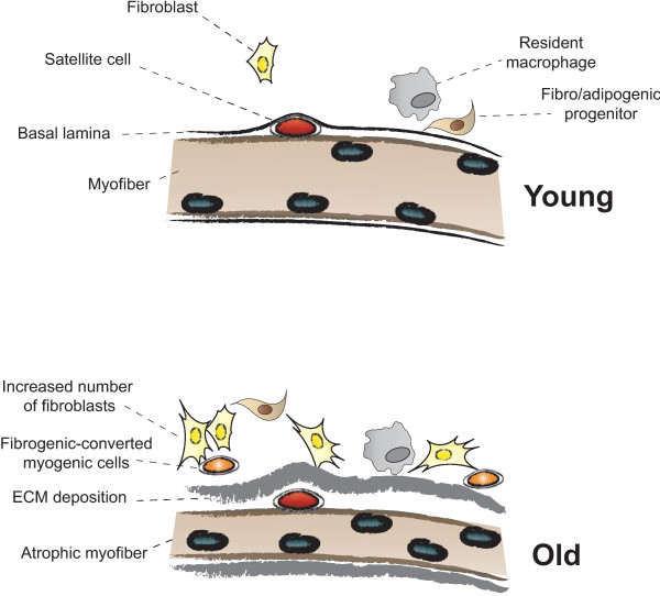
“Whether FAP conversion also occurs during aging, and whether Basal lamina ECM deposition Satellite cell Fibro/adipogenic progenitor Resident macrophage Fibroblast Increased number of fibroblasts Fibrogenic-converted myogenic cells Myofiber Atrophic myofiber Young Old Figure 5 Myofiber growth and extracellular matrix (…”
BackgroundInfluential
Cited by
Sorted by citations
Items 301–400 of 854
Qian, W., Xia, S., Yang, X., Yu, J., Guo, B., Lin, Z., Wei, R., Mao, M., Zhang, Z., Zhao, G., et al. (2022) · Frontiers in Molecular Biosciences · doi:10.3389/fmolb.2021.800747 · Cited by 31
“At present, the mainstream theory about the muscle degeneration and fibrosis causes of Duchenne muscular dystrophy (Mann et al., 2011) is that the integrity of the dystrophin protein in muscle cell membrane is lost and destroyed.”
BackgroundIrisin ameliorates D-galactose-induced skeletal muscle fibrosis via the PI3K/Akt pathway
Wu, Y., Wu, Y., Yu, J., Zhang, Y., Li, Y., Fu, R., Sun, Y., Zhao, K., & Xiao, Q. (2022) · European Journal of Pharmacology · doi:10.1016/j.ejphar.2022.175476 · Cited by 32
Li, J., Fredericks, M., Cannell, M., Wang, K., Sako, D., Maguire, M. C., Grenha, R., Liharska, K., Krishnan, L., Bloom, T., et al. (2021) · Journal of Clinical Investigation · doi:10.1172/jci138634 · Cited by 31
“Dystrophin deficiency weakens membrane integrity, leading to myofiber damage during muscle contraction, and eventually muscle is replaced by fat and connective tissue (7).”
BackgroundPerdiguero, E., Kharraz, Y., Serrano, A. L., & Muñoz-Cánoves, P. (2012) · Cell Cycle · doi:10.4161/cc.11.5.19374 · Cited by 30
Single-Nuclei RNA-Sequencing of the Gastrocnemius Muscle in Peripheral Artery Disease
Pass, C. G., Palzkill, V. R., Tan, J., Kim, K., Thome, T., Yang, Q., Fazzone, B. J., Robinson, S. T., O’Malley, K. A., Yue, F., et al. (2023) · Circulation Research · doi:10.1161/circresaha.123.323161 · Cited by 31
Milner, D., & Cameron, J. A. (2013) · Current Topics in Microbiology and Immunology · doi:10.1007/82_2012_292 · Cited by 28
“A balanced progression of both of these processes is a prerequisite for optimal recovery of muscle function (Jarvinen et al. 2005; Serrano et al. 2011; Murphy et al. 2011; Mann et al. 2011).”
BackgroundA TALEN-Exon Skipping Design for a Bethlem Myopathy Model in Zebrafish
Radev, Z., Hermel, J., Elipot, Y., Bretaud, S., Arnould, S., Duchateau, P. N., Ruggiero, F., Joly, J., & Sohm, F. (2015) · PLoS ONE · doi:10.1371/journal.pone.0133986 · Cited by 30
“The presence of fibrosis, essentially composed of collagen, a characteristic of all muscular dystrophies [69], is present in UCMD patient biopsies [23,67] and was also described in the Col6a3 deficient mouse model [34].”
BackgroundNorris, A. M., Fierman, K. E., Campbell, J., Pitale, R., Shahraj, M., & Kopinke, D. (2024) · Skeletal Muscle · doi:10.1186/s13395-024-00344-4 · Cited by 28
“In humans, fibrosis and IMAT form simultaneously [16, 45–48, 53, 79, 80].”
BackgroundSong, X., Liu, F., Chen, M., Zhu, M., Zheng, H., Wang, W., Chen, D., Li, M., & Chen, S. (2024) · Heliyon · doi:10.1016/j.heliyon.2024.e33062 · Cited by 29
Fibrosis and inflammation are greater in muscles of beta-sarcoglycan-null mouse than mdx mouse
Gibertini, S., Zanotti, S., Savadori, P., Curcio, M., Saredi, S., Salerno, F., Andreetta, F., Bernasconi, P., Mantegazza, R., & Mora, M. (2014) · Cell and Tissue Research · doi:10.1007/s00441-014-1854-4 · Cited by 29
Hord, J. M., Botchlett, R. E., & Lawler, J. M. (2016) · Experimental Gerontology · doi:10.1016/j.exger.2016.08.006 · Cited by 29
Bruno, G., Cencetti, F., Bernacchioni, C., Donati, C., Blankenbach, K. V., Thomas, D., zu Heringdorf, D. M., & Bruni, P. (2018) · Cellular Signalling · doi:10.1016/j.cellsig.2018.02.001 · Cited by 29
Characterization of a novel murine Sost ERT2 Cre model targeting osteocytes
Maurel, D. B., Matsumoto, T., Vallejo, J. A., Johnson, M. L., Dallas, S. L., Kitase, Y., Brotto, M. A. P., Wacker, M., Harris, M. A., Harris, S. E., et al. (2019) · Bone Research · doi:10.1038/s41413-018-0037-4 · Cited by 29
miR-146a deficiency does not aggravate muscular dystrophy in mdx mice
Bronisz-Budzyńska, I., Chwalenia, K., Mucha, O., Podkalicka, P., Karolina-Bukowska-Strakova, Józkowicz, A., Łoboda, A., Kozakowska, M., & Dulak, J. (2019) · Skeletal Muscle · doi:10.1186/s13395-019-0207-0 · Cited by 29
Squecco, R., Chellini, F., Idrizaj, E., Tani, A., Garella, R., Pancani, S., Pavan, P., Bambi, F., Zecchi‐Orlandini, S., & Sassoli, C. (2020) · Cells · doi:10.3390/cells9051199 · Cited by 29
Hu, L.-Y., Mileti, C. J., Loomis, T., Brashear, S. E., Ahmad, S., Chellakudam, R. R., Wohlgemuth, R. P., Gionet‐Gonzales, M., Leach, J. K., & Smith, L. (2021) · American Journal of Physiology-Cell Physiology · doi:10.1152/ajpcell.00065.2021 · Cited by 34
Biomechanical Origins of Muscle Stem Cell Signal Transduction
Morrissey, J. B., Cheng, R. Y., Davoudi, S., & Gilbert, P. M. (2015) · Journal of Molecular Biology · doi:10.1016/j.jmb.2015.05.004 · Cited by 29
Identification and Function of Fibrocytes in Skeletal Muscle Injury Repair and Muscular Dystrophy
Wang, X., Zhao, W., Ransohoff, R. M., & Zhou, L. (2016) · The Journal of Immunology · doi:10.4049/jimmunol.1601308 · Cited by 29
Wang, Y. X., Holbrook, C. A., Hamilton, J. N., Garoussian, J., Afshar, M., Su, S., Schürch, C. M., Lee, M. Y., Goltsev, Y., Kundaje, A. B., et al. (2022) · bioRxiv (Cold Spring Harbor Laboratory) · doi:10.1101/2022.06.10.494732 · Cited by 31
“This leads to molecular dysregulation that disrupts the signals that 16 instruct MuSCs to proliferate and orchestrate the complex cellular symphony that underlies the 17 regenerative process, resulting in fatty-fibrotic scarring and progressive replacement of muscle 18 cells (Blau et al., 2015; Mann et al., 2011; Muño…”
BackgroundHighly Stretchable Piezoelectric Elastomer for Accelerated Repairing of Skeletal Muscles Loss
Fang, Q., Wang, D., Lin, W., Ge, Z., Deng, X., Zhao, R., Tang, Y., Liu, W., Xiong, Z., Duan, A., et al. (2024) · Advanced Functional Materials · doi:10.1002/adfm.202313055 · Cited by 28
Ferreira, M. M., Dewi, R. E., & Heilshorn, S. C. (2015) · Integrative Biology · doi:10.1039/c5ib00060b · Cited by 28
Krieger, J., Park, B., Lambert, C. R., & Malcuit, C. M. (2018) · PeerJ · doi:10.7717/peerj.4939 · Cited by 28
Adipocyte plasticity in tissue regeneration, repair, and disease
Horsley, V. (2022) · Current Opinion in Genetics & Development · doi:10.1016/j.gde.2022.101968 · Cited by 28
Liu, X., Zhao, L., Gao, Y., Chen, Y., Tian, Q., Son, J. S., Chae, S. A., de Avila, J. M., Zhu, M. J., & Du, . (2022) · Journal of Cachexia Sarcopenia and Muscle · doi:10.1002/jcsm.13150 · Cited by 27
“Muscle regeneration is tightly regulated, and improper regeneration results in the replacement of muscle fibres by fibrotic and fatty tissues, progressively worsening muscle functions.(5,6) OR IG INAL ART ICLE”
BackgroundFoltz, S. J., Luan, J.-N., Call, J. A., Patel, A., Peissig, K. B., Fortunato, M. J., & Beedle, A. M. (2016) · Skeletal Muscle · doi:10.1186/s13395-016-0091-9 · Cited by 29
“However, dystrophic muscle experiences repeated cycles of damage and repair with persistence of the inflammatory phenotype [57, 58].”
BackgroundXie, H., Wang, H., Wu, Z., Li, W., Liu, Y., & Wang, N. (2023) · Frontiers in Nutrition · doi:10.3389/fnut.2023.1100918 · Cited by 29
“M2 macrophages can induce extracellular matrix accumulation and muscle fibrosis and impair the function of MuSCs, so as to affect skeletal muscle regeneration (75, 76).”
BackgroundMohamed, J. S., Hajira, A., Lopez, M. A., & Boriek, A. M. (2015) · Journal of Biological Chemistry · doi:10.1074/jbc.m115.659375 · Cited by 27
“nance, and the vital function of muscles (51, 55).”
BackgroundFarup, J., Dalgas, U., Keytsman, C., Eijnde, B. O., & Wens, I. (2016) · Frontiers in Physiology · doi:10.3389/fphys.2016.00193 · Cited by 28
“Moreover, the aging muscle microenvironment is associated with increased fibrosis and adipocyte infiltration (Conley et al., 1995; Mann et al., 2011).”
D’Souza, D. M., Roubos, S., Larkin, J., Lloyd, J. C., Emmons, R. S., Chen, H., & Lisio, M. D. (2019) · Scientific Reports · doi:10.1038/s41598-019-43204-8 · Cited by 27
D'Agostino, M., Torcinaro, A., Madaro, L., Marchetti, L., Sileno, S., Beji, S., Salis, C., Proietti, D., Imeneo, G., Capogrossi, M. C., et al. (2018) · Oxidative Medicine and Cellular Longevity · doi:10.1155/2018/4814696 · Cited by 26
“Consequently, fibrous and fatty connective tissue overtakes the functional myofibers [15, 19].”
BackgroundInterspecies Organogenesis for Human Transplantation
Crane, A. T., Aravalli, R. N., Asakura, A., Grande, A. W., Krishna, V. D., Carlson, D. F., Cheeran, M. C. ‐., Danczyk, G., Dutton, J. R., Hackett, P. B., et al. (2019) · Cell Transplantation · doi:10.1177/0963689719845351 · Cited by 26
Plasminogen activation in the musculoskeletal acute phase response: Injury, repair, and disease
Gibson, B. H. Y., Duvernay, M. T., Moore‐Lotridge, S. N., Flick, M. J., & Schoenecker, J. G. (2020) · Research and Practice in Thrombosis and Haemostasis · doi:10.1002/rth2.12355 · Cited by 26
The clock regulator Bmal1 protects against muscular dystrophy
Gao, H., Xiong, X., Lin, Y., Chatterjee, S., & Ma, K. (2020) · Experimental Cell Research · doi:10.1016/j.yexcr.2020.112348 · Cited by 25
Aguilar, C., Shcherbina, A., Ricke, D. O., Pop, R., Carrigan, C., Gifford, C., Urso, M., Kottke, M. A., & Meissner, A. (2015) · Scientific Reports · doi:10.1038/srep13885 · Cited by 42
Stager, D. R., McLoon, L. K., & Felius, J. (2013) · Europe PMC (PubMed Central) · Cited by 26
Nowak, R., Kwiecien, M., Tkacz, M. A., & Mazurek, U. (2014) · BioMed Research International · doi:10.1155/2014/594287 · Cited by 26
“In addition to the canonical Smad pathway TGF-βs also signal through noncanonical pathways including mitogen activated kinase (MAPK), nuclear factor κ-B (NF-κ-B), Rho-like GTPase, phosphoinositide 3kinase (PI3K)/Akt, hypoxia/hypoxia-inducible factor-1 (HIF1) pathways [10, 11, 25].”
Age-Related Changes in Skeletal Muscle of Cattle
Costagliola, A., Wojcik, S. P., Pagano, T. B., Biase, D. D., Russo, V., Iovane, V., Grieco, E., Papparella, S., & Paciello, O. (2016) · Veterinary Pathology · doi:10.1177/0300985815624495 · Cited by 27
“Moreover, the progressive decrease of skeletal muscle quality is associated with fibrosis and intracellular deposition of non-contractile material.(37) The pathogenesis of these alterations are associated with specific biological processes, such as oxidative stress.”
BackgroundAlbright, J. A., Testa, E. J., Ibrahim, Z. F., Quinn, M. S., Chang, K., Alsoof, D., Diebo, B. G., Barrett, T. J., & Daniels, A. H. (2023) · The Journal of Arthroplasty · doi:10.1016/j.arth.2023.10.015 · Cited by 25
Ducommun, S., Jannig, P. R., Červenka, I., Murgia, M., Mittenbühler, M. J., Chernogubova, E., Dias, J. M. M., Jude, B., Correia, J. C., Vranken, J. G. V., et al. (2024) · Molecular Metabolism · doi:10.1016/j.molmet.2024.101912 · Cited by 27
Laminin α2 Chain-Deficiency is Associated with microRNA Deregulation in Skeletal Muscle and Plasma
Holmberg, J., Alajbegovic, A., Gawlik, K. I., Elowsson, L., & Durbeej, M. (2014) · Frontiers in Aging Neuroscience · doi:10.3389/fnagi.2014.00155 · Cited by 25
“Dystrophic muscles are characterized by infiltration of inflammatory cells (Mann et al., 2011).”
BackgroundTakeuchi, S., Nakano, S.-I., Nakamura, K., Ozoe, A., Chien, P., Yoshihara, H., Hakuno, F., Matsuwaki, T., Saeki, Y., Takahashi, S., et al. (2016) · Experimental Cell Research · doi:10.1016/j.yexcr.2016.08.023 · Cited by 26
“tissue are thought to cause muscle weakness [4,5].”
BackgroundFleming, J. W., Capel, A. J., Rimington, R. P., Player, D. J., Stolzing, A., & Lewis, M. P. (2019) · Cytoskeleton · doi:10.1002/cm.21553 · Cited by 25
“In such situations the capacity of muscle to heal is impaired, resulting in non-regenerative healing and subsequent fibrosis (Mann et al., 2011; Mueller, Van Velthoven, Fukumoto, Cheung, & Rando, 2016; Olson & Soriano, 2009), adipogenesis (Rose, 2002; Uezumi, Fukada, Yamamoto, Takeda, & Tsuchida, 2010; Uezumi et al.”
BackgroundFernández‐Simón, E., Suárez‐Calvet, X., Carrasco‐Rozas, A., Piñol‐Jurado, P., Fernández, S. L., Pons, G., Bech‐Serra, J. J., de la Torre, C., de Luna, N., Gallardo, E., et al. (2022) · Journal of Cachexia Sarcopenia and Muscle · doi:10.1002/jcsm.12923 · Cited by 25
Xu, H., Bhaskaran, S., Piekarz, K. M., Ranjit, R., Bian, J., Kneis, P., Ellis, A., Bhandari, S., Rice, H. C., & Remmen, H. V. (2022) · Frontiers in Aging Neuroscience · doi:10.3389/fnagi.2022.876816 · Cited by 25
Chronic effects of losartan on the muscles and the serologic profiles of mdx mice
Lee, E. M., Kim, D.-Y., Kim, A.-Y., Lee, E. H., Kim, S., Lee, M., Sung, S., Park, J., & Jeong, K. (2015) · Life Sciences · doi:10.1016/j.lfs.2015.10.023 · Cited by 25
“In conditions of chronic injury, as occurs in muscular dystrophy, chronic inflammatory events result in excessive TGF-β levels, which inhibit myogenic repair and lead to muscles being replaced with fibrotic/scar tissue [11,17].”
de Melo, C. A. V., Alves, A. N., Terena, S. M. L., Fernandes, K., Nunes, F., da Silva, D. D. F. T., Bussadori, S., Deana, A., & Mesquita-Ferrari, R. (2016) · Lasers in Medical Science · doi:10.1007/s10103-016-1888-9 · Cited by 14
Quiescence of human muscle stem cells is favored by culture on natural biopolymeric films
Monge, C., Distasio, N., Rossi, T., Sébastien, M., Sakai, H., Kalman, B., Boudou, T., Tajbakhsh, S., Marty, I., Bigot, A., et al. (2017) · Stem Cell Research & Therapy · doi:10.1186/s13287-017-0556-8 · Cited by 25
Conductive hydrogel luminal filler for peripheral nerve regeneration
Park, J., Kim, J., Choe, G., Jung, Y., & Lee, J. Y. (2025) · Biomaterials · doi:10.1016/j.biomaterials.2025.123103 · Cited by 24
Riparini, G., Mackenzie, M. E., Naz, F., Brooks, S. R., Jiang, K., Deewan, A., Dulek, B., Islam, S., Ko, K. D., Tsai, W. L., et al. (2025) · Nature Aging · doi:10.1038/s43587-025-01002-0 · Cited by 27
“Similar to inflammaging—the chronic, low-grade inflammation associated with aging 58 —fibroaging, which refers to the tendency to develop tissue fibrosis in aging 59 , worsens regenerative capabilities and impairs Article https://doi.org/10.1038/s43587-025-01002-0 skeletal muscle performance 60 .”
Influentialde Freitas, C. E. D., Bertaglia, R. S., Vechetti, I., Mareco, E. A., Salomão, R. A. S., de Paula, T. G., Nai, G. A., Carvalho, R., Pacagnelli, F., & Dal-Pai-Silva, M. (2015) · Photochemistry and Photobiology · doi:10.1111/php.12446 · Cited by 23
What do mouse models of muscular dystrophy tell us about the DAPC and its components?
Whitmore, C. L., & Morgan, J. E. (2014) · International Journal of Experimental Pathology · doi:10.1111/iep.12095 · Cited by 24
“…shown that altering the behaviour of these cells influences muscle pathology; for example, modulating macrophage or T cell populations reduces pathological features such as fibrosis (Morrison et al. 2000; Farini et al. 2007; Villalta et al. 2009; reviewed in (Evans et al. 2009; Mann et al. 2011)).”
BackgroundGoetsch, K. P., Snyman, C., Myburgh, K. H., & Niesler, C. U. (2015) · BioTechniques · doi:10.2144/000114246 · Cited by 24
Choi, Y. H., Kim, S. H., Kim, I. G., Lee, J. H., & Kwon, S. K. (2019) · Acta Biomaterialia · doi:10.1016/j.actbio.2019.03.005 · Cited by 24
Long non‐coding RNA H19 promotes myoblast fibrogenesis via regulating the miR‐20a‐5p‐Tgfbr2 axis
Lin, J., Luo, Z., Liu, S., Chen, Q., Liu, S., & Chen, J. (2021) · Clinical and Experimental Pharmacology and Physiology · doi:10.1111/1440-1681.13489 · Cited by 24
Cho, K., Park, M., Kim, Y., Woo, S., & Ryu, K. (2016) · Journal of Cellular and Molecular Medicine · doi:10.1111/jcmm.12947 · Cited by 26
(-)-Epigallocatechin-3-gallate stimulates myogenic differentiation through TAZ activation
Kim, A. R., Kim, K. M., Byun, M. R., Hwang, J.-H., Park, J. I., Oh, H. T., Jeong, M. G., Hwang, E. S., & Hong, J.-H. (2017) · Biochemical and Biophysical Research Communications · doi:10.1016/j.bbrc.2017.03.049 · Cited by 24
Gallardo, F. S., Córdova-Casanova, A., & Brandan, E. (2021) · Journal of Cell Communication and Signaling · doi:10.1007/s12079-021-00610-w · Cited by 27
“Histologically, degenerating muscle fibers are replaced with fibrofatty tissue (Mann et al. 2011), affecting muscle architecture and functionality.”
BackgroundDecorin the antifibrotic proteoglycan and its progression in therapy
Baghy, K., Szakadáti, H., & Kovalszky, I. (2025) · American Journal of Physiology-Cell Physiology · doi:10.1152/ajpcell.01075.2024 · Cited by 23
Bai, X., Wang, X., Tang, B., Shi, H., Boireau, P., Rosenthal, B. M., Wu, X., Liu, M. Y., & Liu, X. L. (2016) · Veterinary Parasitology · doi:10.1016/j.vetpar.2016.07.033 · Cited by 23
Lack of PKCθ Promotes Regenerative Ability of Muscle Stem Cells in Chronic Muscle Injury
Fiore, P. F., Benedetti, A., Sandonà, M., Madaro, L., Bardi, M. D., Saccone, V., Puri, P., Gargioli, C., Lozanoska‐Ochser, B., & Bouché, M. (2020) · International Journal of Molecular Sciences · doi:10.3390/ijms21030932 · Cited by 24
“In mdx mice, activation of the Notch pathway rescued the self-renewal ability of satellite cells [17].”
BackgroundPaepe, B. D. (2022) · International Journal of Molecular Sciences · doi:10.3390/ijms232113180 · Cited by 24
“Successful regeneration restores damage to muscle fibers, but an imbalance between anabolic and catabolic pathways in favor of the latter may lead to loss of muscle mass and replacement with fibrotic and fatty tissue [2].”
BackgroundKaziród, K., Myszka, M., Dulak, J., & Łoboda, A. (2022) · Cellular and Molecular Life Sciences · doi:10.1007/s00018-022-04636-0 · Cited by 24
“Under dystrophic conditions, the increased and persistent presence of different macrophage phenotypes may contribute to alterations in SC function [26].”
BackgroundArraut, J., Lygrisse, K. A., Singh, V., Fiedler, B., Schwarzkopf, R. M., & Rozell, J. C. (2022) · Archives of Orthopaedic and Trauma Surgery · doi:10.1007/s00402-022-04696-8 · Cited by 23
Bae, J., Jo, A., Cho, S. C., Lee, Y., Kam, T., You, C.-L., Jeong, H., Kim, H., Jeong, M., Jeong, Y., et al. (2023) · Science Translational Medicine · doi:10.1126/scitranslmed.abh3489 · Cited by 23
Donati, C., Cencetti, F., & Bruni, P. (2012) · Biochimica et Biophysica Acta (BBA) - Molecular and Cell Biology of Lipids · doi:10.1016/j.bbalip.2012.06.013 · Cited by 23
Michielin, F., Serena, E., Pavan, P. G., & Elvassore, N. (2015) · RSC Advances · doi:10.1039/c5ra16957g · Cited by 23
Regenerative Repair of Volumetric Muscle Loss Injury is Sensitive to Age
Kim, J. T., Kasukonis, B. M., Dunlap, G., Perry, R. A., Washington, T. A., & Wolchok, J. C. (2019) · Tissue Engineering Part A · doi:10.1089/ten.tea.2019.0034 · Cited by 23
Muscular dystrophy in PTFR/cavin-1 null mice
Ding, S.-Y., Liu, L., & Pilch, P. F. (2017) · JCI Insight · doi:10.1172/jci.insight.91023 · Cited by 22
“which is associated with disruption of muscle integrity and function (38).”
BackgroundThummarati, P., & Kino‐oka, M. (2020) · Frontiers in Bioengineering and Biotechnology · doi:10.3389/fbioe.2020.578140 · Cited by 22
Arrighi, N., Moratal, C., Savary, G., Fassy, J., Nottet, N., Pons, N., Clément, N., Fellah, S., Larrue, R., Magnone, V., et al. (2021) · Cells · doi:10.3390/cells10071832 · Cited by 25
Cold and hot fibrosis define clinically distinct cardiac pathologies
Miyara, S., Adler, M., Umansky, K. B., Häußler, D., Bassat, E., Divinsky, Y., Elkahal, J., Kain, D., Lendengolts, D., Flores, R. O. R., et al. (2025) · Cell Systems · doi:10.1016/j.cels.2025.101198 · Cited by 28
Inflammatory Genes and Psychological Factors Predict Induced Shoulder Pain Phenotype
George, S. Z., Parr, J. J., Wallace, M. R., Wu, S. S., Borsa, P. A., Dai, Y. E., & Fillingim, R. B. (2014) · Medicine & Science in Sports & Exercise · doi:10.1249/mss.0000000000000328 · Cited by 22
“Yet, many individuals experience robust inflammatory responses and substantial pain even in the absence of severe injury (23).”
BackgroundJiang, B., Zu, W., Xu, J., Xiong, Z., Zhang, Y., Gao, S., Ge, S.-X., & Zhang, L. (2018) · International Journal of Biological Macromolecules · doi:10.1016/j.ijbiomac.2018.02.077 · Cited by 22
Liu, G., Wei, J., Xiao, W., Xie, W., Ru, Q., Chen, L., Wu, Y., Mobasheri, A., & Li, Y. (2023) · Biomedicine & Pharmacotherapy · doi:10.1016/j.biopha.2023.115884 · Cited by 23
Cripto shapes macrophage plasticity and restricts EndMT in injured and diseased skeletal muscle
Iavarone, F., Guardiola, O., Scagliola, A., Andolfi, G., Esposito, F., Serrano, A. L., Perdiguero, E., Brunelli, S., Muñoz‐Cánoves, P., & Minchiotti, G. (2020) · EMBO Reports · doi:10.15252/embr.201949075 · Cited by 21
“(Fig 5B left panels and C), in line with the idea that this is the most severely affected muscle in mdx mice [11].”
P2Y2 promotes fibroblasts activation and skeletal muscle fibrosis through AKT, ERK, and PKC
Chen, M., Chen, H., Gu, Y., Sun, P., Sun, J., Yu, H., Zheng, H., & Chen, D. (2021) · BMC Musculoskeletal Disorders · doi:10.1186/s12891-021-04569-y · Cited by 21
“The muscle damage induces an inflammatory response cascade involving the inflitration of inflammatory cells into the injured tissue [18, 19].”
BackgroundMo, Y., Kang, H., Bang, J.-Y., Shin, J. W., Kim, H. Y., Cho, S., & Kang, H. (2022) · Scientific Reports · doi:10.1038/s41598-022-14846-y · Cited by 20
Chong, G. L. W., Böhmert, B., Lee, L. E. J., Bols, N. C., & Dowd, G. C. (2022) · In Vitro Cellular & Developmental Biology - Animal · doi:10.1007/s11626-022-00734-2 · Cited by 21
“In some situations, excessive TGFβinduced deposition of extracellular matrix components can lead to fibrosis, a condition which severely impairs muscle function via reduced mobility and contractile function (Mann et al. 2011).”
BackgroundKim, H., Ahn, M., Choi, S., Kim, M., Sim, K., Kim, J. H., Moon, C., & Shin, T. (2013) · Brain Research · doi:10.1016/j.brainres.2013.01.043 · Cited by 21
T lymphocytes are not required for the development of fatty degeneration after rotator cuff tear
Gumucio, J., Flood, M. D., Harning, J. A., Phan, A. C., Roche, S. M., Lynch, E. B., Bedi, A., & Mendias, C. (2014) · Bone & Joint Research · doi:10.1302/2046-3758.39.2000294 · Cited by 18
An advanced magnetic resonance imaging perspective on the etiology of deep tissue injury
Nelissen, J. L., Traa, W. A., de Boer, H. H., de Graaf, L., Mazzoli, V., Savci-Heijink, C. D., Nicolay, K., Froeling, M., Bader, D. L., Nederveen, A. J., et al. (2018) · Journal of Applied Physiology · doi:10.1152/japplphysiol.00891.2017 · Cited by 21
Lee, J. E., Shah, V. K., Lee, D. W., Oh, M., & Choi, J. J. (2019) · Journal of Pharmacological Sciences · doi:10.1016/j.jphs.2019.03.009 · Cited by 21
Barajaa, M. A., Ghosh, D., & Laurencin, C. T. (2023) · Regenerative Engineering and Translational Medicine · doi:10.1007/s40883-023-00328-8 · Cited by 24
“Remodeling and Maturation Phase Post-regenerative phase, the temporary ECM deposited by the SMCT fibroblasts gets remodeled, thus, marking the beginning of the remodeling phase [128, 227].”
Li, Y., Fan, Y., Pan, H., Qian, H., Qi, X., Wu, G., Zhang, H., Xu, M., Rao, Z., Wang, L., et al. (2018) · International Journal of Biological Macromolecules · doi:10.1016/j.ijbiomac.2018.05.188 · Cited by 20
Effects of Geniposide from Gardenia Fruit Pomace on Skeletal-Muscle Fibrosis
Pan, H., Li, Y., Qian, H., Qi, X., Wu, G., Zhang, H., Xu, M., Rao, Z., Li, J., Wang, L., et al. (2018) · Journal of Agricultural and Food Chemistry · doi:10.1021/acs.jafc.8b00739 · Cited by 20
Alonso‐Martín, S., Aurade, F., Mademtzoglou, D., Rochat, A., Zammit, P. S., & Relaix, F. (2018) · eLife · doi:10.7554/elife.26039 · Cited by 22
“Histological analysis of TA muscles in Pax3;Sox17 mice at d7 after CTX-induced regeneration revealed cell infiltration, fat accumulation and fibrosis, that were absent in regenerating muscles of control Sox17 mice (Figure 5G), suggesting abnormal regeneration and impaired satellite cell function (Mann et al., 2011; Sa…”
BackgroundRaz, V., Sterrenburg, E., Routledge, S. J. E., Venema, A., van der Sluijs, B. M., Trollet, C., Dickson, G., van Engelen, B. G. M., van der Maarel, S. M., & Antoniou, M. N. (2013) · BMC Neurology · doi:10.1186/1471-2377-13-70 · Cited by 20
Kang, H., Bang, J., Mo, Y., Shin, J. W., Bae, B., Cho, S., Kim, H. Y., & Kang, H. (2021) · Clinical & Experimental Allergy · doi:10.1111/cea.14077 · Cited by 19
Activation of the ATX/LPA/LPARs axis induces a fibrotic response in skeletal muscle
Córdova-Casanova, A., Cruz-Soca, M., Chun, J. J. M., Casar, J. C., & Brandan, E. (2022) · Matrix Biology · doi:10.1016/j.matbio.2022.03.008 · Cited by 20
“Fibrosis is commonly described as the accumulation of extracellular matrix (ECM) in a tissue due to an imbalance between its synthesis and degradation rates [1,2].”
FGF8 rescues motor deficits in zebrafish model of limb-girdle muscular dystrophy R18
Ulhaq, Z. S., Ogino, Y., & Tse, W. K. F. (2023) · Biochemical and Biophysical Research Communications · doi:10.1016/j.bbrc.2023.02.046 · Cited by 19
Kim, J., & Lee, J. (2016) · Journal of Exercise Rehabilitation · doi:10.12965/jer.1632640.320 · Cited by 19
“MMP can degrade ECM alone or in combination with the plasminogen/plasmin system to facilitate the migration of inflammatory and myogenic cells, such as satellite cells, from ECM to the site of injury (Mann et al., 2011).”
BackgroundBrito, A., Alves, A. N., Ribeiro, B. G., Barbosa, D. V. D. E., Magalhães, E. M. R., Fernandes, K. P. S., Bussadori, S. K., Goulardins, J. B., & Mesquita‐Ferrari, R. A. (2017) · Lasers in Medical Science · doi:10.1007/s10103-017-2392-6 · Cited by 19
“Thus, muscle regeneration is slower in older animals [6, 39, 40].”
BackgroundHogan, K. J., Smoak, M. M., Koons, G. L., Perez, M. R., Bedell, M. L., Jiang, E. Y., Young, S. W., & Mikos, A. G. (2022) · Journal of Biomedical Materials Research Part A · doi:10.1002/jbm.a.37355 · Cited by 19
Meisner, J. K., Annex, B. H., & Price, R. J. (2014) · Journal of Vascular Surgery · doi:10.1016/j.jvs.2014.01.038 · Cited by 19
Musiał-Wysocka, A., Kot, M., Sułkowski, M., & Majka, M. (2019) · International Journal of Molecular Sciences · doi:10.3390/ijms20184632 · Cited by 19
“VEGF as a key mediator of angiogenesis; it facilitates the formation of new blood vessels, the remodeling process, and stimulates mitogenesis and survival the of endothelial cells [24,25].”
BackgroundByun, W. S., Lee, J., & Baek, J.-H. (2024) · Inflammation and Regeneration · doi:10.1186/s41232-024-00354-1 · Cited by 20
“At this stage, the extracellular matrix (ECM) is constantly remodeled in response to the secretion of its constituents, fibrogenic cytokines, and growth factors from interstitial fibroblasts and fibro-adipogenic progenitors (FAPs) [8, 9].”
BackgroundTreating Muscular Dystrophy by Stimulating Intrinsic Repair
Wang, Y. X., Bentzinger, C. F., & Rudnicki, M. A. (2013) · Regenerative Medicine · doi:10.2217/rme.13.27 · Cited by 18
Noehren, B. W., Hardy, P., Andersen, A. H., Brightwell, C. R., Fry, J. L., Vandsburger, M. H., Thompson, K. L., & Fry, C. S. (2021) · The Journal of Physiology · doi:10.1113/jp281964 · Cited by 18
“Acute injuries (Brightwell et al. 2020; Lee et al. 2020), chronic illness (Abramowitz et al. 2018), ageing (Brack et al. 2007) and dystrophies (Mann et al. 2011; Lieber & Ward, 2013) all show dysregulation of the muscle ECM that contributes to disease pathophysiology.”
BackgroundYedigaryan, L., Martínez‐Sarrà, E., Giacomazzi, G., Giarratana, N., van der Veer, B. K., Rotini, A., Querceto, S., Grosemans, H., Cortés-Calabuig, Á., Salucci, S., et al. (2022) · Frontiers in Immunology · doi:10.3389/fimmu.2022.977617 · Cited by 18
“Fibrosis is associated with poor recovery after injury in aged individuals (56, 57).”
Goldman, S. M., Henderson, B. E. P., & Corona, B. T. (2017) · Stem Cell Research & Therapy · doi:10.1186/s13287-017-0589-z · Cited by 18
“Furthermore, fractionation would allow the assessment of both of these myogenic stem cell populations without the added complication of the immune cell populations which might drive a fibrotic rather than myogenic response [21, 22].”
Metadata
- Authors
- Mann, C. J., Perdiguero, E., Kharraz, Y., Aguilar, S., Pessina, P., Serrano, A. L., & Muñoz-Cánoves, P.
- Affiliations
- Universitat Pompeu Fabra (ES)
- Venue
- Skeletal Muscle
- Published
- 2011-05-04
- Volume
- 1
- Issue
- 1
- Pages
- 21-21
- ISSN
- 2044-5040 (ISSN-L)
- PMC
- PMC3156644
- OpenAlex
- W2076787014
- Cited by
- 853
- References
- 129
- Citations by year
- 2026: 37 · 2025: 59 · 2024: 55 · 2023: 53 · 2022: 59 · 2021: 71 · 2020: 81 · 2019: 85 · 2018: 76 · 2017: 51 · 2016: 68 · 2015: 48 · 2014: 49 · 2013: 33 · 2012: 27
- FWCI
- 14.30
- Type
- journal article
- Language
- en
- Open access
- gold
- doi.org
- Licence
- cc-by
- Locations
- Skeletal Muscle (publishedVersion, cc-by)
- Europe PMC (PubMed Central) (publishedVersion, cc-by)
- PubMed Central (publishedVersion, cc-by)
- PubMed (publishedVersion)
- RECERCAT (Consorci de Serveis Universitaris de Catalunya) (publishedVersion, other-oa)
- DOAJ (DOAJ: Directory of Open Access Journals) (submittedVersion, cc-by-sa)
- Repositori digital de la UPF (Universitat Pompeu Fabra) (submittedVersion, cc-by)
- Funders
- Ministerio de Ciencia e Innovación (PLE2009)
- Centro de Investigación Biomédica en Red sobre Enfermedades Neurodegenerativas
- Topics
- Life Sciences › Biochemistry, Genetics and Molecular Biology › Molecular Biology › Muscle Physiology and Disorders
- Health Sciences › Medicine › Cardiology and Cardiovascular Medicine › Cardiac Fibrosis and Remodeling
- Health Sciences › Medicine › Surgery › Wound Healing and Treatments
- Keywords
- muscle fibrosis, muscle repair, muscular dystrophy, satellite cells, muscle stem cells, fibroblast activation, inflammatory cell infiltration, extracellular matrix, wound healing, microenvironment
About these checks
Registry, retraction, Cites and statistics results are computed, not judged, with one exception: a printed reference that no index lists is read once by a model before it counts as a citation error. Claims are extracted by a model and kept only when their quote appears verbatim in the text; each cited source is then checked, with a model’s help, for whether it says what the claim relies on. Those verdicts can be wrong — read the claim’s quote and the cited source before relying on one.
Statistics and arithmetic are recomputed, not judged.
Confidence bands are shown only for fields calibrated on our annotated test set. None is calibrated yet, so no band is shown.
What counts as a claim ›How we measure confidence ›How we check this ›
Are you an author, or do you see a problem with this page? Request a review
