

The DOI matches its Crossref record; 129 of the 129 references it cites were found in a registry or an index.
Aberrant repair and fibrosis development in skeletal muscle
Open accessAbstract
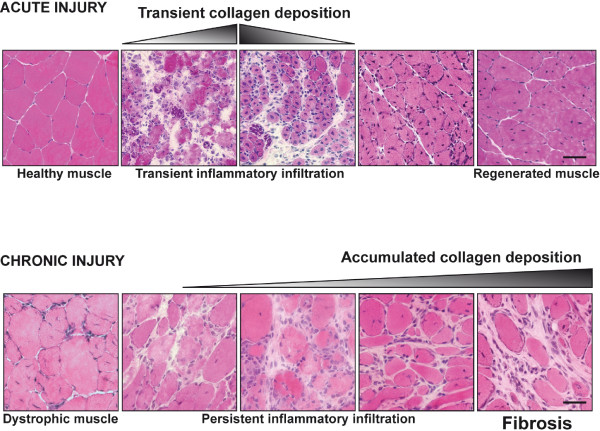
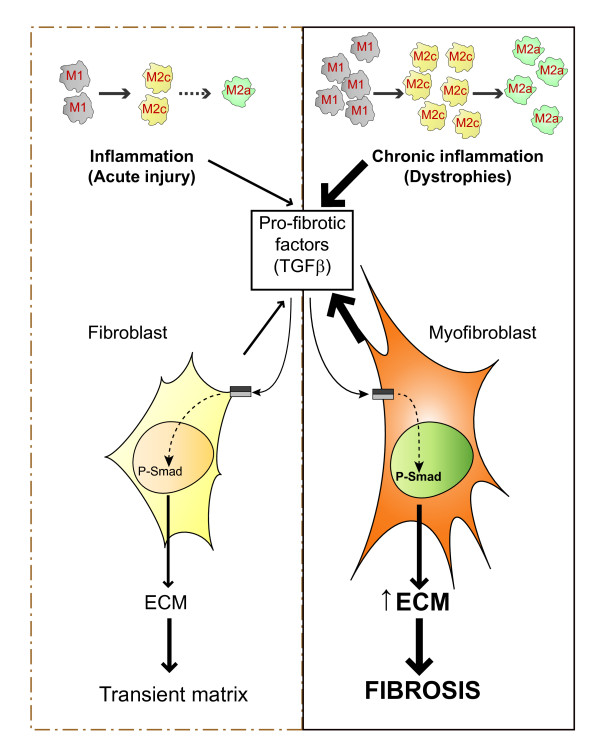
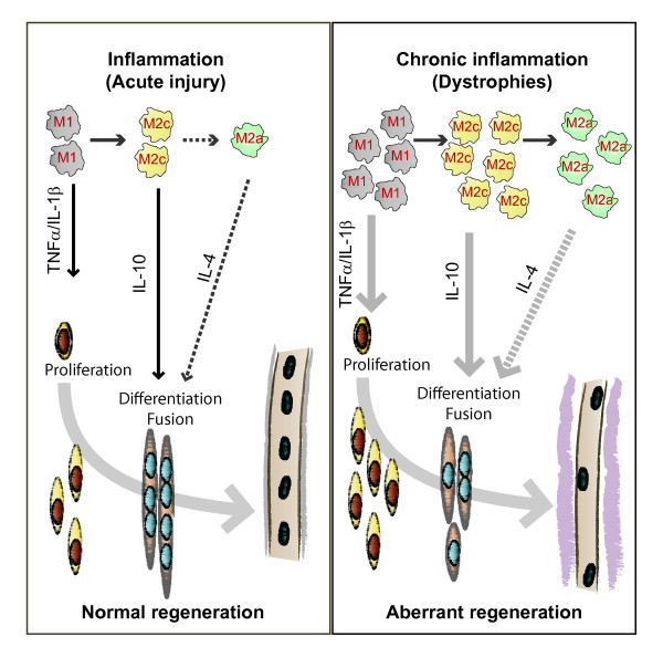
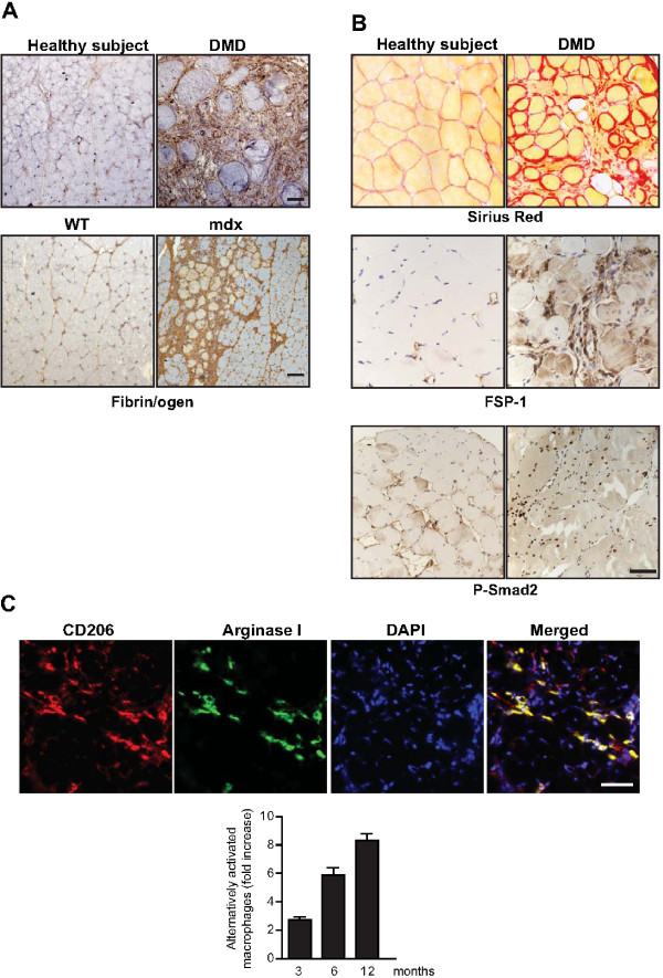
The repair process of damaged tissue involves the coordinated activities of several cell types in response to local and systemic signals. Following acute tissue injury, infiltrating inflammatory cells and resident stem cells orchestrate their activities to restore tissue homeostasis. However, during chronic tissue damage, such as in muscular dystrophies, the inflammatory-cell infiltration and fibroblast activation persists, while the reparative capacity of stem cells (satellite cells) is attenuated. Abnormal dystrophic muscle repair and its end stage, fibrosis, represent the final common pathway of virtually all chronic neurodegenerative muscular diseases. As our understanding of the pathogenesis of muscle fibrosis has progressed, it has become evident that the muscle provides a useful model for the regulation of tissue repair by the local microenvironment, showing interplay among muscle-specific stem cells, inflammatory cells, fibroblasts and extracellular matrix components of the mammalian wound-healing response. This article reviews the emerging findings of the mechanisms that underlie normal versus aberrant muscle-tissue repair.
Overview
The claims and statistics of this paper haven’t been checked yet.
Run the check — free, no account needed. It reads the open-access text and checks the sources it cites.
Integrity signals
Registry
Cites
Statistics
Figures
Verification
Claims the paper makes
The claims and statistics of this paper haven’t been checked yet.
Run the check — free, no account needed. It reads the open-access text and checks the sources it cites.
Claims other papers make about this one
Each is checked against this paper’s own text: does it say what the citing paper says it does?
In the citing paper
“Previous work has shown that the accumulation of inflammatory macrophages and age-related declines in interferon-gamma signaling delay muscle repair following injury”
From Skeletal muscle aging, cellular senescence, and senotherapeutics: current knowledge and future directions (2021) · cites this paper as Mann et al., 2011
Self-check
Percentages, sums and reported differences re-computed from the paper’s own numbers.
The self-check needs the paper’s full text.
Citations
Cites
1 retracted
RETRACTED ARTICLE: The role of nitric oxide during healing of trauma to the skeletal muscle
Filippin, L. I., Cuevas, M. J., Lima‐Cabello, E., Marroni, N. P., González‐Gallego, J., & Xavier, R. M. (2010) · Inflammation Research · doi:10.1007/s00011-010-0277-2 · Cited by 34 · cited 4×
“In addition, NO, which is released by local and invading cells during injury, can act as a pro-inflammatory molecule, and is able to activate MMP-2, which in turn releases HGF. Importantly, blockers of NOS function, such as nitro- Larginine methyl ester (L-NAME), have been shown to decrease the acute inflammatory reac…”
MethodInfluentialRetractedThe chemokine system in diverse forms of macrophage activation and polarization
MANTOVANI, A., SICA, A., SOZZANI, S., ALLAVENA, P., VECCHI, A., & LOCATI, M. (2004) · Trends in Immunology · doi:10.1016/j.it.2004.09.015 · Cited by 6,534 · cited 2×
“A nomenclature for polarized macrophages has been proposed [24,25] and they are now referred to as classically and alternatively activated macrophages, or M1 and M2 macrophages, respectively (Figure 2, Figure 3).”
BackgroundMechanisms of TGF-β Signaling from Cell Membrane to the Nucleus
Shi, Y., & Massagué, J. (2003) · Cell · doi:10.1016/s0092-8674(03)00432-x · Cited by 5,860
Visse, R., & Nagase, H. (2003) · Circulation Research · doi:10.1161/01.res.0000070112.80711.3d · Cited by 4,575 · cited 1×
“Activation requires proteolytic cleavage of the inactive precursor by either membrane-type (MT)1MMP [105] or plasmin, and cleavage of their corresponding inhibitors [106].”
Cellular and molecular mechanisms of fibrosis
Wynn, T. (2008) · The Journal of Pathology · doi:10.1002/path.2277 · Cited by 4,495
SATELLITE CELL OF SKELETAL MUSCLE FIBERS
Mauro, A. (1961) · The Journal of Cell Biology · doi:10.1083/jcb.9.2.493 · Cited by 3,683
Hinz, B., Phan, S. H., Thannickal, V. J., Galli, A., Bochaton-Piallat, M.-L., & Gabbiani, G. (2007) · The American Journal of Pathology · doi:10.2353/ajpath.2007.070112 · Cited by 2,068 · cited 1×
“These cells are associated with tissue repair and fibrosis in many tissues and organs, including muscle, skin, liver, lung, bone and cartilage [56].”
BackgroundArnold, L., Henry, A., Poron, F., Baba-Amer, Y., van Rooijen, N., Plonquet, A., Gherardi, R. K., & Chazaud, B. (2007) · The Journal of Experimental Medicine · doi:10.1084/jem.20070075 · Cited by 2,002 · cited 3×
“An important development in our understanding of muscle repair and fibrosis was the demonstration that a heterogeneous population of macrophages exists in regenerating muscle after injury, exhibiting opposing activities (either pro-inflammatory or anti-inflammatory) and different kinetics [23].”
BackgroundInfluentialFibrotic disease and the TH1/TH2 paradigm
Wynn, T. A. (2004) · Nature Reviews Immunology · doi:10.1038/nri1412 · Cited by 1,765
Muscle injury activates resident fibro/adipogenic progenitors that facilitate myogenesis
Joe, A. W. B., Yi, L., Natarajan, A., Le Grand, F., So, L., Wang, J., Rudnicki, M. A., & Rossi, F. M. V. (2010) · Nature Cell Biology · doi:10.1038/ncb2015 · Cited by 1,690 · cited 7×
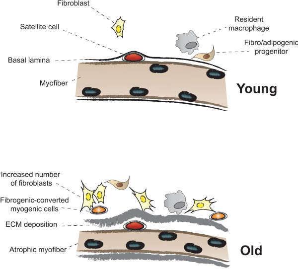
“Whether FAP conversion also occurs during aging, and whether Basal lamina ECM deposition Satellite cell Fibro/adipogenic progenitor Resident macrophage Fibroblast Increased number of fibroblasts Fibrogenic-converted myogenic cells Myofiber Atrophic myofiber Young Old Figure 5 Myofiber growth and extracellular matrix (…”
BackgroundInfluential
Cited by
Sorted by citations
Items 101–200 of 854
Fry, C. S., Johnson, D. L., Ireland, M. L., & Noehren, B. W. (2016) · Journal of Orthopaedic Research® · doi:10.1002/jor.23502 · Cited by 85
Willett, N. J., Li, M.-T. A., Uhrig, B. A., Boerckel, J. D., Huebsch, N., Lundgren, T. S., Warren, G. L., & Guldberg, R. E. (2012) · Tissue Engineering Part C Methods · doi:10.1089/ten.tec.2012.0290 · Cited by 85
“48,49 These factors may reach the bone defect region and could direct a fibrotic response instead of bone regeneration.”
Musculoskeletal senescence: a moving target ready to be eliminated
Baar, M. P., Perdiguero, E., Muñoz-Cánoves, P., & de Keizer, P. L. (2018) · Current Opinion in Pharmacology · doi:10.1016/j.coph.2018.05.007 · Cited by 83
“Most dystrophinopathies also feature increased muscle fibrosis [83], which aggravates disease progression by substituting muscle with scar tissue, and it is plausible that anti-senescence cocktails will also halt fibrosis and improve patient health status.”
BackgroundMacDonald, E. M., & Cohn, R. D. (2012) · Current Opinion in Rheumatology · doi:10.1097/bor.0b013e328358df34 · Cited by 84
“A surplus of extracellular matrix (ECM) protein produced by the muscle fibroblasts, called myofibroblasts, during the initial stages of muscle fiber necrosis also inhibits the normal regeneration process [9,19].”
Opposing roles of miR-21 and miR-29 in the progression of fibrosis in Duchenne muscular dystrophy
Zanotti, S., Gibertini, S., Curcio, M., Savadori, P., Pasanisi, M. B., Morandi, L. O., Cornelio, F., Mantegazza, R., & Mora, M. (2015) · Biochimica et Biophysica Acta (BBA) - Molecular Basis of Disease · doi:10.1016/j.bbadis.2015.04.013 · Cited by 85
Skeletal muscle as a regulator of the longevity protein, Klotho
Avin, K. G., Coen, P. M., Huang, W., Stolz, D. B., Sowa, G., Dubé, J. J., Goodpaster, B. H., O'Doherty, R. M., & Ambrosio, F. (2014) · Frontiers in Physiology · doi:10.3389/fphys.2014.00189 · Cited by 86
“With age, these cells demonstrate a decline in regenerative potential owing to altered cellular proliferation and myogenic differentiation (reviewed in Mann et al., 2011; Conboy and Rando, 2012).”
BackgroundDong, J., Dong, Y., Chen, Z., Mitch, W. E., & Zhang, L. (2016) · Kidney International · doi:10.1016/j.kint.2016.07.029 · Cited by 83
“DISCUSSION Fibrosis in skeletal muscles develops in several chronic illnesses including CKD and muscular dystrophies.(2,28,29) In these disorders, myofibers are replaced by extracellular matrix, impairing skeletal muscle functions and counteracting cell and gene therapies for muscle disorders.”
Numb is required to prevent p53-dependent senescence following skeletal muscle injury
Le Roux, I., Konge, J., Le Cam, L., Flamant, P., & Tajbakhsh, S. (2015) · Nature Communications · doi:10.1038/ncomms9528 · Cited by 81
Armutçu, F. (2019) · Inflammation Research · doi:10.1007/s00011-019-01271-7 · Cited by 80
Passive stiffness of fibrotic skeletal muscle in mdx mice relates to collagen architecture
Brashear, S. E., Wohlgemuth, R. P., Gonzalez, G., & Smith, L. (2020) · The Journal of Physiology · doi:10.1113/jp280656 · Cited by 78
“However, fibrotic muscle is also characterized by a lack of regeneration (Mann et al. 2011; Smith & Barton, 2018).”
BackgroundDuchesne, É., Dufresne, S. S., & Dumont, N. A. (2017) · Physical Therapy · doi:10.1093/ptj/pzx056 · Cited by 85
Advances in biomaterials for skeletal muscle engineering and obstacles still to overcome
Smoak, M. M., & Mikos, A. G. (2020) · Materials Today Bio · doi:10.1016/j.mtbio.2020.100069 · Cited by 81
Smith, L., Cho, S., & Discher, D. E. (2017) · Seminars in Cell and Developmental Biology · doi:10.1016/j.semcdb.2017.05.025 · Cited by 82
“With the proper mixing ratios, a surface coverage of collagen fiber bundles of ∼30% approximates the extent of fibrosis seen, for example, in muscle cross sections [49, 50].”
BackgroundBioinspired electrospun dECM scaffolds guide cell growth and control the formation of myotubes
Smoak, M. M., Hogan, K. J., Grande-Allen, K. J., & Mikos, A. G. (2021) · Science Advances · doi:10.1126/sciadv.abg4123 · Cited by 80
Tiskratok, W., Chuinsiri, N., Limraksasin, P., Kyawsoewin, M., & Jitprasertwong, P. (2025) · Polymers · doi:10.3390/polym17060822 · Cited by 99
“Skeletal muscle fibrosis results from various injuries or chronic conditions that lead to increased ECM deposition and stiffness [139].”
The super super-healing MRL mouse strain
Heydemann, A. (2012) · Frontiers in Biology · doi:10.1007/s11515-012-1192-4 · Cited by 76
“In keeping with a completion of healing phases, MRL/MpJear hole wounds analyzed at 7, 14 and 28 days after injury had fewer differences of expression in inflammation-linked genes (Masinde et al., 2005).”
BackgroundCalcaterra, V., Regalbuto, C., Porri, D., Pelizzo, G., Mazzon, E., Vinci, F., Zuccotti, G., Fabiano, V., & Cena, H. (2020) · Biomolecules · doi:10.3390/biom10091324 · Cited by 84
Nawaz, A., Bilal, M., Fujisaka, S., Kado, T., Aslam, M. R., Ahmed, S., Okabe, K., Igarashi, Y., Watanabe, Y., Kuwano, T., et al. (2022) · Nature Communications · doi:10.1038/s41467-022-34191-y · Cited by 81
Böhm, A., Hoffmann, C., Irmler, M., Schneeweiß, P., Schnauder, G., Sailer, C., Schmid, V., Hudemann, J., Machann, J., Schick, F., et al. (2016) · Diabetes · doi:10.2337/db15-1723 · Cited by 84
“This process possibly took place in all participants of our study after the first training sessions, as part of the normal regeneration program of an untrained muscle (32,35).”
BackgroundChapman, M., Mukund, K., Subramaniam, S., Brenner, D. A., & Lieber, R. L. (2016) · American Journal of Physiology-Cell Physiology · doi:10.1152/ajpcell.00226.2016 · Cited by 83
James, G., Sluka, K., Blomster, L. V., Hall, L. M., Schmid, A., Shu, C. C., Little, C., Melrose, J., & Hodges, P. (2018) · European spine journal · doi:10.1007/s00586-018-5652-7 · Cited by 72
“1 3 Persistence of M1 macrophages past their transient active phase is proposed in numerous inflammatory myopathies [25, 26, 43].”
BackgroundForcina, L., Miano, C., Scicchitano, B. M., & Musarò, A. (2019) · Cells · doi:10.3390/cells8030232 · Cited by 82
Hwang, A. B., & Brack, A. S. (2017) · Current topics in developmental biology/Current Topics in Developmental Biology · doi:10.1016/bs.ctdb.2017.08.008 · Cited by 83
Alves, A. N., Fernandes, K., Melo, C., Yamaguchi, R., França, C., Teixeira, D. F., Bussadori, S., Nunes, F., & Mesquita-Ferrari, R. (2014) · Lasers in Medical Science · doi:10.1007/s10103-013-1428-9 · Cited by 76
“Scar tissue develops as a result of the excessive deposition of ECM and constitutes a mechanical barrier to cell migration and fusion, limiting infusion at the site of vascular injury and preventing normal regeneration following tissue injury [9, 13, 14].”
BackgroundInfluentialKim, J., & Lee, J. (2017) · Journal of Exercise Rehabilitation · doi:10.12965/jer.1735072.536 · Cited by 77
“TGF-β also regulates ECM remodeling and can stimulate the fibroblasts that produce ECM proteins (Gumucio et al., 2015; Mann et al., 2011).”
BackgroundWada, E., Tanihata, J., Iwamura, A., Takeda, S., Hayashi, Y. K., & Matsuda, R. (2017) · Skeletal Muscle · doi:10.1186/s13395-017-0140-z · Cited by 75
“Therefore, the skeletal muscle in DMD patients is eventually replaced with non-functional tissues [4, 5], and hence preventing the accumulation of connective and adipose tissues is an important factor for delaying disease progression.”
Glucocorticoids increase adipocytes in muscle by affecting IL‐4 regulated FAP activity
Dong, Y.-J., Silva, K., Dong, Y.-L., & Zhang, L.-P. (2014) · The FASEB Journal · doi:10.1096/fj.14-254011 · Cited by 66
Induction of Acute Skeletal Muscle Regeneration by Cardiotoxin Injection
Guardiola, O., Andolfi, G., Tirone, M., Iavarone, F., Brunelli, S., & Minchiotti, G. (2017) · Journal of Visualized Experiments · doi:10.3791/54515 · Cited by 73
Tracking disease progression non‐invasively in Duchenne and Becker muscular dystrophies
Spitali, P., Hettne, K. M., Tsonaka, R., Charrout, M., van den Bergen, J. C., Koeks, Z., Kan, H. E., Hooijmans, M. T., Roos, A., Straub, V. W., et al. (2018) · Journal of Cachexia Sarcopenia and Muscle · doi:10.1002/jcsm.12304 · Cited by 73
von Walden, F., Gantelius, S., Liu, C., Borgström, H., Björk, L., Gremark, O., Stål, P., Nader, G. A., & Pontén, E. (2018) · Muscle & Nerve · doi:10.1002/mus.26130 · Cited by 71
“Although increased collagen content of spastic skeletal muscle is a well-known phenomenon,(22) the lack of classical signs of skeletal muscle inflammation such as inflammatory cell infiltration and endomysial fibrosis sets it apart from Duchenne muscular dystrophy and myositis.(51) The underlying mechanisms of contrac…”
BackgroundValle-Tenney, R., Rebolledo, D. L., Lipson, K. E., & Brandan, E. (2019) · Matrix Biology · doi:10.1016/j.matbio.2019.09.003 · Cited by 72
Sarcospan: a small protein with large potential for Duchenne muscular dystrophy
Marshall, J. L., & Crosbie, R. H. (2013) · Skeletal Muscle · doi:10.1186/2044-5040-3-1 · Cited by 71
“yofiber necrosis followed by regeneration (for review, [125]).”
Backgroundα-smooth muscle actin is not a marker of fibrogenic cell activity in skeletal muscle fibrosis
Zhao, W., Wang, X., Sun, K.-H., & Zhou, L. (2018) · PLoS ONE · doi:10.1371/journal.pone.0191031 · Cited by 73
Bersini, S., Gilardi, M., Ugolini, G. S., Sansoni, V., Talò, G., Perego, S., Zanotti, S., Ostano, P., Mora, M., Soncini, M., et al. (2018) · Cell Reports · doi:10.1016/j.celrep.2018.11.092 · Cited by 73
“, 2013) or pathological tissue remodeling (Mann et al., 2011).”
Philips, C., Terrie, L., & Thorrez, L. (2022) · Biomaterials · doi:10.1016/j.biomaterials.2022.121436 · Cited by 69
Zhu, H., Xiao, F., Wang, G., Wei, X., Jiang, L., Chen, Y., Zhu, L., Wang, H., Diao, Y., Wang, H., et al. (2016) · Cell Reports · doi:10.1016/j.celrep.2016.07.041 · Cited by 68
“Muscle fibrosis is another disease hallmark in late-stage DMD patients and is characterized by enhanced expression and deposition of ECM proteins, with collagen (especially types I and III) being the most abundant ECM component (Mann et al., 2011; Wynn, 2008).”
InfluentialLinard, C., Brachet, M., L’Homme, B., Strup-Perrot, C., Busson, É., Bonneau, M., Lataillade, J., Bey, É., & Benderitter, M. (2018) · Stem Cell Research & Therapy · doi:10.1186/s13287-018-1051-6 · Cited by 73
The TWEAK-Fn14 Pathway: A Potent Regulator of Skeletal Muscle Biology in Health and Disease
Tajrishi, M. M., Zheng, T. S., Burkly, L., & Kumar, A. (2013) · Cytokine & growth factor reviews · doi:10.1016/j.cytogfr.2013.12.004 · Cited by 60
“tion and compromises muscle function, and is therefore a significant medical issue [2,57].”
SIRT1 signaling as potential modulator of skeletal muscle diseases
Tonkin, J., Villarroya, F., Puri, P. L., & Vinciguerra, M. (2012) · Current Opinion in Pharmacology · doi:10.1016/j.coph.2012.02.010 · Cited by 60
“Moreover, the final outcome of most SMDs is the fibroadipogenic degeneration, which can be also observed in aged muscles [2].”
BackgroundHIF-hypoxia signaling in skeletal muscle physiology and fibrosis
Valle-Tenney, R., Rebolledo, D. L., Acuña, M. J., & Brandan, E. (2020) · Journal of Cell Communication and Signaling · doi:10.1007/s12079-020-00553-8 · Cited by 70
“The fibrotic environment in skeletal muscle is characterized by the elevated deposition of extracellular matrix (ECM) proteins, overexpression of pro-fibrotic factors, myofibroblast over-activation and persistent inflammation (Mahdy 2018; Mann et al. 2011; Wynn and Ramalingam 2012).”
BackgroundMacrophages decide between regeneration and fibrosis in muscle
Muñoz‐Cánoves, P., & Serrano, A. L. (2015) · Trends in Endocrinology and Metabolism · doi:10.1016/j.tem.2015.07.005 · Cited by 67
Kasukonis, B. M., Kim, J., Brown, L. A., Jones, J. D., Ahmadi, S., Washington, T. A., & Wolchok, J. C. (2016) · Tissue Engineering Part A · doi:10.1089/ten.tea.2016.0134 · Cited by 67
“ongoing fibrosis.(46) To determine whether the expression patterns we observed at 12-weeks are beneficial or pathological, longer recovery time points may need to be explored to determine whether contractile recovery is maintained.”
BackgroundLaminin-111 enriched fibrin hydrogels for skeletal muscle regeneration
Marcinczyk, M., Elmashhady, H., Talovic, M., Dunn, A. J., Bugis, F., & Garg, K. (2017) · Biomaterials · doi:10.1016/j.biomaterials.2017.07.003 · Cited by 69
Abramowitz, M. K., Paredes, W. M., Zhang, K., Brightwell, C. R., Newsom, J. N., Kwon, H., Custodio, M. J., Buttar, R. S., Farooq, H., Zaidi, B., et al. (2018) · American Journal of Physiology-Renal Physiology · doi:10.1152/ajprenal.00314.2018 · Cited by 68
“Nevertheless, our findings agree with animal model data demonstrating the central role of macrophages and FAPs in the repair of muscle ECM and prevention of fibrosis (35).”
BackgroundInfluentialHerrero, L. J., Foo, S., Sheng, K., Chen, W., Forwood, M. R., Bucala, R., & Mahalingam, S. (2015) · Journal of Virology · doi:10.1128/jvi.00224-15 · Cited by 70
“PPS treatment protected the morphology of striations within the skeletal muscle, with sections of collagen formation characteristic of muscle repair and fibrosis (33).”
BackgroundThyroid hormone signaling and deiodinase actions in muscle stem/progenitor cells
Ambrosio, R., Stefano, M. A. D., Girolamo, D. D., & Salvatore, D. (2017) · Molecular and Cellular Endocrinology · doi:10.1016/j.mce.2017.06.014 · Cited by 67
Dalle, S., & Koppo, K. (2020) · Experimental Gerontology · doi:10.1016/j.exger.2020.110964 · Cited by 68
CX 3 CR1 deficiency delays acute skeletal muscle injury repair by impairing macrophage functions
Zhao, W., Lu, H., Wang, X., Ransohoff, R. M., & Zhou, L. (2015) · The FASEB Journal · doi:10.1096/fj.14-270090 · Cited by 70
Bodle, B. C., Alvarado, C., Shirley, R. B., Mercier, Y., & Lee, J. T. (2018) · Poultry Science · doi:10.3382/ps/pey166 · Cited by 65
Rapid Quantification of Cellular Proliferation and Migration Using ImageJ
Venter, C., & Niesler, C. U. (2019) · BioTechniques · doi:10.2144/btn-2018-0132 · Cited by 65
Identification and characterization
Bernstein, J., & Reutzel‐Edens, S. M. (2019) · urn:isbn:978-1-118-41628-0 · Cited by 112
Diabetic sarcopenia: metabolic and molecular appraisal
Giha, H. A., Alamin, O. A. O., & Sater, M. S. (2022) · Acta Diabetologica · doi:10.1007/s00592-022-01883-2 · Cited by 66
“The pathological fibrosis in T2DM results from substantial remodeling of the muscle ECM accompanied by an accumulation of ECM due to inhibited degradation [15].”
BackgroundFollistatin: A Novel Therapeutic for the Improvement of Muscle Regeneration
Yaden, B. C., Croy, J. E., Wang, Y., Wilson, J. M., Datta‐Mannan, A., Shetler, P. K., Milner, A., Bryant, H. U., Andrews, J., Dai, G., et al. (2014) · Journal of Pharmacology and Experimental Therapeutics · doi:10.1124/jpet.113.211169 · Cited by 65
“Prolonged inflammation can have a deleterious effect on skeletal muscle repair by impeding proper satellite stem cell activation and activating myofibroblast differentiation, both of which result in scar formation (Mann et al., 2011).”
BackgroundZhang, Y., Xu, H., Hu, X., Zhang, C., Chu, T., & Zhou, Y. (2014) · International Journal of Rheumatic Diseases · doi:10.1111/1756-185x.12305 · Cited by 62
Lysyl oxidase‐like 2 inhibitor rescues D‐galactose‐induced skeletal muscle fibrosis
Wu, Y., Wu, Y., Yang, Y., Yu, J., Wu, J., Liao, Z., Guo, A., Sun, Y., Zhao, Y., Chen, J., et al. (2022) · Aging Cell · doi:10.1111/acel.13659 · Cited by 62
“During normal aging, muscle cells increase the level of TGF- β1 and change to a more fibrotic phenotype (Mann et al., 2011).”
BackgroundLemoine, G. G., Scott‐Boyer, M., Ambroise, B., Périn, O., & Droit, A. (2021) · BMC Bioinformatics · doi:10.1186/s12859-021-04179-4 · Cited by 61
Satellite cell contribution to disease pathology in Duchenne muscular dystrophy
Kodippili, K., & Rudnicki, M. A. (2023) · Frontiers in Physiology · doi:10.3389/fphys.2023.1180980 · Cited by 66
“TGFβ activity and the expression of its downstream signaling mediators (such as activated SMAD2/3) increase with age in mdx mice, contributing to impaired muscle regeneration and function (Vidal et al., 2008; Mann et al., 2011; Kharraz et al., 2014).”
BackgroundCell Therapy for Stress Urinary Incontinence
Hart, M. L., Izeta, A., Herrera‐Imbroda, B., Amend, B., & Brinchmann, J. E. (2015) · Tissue Engineering Part B Reviews · doi:10.1089/ten.teb.2014.0627 · Cited by 63
“86 At this stage, we believe that more information about the myogenic differentiation capability of the various MSC populations is required and, in the meantime, striated muscle repair is unlikely to be achieved using MSCs from any source.”
The emerging biology of muscle stem cells: Implications for cell‐based therapies
Bentzinger, C. F., Wang, Y. X., von Maltzahn, J., & Rudnicki, M. A. (2012) · BioEssays · doi:10.1002/bies.201200063 · Cited by 67
“Moreover, the persistent inflammation which is characteristic for many forms of muscular dystrophy can provoke an excessive accumulation of ECM resulting in permanent fibrotic scar formation that impedes the differentiation of myogenic progenitors [13].”
BackgroundConnective tissue regeneration in skeletal muscle after eccentric contraction-induced injury
Mackey, A. L., & Kjær, M. (2016) · Journal of Applied Physiology · doi:10.1152/japplphysiol.00577.2016 · Cited by 61
“True scar tissue forms after a classic muscle strain rupture of the muscle-tendon interface or following contusion injury, where a hematoma initially forms and fibrogenic/adipogenic progenitors are activated to form fibrotic scar tissue infiltrated with adipocytes (33, 39), and the effects of growth hormone on this pr…”
BackgroundLarouche, J. A., Greising, S. M., Corona, B. T., & Aguilar, C. A. (2018) · Cell Death and Disease · doi:10.1038/s41419-018-0455-7 · Cited by 60
Lim, J. P., Chong, M. S., Tay, L. B. G., Yang, Y. X., Leung, B. P. L., Yeo, A., Yew, S., Tan, C. H., & Lim, W. S. (2018) · Archives of Gerontology and Geriatrics · doi:10.1016/j.archger.2018.11.006 · Cited by 62
Ha, D. M., Carpenter, L. C., Koutakis, P., Swanson, S. A., Zhu, Z. H., Hanna, M., DeSpiegelaere, H. K., Pipinos, I. I., & Casale, G. P. (2016) · Journal of Translational Medicine · doi:10.1186/s12967-016-0790-3 · Cited by 60
Blocking CTGF/CCN2 reduces established skeletal muscle fibrosis in a rat model of overuse injury
Barbe, M. F., Hilliard, B. A., Amin, M., Harris, M. Y., Hobson, L. J., Cruz, G. E., & Popoff, S. N. (2020) · The FASEB Journal · doi:10.1096/fj.202000240rr · Cited by 59
Matrix stiffness and architecture drive fibro-adipogenic progenitors’ activation into myofibroblasts
Loomis, T., Hu, L.-Y., Wohlgemuth, R. P., Chellakudam, R. R., Muralidharan, P. D., & Smith, L. (2022) · Scientific Reports · doi:10.1038/s41598-022-17852-2 · Cited by 61
ACE2 Is Augmented in Dystrophic Skeletal Muscle and Plays a Role in Decreasing Associated Fibrosis
Riquelme, C. A., Acuña, M. J., Torrejón, J., Rebolledo, D. L., Cabrera, D., Santos, R. A. S., & Brandan, E. (2014) · PLoS ONE · doi:10.1371/journal.pone.0093449 · Cited by 60
“Inflammation is a hallmark of dystrophic muscle and is associated with fibrotic accumulation [9,10].”
BackgroundNguyen, Q., Lim, K. R. Q., & Yokota, T. (2019) · The Application of Clinical Genetics · doi:10.2147/tacg.s187481 · Cited by 59
“(called satellite cells) coordinate their activities to restore tissue homeostasis.(50) However, in situations”
BackgroundIn vivo analysis of γH2AX+ cells in skeletal muscle from aged and obese humans
Dungan, C. M., Peck, B. D., Walton, R. G., Huang, Z., Bamman, M. M., Kern, P. A., & Peterson, C. A. (2020) · The FASEB Journal · doi:10.1096/fj.202000111rr · Cited by 61
Increased sphingosine-1-phosphate improves muscle regeneration in acutely injured mdx mice
Ieronimakis, N. M., Pantoja, M., Hays, A. L., Dosey, T. L., Qi, J., Fischer, K. A., Hoofnagle, A. N., Sadı́lek, M., Chamberlain, J. S., Ruohola‐Baker, H., et al. (2013) · Skeletal Muscle · doi:10.1186/2044-5040-3-20 · Cited by 57
“In addition, when regeneration is impaired, fibrosis and fat accumulate in place of muscle following acute injury [34,35].”
BackgroundKlyne, D. M., Barbe, M. F., James, G., & Hodges, P. W. (2021) · International Journal of Molecular Sciences · doi:10.3390/ijms22147299 · Cited by 57
Cencetti, F., Bernacchioni, C., Tonelli, F., Roberts, E., Donati, C., & Bruni, P. (2013) · The FASEB Journal · doi:10.1096/fj.13-228528 · Cited by 59
Wang, X., Zhao, W., Ransohoff, R. M., & Zhou, L. (2018) · Journal of Neuroimmunology · doi:10.1016/j.jneuroim.2018.01.004 · Cited by 58
“Depleting intramuscular macrophages, or disrupting the recruitment or function of monocytes and macrophages invariably causes poor acute muscle injury repair (Tidball, 2005; Summan et al., 2006; Arnold et al., 2007; Contreras-Shannon et al., 2007; Sun et al., 2009; Lu et al., 2011a, 2011b; Mann et al., 2011; Wang et a…”
Haavik, H., Kumari, N., Holt, K. R., Niazi, I. K., Amjad, I., Pujari, A. N., Türker, K. S., & Murphy, B. A. (2021) · European Journal of Applied Physiology · doi:10.1007/s00421-021-04727-z · Cited by 60
Role of Wnt Signaling in Tissue Fibrosis, Lessons from Skeletal Muscle and Kidney
Cisternas, P., Vio, C. P., & Inestrosa, N. C. (2014) · Current Molecular Medicine · doi:10.2174/1566524014666140414210346 · Cited by 55
Regulatory T Cells Promote Myositis and Muscle Damage in Toxoplasma gondii Infection
Jin, R. M., Blair, S. J., Warunek, J., Heffner, R. R., Blader, I. J., & Wohlfert, E. A. (2016) · The Journal of Immunology · doi:10.4049/jimmunol.1600914 · Cited by 55
“Prolonged IM/M1 presence during muscle injury may lead to delayed or aberrant repair (7–13), whereas exuberant M2 activity may cause muscle fibrosis (6).”
BackgroundRahman, F. A., Angus, S. A., Stokes, K., Karpowicz, P. A., & Krause, M. P. (2020) · International Journal of Molecular Sciences · doi:10.3390/ijms21134575 · Cited by 57
Xiao, W., Liu, Y., & Chen, P. (2016) · Inflammation · doi:10.1007/s10753-016-0438-8 · Cited by 49
“Myostatin is a TGF-beta family member that is specifically expressed in the skeletal-muscle lineage, where its most characteristic role is negative regulation of muscle growth [25].”
BackgroundInfluentialCharacterising the Influence of Genetics on Breast Muscle Myopathies in Broiler Chickens
Bailey, R. A., Souza, E., & Avendaño, S. (2020) · Frontiers in Physiology · doi:10.3389/fphys.2020.01041 · Cited by 53
“Satellite cells are extremely active post hatch and proliferate to increase their population within the muscle, however, as the bird gets older the activity of these cells decrease which could have an impact on their ability to support the muscle later in life (Mann et al., 2011; Harthan et al., 2013; Daughtry et al.,…”
BackgroundCareccia, G., Mangiavini, L., & Cirillo, F. (2023) · International Journal of Molecular Sciences · doi:10.3390/ijms25010512 · Cited by 66
“The deposition and secretion of type I and III collagen, fibronectin, elastin, proteoglycans, and laminin are essential for maintaining the overall integrity of the tissue and providing a scaffold for newly formed fibers [51].”
BackgroundBiomaterials‐Based Technologies in Skeletal Muscle Tissue Engineering
Luo, W., Zhang, H., Wan, R., Cai, Y., Liu, Y., Wu, Y., Yang, Y., Chen, J., Zhang, D., Luo, Z., et al. (2024) · Advanced Healthcare Materials · doi:10.1002/adhm.202304196 · Cited by 53
Goody, M. F., Sher, R. B., & Henry, C. A. (2015) · Developmental Biology · doi:10.1016/j.ydbio.2015.01.002 · Cited by 56
Sions, J. M., Velasco, T. O., Teyhen, D. S., & Hicks, G. E. (2014) · Journal of Orthopaedic and Sports Physical Therapy · doi:10.2519/jospt.2014.4584 · Cited by 52
Rao, N., Evans, S. C., Stewart, D. J., Spencer, K. H., Sheikh, F., Hui, E. E., & Christman, K. L. (2012) · Biomedical Microdevices · doi:10.1007/s10544-012-9709-9 · Cited by 52
Liu, B., Hutchison, A. T., Thompson, C. H., Lange, K. M., & Heilbronn, L. K. (2019) · Obesity Research & Clinical Practice · doi:10.1016/j.orcp.2019.07.001 · Cited by 54
Macrophages fine tune satellite cell fate in dystrophic skeletal muscle of mdx mice
Madaro, L., Torcinaro, A., Bardi, M. D., Contino, F. F., Pelizzola, M., Diaferia, G. R., Imeneo, G., Bouché, M., Puri, P., & Santa, F. D. (2019) · PLoS Genetics · doi:10.1371/journal.pgen.1008408 · Cited by 57
Gumucio, J. P., Flood, M. D., Phan, A. C., Brooks, S. V., & Mendias, C. L. (2013) · Journal of Applied Physiology · doi:10.1152/japplphysiol.00374.2013 · Cited by 52
The effect of calorie restriction on mouse skeletal muscle is sex, strain and time-dependent
Boldrin, L., Ross, J. A., Whitmore, C. L., Doreste, B., Beaver, C., Eddaoudi, A., Pearce, D. J., & Morgan, J. E. (2017) · Scientific Reports · doi:10.1038/s41598-017-04896-y · Cited by 51
Skeletal Muscle Metabolism in Duchenne and Becker Muscular Dystrophy—Implications for Therapies
Heydemann, A. (2018) · Nutrients · doi:10.3390/nu10060796 · Cited by 43
Long-Term Evaluation of Functional Outcomes Following Rat Volumetric Muscle Loss Injury and Repair
Mintz, E. L., Passipieri, J. A., Franklin, I. R., Toscano, V. M., Afferton, E. C., Sharma, P. R., & Christ, G. J. (2019) · Tissue Engineering Part A · doi:10.1089/ten.tea.2019.0126 · Cited by 50
“8), indicative of a chronic remodeling response.(42,43)”
MMP-14 in skeletal muscle repair
Snyman, C., & Niesler, C. U. (2015) · Journal of Muscle Research and Cell Motility · doi:10.1007/s10974-015-9414-4 · Cited by 51
Anderson, J. E. (2021) · European Journal of Applied Physiology · doi:10.1007/s00421-021-04865-4 · Cited by 53
“1980); that impact is now well established in physiological experiments, computational modeling (Mann et al. 2011; Martin et al. 2016; Virgilio et al. 2021), and the clinical literature (Best et al.”
BackgroundSatellite cell activation and apoptosis in skeletal muscle from severely burned children
Fry, C. S., Porter, C. C., Sidossis, L. S., Nieten, C., Reidy, P. T., Hundeshagen, G., Mlcak, R. P., Rasmussen, B. B., Lee, J. O., Suman-Vejas, O. E., et al. (2016) · The Journal of Physiology · doi:10.1113/jp272520 · Cited by 53
“In addition to functional deficits, enhanced deposition of ECM can alter satellite cell activity and self-renewal (Brack et al. 2007; Mann et al. 2011).”
BackgroundSaclier, M., Lapi, M., Bonfanti, C., Rossi, G., Antonini, S., & Messina, G. (2020) · Cells · doi:10.3390/cells9030708 · Cited by 53
Contreras, O., Soliman, H. S., Théret, M., Rossi, F. M. V., & Brandan, E. (2020) · Journal of Cell Science · doi:10.1242/jcs.242297 · Cited by 52
“Thus, it is recognized that dysregulated TGF-β activity is a driver for reduced muscle regeneration, impaired tissue function and fibrosis (Juban et al., 2018; Mann et al., 2011; Pessina et al., 2015; Vidal et al., 2008).”
BackgroundTemporal changes in the muscle extracellular matrix due to volumetric muscle loss injury
Hoffman, D. B., Raymond‐Pope, C. J., Sorensen, J. R., Corona, B. T., & Greising, S. M. (2021) · Connective Tissue Research · doi:10.1080/03008207.2021.1886285 · Cited by 49
“period of inflammation and ECM remodeling, which is ultimately resolved and replaced by repaired and/or regenerated skeletal muscle fibers.(2) However, in more”
BackgroundShi, S., de Gorter, D. J. J., Hoogaars, W. M. H., Hoen, P. A. C. ’., & ten Dijke, P. (2012) · Cellular and Molecular Life Sciences · doi:10.1007/s00018-012-1054-x · Cited by 49
“The inflammatory cells remove the damaged or dead fibers, which are then replaced by the activated satellite cells and the mrSCs [161].”
BackgroundBersini, S., Gilardi, M., Mora, M., Krol, S., Arrigoni, C., Candrian, C., Zanotti, S., & Moretti, M. (2018) · Advanced Drug Delivery Reviews · doi:10.1016/j.addr.2018.02.009 · Cited by 56
Origin of fibrosing cells in systemic sclerosis
Ebmeier, S., & Horsley, V. (2015) · Current Opinion in Rheumatology · doi:10.1097/bor.0000000000000217 · Cited by 48
Marck, A., Berthelot, G., Foulonneau, V., Marc, A., Antero, J., Noirez, P., Bronikowski, A. M., Morgan, T. J., Garland, T., Carter, P. A., et al. (2016) · The Journals of Gerontology Series A · doi:10.1093/gerona/glw136 · Cited by 52
Tichy, E. D., Sidibe, D. K., Tierney, M. T., Stec, M. J., Sharifi‐Sanjani, M., Hosalkar, H. S., Mubarak, S. J., Johnson, F. B., Sacco, A., & Mourkioti, F. (2017) · Stem Cell Reports · doi:10.1016/j.stemcr.2017.08.003 · Cited by 49
“Many muscle diseases, including muscle dystrophies such as Duchenne muscular dystrophy (DMD), present with multiple rounds of muscle damage and repair (Mann et al., 2011).”
Background
Metadata
- Authors
- Mann, C. J., Perdiguero, E., Kharraz, Y., Aguilar, S., Pessina, P., Serrano, A. L., & Muñoz-Cánoves, P.
- Affiliations
- Universitat Pompeu Fabra (ES)
- Venue
- Skeletal Muscle
- Published
- 2011-05-04
- Volume
- 1
- Issue
- 1
- Pages
- 21-21
- ISSN
- 2044-5040 (ISSN-L)
- PMC
- PMC3156644
- OpenAlex
- W2076787014
- Cited by
- 853
- References
- 129
- Citations by year
- 2026: 37 · 2025: 59 · 2024: 55 · 2023: 53 · 2022: 59 · 2021: 71 · 2020: 81 · 2019: 85 · 2018: 76 · 2017: 51 · 2016: 68 · 2015: 48 · 2014: 49 · 2013: 33 · 2012: 27
- FWCI
- 14.30
- Type
- journal article
- Language
- en
- Open access
- gold
- doi.org
- Licence
- cc-by
- Locations
- Skeletal Muscle (publishedVersion, cc-by)
- Europe PMC (PubMed Central) (publishedVersion, cc-by)
- PubMed Central (publishedVersion, cc-by)
- PubMed (publishedVersion)
- RECERCAT (Consorci de Serveis Universitaris de Catalunya) (publishedVersion, other-oa)
- DOAJ (DOAJ: Directory of Open Access Journals) (submittedVersion, cc-by-sa)
- Repositori digital de la UPF (Universitat Pompeu Fabra) (submittedVersion, cc-by)
- Funders
- Ministerio de Ciencia e Innovación (PLE2009)
- Centro de Investigación Biomédica en Red sobre Enfermedades Neurodegenerativas
- Topics
- Life Sciences › Biochemistry, Genetics and Molecular Biology › Molecular Biology › Muscle Physiology and Disorders
- Health Sciences › Medicine › Cardiology and Cardiovascular Medicine › Cardiac Fibrosis and Remodeling
- Health Sciences › Medicine › Surgery › Wound Healing and Treatments
- Keywords
- muscle fibrosis, muscle repair, muscular dystrophy, satellite cells, muscle stem cells, fibroblast activation, inflammatory cell infiltration, extracellular matrix, wound healing, microenvironment
About these checks
Registry, retraction, Cites and statistics results are computed, not judged, with one exception: a printed reference that no index lists is read once by a model before it counts as a citation error. Claims are extracted by a model and kept only when their quote appears verbatim in the text; each cited source is then checked, with a model’s help, for whether it says what the claim relies on. Those verdicts can be wrong — read the claim’s quote and the cited source before relying on one.
Statistics and arithmetic are recomputed, not judged.
Confidence bands are shown only for fields calibrated on our annotated test set. None is calibrated yet, so no band is shown.
What counts as a claim ›How we measure confidence ›How we check this ›
Are you an author, or do you see a problem with this page? Request a review
