

The DOI matches its Crossref record; 129 of the 129 references it cites were found in a registry or an index.
Aberrant repair and fibrosis development in skeletal muscle
Open accessAbstract
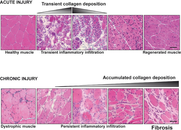
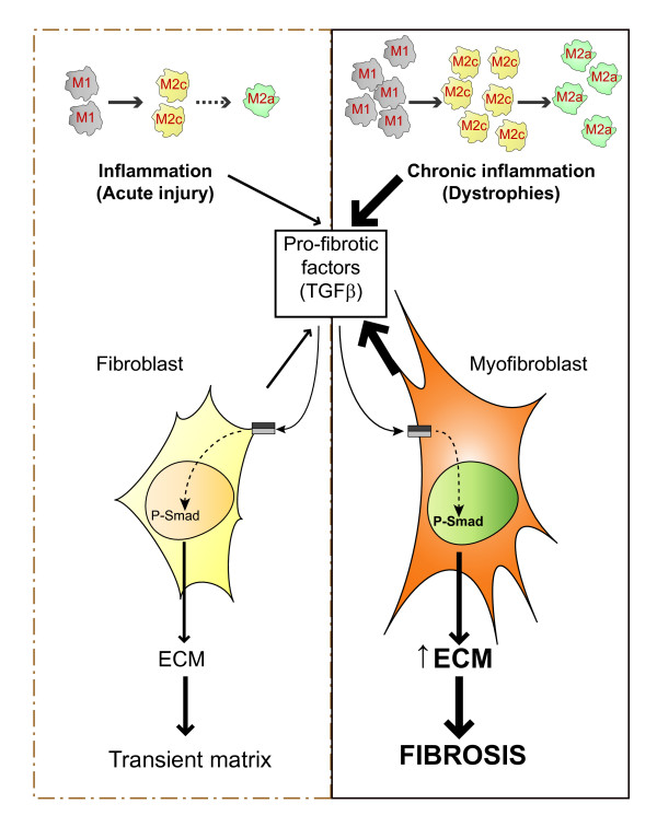
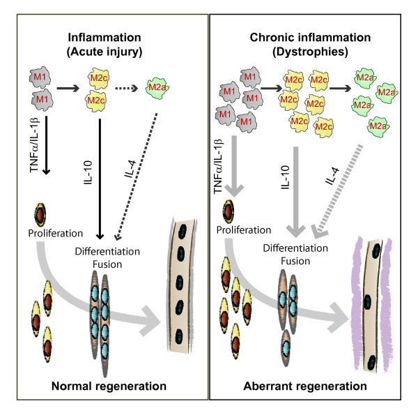
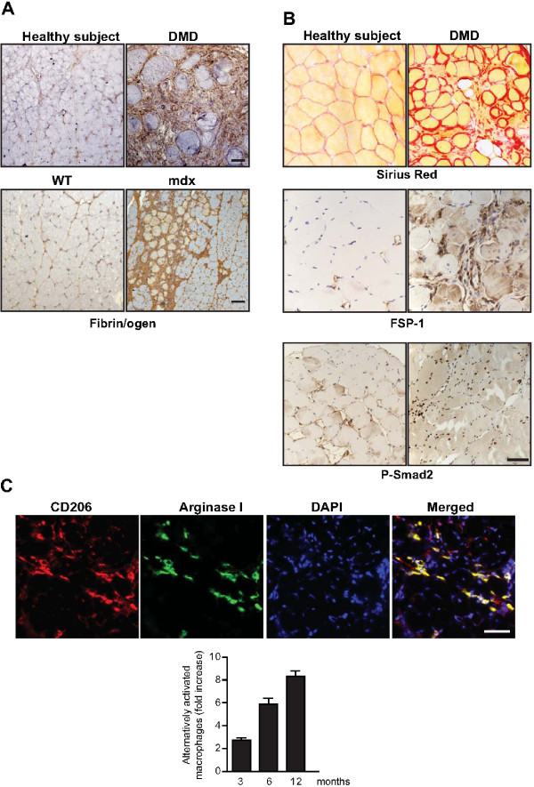
The repair process of damaged tissue involves the coordinated activities of several cell types in response to local and systemic signals. Following acute tissue injury, infiltrating inflammatory cells and resident stem cells orchestrate their activities to restore tissue homeostasis. However, during chronic tissue damage, such as in muscular dystrophies, the inflammatory-cell infiltration and fibroblast activation persists, while the reparative capacity of stem cells (satellite cells) is attenuated. Abnormal dystrophic muscle repair and its end stage, fibrosis, represent the final common pathway of virtually all chronic neurodegenerative muscular diseases. As our understanding of the pathogenesis of muscle fibrosis has progressed, it has become evident that the muscle provides a useful model for the regulation of tissue repair by the local microenvironment, showing interplay among muscle-specific stem cells, inflammatory cells, fibroblasts and extracellular matrix components of the mammalian wound-healing response. This article reviews the emerging findings of the mechanisms that underlie normal versus aberrant muscle-tissue repair.
Overview
The claims and statistics of this paper haven’t been checked yet.
Run the check — free, no account needed. It reads the open-access text and checks the sources it cites.
Integrity signals
Registry
Cites
Statistics
Figures
Verification
Claims the paper makes
The claims and statistics of this paper haven’t been checked yet.
Run the check — free, no account needed. It reads the open-access text and checks the sources it cites.
Claims other papers make about this one
Each is checked against this paper’s own text: does it say what the citing paper says it does?
In the citing paper
“Previous work has shown that the accumulation of inflammatory macrophages and age-related declines in interferon-gamma signaling delay muscle repair following injury”
From Skeletal muscle aging, cellular senescence, and senotherapeutics: current knowledge and future directions (2021) · cites this paper as Mann et al., 2011
Self-check
Percentages, sums and reported differences re-computed from the paper’s own numbers.
The self-check needs the paper’s full text.
Citations
Cites
1 retracted
RETRACTED ARTICLE: The role of nitric oxide during healing of trauma to the skeletal muscle
Filippin, L. I., Cuevas, M. J., Lima‐Cabello, E., Marroni, N. P., González‐Gallego, J., & Xavier, R. M. (2010) · Inflammation Research · doi:10.1007/s00011-010-0277-2 · Cited by 34 · cited 4×
“In addition, NO, which is released by local and invading cells during injury, can act as a pro-inflammatory molecule, and is able to activate MMP-2, which in turn releases HGF. Importantly, blockers of NOS function, such as nitro- Larginine methyl ester (L-NAME), have been shown to decrease the acute inflammatory reac…”
MethodInfluentialRetractedThe chemokine system in diverse forms of macrophage activation and polarization
MANTOVANI, A., SICA, A., SOZZANI, S., ALLAVENA, P., VECCHI, A., & LOCATI, M. (2004) · Trends in Immunology · doi:10.1016/j.it.2004.09.015 · Cited by 6,534 · cited 2×
“A nomenclature for polarized macrophages has been proposed [24,25] and they are now referred to as classically and alternatively activated macrophages, or M1 and M2 macrophages, respectively (Figure 2, Figure 3).”
BackgroundMechanisms of TGF-β Signaling from Cell Membrane to the Nucleus
Shi, Y., & Massagué, J. (2003) · Cell · doi:10.1016/s0092-8674(03)00432-x · Cited by 5,860
Visse, R., & Nagase, H. (2003) · Circulation Research · doi:10.1161/01.res.0000070112.80711.3d · Cited by 4,575 · cited 1×
“Activation requires proteolytic cleavage of the inactive precursor by either membrane-type (MT)1MMP [105] or plasmin, and cleavage of their corresponding inhibitors [106].”
Cellular and molecular mechanisms of fibrosis
Wynn, T. (2008) · The Journal of Pathology · doi:10.1002/path.2277 · Cited by 4,495
SATELLITE CELL OF SKELETAL MUSCLE FIBERS
Mauro, A. (1961) · The Journal of Cell Biology · doi:10.1083/jcb.9.2.493 · Cited by 3,683
Hinz, B., Phan, S. H., Thannickal, V. J., Galli, A., Bochaton-Piallat, M.-L., & Gabbiani, G. (2007) · The American Journal of Pathology · doi:10.2353/ajpath.2007.070112 · Cited by 2,068 · cited 1×
“These cells are associated with tissue repair and fibrosis in many tissues and organs, including muscle, skin, liver, lung, bone and cartilage [56].”
BackgroundArnold, L., Henry, A., Poron, F., Baba-Amer, Y., van Rooijen, N., Plonquet, A., Gherardi, R. K., & Chazaud, B. (2007) · The Journal of Experimental Medicine · doi:10.1084/jem.20070075 · Cited by 2,002 · cited 3×
“An important development in our understanding of muscle repair and fibrosis was the demonstration that a heterogeneous population of macrophages exists in regenerating muscle after injury, exhibiting opposing activities (either pro-inflammatory or anti-inflammatory) and different kinetics [23].”
BackgroundInfluentialFibrotic disease and the TH1/TH2 paradigm
Wynn, T. A. (2004) · Nature Reviews Immunology · doi:10.1038/nri1412 · Cited by 1,765
Muscle injury activates resident fibro/adipogenic progenitors that facilitate myogenesis
Joe, A. W. B., Yi, L., Natarajan, A., Le Grand, F., So, L., Wang, J., Rudnicki, M. A., & Rossi, F. M. V. (2010) · Nature Cell Biology · doi:10.1038/ncb2015 · Cited by 1,690 · cited 7×
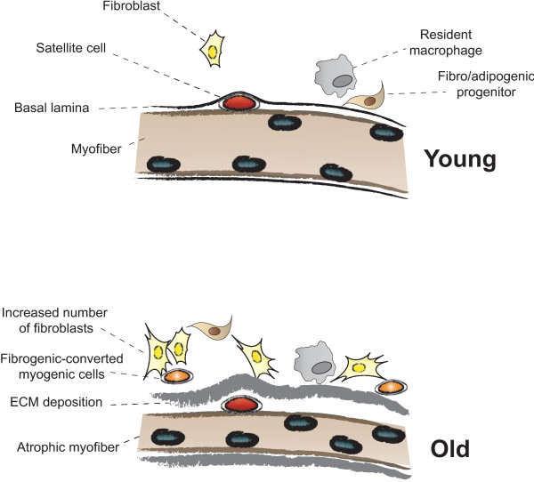
“Whether FAP conversion also occurs during aging, and whether Basal lamina ECM deposition Satellite cell Fibro/adipogenic progenitor Resident macrophage Fibroblast Increased number of fibroblasts Fibrogenic-converted myogenic cells Myofiber Atrophic myofiber Young Old Figure 5 Myofiber growth and extracellular matrix (…”
BackgroundInfluential
Cited by
Sorted by citations
Items 801–854 of 854
The usefulness and limitations of muscle quality assessment for sarcopenia diagnosis
Yamada, Y., & Yoshida, T. (2025) · 体力科學 · doi:10.7600/jspfsm.74.215
Krugom, S. V. (2025) · Stomatology for all / International Dental Review · doi:10.35556/idr-2025-2(111)42-46
Fornaro, M., Giannini, M., D’Abbicco, D., Fiorella, M. L., Errede, M., Virgintino, D., Valente, O., Frigeri, A., Iannone, F., & Girolamo, F. (2025) · Lara D. Veeken · doi:10.1093/rheumatology/keaf436
Liu, X., Carbajal, E. E. P., Hwang, Y.-C., Gilman, B. W., Bliss, S. A., Morrissey, C. S., & Wosczyna, M. N. (2025) · bioRxiv (Cold Spring Harbor Laboratory) · doi:10.1101/2025.10.02.678862
Dapper, H., Waltenberger, M., Pigorsch, S. U., Combs, S. E., Bauermeister, K., & Bauermeister, W. (2026) · Physics and Imaging in Radiation Oncology · doi:10.1016/j.phro.2026.101005
“Essentially, fibrosis results from a misdirected wound-healing process [5].”
Efeito dos ácidos linoleico e oleico na regeneração do músculo gastrocnêmio de ratos após laceração.
Teixeira, P. S. A. (2014) · Digital Library of Theses and Dissertations (Universidade de São Paulo) · doi:10.11606/t.42.2014.tde-11032015-142555
Development of approaches to improve the regenerative potential of muscle stem cells
Beckman, S. (2012) · D-Scholarship@Pitt (University of Pittsburgh)
Barbe, M. F., Harris, M. Y., Cruz, G. E., Amin, M., Billett, N. M., Dorotan, J. T., Day, E. P., Kim, S. Y., & Bove, G. M. (2021) · Figshare · doi:10.6084/m9.figshare.14545189
Lemoine, G. G., Scott‐Boyer, M., Ambroise, B., Périn, O., & Droit, A. (2021) · Figshare · doi:10.6084/m9.figshare.14677009
Kakutani, N., Takada, S., Nambu, H., Matsumoto, J., Furihata, T., Yokota, T., Fukushima, A., & Kinugawa, S. (2020) · Figshare · doi:10.6084/m9.figshare.12197475
Norris, A. M., Fierman, K. E., Campbell, J., Pitale, R., Shahraj, M., & Kopinke, D. (2024) · Figshare · doi:10.6084/m9.figshare.25929164
Ragozzino, E., Bortolani, S., Pietro, L. D., Papait, A., Parolini, O., Monforte, M., Tasca, G., & Ricci, E. (2024) · Figshare · doi:10.6084/m9.figshare.24329950
Ollewagen, T., Myburgh, K. H., van de Vyver, M., & Smith, C. (2021) · Figshare · doi:10.6084/m9.figshare.14157658
Chen, M., Chen, H., Gu, Y., Sun, P., Sun, J., Yu, H., Zheng, H., & Chen, D. (2021) · Figshare · doi:10.6084/m9.figshare.15153998
Additional file 1 of Bioengineered human skeletal muscle capable of functional regeneration
Fleming, J. W., Capel, A. J., Rimington, R. P., Wheeler, P., Leonard, A. N., Bishop, N. C., Davies, O. G., & Lewis, M. P. (2020) · Figshare · doi:10.6084/m9.figshare.13120938
LEM-Domain-Containing Inner Nuclear Membrane Proteins: Emerging Regulators of Intranuclear Signaling
Lee, B., Lee, H., & Shim, J. (2026) · International Journal of Molecular Sciences · doi:10.3390/ijms27020942
Okuyama, H., Brown, M., Munipalle, M., Nejati, S., Huo, R., Sakata, H., Mizuta, R., Sasaki, Y., Akiyoshi, K., Ohnishi, H., et al. (2026) · Biomaterials · doi:10.1016/j.biomaterials.2026.124044
“Muscle regeneration is often accompanied by fibrosis, which involves excessive collagen and ECM deposition [86], leading to increased stiffness and impaired function.”
Hanson, L. R., Scalise, K. L., Esch, R. M., Nevo, L., & Goldhamer, D. J. (2026) · Bone · doi:10.1016/j.bone.2026.117829
Deng, Z., Luo, D., Zhou, J., Zhang, X., Ren, H., Deng, W., & Gong, L. (2026) · BMC Geriatrics · doi:10.1186/s12877-026-07241-z
“Skeletal muscle atrophy has also become a significant health concern for elderly patients [5, 6], as aging is often accompanied by systemic chronic inflammation and a reduced capacity for skeletal muscle repair, leading to muscle fibrosis and fat deposition [7].”
Characterization of the dystrophic muscle by ²³Na NMR and ¹H NMR T₂ spectrum
Gerhalter, T. (2018) · theses.fr (ABES) · doi:10.70675/d7ddb629z3f9fz436bz96bezd0b545bdf370
The extracellular matrix as a biomaterial to optimize skeletal muscle regeneration
Trignol, A. (2019) · theses.fr (ABES) · doi:10.70675/0610b575z71faz4ea8zb917z1c4ad5f29d23
Fibrose musculaire : acteurs cellulaires et stratégies thérapeutiques
Bensalah, M. (2019) · theses.fr (ABES) · doi:10.70675/26f11f97zd40az491cz93fcz9608314a2b03
Sastourné-Arrey, Q. (2019) · theses.fr (ABES) · doi:10.70675/df26e053z6d2cz4b97z931dz7e7acceec691
Les cellules lymphoïdes dans la biologie et la pathologie musculaire
Saidj, T. (2022) · theses.fr (ABES) · doi:10.70675/8bd8c2daz45ffz4bcfzad71z3f11c2e9dead
Al-waqeerah, A., He, H., Bashah, A., Ghaleb, E., Wang, N., & Gao, L. (2026) · Respiratory Research · doi:10.1186/s12931-026-03661-y
“While it serves an essential role in preserving tissue architecture and maintaining organ function in the acute phase following injury, dysregulation of this process can result in pathological fibrosis [2, 3].”
InfluentialChatterjee, N., Pandita, U., & Misra, S. K. (2026) · ACS Biomaterials Science & Engineering · doi:10.1021/acsbiomaterials.5c02171
Ageing‐Associated Dysregulation of Myogenic Differentiation in Inclusion Body Myositis
de Vries, G., Ridder, W. D., & Baets, J. (2026) · Journal of Cachexia Sarcopenia and Muscle · doi:10.1002/jcsm.70301
“…cells in the muscle niche is associated with degenerative phenotypes, remodelling of the extracellular matrix (ECM) and ageing of the surrounding tissue through the adoption of the SASP, characterised by the secretion of pro-inflammatory cytokines, chemokines and growth factors [54, 71, 72].”
Guo, C., Wei, X., Li, W., Wang, G., Gao, B., Zhang, L., & Gao, B. (2026) · Journal of Nanobiotechnology · doi:10.1186/s12951-026-04610-z
Zotti, M. G., Mekhail, D., Kennedy, B. A., Wyatt, M., & Bonev, V. (2026) · BMC Musculoskeletal Disorders · doi:10.1186/s12891-026-10084-9
“Other alternative theories suggest that myopathy and inflammation are responsible for LMA; however, less is published on this than the main theories of dener-vation and arthrogenic inhibition, which may commonly co-exist [24].”
Camman, M., Nieswic, N., Mauviel, M., Joanne, P., Brun, J., Villerabel, L., Marcellan, A., Agbulut, O., & Hélary, C. (2026) · Biomaterials Science · doi:10.1039/d6bm00046k
Fernandes, J. D. S., Noronha, J. A., Brito, B. F., Guimarães, G. K. S., Palomino, G. J. Q., Llacho, C. P. H., & da Silva, L. D. M. (2026) · Reproduction in Domestic Animals · doi:10.1111/rda.70253
Mendanha, M., Soares, R. A., Luís, C., Pedrosa, S. S., Ferreira, C., Pinto-Ribeiro, I., Madureira, A. R., & Queirós, L. (2026) · Aesthetic Surgery Journal · doi:10.1093/asj/sjag121
Ghosh, S., Arshi, M. U., Ghosh, S., Jana, A., Nandi, S., Purohit, N., Ram, H., & Ghosh, S. (2026) · ACS Applied Bio Materials · doi:10.1021/acsabm.6c00163
Functional primary human 3D skeletal muscle models enable exercise and metabolic researchPreprint
Dreher, S. I., Schoeler, R., Zorn, K. S., Martin, J., Kuehnle, J., Elsner, K., Behle, I., Goj, T., Ruoff, L., Leffek, K., et al. (2026) · bioRxiv (Cold Spring Harbor Laboratory) · doi:10.64898/2026.06.29.735246
Madden, M. C., Evanchik, M. J., Lane, J., Donovan, J., Russell, A. J., & Silverman, J. A. (2026) · The Journal of Clinical Pharmacology · doi:10.1002/jcph.70234
Wang, Z., Sang, L. L., Youn, A., Liu, M., Feeley, B. T., & Liu, X. (2026) · Bone and Joint Research · doi:10.1302/2046-3758.158.bjr-2025-0112.r2
Li, Z., Lin, C., Tang, L., Xu, Y., Liang, J., Pan, D., Huang, Z., Xie, X., Wang, Y., Qin, S., et al. (2026) · International Journal of Molecular Sciences · doi:10.3390/ijms27177527
“Abnormal collagen deposition and fibrosis may disrupt the normal regenerative microenvironment and are associated with imbalanced macrophage polarization and impaired repair [96].”
Chen, Y., Ma, K., Huang, T., Gao, J., & Feng, X. (2026) · Functional & Integrative Genomics · doi:10.1007/s10142-026-02023-7
“In aging muscle, fibroblast expansion and matrix accumulation can have several pathophysiologic consequences: increasing tissue stiffness, altering mechanical signaling, disturbing satellite-cell behavior, and reducing the efficiency of regenerative repair(Blau et al. 2015, Mann et al. 2011 ).”
Matsunaga, H., Yonemitsu, R., Ideo, K., Ide, J., Shimada, M., Tateyama, M., Tian, X., Takata, S., Takata, K., Tanimura, S., et al. (2026) · PLoS ONE · doi:10.1371/journal.pone.0357829
“Although tissue fibrosis in vivo after I/R is induced by various cell types [59], including myoblasts, our in vitro C2C12 myoblast model suggests that myoblasts may contribute to profibrotic signaling but may not account fully for collagen accumulation in vivo .”
Chen, T., Song, J., & Ma, L. (2026) · Frontiers in Cell and Developmental Biology · doi:10.3389/fcell.2026.1928020
“…fibrosis reflects dysregulated repair, including TGF-beta-associated signaling, persistent fibroblast/FAP activation, and abnormal extracellular-matrix turnover (Gillies and Lieber, 2011; Lieber and Ward, 2013; Mann et al., 2011; Serrano and Muñoz-Cánoves, 2010; Bentzinger et al., 2012).”
Zou, X., Li, Y., Zhang, X., Niu, H., Zhou, H., Liu, L., Cui, G., Wang, B., & Wu, R. (2026) · Microbiology Spectrum · doi:10.1128/spectrum.00383-26
“Persistent inflammation is a key driver of tissue remodeling, extracellular matrix deposition, and fibrosis (11, 42, 43).”
UA-30 mitigates systemic aging and motor decline by inhibiting RalA signaling in naturally aged mice
Zhang, B., He, Y., Zhang, R., Su, T., Liu, R., Li, X., Zhang, X., Xiong, B., Yang, C., Liu, X., et al. (2026) · The Journal of Experimental Medicine · doi:10.1084/jem.20260071
Therapeutic Potential of SAT-3247 in a Canine Model of Duchenne Muscular DystrophyPreprint
Rayagiri, S., Smid, J. K., Lambert, P., Jaquith, J., Murray, T., Mitchell, R., Gleeson, F., Monast, J. G., Teixeira, J. A., Yue, Y., et al. (2026) · Research Square · doi:10.21203/rs.3.rs-11220671/v1
Zhang, T., Ya, Z., Ji, D.-J., Chu, Y.-Z., Su, X.-J., Huang, S.-P., Chen, Q., Yan, J.-T., Zhu, A., Xie, J., et al. (2026) · Tissue and Cell · doi:10.1016/j.tice.2026.104008
FAPs orchestrate ANTXR2-mediated fine-tuning of collagen VI to ensure skeletal muscle function
Metti, S., Bracq, L. J., Signor, M., Facchinello, N., Gambarotto, L., Nogara, L., Pigato, G., Abrami, L., Kunz, B., Duc, J., et al. (2026) · The Journal of Cell Biology · doi:10.1083/jcb.202606056
Surgical management of hypertonia in children
Houskamp, E., Mossner, J., & Raskin, J. S. (2026) · Elsevier eBooks · doi:10.1016/b978-0-443-23540-5.00013-8
The role of Xin in skeletal muscle regeneration
Nissar, A. A. (2012) · MacSphere (McMaster University) · doi:10.71548/18079
Metadata
- Authors
- Mann, C. J., Perdiguero, E., Kharraz, Y., Aguilar, S., Pessina, P., Serrano, A. L., & Muñoz-Cánoves, P.
- Affiliations
- Universitat Pompeu Fabra (ES)
- Venue
- Skeletal Muscle
- Published
- 2011-05-04
- Volume
- 1
- Issue
- 1
- Pages
- 21-21
- ISSN
- 2044-5040 (ISSN-L)
- PMC
- PMC3156644
- OpenAlex
- W2076787014
- Cited by
- 853
- References
- 129
- Citations by year
- 2026: 37 · 2025: 59 · 2024: 55 · 2023: 53 · 2022: 59 · 2021: 71 · 2020: 81 · 2019: 85 · 2018: 76 · 2017: 51 · 2016: 68 · 2015: 48 · 2014: 49 · 2013: 33 · 2012: 27
- FWCI
- 14.30
- Type
- journal article
- Language
- en
- Open access
- gold
- doi.org
- Licence
- cc-by
- Locations
- Skeletal Muscle (publishedVersion, cc-by)
- Europe PMC (PubMed Central) (publishedVersion, cc-by)
- PubMed Central (publishedVersion, cc-by)
- PubMed (publishedVersion)
- RECERCAT (Consorci de Serveis Universitaris de Catalunya) (publishedVersion, other-oa)
- DOAJ (DOAJ: Directory of Open Access Journals) (submittedVersion, cc-by-sa)
- Repositori digital de la UPF (Universitat Pompeu Fabra) (submittedVersion, cc-by)
- Funders
- Ministerio de Ciencia e Innovación (PLE2009)
- Centro de Investigación Biomédica en Red sobre Enfermedades Neurodegenerativas
- Topics
- Life Sciences › Biochemistry, Genetics and Molecular Biology › Molecular Biology › Muscle Physiology and Disorders
- Health Sciences › Medicine › Cardiology and Cardiovascular Medicine › Cardiac Fibrosis and Remodeling
- Health Sciences › Medicine › Surgery › Wound Healing and Treatments
- Keywords
- muscle fibrosis, muscle repair, muscular dystrophy, satellite cells, muscle stem cells, fibroblast activation, inflammatory cell infiltration, extracellular matrix, wound healing, microenvironment
About these checks
Registry, retraction, Cites and statistics results are computed, not judged, with one exception: a printed reference that no index lists is read once by a model before it counts as a citation error. Claims are extracted by a model and kept only when their quote appears verbatim in the text; each cited source is then checked, with a model’s help, for whether it says what the claim relies on. Those verdicts can be wrong — read the claim’s quote and the cited source before relying on one.
Statistics and arithmetic are recomputed, not judged.
Confidence bands are shown only for fields calibrated on our annotated test set. None is calibrated yet, so no band is shown.
What counts as a claim ›How we measure confidence ›How we check this ›
Are you an author, or do you see a problem with this page? Request a review
