

The DOI matches its Crossref record; 129 of the 129 references it cites were found in a registry or an index.
Aberrant repair and fibrosis development in skeletal muscle
Open accessAbstract
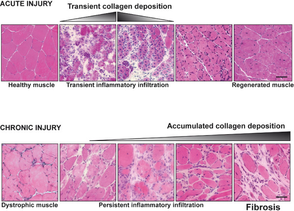
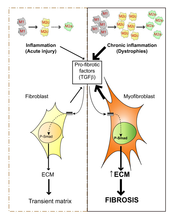
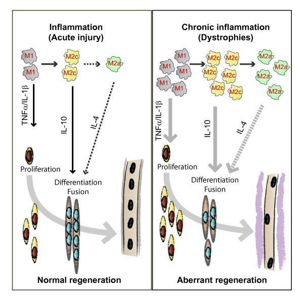
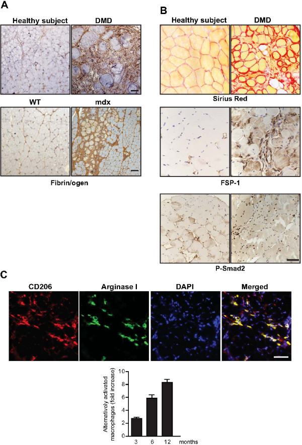
The repair process of damaged tissue involves the coordinated activities of several cell types in response to local and systemic signals. Following acute tissue injury, infiltrating inflammatory cells and resident stem cells orchestrate their activities to restore tissue homeostasis. However, during chronic tissue damage, such as in muscular dystrophies, the inflammatory-cell infiltration and fibroblast activation persists, while the reparative capacity of stem cells (satellite cells) is attenuated. Abnormal dystrophic muscle repair and its end stage, fibrosis, represent the final common pathway of virtually all chronic neurodegenerative muscular diseases. As our understanding of the pathogenesis of muscle fibrosis has progressed, it has become evident that the muscle provides a useful model for the regulation of tissue repair by the local microenvironment, showing interplay among muscle-specific stem cells, inflammatory cells, fibroblasts and extracellular matrix components of the mammalian wound-healing response. This article reviews the emerging findings of the mechanisms that underlie normal versus aberrant muscle-tissue repair.
Overview
The claims and statistics of this paper haven’t been checked yet.
Run the check — free, no account needed. It reads the open-access text and checks the sources it cites.
Integrity signals
Registry
Cites
Statistics
Figures
Verification
Claims the paper makes
The claims and statistics of this paper haven’t been checked yet.
Run the check — free, no account needed. It reads the open-access text and checks the sources it cites.
Claims other papers make about this one
Each is checked against this paper’s own text: does it say what the citing paper says it does?
In the citing paper
“Previous work has shown that the accumulation of inflammatory macrophages and age-related declines in interferon-gamma signaling delay muscle repair following injury”
From Skeletal muscle aging, cellular senescence, and senotherapeutics: current knowledge and future directions (2021) · cites this paper as Mann et al., 2011
Self-check
Percentages, sums and reported differences re-computed from the paper’s own numbers.
The self-check needs the paper’s full text.
Citations
Cites
1 retracted
RETRACTED ARTICLE: The role of nitric oxide during healing of trauma to the skeletal muscle
Filippin, L. I., Cuevas, M. J., Lima‐Cabello, E., Marroni, N. P., González‐Gallego, J., & Xavier, R. M. (2010) · Inflammation Research · doi:10.1007/s00011-010-0277-2 · Cited by 34 · cited 4×
“In addition, NO, which is released by local and invading cells during injury, can act as a pro-inflammatory molecule, and is able to activate MMP-2, which in turn releases HGF. Importantly, blockers of NOS function, such as nitro- Larginine methyl ester (L-NAME), have been shown to decrease the acute inflammatory reac…”
MethodInfluentialRetractedThe chemokine system in diverse forms of macrophage activation and polarization
MANTOVANI, A., SICA, A., SOZZANI, S., ALLAVENA, P., VECCHI, A., & LOCATI, M. (2004) · Trends in Immunology · doi:10.1016/j.it.2004.09.015 · Cited by 6,534 · cited 2×
“A nomenclature for polarized macrophages has been proposed [24,25] and they are now referred to as classically and alternatively activated macrophages, or M1 and M2 macrophages, respectively (Figure 2, Figure 3).”
BackgroundMechanisms of TGF-β Signaling from Cell Membrane to the Nucleus
Shi, Y., & Massagué, J. (2003) · Cell · doi:10.1016/s0092-8674(03)00432-x · Cited by 5,860
Visse, R., & Nagase, H. (2003) · Circulation Research · doi:10.1161/01.res.0000070112.80711.3d · Cited by 4,575 · cited 1×
“Activation requires proteolytic cleavage of the inactive precursor by either membrane-type (MT)1MMP [105] or plasmin, and cleavage of their corresponding inhibitors [106].”
Cellular and molecular mechanisms of fibrosis
Wynn, T. (2008) · The Journal of Pathology · doi:10.1002/path.2277 · Cited by 4,495
SATELLITE CELL OF SKELETAL MUSCLE FIBERS
Mauro, A. (1961) · The Journal of Cell Biology · doi:10.1083/jcb.9.2.493 · Cited by 3,683
Hinz, B., Phan, S. H., Thannickal, V. J., Galli, A., Bochaton-Piallat, M.-L., & Gabbiani, G. (2007) · The American Journal of Pathology · doi:10.2353/ajpath.2007.070112 · Cited by 2,068 · cited 1×
“These cells are associated with tissue repair and fibrosis in many tissues and organs, including muscle, skin, liver, lung, bone and cartilage [56].”
BackgroundArnold, L., Henry, A., Poron, F., Baba-Amer, Y., van Rooijen, N., Plonquet, A., Gherardi, R. K., & Chazaud, B. (2007) · The Journal of Experimental Medicine · doi:10.1084/jem.20070075 · Cited by 2,002 · cited 3×
“An important development in our understanding of muscle repair and fibrosis was the demonstration that a heterogeneous population of macrophages exists in regenerating muscle after injury, exhibiting opposing activities (either pro-inflammatory or anti-inflammatory) and different kinetics [23].”
BackgroundInfluentialFibrotic disease and the TH1/TH2 paradigm
Wynn, T. A. (2004) · Nature Reviews Immunology · doi:10.1038/nri1412 · Cited by 1,765
Muscle injury activates resident fibro/adipogenic progenitors that facilitate myogenesis
Joe, A. W. B., Yi, L., Natarajan, A., Le Grand, F., So, L., Wang, J., Rudnicki, M. A., & Rossi, F. M. V. (2010) · Nature Cell Biology · doi:10.1038/ncb2015 · Cited by 1,690 · cited 7×
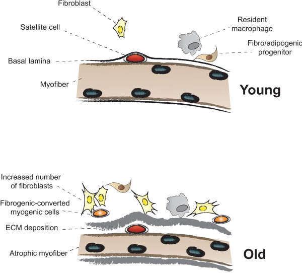
“Whether FAP conversion also occurs during aging, and whether Basal lamina ECM deposition Satellite cell Fibro/adipogenic progenitor Resident macrophage Fibroblast Increased number of fibroblasts Fibrogenic-converted myogenic cells Myofiber Atrophic myofiber Young Old Figure 5 Myofiber growth and extracellular matrix (…”
BackgroundInfluential
Cited by
Sorted by citations
Items 701–800 of 854
Signaling mechanisms in regenerative myogenesis.
Hindi, S. M. (2014) · doi:10.18297/etd/1740
“Fibrosis is a pathogenic factor characterized by chronic inflammation with persistent production of profibrotic cytokines and excessive deposition of ECM proteins, including collagens and fibronectin which can impair tissue function (124).”
Zadegan, S. A., Firouzi, M., Nabian, M. H., Zanjani, L. O., Nategh, M., & Kamrani, R. S. (2015) · DOAJ (DOAJ: Directory of Open Access Journals)
Hitler, D., Arumugam, P., Mathivanan, N., & Vellaichamy, E. (2014)
Normale Entwicklung, Anatomie und Funktion
Döderlein, L. (2015) · Springer eBooks · doi:10.1007/978-3-642-35319-2_2
Huntsman, H. D. (2014) · Illinois Digital Environment for Access to Learning and Scholarship (University of Illinois at Urbana-Champaign)
Epigenetic and genetic mechanisms through which Myostatin regulates muscle wasting
Lua, G. B. J. (2016) · DR-NTU (Nanyang Technological University) · doi:10.32657/10356/65954
Mautner, K., & Blazuk, J. (2015)
Application of bioresorbable polymers in muscular system
Villa, C., Erratico, S., Belicchi, M., & Torrente, Y. (2016) · Elsevier eBooks · doi:10.1016/b978-0-08-100262-9.00020-3
The Role of the Ubiquitin Ligase Nedd4-1 in Skeletal Muscle Atrophy
Nagpal, P. (2012) · TSpace (University of Toronto)
The Science of Neuromuscular Healing
Piraino, A. (2017) · Orthopaedic Section, APTA, Inc. eBooks · doi:10.17832/isc.2017.27.4.1
Neuromuscular Tissue Engineering
Avin, K. G., Badylak, S. F., & Ambrosio, F. (2014) · Orthopaedic Section, APTA, Inc. eBooks · doi:10.17832/isc.2014.23.2.2
Pollock‐Tahiri, E. (2015) · TSpace (University of Toronto)
Edmunds, K. J. (2017) · Opin vísindi (Opin vísindi)
Determinants of Upper Genital Tract Complications in a Chlamydial Urogenital Mouse Model
Jones, E. (2013) · doi:10.21007/etd.cghs.2013.0156
“Recent reports have linked IL-4 to tissue repair and wound healing by alternatively activating macrophages and stimulating the expansion of IL-4 producing cells [196] [197] [198] [199] [200].”
BackgroundNascimento, T. L. (2018) · Digital Library of Theses and Dissertations (Universidade de São Paulo) · doi:10.11606/t.42.2018.tde-08082018-084036
Mallender, M. (2018) · Scholarship at UWindsor (University of Windsor)
Low-intensity laser favors muscle regeneration in a malnourished and recovered experimental model
da Silva, M. C., da Silveira, R. S., Matsumura, C. Y., & Pertille, A. (2018) · Fisioterapia e Pesquisa · doi:10.1590/1809-2950/17527425022018
Nuclear localized Akt limits skeletal muscle derived fibrotic signaling
Guadagnin, E., Bagchi, D., Sinha, I., & Neppl, R. L. (2018) · Biochemical and Biophysical Research Communications · doi:10.1016/j.bbrc.2018.11.202
Chen, J. J. (2018) · TSpace (University of Toronto)
Proteomics Exploration in Skeletal Muscle Following Physical Trauma
Liu, L. (2019) · Queensland University of Technology · doi:10.5204/thesis.eprints.131581
Characterization of Wilms' Tumour 1 (Wt1) as a Biomarker for Fibrosis in Duchenne Muscular Dystrophy
Murphy, P. J. (2019) · Scholarship@Western (Western University)
Matviienko, T., Bogutskaya, K. I., Nozdrenko, D. M., & Prilutsky, Y. I. (2019) · Fiziolohichnyĭ zhurnal · doi:10.15407/fz65.05.077
GABINETE DE LEITURA DO GRÊMIO LITERÁRIO PORTUGUÊS: SABERES E PRÁTICAS EDUCATIVAS (1868-1889)
Boulhosa, M. J. D. S. C. (2019) · Anais do VIII Seminário de Integração Científica da Universidade do Estado do Pará · doi:10.31792/21759766.viiisic.2019.462-466
da Paz, T. Y. B. (2019) · Anais do VIII Seminário de Integração Científica da Universidade do Estado do Pará · doi:10.31792/21759766.viiisic.2019.307-311
Cerdeira, C. D. L., Vieira, N. G. D. N., & Santana, J. V. (2019) · Anais do VIII Seminário de Integração Científica da Universidade do Estado do Pará · doi:10.31792/21759766.viiisic.2019.109
dos Santos, A. T. M. (2019) · Anais do VIII Seminário de Integração Científica da Universidade do Estado do Pará · doi:10.31792/21759766.viiisic.2019.186-189
da Trindade, L. R. (2019) · Anais do VIII Seminário de Integração Científica da Universidade do Estado do Pará · doi:10.31792/21759766.viiisic.2019.117-122
Lima, D. E. D. O. B. (2019) · Anais do VIII Seminário de Integração Científica da Universidade do Estado do Pará · doi:10.31792/21759766.viiisic.2019.489-492
Heinen, I. L. S. (2019) · Anais do VIII Seminário de Integração Científica da Universidade do Estado do Pará · doi:10.31792/21759766.viiisic.2019.382-387
AÇÃO INIBITÓRIA DO EXTRATO DE AÇAÍ NA INFECÇÃO DO VÍRUS DO NILO OCIDENTAL
Teixeira, B. N. (2019) · Anais do VIII Seminário de Integração Científica da Universidade do Estado do Pará · doi:10.31792/21759766.viiisic.2019.208-211
da Silva, C. I. M. (2019) · Anais do VIII Seminário de Integração Científica da Universidade do Estado do Pará · doi:10.31792/21759766.viiisic.2019.223-225
Santos, E. D. F., & Carvalho, V. M. T. (2019) · Anais do VIII Seminário de Integração Científica da Universidade do Estado do Pará · doi:10.31792/21759766.viiisic.2019.141-143
PREVALÊNCIA E FATORES DE RISCO ASSOCIADOS ÀS ALTERAÇÕES POSTURAIS ENTRE ESCOLARES
Veloso, S. M. A., Pereira, J. D. S., & Franco, V. P. (2019) · Anais do VIII Seminário de Integração Científica da Universidade do Estado do Pará · doi:10.31792/21759766.viiisic.2019.61-65
Costa, I. S., & Barbosa, S. N. D. S. (2019) · Anais do VIII Seminário de Integração Científica da Universidade do Estado do Pará · doi:10.31792/21759766.viiisic.2019.132
NA BARRA DE SUA SAIA: D. MARIA PADILHA, A MULHER QUE EDUCA
Fares, J. A., & Soares, M. D. M. (2019) · Anais do VIII Seminário de Integração Científica da Universidade do Estado do Pará · doi:10.31792/21759766.viiisic.2019.457-461
Barros, M. C., & da Conceição, D. R. (2019) · Anais do VIII Seminário de Integração Científica da Universidade do Estado do Pará · doi:10.31792/21759766.viiisic.2019.400-404
A MORTE CELULAR EM LESÕES CUTÂNEAS DA DOENÇA DE HANSEN
Virgolino, G. L., Teixeira, V. F., Neto, F. D. L., da Cruz, M. F. S., & Quaresma, J. A. S. (2019) · Anais do VIII Seminário de Integração Científica da Universidade do Estado do Pará · doi:10.31792/21759766.viiisic.2019.66-68
de Santi, C. A. N., Barbosa, L. N., & Macedo, D. B. (2019) · Anais do VIII Seminário de Integração Científica da Universidade do Estado do Pará · doi:10.31792/21759766.viiisic.2019.28
ASPECTOS EPIDEMIOLÓGICOS DA MALÁRIA NO MUNICÍPIO DE TUCURUÍPA
Peixoto, M. C. D. S. (2019) · Anais do VIII Seminário de Integração Científica da Universidade do Estado do Pará · doi:10.31792/21759766.viiisic.2019.275-279
de Oliveira, E. M., & da Silva, L. B. M. (2019) · Anais do VIII Seminário de Integração Científica da Universidade do Estado do Pará · doi:10.31792/21759766.viiisic.2019.35-36
PLANTAS ALIMENTÍCIAS NÃO CONVENCIONAIS (PANC´S) DA ILHA DO MOSQUEIRO, BELÉM-PARÁ
Corrêa, C. N., & dos Santos, K. R. (2019) · Anais do VIII Seminário de Integração Científica da Universidade do Estado do Pará · doi:10.31792/21759766.viiisic.2019.145
Rocha, M. P., Cardoso, B. C., Leão, L. D. A., & Costa, N. P. (2019) · Anais do VIII Seminário de Integração Científica da Universidade do Estado do Pará · doi:10.31792/21759766.viiisic.2019.83
A CRÍTICA DA RELIGIÃO EM KANT: CONTRIBUIÇÕES PARA O DEBATE ACERCA DA INTOLERÂNCIA RELIGIOSA
dos Santos, H. H. S. (2019) · Anais do VIII Seminário de Integração Científica da Universidade do Estado do Pará · doi:10.31792/21759766.viiisic.2019.378-381
Lopes, N. S., & Fayal, S. P. (2019) · Anais do VIII Seminário de Integração Científica da Universidade do Estado do Pará · doi:10.31792/21759766.viiisic.2019.42
ESTUDO DA MORTALIDADE NEONATAL EM HOSPITAIS PÚBLICOS DE ALTA E MÉDIA COMPLEXIDADE NO BAIXO AMAZONAS
de Lima, S. M. M., Siqueira, V. A., & Pimentel, S. K. S. (2019) · Anais do VIII Seminário de Integração Científica da Universidade do Estado do Pará · doi:10.31792/21759766.viiisic.2019.90
Silva, M. J. R. B., & de Assunção, L. A. (2019) · Anais do VIII Seminário de Integração Científica da Universidade do Estado do Pará · doi:10.31792/21759766.viiisic.2019.89
UM OLHAR SOBRE A SAÚDE MENTAL DE ACADÊMICOS DA ÁREA DA SAÚDE DE UMA UNIVERSIDADE PÚBLICA
de Melo, :. M. P. M., Costa, F. D. S., Júnior, H. C. F. D. S., & Najjar, E. C. A. (2019) · Anais do VIII Seminário de Integração Científica da Universidade do Estado do Pará · doi:10.31792/21759766.viiisic.2019.52-53
Xavier, L. H. P., & Santos, M. R. S. (2019) · Anais do VIII Seminário de Integração Científica da Universidade do Estado do Pará · doi:10.31792/21759766.viiisic.2019.169-172
RELIGIÃO E PODER: A VISÃO CELULAR NA PRIMEIRA IGREJA BATISTA EM ICOARACI
Batista, W. A. (2019) · Anais do VIII Seminário de Integração Científica da Universidade do Estado do Pará · doi:10.31792/21759766.viiisic.2019.419-423
DESENVOLVIMENTO DE MATERIAIS BIOCOMPÓSITOS A BASE DE PLÁSTICO E PAPEL PÓS CONSUMO
Cardoso, J. F., & Teixeira, I. L. C. (2019) · Anais do VIII Seminário de Integração Científica da Universidade do Estado do Pará · doi:10.31792/21759766.viiisic.2019.139
Les plasmas riches en plaquettes dans les lésions myo-aponévrotiques
Dalmais, E. (2020) · Journal de Traumatologie du Sport · doi:10.1016/j.jts.2019.12.004
The role of platelets in skeletal muscle regeneration
Scully, D. (2019) · Repository@Hull (Worktribe) (University of Hull)
Qiu, X., Zeng, S., Xiong, Z., Tang, S., Tang, G., & Li, S. (2020) · Journal of Craniofacial Surgery · doi:10.1097/scs.0000000000006827
“During skeletal muscle fibrosis, myofibroblasts over-activate, proliferate, and produce a large amount of collagen, which is the key factor in this process.(6,7) In addition, myoblasts also play an important role in skeletal muscle regeneration.”
BackgroundMinimal Consequences of CMAH and DBA/2 Backgrounds on a FKRP Deficient Model
Vaubourg, C., Gicquel, E., Richard, I., Lostal, W., & Bellec, J. (2020) · Journal of Neuromuscular Diseases · doi:10.3233/jnd-200487
“TGF (cid:2) 1, acting via the Smad2/3/4 pathway, induces the expression of genes coding for extracellular matrix (ECM) components and enzymes inhibiting ECM degradation [27].”
Saad, S., eldeen, E. D., Soliman, A., Din, R. S. E., & Hanna, G. F. B. (2020) · The Egyptian Journal of Histology · doi:10.21608/ejh.2020.43345.1359
Nutritional Strategies to Mitigate Woody Breast and White Striping
Bodle, B. C. (2017) · OakTrust (Texas A&M University Libraries)
Kapoor, S., Subba, P., Sudheer Shenoy, P., & Bose, B. (2021) · Stem Cell Reviews and Reports · doi:10.1007/s12015-021-10134-w
Effect of vitamin D on Becker muscular dystrophy: A review
Alasady, S. H. (2021) · Tropical Journal of Pharmaceutical Research · doi:10.4314/tjpr.v19i12.30
“tumor necrosis factor alpha (TNFα), interleukin (IL)-1 and IL-4) are major mediators of inflammatory responses involved in tissue lesions [13,16,17].”
BackgroundHubbert, K., & Clement, R. D. (2021) · JBJS Journal of Orthopaedics for Physician Assistants · doi:10.2106/jbjs.jopa.20.00030
Kang, H., Mo, Y., Shin, J., Kim, H. Y., Cho, S., & Kang, H. (2020) · Research Square · doi:10.21203/rs.3.rs-91543/v1
“Additionally, M2c macrophages have been reported to be crucial in the development of pulmonary brosis [46, 47], and in vitro experiments have con rmed that ASCs can regulate M2c macrophage activation [48].”
BackgroundHoffman, D. B. (2020) · University of Minnesota Digital Conservancy (University of Minnesota)
Regenerative Medicine (Biological) Therapies for Acute Muscle Injury
Lauf, K., van der Made, A. D., Reurink, G., Tol, J. L., & Kerkhoffs, G. M. M. J. (2021) · Springer eBooks · doi:10.1007/978-3-030-60216-1_17
Kelly, K., McNeely, C., & Seda, S. (2020) · PubMed Central · doi:10.34917/2143862
Rossi, F. M. V., & Judson, R. N. (2013) · Faculty Opinions – Post-Publication Peer Review of the Biomedical Literature · doi:10.3410/f.718017737.793479334
Kennedy, B. K., & Tsai, S. (2020) · Faculty Opinions – Post-Publication Peer Review of the Biomedical Literature · doi:10.3410/f.726972164.793576522
Avian Muscle Development and Growth Mechanisms: Association with Muscle Myopathies and Meat Quality
Velleman, S. G., & Petracci, M. (2020) · Frontiers research topics · doi:10.3389/978-2-88966-314-9
Phillips, S. M. (2020) · Faculty Opinions – Post-Publication Peer Review of the Biomedical Literature · doi:10.3410/f.726972164.793571163
(2014) · Elsevier eBooks · doi:10.1016/b978-1-907568-42-8.50016-2
Elsy, B., & Khan, A. A. (2022) · Asian Pacific Journal of Health Sciences · doi:10.21276/apjhs.2022.9.1.44
Injury and Self‐Repair of Musculoskeletal Tissues
Gallagher, S., & Barbe, M. F. (2022) · doi:10.1002/9781119640172.ch11
AGE-ASSOCIATION FEATURES OF FAT AND LEAN MASS IN MEN
Musiіenko, А. S., Grygorieva, N. V., & Bystrytska, M. A. (2022) · Fìzìologìčnij žurnal · doi:10.15407/fz68.02.084
Circulating TGF-β1 Levels: Linking Muscle and Trauma
La, L., & Tuan, R. S. (2022) · Biomarkers in disease · doi:10.1007/978-3-030-87302-8_37-1
Potential Therapeutic Strategies for Muscular Dystrophy
Gaina, G. (2022) · IntechOpen eBooks · doi:10.5772/intechopen.102255
Cell Therapy for Muscular Dystrophy
Sharma, A. K., Sane, H. M., Gokulchandran, N., Paranjape, A., Shaikh, Z., Arjun, K. M., & Badhe, P. B. (2023) · IntechOpen eBooks · doi:10.5772/intechopen.108600
Takenami, L. I., Filha, A. M. S. G., Bomfim, E. S., Lagares, L. S., de Macedo, R. C., de Almeida, L. A. B., & dos Santos, C. P. C. (2023) · Revista Brasileira de Fisiologia do Exercício · doi:10.33233/rbfex.v21i4.5379
SKELETAL MUSCLE SENSITIVITY TO WASTING INDUCED BY UROTHELIAL CARCINOMA
Esteves, M., Duarte, M., Oliveira, P. A., da Costa, R. M. G., Monteiro, M. P., & Duarte, J. A. (2023) · Experimental Oncology · doi:10.15407/exp-oncology.2023.01.107
Harvest of Cell-Only Muscle Fibers Using Thermally Expandable Hydrogels with Adhesive Patterns
Lee, Y. B., Kim, S.-J., Kim, E. M., Byun, H., & Shin, H. (2023) · Tissue Engineering Part C Methods · doi:10.1089/ten.tec.2023.0096
“48,49 Cx43 controls cell–cell communication and is involved in contractility and electrical coupling.”
Compromiso miogénico de las células satélites en las distrofias musculares
Vélez-Caballero, S. H., Cano-Martínez, L. J., & Coral‐Vázquez, R. M. (2023) · Investigación en Discapacidad · doi:10.35366/113830
The role of satellite cell-derived TRIM28 in mechanical load- and injury-induced myogenesisPreprint
Lin, K., Hibbert, J. E., Lemens, J. L., Torbey, M. M., Steinert, N. D., Flejsierowicz, P. M., Melka, K. M., Lares, M., Setaluri, V., & Hornberger, T. A. (2023) · bioRxiv (Cold Spring Harbor Laboratory) · doi:10.1101/2023.12.20.572566
The role of MMP-14 and MMP-2 in mediating myoblast fusion.
Nkosi, M. S. (2016) · doi:10.29086/10413/22571
Transcriptional Profiling of Muscle in Females With Distal Radius Fracture and Functional Sarcopenia
Kang, J., Baek, J., Lee, J. K., & Hong, S. W. (2024) · The Journals of Gerontology Series A · doi:10.1093/gerona/glae002
“They are involved in extracellular matrix deposition, fibrosis, progressive muscular atrophy, as previously suggested (27).”
BackgroundKarcı, M. T., Özyurt, E., Ercan, M. B., Kenar, S. G., Demirdöğen, F., Özenç, B., Beton, Ö. E., Türkmen, N., Şen, M., Açıkgöz, M., et al. (2022) · Akademisyen Kitabevi eBooks · doi:10.37609/akya.1350
Jiang, X., Zhang, H., Ren, Y., Li, Y., Zhong, L., Guo, J., & Zhang, X. (2023) · Figshare · doi:10.6084/m9.figshare.23902842
Aziz, D., Rizkalla, N., Naguib, M., Habib, E. K., & Bekheet, E. A. (2024) · The Egyptian Journal of Anatomy · doi:10.21608/ejana.2020.22272.1067
“[51] Few numbers of satellite cells were also noted among this group (III) in the healing skeletal muscles and can be recognized between the sarcolemma and the basal lamina.”
BackgroundKlassische Muskelkontrakturen bei Katzen und Hunden
Luginbühl, J., Strässle, M., & Forterre, F. (2024) · kleintier konkret · doi:10.1055/a-2224-3757
Santos, A. D. R. (2024) · Digital Library of Theses and Dissertations (Universidade de São Paulo) · doi:10.11606/t.42.2024.tde-28062024-120145
Albini, S., Palmieri, L., Pili, L., Jaber, A., Hong, A. V., Moula, M., El‐Khoury, R., Brochier, G., Bigot, A., Israeli, D., et al. (2025) · Research Square · doi:10.21203/rs.3.rs-4270736/v1 · Cited by 1
Parga, A. D., & Raheem, I. (2025) · Cureus · doi:10.7759/cureus.78002
“The biopsy also revealed fibrinopurulent debris in the epimysium, suggestive of focal myofasciitis [8].”
Biologic Strategies for Muscle Injuries
Maier, L. P. N., Wuerfel, T., & Marshall, R. P. (2024) · Springer eBooks · doi:10.1007/978-3-030-65430-6_220-1
Zakirova, A. M., Борисовна, М. Т., Fayzullina, R. A., Муратович, В. Х., Мидхатович, З. А., Kadriev, A. G., Ленаровна, Р. Э., Klyushkina, A. I., Kadriev, A. A., & Sherifova, I. A. (2025) · Meditsinskiy sovet = Medical Council · doi:10.21518/ms2025-027
Metadata
- Authors
- Mann, C. J., Perdiguero, E., Kharraz, Y., Aguilar, S., Pessina, P., Serrano, A. L., & Muñoz-Cánoves, P.
- Affiliations
- Universitat Pompeu Fabra (ES)
- Venue
- Skeletal Muscle
- Published
- 2011-05-04
- Volume
- 1
- Issue
- 1
- Pages
- 21-21
- ISSN
- 2044-5040 (ISSN-L)
- PMC
- PMC3156644
- OpenAlex
- W2076787014
- Cited by
- 853
- References
- 129
- Citations by year
- 2026: 37 · 2025: 59 · 2024: 55 · 2023: 53 · 2022: 59 · 2021: 71 · 2020: 81 · 2019: 85 · 2018: 76 · 2017: 51 · 2016: 68 · 2015: 48 · 2014: 49 · 2013: 33 · 2012: 27
- FWCI
- 14.30
- Type
- journal article
- Language
- en
- Open access
- gold
- doi.org
- Licence
- cc-by
- Locations
- Skeletal Muscle (publishedVersion, cc-by)
- Europe PMC (PubMed Central) (publishedVersion, cc-by)
- PubMed Central (publishedVersion, cc-by)
- PubMed (publishedVersion)
- RECERCAT (Consorci de Serveis Universitaris de Catalunya) (publishedVersion, other-oa)
- DOAJ (DOAJ: Directory of Open Access Journals) (submittedVersion, cc-by-sa)
- Repositori digital de la UPF (Universitat Pompeu Fabra) (submittedVersion, cc-by)
- Funders
- Ministerio de Ciencia e Innovación (PLE2009)
- Centro de Investigación Biomédica en Red sobre Enfermedades Neurodegenerativas
- Topics
- Life Sciences › Biochemistry, Genetics and Molecular Biology › Molecular Biology › Muscle Physiology and Disorders
- Health Sciences › Medicine › Cardiology and Cardiovascular Medicine › Cardiac Fibrosis and Remodeling
- Health Sciences › Medicine › Surgery › Wound Healing and Treatments
- Keywords
- muscle fibrosis, muscle repair, muscular dystrophy, satellite cells, muscle stem cells, fibroblast activation, inflammatory cell infiltration, extracellular matrix, wound healing, microenvironment
About these checks
Registry, retraction, Cites and statistics results are computed, not judged, with one exception: a printed reference that no index lists is read once by a model before it counts as a citation error. Claims are extracted by a model and kept only when their quote appears verbatim in the text; each cited source is then checked, with a model’s help, for whether it says what the claim relies on. Those verdicts can be wrong — read the claim’s quote and the cited source before relying on one.
Statistics and arithmetic are recomputed, not judged.
Confidence bands are shown only for fields calibrated on our annotated test set. None is calibrated yet, so no band is shown.
What counts as a claim ›How we measure confidence ›How we check this ›
Are you an author, or do you see a problem with this page? Request a review
