

The DOI matches its Crossref record; 57 of the 57 references it cites were found in a registry or an index; 43 of the 72 sources its claims cite were checked for support.
Seizure protein 6 and its homolog seizure 6-like protein are physiological substrates of BACE1 in neurons
Open accessAbstract
BACKGROUND: The protease BACE1 (beta-site APP cleaving enzyme) is a major drug target in Alzheimer's disease. However, BACE1 therapeutic inhibition may cause unwanted adverse effects due to its additional functions in the nervous system, such as in myelination and neuronal connectivity. Additionally, recent proteomic studies investigating BACE1 inhibition in cell lines and cultured murine neurons identified a wider range of neuronal membrane proteins as potential BACE1 substrates, including seizure protein 6 (SEZ6) and its homolog SEZ6L. METHODS AND RESULTS: We generated antibodies against SEZ6 and SEZ6L and validated these proteins as BACE1 substrates in vitro and in vivo. Levels of the soluble, BACE1-cleaved ectodomain of both proteins (sSEZ6, sSEZ6L) were strongly reduced upon BACE1 inhibition in primary neurons and also in vivo in brains of BACE1-deficient mice. BACE1 inhibition increased neuronal surface levels of SEZ6 and SEZ6L as shown by cell surface biotinylation, demonstrating that BACE1 controls surface expression of both proteins. Moreover, mass spectrometric analysis revealed that the BACE1 cleavage site in SEZ6 is located in close proximity to the membrane, similar to the corresponding cleavage site in SEZ6L. Finally, an improved method was developed for the proteomic analysis of murine cerebrospinal fluid (CSF) and was applied to CSF from BACE-deficient mice. Hereby, SEZ6 and SEZ6L were validated as BACE1 substrates in vivo by strongly reduced levels in the CSF of BACE1-deficient mice. CONCLUSIONS: This study demonstrates that SEZ6 and SEZ6L are physiological BACE1 substrates in the murine brain and suggests that sSEZ6 and sSEZ6L levels in CSF are suitable markers to monitor BACE1 inhibition in mice.
Overview
AI snapshot
Written from the paper’s full text.
TL;DR
SEZ6 and SEZ6L are validated as physiological BACE1 substrates in murine neurons and brain, with BACE1 cleavage controlling their neuronal surface levels and generating soluble ectodomains detectable in CSF. The authors also improve murine CSF proteomics and show sSEZ6/sSEZ6L are strongly reduced in BACE-deficient CSF, suggesting them as BACE1 activity markers in mice.
Key findings
- BACE1 inhibition or deficiency strongly reduced soluble SEZ6/SEZ6L ectodomains (sSEZ6, sSEZ6L) in primary neurons and mouse brain, while full-length SEZ6/SEZ6L increased; in CSF from BACE1/BACE2 double knockout mice, SEZ6 was reduced to 13% of WT (p = 5.99E-06) and SEZ6L to 20% of WT (p = 5.10E-05), the strongest reductions among identified BACE1 substrates and significant after Benjamini-Hochberg FDR correction.
- BACE1 activity negatively controls neuronal surface levels of SEZ6 and SEZ6L: cell surface biotinylation detected mature forms at the surface, and the BACE inhibitor C3 increased surface SEZ6/SEZ6L, whereas the ADAM10 substrate LDLR was unchanged.
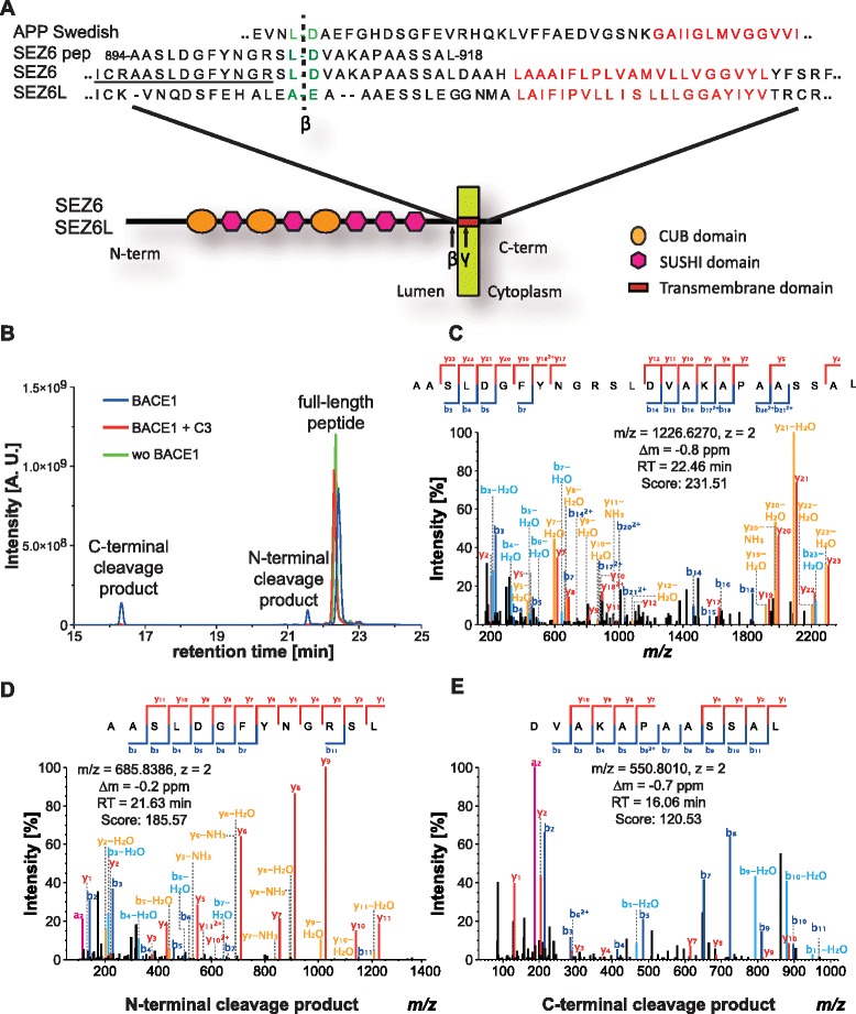
- The BACE1 cleavage site in SEZ6 was mapped to the peptide bond between leucine 906 and aspartate 907, 16 amino acids from the transmembrane domain, similar to SEZ6L (14 amino acids) and with the same P1/P1′ L-D residues as Swedish mutant APP; SEZ6 also accumulated a ~13 kDa C-terminal fragment upon γ-secretase inhibition with DAPT.
- In pancreatic MIN6 cells, SEZ6 and SEZ6L were cleaved by BACE2 rather than BACE1, and BACE1-selective inhibitors affected sSEZ6/sSEZ6L less than Aβ42, following BACE2 inhibition curves; an improved SDC-based CSF digestion protocol reduced average missed cleavages by 58.1% (0.26 vs 0.62), increased protein identifications by 10.3%, quantifications by 7.5%, and quantified membrane proteins from 124 to 135.
Methods
- Rat monoclonal antibodies against murine SEZ6 (clone 14E5) and SEZ6L (clone 21D9) were generated and validated by immunoblot and immunohistochemistry using WT, SEZ6 KO, SEZ6L2 KO, and SEZ6 family triple knockout mouse brains.
- Primary murine neurons (E15/E16), BACE1 KO mouse brains, HEK293T cells stably expressing tagged SEZ6, and MIN6 pancreatic β cells were used with BACE inhibitors (C3, DAPT, nonselective and BACE1-selective compounds), siRNA knockdown, deglycosylation, cell surface biotinylation, and Western blotting.
- BACE1 cleavage site was determined by in vitro digestion of a synthetic SEZ6 peptide with recombinant BACE1 followed by nanoLC-MS/MS; CSF proteomics used 5 µL CSF from seven 4-month-old WT and seven BACE1/BACE2 double knockout mice, SDC-assisted digestion, LC-MS/MS, MaxQuant label-free quantification, Welch’s t-test (p < 0.05), and Benjamini-Hochberg FDR correction.
Limitations
- The paper states that future studies are needed to determine how BACE1 alters SEZ6/SEZ6L function and whether such changes contribute to BACE1-deficient phenotypes, and that sSEZ6/sSEZ6L use as human companion diagnostics requires confirmation in human CSF.
- The paper states that SEZ6L-/- mouse brains were not available, so SEZ6 family triple knockout brains were used instead for SEZ6L antibody specificity.
- Evident from the text: the validation is largely murine and in vitro/cell-line based, with no human CSF data; the CSF proteomic comparison used n = 7 per genotype; and the SEZ6 cleavage site was mapped using a synthetic peptide rather than endogenous full-length protein.
Model-written summary. Check it against the paper before relying on it.
Integrity signals
Registry
Cites
Statistics
Figures
Verification
Claims the paper makes
56 Own result · 30 Own interpretation · 15 Partly supported · 11 Supported · 9 Own method · 7 No full text of the sources · 3 Supported so far · 3 Not checked yet · 1 Mixed support · 1 Couldn’t confirm · 1 Sources not found · 1 No basis given · accuracy not yet measured
Every source the paper cites for this claim was checked and supports it.
In the paper
“In contrast, the two type I membrane proteins SEZ6 and its homolog SEZ6L appeared to be almost exclusively cleaved by BACE1 in neurons [15], making them potential biomarkers for BACE activity in vivo.”
§ Seizure protein 6 and its homolog seizure 6-like protein are physiological substrates of BACE1 in neurons > Background
What the sources say
- Supportedcites [15] Kuhn PH, Koroniak K, Hogl S, Colombo A, Zeitschel U, Willem M, Volbracht C, Schepers U, Imhof A, Hoffmeister A, et al. Secretome protein enrichment identifies physiological BACE1 protease substrates in neurons. Embo J. 2012;31:3157–68. source
The passage labeled “Isolation of primary neurons” (page not supplied) explicitly calls SEZ6L1 and SEZ6 nearly exclusive BACE1 substrates and contrasts them with proteins also cleaved by other proteases. The Discussion identifies SEZ6L1 as a homologue of SEZ6. (LLM, conf=0.94)
hedgeddirect- Asserted: passes — The authors put it forward as true.
- Checkable: passes — It can be checked against evidence.
- Load-bearing: passes — The paper’s conclusions depend on it.
- Based: passes — The paper gives a reason why it holds.
- Supported
Every source the paper cites for this claim was checked and supports it.
In the paper
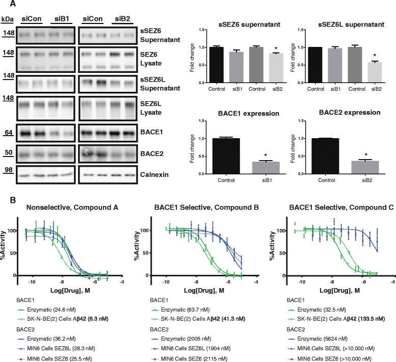
“In agreement with the previous study [17], sSEZ6L was reduced upon knock-down of BACE2, but not of BACE1.”
§ Seizure protein 6 and its homolog seizure 6-like protein are physiological substrates of BACE1 in neurons > Results > BACE1 cleavage of SEZ6 and SEZ6L in pancreatic MIN6 cells
What the sources say
- Supportedcites [17] Stutzer I, Selevsek N, Esterhazy D, Schmidt A, Aebersold R, Stoffel M. Systematic proteomic analysis identifies beta-site amyloid precursor protein cleaving enzyme 2 and 1 (BACE2 and BACE1) substrates in pancreatic beta-cells. J Biol Chem. 2013;288:10536–47. source
The ‘Validation of β- and γ-Secretase Substrates in MIN6 Cells’ passage reports that silencing BACE2, but not BACE1, markedly reduced shed SEZ6L in MIN6 cell supernatants. This directly supports the attributed claim. (LLM, conf=0.95)
plaindirect- Asserted: passes — The authors put it forward as true.
- Checkable: passes — It can be checked against evidence.
- Load-bearing: passes — The paper’s conclusions depend on it.
- Based: passes — The paper gives a reason why it holds.
- Supported
The cited sources support only part of this claim — read each source’s verdict below.
In the paper
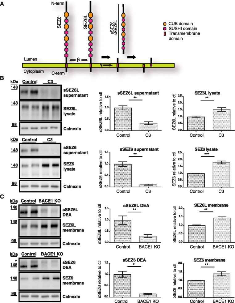
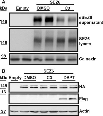
“In agreement with our previous study on SEZ6 [15], similar results were obtained for sSEZ6 and full-length SEZ6 both in C3-treated neurons and in BACE1 KO mouse brains (Fig. 2b and c).”
§ Seizure protein 6 and its homolog seizure 6-like protein are physiological substrates of BACE1 in neurons > Results > BACE1 cleavage of SEZ6 and SEZ6L in primary neurons and mouse brain
What the sources say
- Partly supportedcites [15] Kuhn PH, Koroniak K, Hogl S, Colombo A, Zeitschel U, Willem M, Volbracht C, Schepers U, Imhof A, Hoffmeister A, et al. Secretome protein enrichment identifies physiological BACE1 protease substrates in neurons. Embo J. 2012;31:3157–68. sourceExperimental — accuracy not yet measured
The 'Validation of BACE1 substrates in primary neurons' section reports reduced SEZ6 shedding and increased full-length protein after C3 treatment. The 'Validation of BACE1 substrates in BACE1-deficient mouse brains' section reports reduced SEZ6 shedding in knockout brains, but the retrieved passages do not establish the claimed result for full-length SEZ6 in those brains; the passage showing increased full-length SEZ6 in brain concerns inhibitor-treated mice instead. Page numbers were not available in our copy. (LLM, conf=0.78)
plain- Asserted: passes — The authors put it forward as true.
- Checkable: passes — It can be checked against evidence.
- Load-bearing: passes — The paper’s conclusions depend on it.
- Based: passes — The paper gives a reason why it holds.
- Partly supported
Every source the paper cites for this claim was checked and supports it.
In the paper
“After the proteomic identification of SEZ6 as a BACE1 substrate candidate, SEZ6 was also shown to undergo reduced cleavage in BACE1-/- mouse brains [15].”
§ Seizure protein 6 and its homolog seizure 6-like protein are physiological substrates of BACE1 in neurons > Background
What the sources say
- Supportedcites [15] Kuhn PH, Koroniak K, Hogl S, Colombo A, Zeitschel U, Willem M, Volbracht C, Schepers U, Imhof A, Hoffmeister A, et al. Secretome protein enrichment identifies physiological BACE1 protease substrates in neurons. Embo J. 2012;31:3157–68. source
In “Validation of BACE1 substrates in BACE1-deficient mouse brains,” the paper reports reduced SEZ6 shedding in P7 BACE1-deficient brains and concludes from its P7 and P60 brain analyses that SEZ6 is a physiological BACE1 substrate in vivo. This supports the citing sentence’s description of reduced cleavage in BACE1-/- mouse brains. (LLM, conf=0.94)
plaindirect- Asserted: passes — The authors put it forward as true.
- Checkable: passes — It can be checked against evidence.
- Load-bearing: passes — The paper’s conclusions depend on it.
- Based: passes — The paper gives a reason why it holds.
- Supported
The cited sources support only part of this claim — read each source’s verdict below.
In the paper
“The third family member, SEZ6L2, appeared to be mostly cleaved by proteases other than BACE1 [15, 17].”
§ Seizure protein 6 and its homolog seizure 6-like protein are physiological substrates of BACE1 in neurons > Background
What the sources say
- Supportedcites [15] Kuhn PH, Koroniak K, Hogl S, Colombo A, Zeitschel U, Willem M, Volbracht C, Schepers U, Imhof A, Hoffmeister A, et al. Secretome protein enrichment identifies physiological BACE1 protease substrates in neurons. Embo J. 2012;31:3157–68. source
The Discussion (page not specified in our copy) says SEZ6L2 is a BACE1 substrate but requires BACE1 to a much lower extent for its total shedding than the other SEZ6 family members discussed. That supports the citing sentence’s hedged conclusion that most SEZ6L2 cleavage appeared to involve other proteases. (LLM, conf=0.88)
- Partly supportedcites [17] Stutzer I, Selevsek N, Esterhazy D, Schmidt A, Aebersold R, Stoffel M. Systematic proteomic analysis identifies beta-site amyloid precursor protein cleaving enzyme 2 and 1 (BACE2 and BACE1) substrates in pancreatic beta-cells. J Biol Chem. 2013;288:10536–47. sourceExperimental — accuracy not yet measured
The validation results identify BACE2, rather than BACE1, as the rate-limiting protease for SEZ6L2 shedding in pancreatic islets. The Discussion describes SEZ6L2 as a BACE2-specific substrate in pancreatic islets but notes that cleavage of the seizure 6 protein family can differ by tissue. The unqualified claim extends beyond the pancreatic-cell setting studied here. (LLM, conf=0.86)
hedgeddirect- Asserted: passes — The authors put it forward as true.
- Checkable: passes — It can be checked against evidence.
- Load-bearing: unclear — It is unclear whether the paper’s conclusions depend on it.
- Based: passes — The paper gives a reason why it holds.
- Supported
Every source the paper cites for this claim was checked and supports it.
In the paper
“In the previous proteomic study which identified SEZ6 as a BACE1 substrate candidate, several tryptic peptides of the secreted SEZ6 ectodomain were identified.”
§ Seizure protein 6 and its homolog seizure 6-like protein are physiological substrates of BACE1 in neurons > Results > BACE1 cleaves SEZ6 within its juxtamembrane domain
What the sources say
- Supportedcites [15] Kuhn PH, Koroniak K, Hogl S, Colombo A, Zeitschel U, Willem M, Volbracht C, Schepers U, Imhof A, Hoffmeister A, et al. Secretome protein enrichment identifies physiological BACE1 protease substrates in neurons. Embo J. 2012;31:3157–68. source
The Discussion identifies SEZ6 as a top BACE1 substrate candidate and refers to an identified SEZ6 peptide. The sheddome results state that identified membrane-protein peptides came from ectodomains; the methods describe trypsin digestion, and the secretome analysis included only proteins detected by at least two peptides. Together, these support the claim about several tryptic peptides from the shed SEZ6 ectodomain. (LLM, conf=0.90)
plain- Asserted: passes — The authors put it forward as true.
- Checkable: passes — It can be checked against evidence.
- Load-bearing: passes — The paper’s conclusions depend on it.
- Based: passes — The paper gives a reason why it holds.
- Supported
The cited sources support only part of this claim — read each source’s verdict below.
In the paper
“The most C-terminal of these peptides encompassed amino acids 894 to 904 (AASLDGFYNGR) of murine SEZ6 (Fig. 5a).”
§ Seizure protein 6 and its homolog seizure 6-like protein are physiological substrates of BACE1 in neurons > Results > BACE1 cleaves SEZ6 within its juxtamembrane domain
What the sources say
- Partly supportedcites [15] Kuhn PH, Koroniak K, Hogl S, Colombo A, Zeitschel U, Willem M, Volbracht C, Schepers U, Imhof A, Hoffmeister A, et al. Secretome protein enrichment identifies physiological BACE1 protease substrates in neurons. Embo J. 2012;31:3157–68. sourceExperimental — accuracy not yet measured
The Discussion (page unavailable) identifies a most C-terminal SEZ6 peptide and places it before the transmembrane domain, supporting the peptide’s role in the claim. Our copy does not give its amino-acid range or the sequence AASLDGFYNGR, so those specifics cannot be verified. (LLM, conf=0.86)
plain- Asserted: passes — The authors put it forward as true.
- Checkable: passes — It can be checked against evidence.
- Load-bearing: passes — The paper’s conclusions depend on it.
- Based: passes — The paper gives a reason why it holds.
- Partly supported
Every source the paper cites for this claim was checked and supports it.
In the paper
“The accumulation of C-terminal fragments upon pharmacological inhibition of γ-secretase with DAPT can be used to identify γ-secretase substrates [42].”
§ Seizure protein 6 and its homolog seizure 6-like protein are physiological substrates of BACE1 in neurons > Results > SEZ6 is a substrate for γ-secretase
What the sources say
- Supportedcites [42] Hemming ML, Elias JE, Gygi SP, Selkoe DJ. Proteomic profiling of gamma-secretase substrates and mapping of substrate requirements. PLoS Biol. 2008;6:e257. source
The Results section, under “Identification of γ-Secretase Substrates Using Quantitative Proteomics and γ-Secretase Inhibition” (Figure 1 and accompanying text), describes using DAPT-induced accumulation of C-terminal fragments as the spectral pattern for identifying substrates. It also reports that DAPT increased levels of the APP C-terminal fragment, a known γ-secretase substrate. (LLM, conf=0.94)
plaindirect- Asserted: passes — The authors put it forward as true.
- Checkable: passes — It can be checked against evidence.
- Load-bearing: passes — The paper’s conclusions depend on it.
- Based: passes — The paper gives a reason why it holds.
- Supported
Every source the paper cites for this claim was checked and supports it.
In the paper
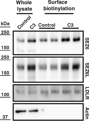
“As a control, surface levels of the LDL-receptor (LDLR), which is a substrate of ADAM10, but not of BACE1 [38], were not altered upon BACE inhibition.”
§ Seizure protein 6 and its homolog seizure 6-like protein are physiological substrates of BACE1 in neurons > Results > BACE1 inhibition increases neuronal cell surface levels of SEZ6 and SEZ6L
What the sources say
- Supportedcites [38] Kuhn PH, Colombo AV, Schusser B, Dreymueller D, Wetzel S, Schepers U, Herber J, Ludwig A, Kremmer E, Montag D, et al. Systematic substrate identification indicates a central role for the metalloprotease ADAM10 in axon targeting and synapse function. Elife. 2016; 5, doi: 10.7554/eLife.12748 . source
The citation supports the sentence’s identification of LDLR as an ADAM10 substrate, but not a BACE1 substrate. In Results (‘Cross-talk between ADAM10 and BACE1’), LDLR ectodomain levels fell upon Adam10 deletion, while BACE1 inhibition had no additional effect. The citing authors’ observation about surface levels is not itself attributed to this source. (LLM, conf=0.91)
plain- Asserted: passes — The authors put it forward as true.
- Checkable: passes — It can be checked against evidence.
- Load-bearing: unclear — It is unclear whether the paper’s conclusions depend on it.
- Based: passes — The paper gives a reason why it holds.
- Supported
Every source the paper cites for this claim was checked and supports it.
In the paper
“A previous study reported that in pancreatic cells SEZ6L is predominantly cleaved by BACE2, but not by BACE1 [17].”
§ Seizure protein 6 and its homolog seizure 6-like protein are physiological substrates of BACE1 in neurons > Discussion
What the sources say
- Supportedcites [17] Stutzer I, Selevsek N, Esterhazy D, Schmidt A, Aebersold R, Stoffel M. Systematic proteomic analysis identifies beta-site amyloid precursor protein cleaving enzyme 2 and 1 (BACE2 and BACE1) substrates in pancreatic beta-cells. J Biol Chem. 2013;288:10536–47. source
The section “Validation and Characterization of SEZ6L and SEZ6L2 as Specific BACE2 Substrates in Pancreatic β-Cells” reports that loss of BACE2 nearly eliminated SEZ6L shedding from pancreatic islets, while loss of BACE1 did not. The Discussion likewise identifies SEZ6L as a BACE2-specific substrate in pancreatic islets. Page numbers are not provided in our copy. (LLM, conf=0.94)
plaindirect- Asserted: passes — The authors put it forward as true.
- Checkable: passes — It can be checked against evidence.
- Load-bearing: passes — The paper’s conclusions depend on it.
- Based: passes — The paper gives a reason why it holds.
- Supported
- Probably not a substantial claim. 36
- It may state two separate claims as one. 38
- The quote only partly states this claim. 3
Some cited sources support this claim and some don’t — read each source’s verdict below.
In the paper
“because BACE1 also negatively regulates surface levels and/or function of two other substrates, contactin-2 and CHL1 [12, 15, 53].”
§ Seizure protein 6 and its homolog seizure 6-like protein are physiological substrates of BACE1 in neurons > Discussion
What the sources say
- Disputedcites [12] Barao S, Gartner A, Leyva-Diaz E, Demyanenko G, Munck S, Vanhoutvin T, Zhou L, Schachner M, Lopez-Bendito G, Maness PF, De Strooper B. Antagonistic effects of BACE1 and APH1B-gamma-secretase control axonal guidance by regulating growth cone collapse. Cell Rep. 2015;12:1367–76. sourceExperimental — accuracy not yet measured
The Discussion (page unavailable) says BACE1 cleavage of CHL1 is necessary for Sema3A-induced growth cone collapse, and that the resulting CHL1 fragment transmits the signal. That is contrary to the claim that BACE1 negatively regulates CHL1 function. Our copy does not address contactin-2 or establish a reduction in surface levels. (LLM, conf=0.84)
Checked by a reasoning model — may be wrong.
- Partly supportedcites [15] Kuhn PH, Koroniak K, Hogl S, Colombo A, Zeitschel U, Willem M, Volbracht C, Schepers U, Imhof A, Hoffmeister A, et al. Secretome protein enrichment identifies physiological BACE1 protease substrates in neurons. Embo J. 2012;31:3157–68. sourceExperimental — accuracy not yet measured
The section “BACE1 inhibition increases cell surface levels of contactin-2” directly states that BACE1 negatively regulates contactin-2 at the neuronal plasma membrane. “Validation of BACE1 substrates in primary neurons” reports reduced CHL1 shedding and increased full-length protein in cell lysates after BACE1 inhibition, but does not establish reduced CHL1 surface levels or function. (LLM, conf=0.86)
- Supportedcites [53] Gautam V, D’Avanzo C, Hebisch M, Kovacs DM, Kim DY. BACE1 activity regulates cell surface contactin-2 levels. Mol Neurodegener. 2014;9:4. source
The Results and discussion section (Figure 5) reports that BACE1 overexpression decreased surface contactin-2 in mouse primary neurons, while BACE1 inhibition increased it. This supports the contactin-2 part attributed to this paper; the same section identifies Hitt et al. as the source of the CHL1 finding, which the citing sentence supports with multiple citations. (LLM, conf=0.91)
plaindirect- Asserted: passes — The authors put it forward as true.
- Checkable: passes — It can be checked against evidence.
- Load-bearing: fails — The paper’s conclusions may not depend on it.
- Based: passes — The paper gives a reason why it holds.
Probably not a substantial claim. The paper’s conclusions may not depend on it.
- Disputed
The sources with a verdict support this claim; the rest weren’t checked yet, have no full text we could read, or their verdicts aren’t rated yet.
In the paper
“BACE1 cleaves the amyloid precursor protein (APP) and thus catalyzes the first step in generation of the amyloid β peptide (Aβ) [2–5]”
§ Seizure protein 6 and its homolog seizure 6-like protein are physiological substrates of BACE1 in neurons > Background
What the sources say
- Not checked — no full text of the sourcecites [2] Hussain I, Powell D, Howlett DR, Tew DG, Meek TD, Chapman C, Gloger IS, Murphy KE, Southan CD, Ryan DM, et al. Identification of a novel aspartic protease (Asp 2) as beta-secretase. Mol Cell Neurosci. 1999;14:419–27. source
- Not checked — no full text of the sourcecites [3] Sinha S, Anderson JP, Barbour R, Basi GS, Caccavello R, Davis D, Doan M, Dovey HF, Frigon N, Hong J, et al. Purification and cloning of amyloid precursor protein beta-secretase from human brain. Nature. 1999;402:537–40. source
- Supportedcites [4] Vassar R, Bennett BD, Babu-Khan S, Kahn S, Mendiaz EA, Denis P, Teplow DB, Ross S, Amarante P, Loeloff R, et al. Beta-secretase cleavage of Alzheimer’s amyloid precursor protein by the transmembrane aspartic protease BACE. Science. 1999;286:735–41. source
The introductory passage indexed under Abstract (page not provided) states that β-secretase cleaves APP to initiate Aβ formation, before further cleavage by γ-secretase. The purified-BACE section shows that BACE directly cleaves APP-derived substrates at the β-secretase site. (LLM, conf=0.94)
- Not checked — no full text of the sourcecites [5] Yan R, Bienkowski MJ, Shuck ME, Miao H, Tory MC, Pauley AM, Brashier JR, Stratman NC, Mathews WR, Buhl AE, et al. Membrane-anchored aspartyl protease with Alzheimer’s disease beta-secretase activity. Nature. 1999;402:533–7. source
plaindirect- Asserted: passes — The authors put it forward as true.
- Checkable: passes — It can be checked against evidence.
- Load-bearing: fails — The paper’s conclusions may not depend on it.
- Based: passes — The paper gives a reason why it holds.
Probably not a substantial claim. The paper’s conclusions may not depend on it. It may state two separate claims as one.
The cited sources support only part of this claim — read each source’s verdict below.
In the paper
“The three members of the seizure protein 6 (SEZ6) family, namely SEZ6, SEZ6-like (SEZ6L) and SEZ6-like 2 (SEZ6L2) have been identified as candidate BACE1 substrates in different studies [15, 17], but have not yet been validated in detail.”
§ Seizure protein 6 and its homolog seizure 6-like protein are physiological substrates of BACE1 in neurons > Background
What the sources say
- Partly supportedcites [15] Kuhn PH, Koroniak K, Hogl S, Colombo A, Zeitschel U, Willem M, Volbracht C, Schepers U, Imhof A, Hoffmeister A, et al. Secretome protein enrichment identifies physiological BACE1 protease substrates in neurons. Embo J. 2012;31:3157–68. sourceExperimental — accuracy not yet measured
Table I and the Discussion identify SEZ6, SEZ6L1 and SEZ6L2 as BACE1 substrate candidates, with SEZ6L2 showing less dependence on BACE1 for shedding. However, the sections on validation in primary neurons and BACE1-deficient mouse brains report further validation of SEZ6. The claim that the family members had not yet been validated in detail therefore does not hold for SEZ6 in this source. (LLM, conf=0.87)
- Partly supportedcites [17] Stutzer I, Selevsek N, Esterhazy D, Schmidt A, Aebersold R, Stoffel M. Systematic proteomic analysis identifies beta-site amyloid precursor protein cleaving enzyme 2 and 1 (BACE2 and BACE1) substrates in pancreatic beta-cells. J Biol Chem. 2013;288:10536–47. sourceExperimental — accuracy not yet measured
The Discussion (page unavailable) reports that the SEZ6 family had been shown to be cleaved by BACE1 in primary neurons. But this paper does not identify all three named proteins as candidate BACE1 substrates: it investigates SEZ6L and SEZ6L2 and validates them as BACE2-specific substrates in pancreatic islet β-cells. It also does not establish the claim that the BACE1 candidates had not been validated in detail. (LLM, conf=0.86)
plaincorrelational- Asserted: passes — The authors put it forward as true.
- Checkable: passes — It can be checked against evidence.
- Load-bearing: passes — The paper’s conclusions depend on it.
- Based: passes — The paper gives a reason why it holds.
It may state two separate claims as one.
- Partly supported
The cited sources support only part of this claim — read each source’s verdict below.
In the paper
“The SEZ6 family controls synaptic connectivity and motor coordination in mice [18, 19], but little is known about the functions of these proteins at the molecular level.”
§ Seizure protein 6 and its homolog seizure 6-like protein are physiological substrates of BACE1 in neurons > Background
What the sources say
- Partly supportedcites [18] Gunnersen JM, Kim MH, Fuller SJ, De Silva M, Britto JM, Hammond VE, Davies PJ, Petrou S, Faber ES, Sah P, Tan SS. Sez-6 proteins affect dendritic arborization patterns and excitability of cortical pyramidal neurons. Neuron. 2007;56:621–39. sourceExperimental — accuracy not yet measured
The Abstract reports fewer excitatory synapses and impaired motor coordination in Sez-6-null mice, supporting both effects. This paper studies Sez-6 and its isoforms, however, rather than establishing that the SEZ6 family as a whole controls these outcomes. (LLM, conf=0.86)
- Not checked — no full text of the sourcecites [19] Miyazaki T, Hashimoto K, Uda A, Sakagami H, Nakamura Y, Saito SY, Nishi M, Kume H, Tohgo A, Kaneko I, et al. Disturbance of cerebellar synaptic maturation in mutant mice lacking BSRPs, a novel brain-specific receptor-like protein family. FEBS Lett. 2006;580:4057–64. source
plaindirect- Asserted: passes — The authors put it forward as true.
- Checkable: passes — It can be checked against evidence.
- Load-bearing: fails — The paper’s conclusions may not depend on it.
- Based: passes — The paper gives a reason why it holds.
Probably not a substantial claim. The paper’s conclusions may not depend on it. It may state two separate claims as one.
- Partly supported
Every source the paper cites for this claim was checked and supports it.
In the paper
“The protease BACE1 (beta-site APP cleaving enzyme) is a major drug target in Alzheimer’s disease.”
- Also stated in: § Background · plain
“The β-secretase BACE1 (β-site APP cleaving enzyme) is a key drug target in Alzheimer’s disease (AD) [1].”
§ Seizure protein 6 and its homolog seizure 6-like protein are physiological substrates of BACE1 in neurons > Abstract > Background
What the sources say
- Supportedcites [1] Vassar R, Kuhn PH, Haass C, Kennedy ME, Rajendran L, Wong PC, Lichtenthaler SF. Function, therapeutic potential and cell biology of BACE proteases: current status and future prospects. J Neurochem. 2014;130:4–28. source
The Outlook section says BACE1 has taken center stage in Alzheimer’s disease drug-development programs and that around a dozen companies have BACE inhibitors in clinical trials. This supports calling BACE1 a major drug target. (LLM, conf=0.94)
plain- Asserted: passes — The authors put it forward as true.
- Checkable: passes — It can be checked against evidence.
- Load-bearing: fails — The paper’s conclusions may not depend on it.
- Based: passes — The paper gives a reason why it holds.
Probably not a substantial claim. The paper’s conclusions may not depend on it.
- Also stated in: § Background · plain
The cited sources support only part of this claim — read each source’s verdict below.
In the paper
“More than 40 substrate candidates for BACE1 were identified in recent proteomic studies in murine neurons or cerebrospinal fluid (CSF), but only a few of them have been validated to date with functional or in vitro assays, including L1, CHL1, ENPP5 and PTPRN2 [12, 14–16].”
§ Seizure protein 6 and its homolog seizure 6-like protein are physiological substrates of BACE1 in neurons > Background
What the sources say
- Partly supportedcites [12] Barao S, Gartner A, Leyva-Diaz E, Demyanenko G, Munck S, Vanhoutvin T, Zhou L, Schachner M, Lopez-Bendito G, Maness PF, De Strooper B. Antagonistic effects of BACE1 and APH1B-gamma-secretase control axonal guidance by regulating growth cone collapse. Cell Rep. 2015;12:1367–76. sourceExperimental — accuracy not yet measured
The Introduction (page unavailable) reports a long list of potential BACE1 substrates, and the paper investigates the functional consequences of BACE1 processing of CHL1. It does not give the figure of more than 40, specify proteomic studies of cerebrospinal fluid, or address validation of ENPP5 and PTPRN2. (LLM, conf=0.85)
- Not checked — no full text of the sourcecites [14] Dislich B, Wohlrab F, Bachhuber T, Müller SA, Kuhn P-H, Hogl S, Meyer-Luehmann M, Lichtenthaler SF. Label-free quantitative proteomics of mouse cerebrospinal fluid detects β-Site APP Cleaving Enzyme (BACE1) protease substrates in vivo. Mol Cell Proteomics. 2015;14:2550–63. source
- Partly supportedcites [15] Kuhn PH, Koroniak K, Hogl S, Colombo A, Zeitschel U, Willem M, Volbracht C, Schepers U, Imhof A, Hoffmeister A, et al. Secretome protein enrichment identifies physiological BACE1 protease substrates in neurons. Embo J. 2012;31:3157–68. sourceExperimental — accuracy not yet measured
The ‘Identification of neuronal BACE1 sheddome’ section reports 34 candidate membrane-protein substrates in mouse neurons, and the validation sections confirm L1 and CHL1 as physiological substrates. This paper alone does not establish a total of more than 40 substrate candidates across neuronal and CSF studies, or validate ENPP5 and PTPRN2. (LLM, conf=0.85)
- Not checked — no full text of the sourcecites [16] Zhou L, Barao S, Laga M, Bockstael K, Borgers M, Gijsen H, Annaert W, Moechars D, Mercken M, Gevaert K, De Strooper B. The neural cell adhesion molecules L1 and CHL1 are cleaved by BACE1 protease in vivo. J Biol Chem. 2012;287:25927–40. source
plain- Asserted: passes — The authors put it forward as true.
- Checkable: passes — It can be checked against evidence.
- Load-bearing: fails — The paper’s conclusions may not depend on it.
- Based: passes — The paper gives a reason why it holds.
Probably not a substantial claim. The paper’s conclusions may not depend on it. It may state two separate claims as one.
- Partly supported
The cited sources support only part of this claim — read each source’s verdict below.
In the paper
“BACE1 is highly expressed in the nervous system and contributes to additional physiological processes besides its role in AD, e.g. through neuregulin-1 cleavage in myelination and CHL1 cleavage in axon targeting [7–12].”
§ Seizure protein 6 and its homolog seizure 6-like protein are physiological substrates of BACE1 in neurons > Background
What the sources say
- Not checked — no full text of the sourcecites [7] Willem M, Garratt AN, Novak B, Citron M, Kaufmann S, Rittger A, DeStrooper B, Saftig P, Birchmeier C, Haass C. Control of peripheral nerve myelination by the beta-secretase BACE1. Science. 2006;314:664–6. source
- Not checked — no full text of the sourcecites [8] Hu X, Hicks CW, He W, Wong P, Macklin WB, Trapp BD, Yan R. Bace1 modulates myelination in the central and peripheral nervous system. Nat Neurosci. 2006;9:1520–5. source
- Partly supportedcites [9] Fleck D, van Bebber F, Colombo A, Galante C, Schwenk BM, Rabe L, Hampel H, Novak B, Kremmer E, Tahirovic S, et al. Dual cleavage of neuregulin 1 type III by BACE1 and ADAM17 liberates its EGF-like domain and allows paracrine signaling. J Neurosci. 2013;33:7856–69. sourceExperimental — accuracy not yet measured
The Introduction says BACE1 processes neuregulin-1 type III, a regulator of peripheral nervous system myelination, and the Discussion reports that a BACE1-liberated neuregulin domain promotes myelination in zebrafish. The passages do not support the sentence’s separate claims that BACE1 is highly expressed in the nervous system or that CHL1 cleavage contributes to axon targeting. (LLM, conf=0.86)
- Partly supportedcites [10] Cheret C, Willem M, Fricker FR, Wende H, Wulf-Goldenberg A, Tahirovic S, Nave KA, Saftig P, Haass C, Garratt AN, et al. Bace1 and Neuregulin-1 cooperate to control formation and maintenance of muscle spindles. Embo J. 2013;32:2015–28. sourceExperimental — accuracy not yet measured
The Introduction reports that Bace1-dependent shedding of neuregulin-1 is implicated in peripheral nervous system myelination, and the Results say Bace1 is broadly expressed in sensory neurons. The copy does not establish high expression throughout the nervous system. Its References list a study of CHL1 cleavage by Bace1, but no passage links that cleavage to axon targeting. (LLM, conf=0.88)
- Supportedcites [11] Hitt B, Riordan SM, Kukreja L, Eimer WA, Rajapaksha TW, Vassar R. beta-Site amyloid precursor protein (APP)-cleaving enzyme 1 (BACE1)-deficient mice exhibit a close homolog of L1 (CHL1) loss-of-function phenotype involving axon guidance defects. J Biol Chem. 2012;287:38408–25. source
The Introduction reports robust BACE1 expression in hippocampal mossy-fiber terminals and describes reduced neuregulin-1 cleavage as a cause of hypomyelination. The Abstract and Introduction report BACE1-dependent CHL1 processing in the hippocampus and olfactory bulb and link its loss to axon-guidance defects. Together, these passages support the cited examples of nervous-system functions beyond BACE1’s role in Alzheimer disease. (LLM, conf=0.86)
- Partly supportedcites [12] Barao S, Gartner A, Leyva-Diaz E, Demyanenko G, Munck S, Vanhoutvin T, Zhou L, Schachner M, Lopez-Bendito G, Maness PF, De Strooper B. Antagonistic effects of BACE1 and APH1B-gamma-secretase control axonal guidance by regulating growth cone collapse. Cell Rep. 2015;12:1367–76. sourceExperimental — accuracy not yet measured
The Introduction (page unavailable) identifies neuregulin-1 type III as a BACE1 substrate implicated in myelination, and the Discussion reports that BACE1 cleavage of CHL1 is necessary for growth-cone collapse during axonal guidance. The copy does not establish that BACE1 is highly expressed in the nervous system. The Introduction also says the precise link between neuregulin-1 processing and its function is unknown, so the claim's cleavage-to-myelination mechanism is more definite than the source supports. (LLM, conf=0.87)
plaindirect- Asserted: passes — The authors put it forward as true.
- Checkable: passes — It can be checked against evidence.
- Load-bearing: fails — The paper’s conclusions may not depend on it.
- Based: passes — The paper gives a reason why it holds.
Probably not a substantial claim. The paper’s conclusions may not depend on it. It may state two separate claims as one.
Every source the paper cites for this claim was checked and supports it.
In the paper
“However, the proteomic data for SEZ6L have not been validated by other methods and another proteomic study using pancreatic cells and tissue failed to confirm SEZ6L as a BACE1 substrate. Instead, that study demonstrated that SEZ6L is cleaved by the BACE1-homolog BACE2 in pancreas [17].”
§ Seizure protein 6 and its homolog seizure 6-like protein are physiological substrates of BACE1 in neurons > Background
What the sources say
- Supportedcites [17] Stutzer I, Selevsek N, Esterhazy D, Schmidt A, Aebersold R, Stoffel M. Systematic proteomic analysis identifies beta-site amyloid precursor protein cleaving enzyme 2 and 1 (BACE2 and BACE1) substrates in pancreatic beta-cells. J Biol Chem. 2013;288:10536–47. source
In “Validation and Characterization of SEZ6L and SEZ6L2 as Specific BACE2 Substrates in Pancreatic β-Cells” (page not provided), SEZ6L shedding persisted in BACE1-deficient islets but was nearly undetectable in BACE2-deficient islets. The same section concludes that BACE2 is sufficient and required for SEZ6L cleavage in pancreatic β-cells, supporting the cited sentence’s pancreas-specific contrast. (LLM, conf=0.93)
boosteddirect- Asserted: passes — The authors put it forward as true.
- Checkable: passes — It can be checked against evidence.
- Load-bearing: passes — The paper’s conclusions depend on it.
- Based: passes — The paper gives a reason why it holds.
It may state two separate claims as one.
- Supported
Every source the paper cites for this claim was checked and supports it.
In the paper
“which is very efficiently cleaved by BACE1 [4].”
§ Seizure protein 6 and its homolog seizure 6-like protein are physiological substrates of BACE1 in neurons > Results > BACE1 cleaves SEZ6 within its juxtamembrane domain
What the sources say
- Supportedcites [4] Vassar R, Bennett BD, Babu-Khan S, Kahn S, Mendiaz EA, Denis P, Teplow DB, Ross S, Amarante P, Loeloff R, et al. Beta-secretase cleavage of Alzheimer’s amyloid precursor protein by the transmembrane aspartic protease BACE. Science. 1999;286:735–41. source
The section “Purified BACE cleaves APP substrates and exhibits the properties of β-secretase” reports that purified BACE cleaves a Swedish-mutant APP peptide with much higher specific activity than a wild-type APP peptide. The cell experiments also report increased β-secretase cleavage of Swedish-mutant APP when BACE is overexpressed. The supplied passages have no page numbers. (LLM, conf=0.93)
boosteddirect- Asserted: passes — The authors put it forward as true.
- Checkable: passes — It can be checked against evidence.
- Load-bearing: fails — The paper’s conclusions may not depend on it.
- Based: passes — The paper gives a reason why it holds.
Probably not a substantial claim. The paper’s conclusions may not depend on it.
- Supported
The cited sources support only part of this claim — read each source’s verdict below.
In the paper
“The previously identified cleavage site in SEZ6L [15] is not identical, but similar to SEZ6, as it also has a hydrophobic amino acid in the P1 and a negatively charged amino acid in the P1’ position (Fig. 5a).”
§ Seizure protein 6 and its homolog seizure 6-like protein are physiological substrates of BACE1 in neurons > Results > BACE1 cleaves SEZ6 within its juxtamembrane domain
What the sources say
- Partly supportedcites [15] Kuhn PH, Koroniak K, Hogl S, Colombo A, Zeitschel U, Willem M, Volbracht C, Schepers U, Imhof A, Hoffmeister A, et al. Secretome protein enrichment identifies physiological BACE1 protease substrates in neurons. Embo J. 2012;31:3157–68. sourceExperimental — accuracy not yet measured
The Identification of neuronal BACE1 sheddome section reports a SEZ6L1 semi-tryptic peptide consistent with direct BACE1 cleavage. The Discussion proposes a leucine–aspartate cleavage site for SEZ6, but our copy does not give the amino acids flanking the SEZ6L1 site or establish the claimed similarity between the two sites. (LLM, conf=0.82)
plaincorrelational- Asserted: passes — The authors put it forward as true.
- Checkable: passes — It can be checked against evidence.
- Load-bearing: unclear — It is unclear whether the paper’s conclusions depend on it.
- Based: passes — The paper gives a reason why it holds.
It may state two separate claims as one.
- Partly supported
The cited sources support only part of this claim — read each source’s verdict below.
In the paper
“Recent proteomic studies have identified more than 40 membrane proteins as potential BACE1 substrates [14–16].”
§ Seizure protein 6 and its homolog seizure 6-like protein are physiological substrates of BACE1 in neurons > Discussion
What the sources say
- Not checked — no full text of the sourcecites [14] Dislich B, Wohlrab F, Bachhuber T, Müller SA, Kuhn P-H, Hogl S, Meyer-Luehmann M, Lichtenthaler SF. Label-free quantitative proteomics of mouse cerebrospinal fluid detects β-Site APP Cleaving Enzyme (BACE1) protease substrates in vivo. Mol Cell Proteomics. 2015;14:2550–63. source
- Partly supportedcites [15] Kuhn PH, Koroniak K, Hogl S, Colombo A, Zeitschel U, Willem M, Volbracht C, Schepers U, Imhof A, Hoffmeister A, et al. Secretome protein enrichment identifies physiological BACE1 protease substrates in neurons. Embo J. 2012;31:3157–68. sourceExperimental — accuracy not yet measured
The 'Identification of neuronal BACE1 sheddome' section reports 34 membrane proteins among 40 proteins whose shedding or secretion was reduced. It supports the identification of potential BACE1 substrates, but does not establish the citing sentence’s total of more than 40 membrane proteins; six of its 40 proteins were soluble. (LLM, conf=0.87)
- Not checked — no full text of the sourcecites [16] Zhou L, Barao S, Laga M, Bockstael K, Borgers M, Gijsen H, Annaert W, Moechars D, Mercken M, Gevaert K, De Strooper B. The neural cell adhesion molecules L1 and CHL1 are cleaved by BACE1 protease in vivo. J Biol Chem. 2012;287:25927–40. source
plain- Asserted: passes — The authors put it forward as true.
- Checkable: passes — It can be checked against evidence.
- Load-bearing: fails — The paper’s conclusions may not depend on it.
- Based: passes — The paper gives a reason why it holds.
Probably not a substantial claim. The paper’s conclusions may not depend on it.
The sources with a verdict support this claim; the rest weren’t checked yet, have no full text we could read, or their verdicts aren’t rated yet.
In the paper
“BACE1 is a major drug target in AD, but has additional substrates and thus contributes to various biological processes [1, 13], which may limit its therapeutic potential.”
§ Seizure protein 6 and its homolog seizure 6-like protein are physiological substrates of BACE1 in neurons > Discussion
What the sources say
- Supportedcites [1] Vassar R, Kuhn PH, Haass C, Kennedy ME, Rajendran L, Wong PC, Lichtenthaler SF. Function, therapeutic potential and cell biology of BACE proteases: current status and future prospects. J Neurochem. 2014;130:4–28. source
The Outlook identifies BACE1 as a focus of AD drug development and warns that inhibiting its other functions could cause side effects. The section on therapeutic potential says that newly identified substrates and biological functions have raised safety concerns about chronic BACE inhibition. This supports the claim’s qualified statement that those functions may limit its therapeutic potential. (LLM, conf=0.94)
- Not checked — no full text of the sourcecites [13] Barao S, Moechars D, Lichtenthaler SF, De Strooper B. BACE1 physiological functions may limit its use as therapeutic target for alzheimer’s disease. Trends Neurosci. 2016;39:158–69. source
hedgedconditional- Asserted: passes — The authors put it forward as true.
- Checkable: passes — It can be checked against evidence.
- Load-bearing: passes — The paper’s conclusions depend on it.
- Based: passes — The paper gives a reason why it holds.
It may state two separate claims as one.
- Supported
The cited sources support only part of this claim — read each source’s verdict below.
In the paper
“Notably, both BACE1- and SEZ6-deficient mice have deficits in hippocampal learning paradigms [18, 54–56] and in motor coordination [18, 57].”
§ Seizure protein 6 and its homolog seizure 6-like protein are physiological substrates of BACE1 in neurons > Discussion
What the sources say
- Not checked yetcites [18] Gunnersen JM, Kim MH, Fuller SJ, De Silva M, Britto JM, Hammond VE, Davies PJ, Petrou S, Faber ES, Sah P, Tan SS. Sez-6 proteins affect dendritic arborization patterns and excitability of cortical pyramidal neurons. Neuron. 2007;56:621–39. source
Checked before — its earlier verdict is held back until a new check runs.
- Not checked — no full text of the sourcecites [54] Ohno M, Sametsky EA, Younkin LH, Oakley H, Younkin SG, Citron M, Vassar R, Disterhoft JF. BACE1 deficiency rescues memory deficits and cholinergic dysfunction in a mouse model of Alzheimer’s disease. Neuron. 2004;41:27–33. source
- Not checked — no full text of the sourcecites [55] Ohno M, Chang L, Tseng W, Oakley H, Citron M, Klein WL, Vassar R, Disterhoft JF. Temporal memory deficits in Alzheimer’s mouse models: rescue by genetic deletion of BACE1. Eur J Neurosci. 2006;23:251–60. source
- Partly supportedcites [56] Laird FM, Cai H, Savonenko AV, Farah MH, He K, Melnikova T, Wen H, Chiang HC, Xu G, Koliatsos VE, et al. BACE1, a major determinant of selective vulnerability of the brain to amyloid-beta amyloidogenesis, is essential for cognitive, emotional, and synaptic functions. J Neurosci. 2005;25:11693–709. sourceExperimental — accuracy not yet measured
The section “BACE1 is critical for cognitive and emotional functions” reports deficits in hippocampus-dependent tasks in BACE1-deficient mice. The section on APP and PS1 transgenes reports slower swimming, but does not establish a deficit in motor coordination. This paper also does not study SEZ6-deficient mice; the sentence cites other sources for that part. (LLM, conf=0.85)
- Not checked — no full text of the sourcecites [57] Kobayashi D, Zeller M, Cole T, Buttini M, McConlogue L, Sinha S, Freedman S, Morris RG, Chen KS. BACE1 gene deletion: impact on behavioral function in a model of Alzheimer’s disease. Neurobiol Aging. 2008;29:861–73. source
plaindirect- Asserted: passes — The authors put it forward as true.
- Checkable: passes — It can be checked against evidence.
- Load-bearing: passes — The paper’s conclusions depend on it.
- Based: passes — The paper gives a reason why it holds.
It may state two separate claims as one.
- Not checked yet
The cited sources support only part of this claim — read each source’s verdict below.
In the paper
“Several other previously identified BACE1 substrates, such as CHL1, L1, contactin-2, APP and its homolog APLP2, are not exclusively cleaved by BACE1, but also by other proteases, including ADAM10 [15, 38].”
§ Seizure protein 6 and its homolog seizure 6-like protein are physiological substrates of BACE1 in neurons > Discussion
What the sources say
- Supportedcites [15] Kuhn PH, Koroniak K, Hogl S, Colombo A, Zeitschel U, Willem M, Volbracht C, Schepers U, Imhof A, Hoffmeister A, et al. Secretome protein enrichment identifies physiological BACE1 protease substrates in neurons. Embo J. 2012;31:3157–68. source
The Discussion says that, apart from a few nearly exclusive BACE1 substrates, the others are cleaved to varying degrees by additional proteases. The paper reports partial BACE1-dependent shedding of CHL1, L1 and contactin-2, identifies ADAM10 cleavage of APLP2 and L1, and describes APP cleavage by distinct proteases. Page numbers are not available in our copy. (LLM, conf=0.91)
- Partly supportedcites [38] Kuhn PH, Colombo AV, Schusser B, Dreymueller D, Wetzel S, Schepers U, Herber J, Ludwig A, Kremmer E, Montag D, et al. Systematic substrate identification indicates a central role for the metalloprotease ADAM10 in axon targeting and synapse function. Elife. 2016; 5, doi: 10.7554/eLife.12748 . sourceExperimental — accuracy not yet measured
The Results section (“Cross-talk between ADAM10 and BACE1”; page unavailable) reports cleavage of CHL1, L1, contactin-2 (CNTN2), and APP by both ADAM10 and BACE1. It does not establish the same point for APLP2; the APLP2 mention found in our copy is in a reference about synaptic function, not proteolytic cleavage. (LLM, conf=0.88)
plaindirect- Asserted: passes — The authors put it forward as true.
- Checkable: passes — It can be checked against evidence.
- Load-bearing: fails — The paper’s conclusions may not depend on it.
- Based: passes — The paper gives a reason why it holds.
Probably not a substantial claim. The paper’s conclusions may not depend on it.
- Supported
The cited sources that could be checked couldn’t confirm or dispute this claim.
In the paper
“Hephaestin is known to be expressed in the brain [45].”
§ Seizure protein 6 and its homolog seizure 6-like protein are physiological substrates of BACE1 in neurons > Results > sSEZ6 and sSEZ6L are detected in murine CSF in BACE1-dependent manner
What the sources say
- Could not verifycites [45] Sharma K, Schmitt S, Bergner CG, Tyanova S, Kannaiyan N, Manrique-Hoyos N, Kongi K, Cantuti L, Hanisch U-K, Philips M-A, et al. Cell type- and brain region-resolved mouse brain proteome. Nat Neurosci. 2015;18:1819–31. sourceExperimental — accuracy not yet measured
The Results passages on brain-region and adult mouse brain proteomes describe protein expression generally but do not identify hephaestin. Searches of our copy for “Hephaestin” and “HEPH” found no passage establishing its expression in the brain. (LLM, conf=0.72)
boosted- Asserted: passes — The authors put it forward as true.
- Checkable: passes — It can be checked against evidence.
- Load-bearing: fails — The paper’s conclusions may not depend on it.
- Based: passes — The paper gives a reason why it holds.
Probably not a substantial claim. The paper’s conclusions may not depend on it.
- Could not verify
Every source the paper cites for this claim was checked and supports it.
In the paper
“However, the function of SEZ6 and SEZ6L may not only be exerted by the full-length proteins, but also by sSEZ6 and sSEZ6L or even by the C-terminal fragments resulting from BACE1 cleavage, as recently found for the BACE1 substrate CHL1 [12].”
§ Seizure protein 6 and its homolog seizure 6-like protein are physiological substrates of BACE1 in neurons > Discussion
What the sources say
- Supportedcites [12] Barao S, Gartner A, Leyva-Diaz E, Demyanenko G, Munck S, Vanhoutvin T, Zhou L, Schachner M, Lopez-Bendito G, Maness PF, De Strooper B. Antagonistic effects of BACE1 and APH1B-gamma-secretase control axonal guidance by regulating growth cone collapse. Cell Rep. 2015;12:1367–76. source
The citation supports the sentence’s CHL1 example, not a demonstrated function of SEZ6 or SEZ6L fragments. The Discussion reports that the BACE1-generated, membrane-bound CHL1 C-terminal fragment was necessary and sufficient to restore Sema3A-induced growth cone collapse. The proposed analogous roles for SEZ6 and SEZ6L fragments remain the citing authors’ speculation. (LLM, conf=0.92)
hedgedconditional- Asserted: passes — The authors put it forward as true.
- Checkable: passes — It can be checked against evidence.
- Load-bearing: fails — The paper’s conclusions may not depend on it.
- Based: passes — The paper gives a reason why it holds.
Probably not a substantial claim. The paper’s conclusions may not depend on it. It may state two separate claims as one.
- Supported
The cited sources support only part of this claim — read each source’s verdict below.
In the paper
“For example, the APP homolog APLP2 is cleaved to about 60 % by BACE1 and to 40 % by ADAM10 in neurons, but the percentages may strongly vary for each substrate [38].”
§ Seizure protein 6 and its homolog seizure 6-like protein are physiological substrates of BACE1 in neurons > Discussion
What the sources say
- Partly supportedcites [38] Kuhn PH, Colombo AV, Schusser B, Dreymueller D, Wetzel S, Schepers U, Herber J, Ludwig A, Kremmer E, Montag D, et al. Systematic substrate identification indicates a central role for the metalloprotease ADAM10 in axon targeting and synapse function. Elife. 2016; 5, doi: 10.7554/eLife.12748 . sourceExperimental — accuracy not yet measured
The Results section (‘Cross-talk between ADAM10 and BACE1’; page unavailable) reports that the contribution of the two proteases differs among neuronal substrates, supporting the claim's general qualification. But APLP2 is not among the substrates listed there, and no passage found gives the claimed 60% BACE1/40% ADAM10 split for APLP2. (LLM, conf=0.86)
hedgeddirect- Asserted: passes — The authors put it forward as true.
- Checkable: passes — It can be checked against evidence.
- Load-bearing: fails — The paper’s conclusions may not depend on it.
- Based: passes — The paper gives a reason why it holds.
Probably not a substantial claim. The paper’s conclusions may not depend on it. It may state two separate claims as one.
- Partly supported
The sources with a verdict support this claim; the rest weren’t checked yet, have no full text we could read, or their verdicts aren’t rated yet.
In the paper
“Precedents for such a tissue-specific proteolytic cleavage are the BACE1 substrates CHL1 and L1, which are mostly cleaved by BACE1 in the nervous system, but by ADAM proteases in non-neuronal cells [15, 37].”
§ Seizure protein 6 and its homolog seizure 6-like protein are physiological substrates of BACE1 in neurons > Discussion
What the sources say
- Supportedcites [15] Kuhn PH, Koroniak K, Hogl S, Colombo A, Zeitschel U, Willem M, Volbracht C, Schepers U, Imhof A, Hoffmeister A, et al. Secretome protein enrichment identifies physiological BACE1 protease substrates in neurons. Embo J. 2012;31:3157–68. source
The discussion passage in the provided copy (labelled “Isolation of primary neurons”; page not supplied) explicitly gives CHL1 and L1 as examples of tissue-specific cleavage. It says both are mainly cleaved by BACE1 in neurons, while CHL1 is mainly cleaved by ADAM8 in non-neuronal tissue and L1 by ADAM10 in non-neuronal cells. (LLM, conf=0.95)
- Not checked — no full text of the sourcecites [37] Reiss K, Cornelsen I, Husmann M, Gimpl G, Bhakdi S. Unsaturated fatty acids drive disintegrin and metalloproteinase (ADAM)-dependent cell adhesion, proliferation, and migration by modulating membrane fluidity. J Biol Chem. 2011;286:26931–42. source
plaindirect- Asserted: passes — The authors put it forward as true.
- Checkable: passes — It can be checked against evidence.
- Load-bearing: fails — The paper’s conclusions may not depend on it.
- Based: passes — The paper gives a reason why it holds.
Probably not a substantial claim. The paper’s conclusions may not depend on it.
- Supported
The cited sources support only part of this claim — read each source’s verdict below.
In the paper
“One example is APP. BACE1 inhibition increases the ADAM10 cleavage of APP, such that total APP cleavage is only mildly reduced [36, 47].”
§ Seizure protein 6 and its homolog seizure 6-like protein are physiological substrates of BACE1 in neurons > Discussion
What the sources say
- Supportedcites [36] Colombo A, Wang H, Kuhn PH, Page R, Kremmer E, Dempsey PJ, Crawford HC, Lichtenthaler SF. Constitutive alpha- and beta-secretase cleavages of the amyloid precursor protein are partially coupled in neurons, but not in frequently used cell lines. Neurobiol Dis. 2013;49:137–47. source
In Results > Embryonic primary cortical neurons (page unavailable), pharmacological BACE1 inhibition increased APPsα while reducing total shed APP to 78% of control. Results > ADAM10 mediates the increase of α-cleavage (page unavailable) identifies ADAM10 as responsible for that increase. The finding applies to primary neurons; the paper reports no corresponding increase in the cell lines studied. (LLM, conf=0.91)
- Partly supportedcites [47] May PC, Dean RA, Lowe SL, Martenyi F, Sheehan SM, Boggs LN, Monk SA, Mathes BM, Mergott DJ, Watson BM, et al. Robust central reduction of amyloid-beta in humans with an orally available, non-peptidic beta-secretase inhibitor. J Neurosci. 2011;31:16507–16. sourceExperimental — accuracy not yet measured
The Discussion (page unavailable) reports that BACE1 inhibition reduces the APP cleavage products C99 and sAPPβ. It does not report increased ADAM10 cleavage or establish that total APP cleavage is only mildly reduced. (LLM, conf=0.84)
plaindirect- Asserted: passes — The authors put it forward as true.
- Checkable: passes — It can be checked against evidence.
- Load-bearing: fails — The paper’s conclusions may not depend on it.
- Based: passes — The paper gives a reason why it holds.
Probably not a substantial claim. The paper’s conclusions may not depend on it. It may state two separate claims as one.
- Supported
The cited sources support only part of this claim — read each source’s verdict below.
In the paper
“Moreover, both mouse lines appear to have reduced levels of anxiety and/or cognitive deficits [18, 56], reduced glutamatergic synapse function and reduced dendritic spine densities [18, 58].”
§ Seizure protein 6 and its homolog seizure 6-like protein are physiological substrates of BACE1 in neurons > Discussion
What the sources say
- Supportedcites [18] Gunnersen JM, Kim MH, Fuller SJ, De Silva M, Britto JM, Hammond VE, Davies PJ, Petrou S, Faber ES, Sah P, Tan SS. Sez-6 proteins affect dendritic arborization patterns and excitability of cortical pyramidal neurons. Neuron. 2007;56:621–39. source
The Results report that Sez-6 knockout mice spent more time on the open arms of the elevated plus maze, had reduced dendritic spine density, and showed fewer excitatory synapses. The paper also reports fewer functional excitatory connections and a possible long-term memory deficit. Its anxiety finding is qualified: decreased anxiety was not apparent in the open-field or light/dark tests. This source establishes these findings for the Sez-6 mouse line; the other line is covered by the sentence’s other citations. (LLM, conf=0.88)
- Partly supportedcites [56] Laird FM, Cai H, Savonenko AV, Farah MH, He K, Melnikova T, Wen H, Chiang HC, Xu G, Koliatsos VE, et al. BACE1, a major determinant of selective vulnerability of the brain to amyloid-beta amyloidogenesis, is essential for cognitive, emotional, and synaptic functions. J Neurosci. 2005;25:11693–709. sourceExperimental — accuracy not yet measured
The sections on cognitive and emotional functions report cognitive deficits and low anxiety in BACE1-null mice. The synaptic-plasticity section reports altered paired-pulse facilitation and de-depression, but normal basal AMPA- and NMDA-mediated transmission; it does not establish a general reduction in glutamatergic synapse function. No passage found in our copy reports reduced dendritic spine density, and this paper alone does not establish findings for both mouse lines. (LLM, conf=0.88)
- Not checked — no full text of the sourcecites [58] Savonenko AV, Melnikova T, Laird FM, Stewart KA, Price DL, Wong PC. Alteration of BACE1-dependent NRG1/ErbB4 signaling and schizophrenia-like phenotypes in BACE1-null mice. Proc Natl Acad Sci U S A. 2008;105:5585–90. source
hedgedcorrelational- Asserted: passes — The authors put it forward as true.
- Checkable: passes — It can be checked against evidence.
- Load-bearing: passes — The paper’s conclusions depend on it.
- Based: passes — The paper gives a reason why it holds.
It may state two separate claims as one.
- Supported
The cited sources support only part of this claim — read each source’s verdict below.
In the paper
“After initial BACE1 cleavage, the resulting C-terminal, membrane-bound protein fragments of several membrane proteins, including APP and SEZ6L [17], are further processed within their transmembrane domains by γ-secretase, in a process referred to as regulated intramembrane proteolysis [41]”
§ Seizure protein 6 and its homolog seizure 6-like protein are physiological substrates of BACE1 in neurons > Results > SEZ6 is a substrate for γ-secretase
What the sources say
- Partly supportedcites [17] Stutzer I, Selevsek N, Esterhazy D, Schmidt A, Aebersold R, Stoffel M. Systematic proteomic analysis identifies beta-site amyloid precursor protein cleaving enzyme 2 and 1 (BACE2 and BACE1) substrates in pancreatic beta-cells. J Biol Chem. 2013;288:10536–47. sourceExperimental — accuracy not yet measured
The Discussion identifies APP as a BACE1 substrate, but identifies SEZ6L as a BACE2-specific substrate in pancreatic islets; it also reports BACE1 cleavage of the Seizure 6 family in primary neurons. Thus the paper does not support unqualified BACE1 cleavage of SEZ6L in the studied cells. The later γ-secretase statement is attached to a different citation [41]. (LLM, conf=0.88)
- Supportedcites [41] Lichtenthaler SF, Haass C, Steiner H. Regulated intramembrane proteolysis--lessons from amyloid precursor protein processing. J Neurochem. 2011;117:779–96. source
The RIP of APP section describes a membrane-bound C-terminal fragment formed after β-secretase cleavage and subsequently cleaved by γ-secretase. The Outlook figures identify sequential cleavage of APP by β- and γ-secretase as regulated intramembrane proteolysis. The separate citation [17] accompanies the mention of SEZ6L, so that identification is not attributed to this source. (LLM, conf=0.92)
plaindirect- Asserted: passes — The authors put it forward as true.
- Checkable: passes — It can be checked against evidence.
- Load-bearing: passes — The paper’s conclusions depend on it.
- Based: passes — The paper gives a reason why it holds.
Probably not a substantial claim.
- Partly supported
In the paper
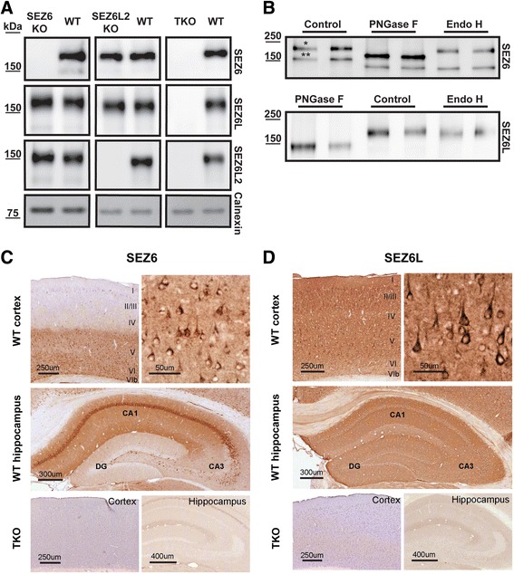
“Similarly, SEZ6L immunoreactivity (Fig. 1d) appeared strong in the neocortex and hippocampus, and protein localization in these areas was consistent with SEZ6L mRNA expression in the Allen Mouse Brain Atlas [34]. SEZ6L localized to pyramidal neurons throughout the cortex, particularly the apical dendrites (Fig. 1d), and appeared relatively lower in layer IV and VI. All regions of the hippocampus”
§ Seizure protein 6 and its homolog seizure 6-like protein are physiological substrates of BACE1 in neurons > Results > Validation of new monoclonal antibodies against SEZ6 and SEZ6L
What the sources say
- Not matchedcites [34] Allen Mouse Brain Atlas [ http://mouse.brain-map.org/ ]
Couldn’t be matched to a paper in Crossref, so it can’t be checked.
hedged- Asserted: passes — The authors put it forward as true.
- Checkable: passes — It can be checked against evidence.
- Load-bearing: unclear — It is unclear whether the paper’s conclusions depend on it.
- Based: passes — The paper gives a reason why it holds.
It may state two separate claims as one.
- Not matched
A result the paper reports from its own data — it cites no other source for it, so there is none to check. The quote shows it is in the paper.
In the paper
“Likewise, in P7 BACE1 KO mouse brains sSEZ6L was strongly reduced in the diethylamine soluble DEA brain fraction, while full-length SEZ6L was increased in the membrane fraction (Fig. 2c).”
§ Seizure protein 6 and its homolog seizure 6-like protein are physiological substrates of BACE1 in neurons > Results > BACE1 cleavage of SEZ6 and SEZ6L in primary neurons and mouse brain
boosteddirect- Asserted: passes — The authors put it forward as true.
- Checkable: passes — It can be checked against evidence.
- Load-bearing: passes — The paper’s conclusions depend on it.
- Based: passes — The paper gives a reason why it holds.
It may state two separate claims as one.
A result the paper reports from its own data — it cites no other source for it, so there is none to check. The quote shows it is in the paper.
In the paper
“Taken together, these results demonstrate that SEZ6 and SEZ6L are N-glycosylated proteins and that the newly generated antibodies specifically detect endogenous SEZ6 and SEZ6L.”
- Also stated in: § Validation of new monoclonal antibodies against SEZ6 and SEZ6L · plain
“Taken together, the newly generated antibodies specifically detect endogenous SEZ6 and SEZ6L by immunohistochemistry as well as Western Blot.”
§ Seizure protein 6 and its homolog seizure 6-like protein are physiological substrates of BACE1 in neurons > Results > Validation of new monoclonal antibodies against SEZ6 and SEZ6L
boosted- Asserted: passes — The authors put it forward as true.
- Checkable: passes — It can be checked against evidence.
- Load-bearing: passes — The paper’s conclusions depend on it.
- Based: passes — The paper gives a reason why it holds.
It may state two separate claims as one.
- Also stated in: § Validation of new monoclonal antibodies against SEZ6 and SEZ6L · plain
A result the paper reports from its own data — it cites no other source for it, so there is none to check. The quote shows it is in the paper.
In the paper
“Interestingly, sSEZ6 was also not reduced upon knock-down of BACE1, but mildly reduced upon knock-down of BACE2.”
§ Seizure protein 6 and its homolog seizure 6-like protein are physiological substrates of BACE1 in neurons > Results > BACE1 cleavage of SEZ6 and SEZ6L in pancreatic MIN6 cells
plaindirect- Asserted: passes — The authors put it forward as true.
- Checkable: passes — It can be checked against evidence.
- Load-bearing: passes — The paper’s conclusions depend on it.
- Based: passes — The paper gives a reason why it holds.
It may state two separate claims as one.
A result the paper reports from its own data — it cites no other source for it, so there is none to check. The quote shows it is in the paper.
In the paper
“This reveals that the 170 kDa band contains complex sugars (referred to as mature SEZ6), whereas the 150 kDa band (referred to as immature SEZ6), contains only high-mannose sugars.”
§ Seizure protein 6 and its homolog seizure 6-like protein are physiological substrates of BACE1 in neurons > Results > Validation of new monoclonal antibodies against SEZ6 and SEZ6L
plaincorrelational- Asserted: passes — The authors put it forward as true.
- Checkable: passes — It can be checked against evidence.
- Load-bearing: unclear — It is unclear whether the paper’s conclusions depend on it.
- Based: passes — The paper gives a reason why it holds.
It may state two separate claims as one.
In the paper
“In fact, treatment of primary neurons with the established BACE1 inhibitor C3 (also known as BACE1 inhibitor IV) [35] strongly reduced sSEZ6L levels compared to the control treatment with a concomitant moderate increase of full-length SEZ6L levels in the cell lysate (Fig. 2b).”
§ Seizure protein 6 and its homolog seizure 6-like protein are physiological substrates of BACE1 in neurons > Results > BACE1 cleavage of SEZ6 and SEZ6L in primary neurons and mouse brain
What the sources say
- Not checked — no full text of the sourcecites [35] Stachel SJ, Coburn CA, Steele TG, Jones KG, Loutzenhiser EF, Gregro AR, Rajapakse HA, Lai MT, Crouthamel MC, Xu M, et al. Structure-based design of potent and selective cell-permeable inhibitors of human beta-secretase (BACE-1). J Med Chem. 2004;47:6447–50. source
boosteddirect- Asserted: passes — The authors put it forward as true.
- Checkable: passes — It can be checked against evidence.
- Load-bearing: passes — The paper’s conclusions depend on it.
- Based: passes — The paper gives a reason why it holds.
It may state two separate claims as one.
A result the paper reports from its own data — it cites no other source for it, so there is none to check. The quote shows it is in the paper.
In the paper
“The non-cleaved, full-length peptide eluted from the nLC column at ~ 22 min (Fig. 5b).”
§ Seizure protein 6 and its homolog seizure 6-like protein are physiological substrates of BACE1 in neurons > Results > BACE1 cleaves SEZ6 within its juxtamembrane domain
plain- Asserted: passes — The authors put it forward as true.
- Checkable: passes — It can be checked against evidence.
- Load-bearing: fails — The paper’s conclusions may not depend on it.
- Based: passes — The paper gives a reason why it holds.
Probably not a substantial claim. The paper’s conclusions may not depend on it.
A result the paper reports from its own data — it cites no other source for it, so there is none to check. The quote shows it is in the paper.
In the paper
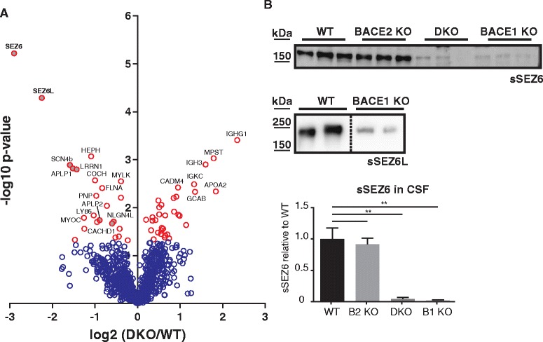
“The same reduction was observed in CSF from BACE1 KO, but not for BACE2 KO mice.”
§ Seizure protein 6 and its homolog seizure 6-like protein are physiological substrates of BACE1 in neurons > Results > sSEZ6 and sSEZ6L are detected in murine CSF in BACE1-dependent manner
plaindirect- Asserted: passes — The authors put it forward as true.
- Checkable: passes — It can be checked against evidence.
- Load-bearing: passes — The paper’s conclusions depend on it.
- Based: passes — The paper gives a reason why it holds.
It may state two separate claims as one.
A result the paper reports from its own data — it cites no other source for it, so there is none to check. The quote shows it is in the paper.
In the paper
“Importantly, we show that neither SEZ6 nor SEZ6L are substrates for BACE1 in the pancreatic cell line, which is in contrast to brain”
§ Seizure protein 6 and its homolog seizure 6-like protein are physiological substrates of BACE1 in neurons > Discussion
boosted- Asserted: passes — The authors put it forward as true.
- Checkable: passes — It can be checked against evidence.
- Load-bearing: passes — The paper’s conclusions depend on it.
- Based: passes — The paper gives a reason why it holds.
It may state two separate claims as one.
A result the paper reports from its own data — it cites no other source for it, so there is none to check. The quote shows it is in the paper.
In the paper
“Upon addition of BACE1, the full-length peptide levels were decreased in the chromatogram and two additional peptides with elution times of ~16 and ~21 min were detected (Fig. 5b).”
§ Seizure protein 6 and its homolog seizure 6-like protein are physiological substrates of BACE1 in neurons > Results > BACE1 cleaves SEZ6 within its juxtamembrane domain
plaindirect- Asserted: passes — The authors put it forward as true.
- Checkable: passes — It can be checked against evidence.
- Load-bearing: passes — The paper’s conclusions depend on it.
- Based: passes — The paper gives a reason why it holds.
It may state two separate claims as one.
A result the paper reports from its own data — it cites no other source for it, so there is none to check. The quote shows it is in the paper.
In the paper
“Among these proteins, SEZ6 (DKO/WT = 13 %, p = 5.99E-06) and SEZ6L (DKO/WT = 20 %, p = 5.10E-05) showed the strongest reduction as well as the highest statistical significance (Fig. 7a).”
§ Seizure protein 6 and its homolog seizure 6-like protein are physiological substrates of BACE1 in neurons > Results > sSEZ6 and sSEZ6L are detected in murine CSF in BACE1-dependent manner
plaindirect- Asserted: passes — The authors put it forward as true.
- Checkable: passes — It can be checked against evidence.
- Load-bearing: passes — The paper’s conclusions depend on it.
- Based: passes — The paper gives a reason why it holds.
It may state two separate claims as one.
A result the paper reports from its own data — it cites no other source for it, so there is none to check. The quote shows it is in the paper.
In the paper
“Subcellular locations of proteins quantified in all replicates of SDC or urea supported digestions were similar (Additional file 1: Figures S2 and S3).”
§ Seizure protein 6 and its homolog seizure 6-like protein are physiological substrates of BACE1 in neurons > Results > sSEZ6 and sSEZ6L are detected in murine CSF in BACE1-dependent manner
plain- Asserted: passes — The authors put it forward as true.
- Checkable: passes — It can be checked against evidence.
- Load-bearing: fails — The paper’s conclusions may not depend on it.
- Based: passes — The paper gives a reason why it holds.
Probably not a substantial claim. The paper’s conclusions may not depend on it.
In the paper
“Importantly, the new workflow allowed the quantification of SEZ6 and SEZ6L, which were not quantified in the previous study [14].”
§ Seizure protein 6 and its homolog seizure 6-like protein are physiological substrates of BACE1 in neurons > Discussion
What the sources say
- Not checked — no full text of the sourcecites [14] Dislich B, Wohlrab F, Bachhuber T, Müller SA, Kuhn P-H, Hogl S, Meyer-Luehmann M, Lichtenthaler SF. Label-free quantitative proteomics of mouse cerebrospinal fluid detects β-Site APP Cleaving Enzyme (BACE1) protease substrates in vivo. Mol Cell Proteomics. 2015;14:2550–63. source
plaindirect- Asserted: passes — The authors put it forward as true.
- Checkable: passes — It can be checked against evidence.
- Load-bearing: passes — The paper’s conclusions depend on it.
- Based: passes — The paper gives a reason why it holds.
Probably not a substantial claim.
A result the paper reports from its own data — it cites no other source for it, so there is none to check. The quote shows it is in the paper.
In the paper
“The deglycosylation experiment (Fig. 1b) had revealed that mature SEZ6 and SEZ6L carry complex N-linked sugars and are resistant to EndoH treatment.”
§ Seizure protein 6 and its homolog seizure 6-like protein are physiological substrates of BACE1 in neurons > Results > BACE1 inhibition increases neuronal cell surface levels of SEZ6 and SEZ6L
plain- Asserted: passes — The authors put it forward as true.
- Checkable: passes — It can be checked against evidence.
- Load-bearing: unclear — It is unclear whether the paper’s conclusions depend on it.
- Based: passes — The paper gives a reason why it holds.
It may state two separate claims as one.
A result the paper reports from its own data — it cites no other source for it, so there is none to check. The quote shows it is in the paper.
In the paper
“Additionally, the average number of identified and quantified proteins was 10.3 and 7.5 % higher for the SDC supported digestion protocol, respectively.”
§ Seizure protein 6 and its homolog seizure 6-like protein are physiological substrates of BACE1 in neurons > Results > sSEZ6 and sSEZ6L are detected in murine CSF in BACE1-dependent manner
plaindirect- Asserted: passes — The authors put it forward as true.
- Checkable: passes — It can be checked against evidence.
- Load-bearing: fails — The paper’s conclusions may not depend on it.
- Based: passes — The paper gives a reason why it holds.
Probably not a substantial claim. The paper’s conclusions may not depend on it.
None of the sources cited for this claim have been checked yet.
In the paper
“We confirm this finding and also extend it to SEZ6.”
§ Seizure protein 6 and its homolog seizure 6-like protein are physiological substrates of BACE1 in neurons > Discussion
plaindirect- Asserted: passes — The authors put it forward as true.
- Checkable: passes — It can be checked against evidence.
- Load-bearing: unclear — It is unclear whether the paper’s conclusions depend on it.
- Based: passes — The paper gives a reason why it holds.
It may state two separate claims as one. The quote only partly states this claim.
A result the paper reports from its own data — it cites no other source for it, so there is none to check. The quote shows it is in the paper.
In the paper
“Peptides were almost exclusively mapped to extracellular domains of transmembrane proteins (Additional file 3: Tables S1-S6).”
§ Seizure protein 6 and its homolog seizure 6-like protein are physiological substrates of BACE1 in neurons > Results > sSEZ6 and sSEZ6L are detected in murine CSF in BACE1-dependent manner
plaincorrelational- Asserted: passes — The authors put it forward as true.
- Checkable: passes — It can be checked against evidence.
- Load-bearing: passes — The paper’s conclusions depend on it.
- Based: passes — The paper gives a reason why it holds.
The quote only partly states this claim.
A result the paper reports from its own data — it cites no other source for it, so there is none to check. The quote shows it is in the paper.
In the paper
“However, cleavage of sFlag-V5-SEZ6 and sSEZ6L was less impacted than Aβ42 upon inhibition with BACE1-selective inhibitors (B and C) and followed the enzymatic inhibition curves of BACE2 rather than BACE1 (Fig. 3b).”
§ Seizure protein 6 and its homolog seizure 6-like protein are physiological substrates of BACE1 in neurons > Results > BACE1 cleavage of SEZ6 and SEZ6L in pancreatic MIN6 cells
plaincorrelational- Asserted: passes — The authors put it forward as true.
- Checkable: passes — It can be checked against evidence.
- Load-bearing: passes — The paper’s conclusions depend on it.
- Based: passes — The paper gives a reason why it holds.
It may state two separate claims as one.
A claim is checked only against the cited paper’s full text, never its abstract, and none is available for its sources.
In the paper
“A concentration of 0.1 % SDC was sufficient to improve the digestion efficiency.”
§ Seizure protein 6 and its homolog seizure 6-like protein are physiological substrates of BACE1 in neurons > Results > sSEZ6 and sSEZ6L are detected in murine CSF in BACE1-dependent manner
What the sources say
- Not checked — no full text of the sourcecites [43] Lin Y, Lin H, Liu Z, Wang K, Yan Y. Improvement of a sample preparation method assisted by sodium deoxycholate for mass-spectrometry-based shotgun membrane proteomics†. J Sep Sci. 2014;37:3321–9. source
- Not checked — no full text of the sourcecites [44] Lin Y, Wang K, Liu Z, Lin H, Yu L. Enhanced SDC-assisted digestion coupled with lipid chromatography-tandem mass spectrometry for shotgun analysis of membrane proteome. J Chromatogr B. 2015;1002:144–51. source
plaindirect- Asserted: passes — The authors put it forward as true.
- Checkable: passes — It can be checked against evidence.
- Load-bearing: unclear — It is unclear whether the paper’s conclusions depend on it.
- Based: passes — The paper gives a reason why it holds.
The quote only partly states this claim.
A result the paper reports from its own data — it cites no other source for it, so there is none to check. The quote shows it is in the paper.
In the paper
“Addition of C3 inhibited the production of both peptides, demonstrating that they are BACE1 cleavage products of the full-length peptide.”
§ Seizure protein 6 and its homolog seizure 6-like protein are physiological substrates of BACE1 in neurons > Results > BACE1 cleaves SEZ6 within its juxtamembrane domain
boosteddirect- Asserted: passes — The authors put it forward as true.
- Checkable: passes — It can be checked against evidence.
- Load-bearing: passes — The paper’s conclusions depend on it.
- Based: passes — The paper gives a reason why it holds.
It may state two separate claims as one.
A result the paper reports from its own data — it cites no other source for it, so there is none to check. The quote shows it is in the paper.
In the paper
“Cleavage of Aβ42 and sFlag-V5-SEZ6 and sSEZ6L were similar after addition of nonselective BACE inhibitor A and was consistent with equipotent inhibition of BACE1 and BACE2 in enzymatic assays.”
§ Seizure protein 6 and its homolog seizure 6-like protein are physiological substrates of BACE1 in neurons > Results > BACE1 cleavage of SEZ6 and SEZ6L in pancreatic MIN6 cells
plaincorrelational- Asserted: passes — The authors put it forward as true.
- Checkable: passes — It can be checked against evidence.
- Load-bearing: unclear — It is unclear whether the paper’s conclusions depend on it.
- Based: passes — The paper gives a reason why it holds.
It may state two separate claims as one.
A result the paper reports from its own data — it cites no other source for it, so there is none to check. The quote shows it is in the paper.
In the paper
“We found opposite expression patterns of BACE1 and BACE2 in MIN6 cells and in neurons, which correlated with the tissue-specific cleavage of SEZ6 and SEZ6L.”
§ Seizure protein 6 and its homolog seizure 6-like protein are physiological substrates of BACE1 in neurons > Discussion
plaincorrelational- Asserted: passes — The authors put it forward as true.
- Checkable: passes — It can be checked against evidence.
- Load-bearing: passes — The paper’s conclusions depend on it.
- Based: passes — The paper gives a reason why it holds.
It may state two separate claims as one.
In the paper
“In contrast to our previous proteomic study of CSF from BACE1 deficient mice [14], we were able to quantify SEZ6 and SEZ6L with the optimized protocol (Additional file 2: Supplementary Data: proteins BACE DKO vs WT CSF).”
§ Seizure protein 6 and its homolog seizure 6-like protein are physiological substrates of BACE1 in neurons > Results > sSEZ6 and sSEZ6L are detected in murine CSF in BACE1-dependent manner
What the sources say
- Not checked — no full text of the sourcecites [14] Dislich B, Wohlrab F, Bachhuber T, Müller SA, Kuhn P-H, Hogl S, Meyer-Luehmann M, Lichtenthaler SF. Label-free quantitative proteomics of mouse cerebrospinal fluid detects β-Site APP Cleaving Enzyme (BACE1) protease substrates in vivo. Mol Cell Proteomics. 2015;14:2550–63. source
plain- Asserted: passes — The authors put it forward as true.
- Checkable: passes — It can be checked against evidence.
- Load-bearing: passes — The paper’s conclusions depend on it.
- Based: passes — The paper gives a reason why it holds.
It may state two separate claims as one.
A result the paper reports from its own data — it cites no other source for it, so there is none to check. The quote shows it is in the paper.
In the paper
“However, digestion efficiency was strongly increased which was detected by the 58.1 % lower number of average missed cleavages per peptide (Table 1).”
§ Seizure protein 6 and its homolog seizure 6-like protein are physiological substrates of BACE1 in neurons > Results > sSEZ6 and sSEZ6L are detected in murine CSF in BACE1-dependent manner
boosteddirect- Asserted: passes — The authors put it forward as true.
- Checkable: passes — It can be checked against evidence.
- Load-bearing: fails — The paper’s conclusions may not depend on it.
- Based: passes — The paper gives a reason why it holds.
Probably not a substantial claim. The paper’s conclusions may not depend on it.
A result the paper reports from its own data — it cites no other source for it, so there is none to check. The quote shows it is in the paper.
In the paper
“The full-length SEZ6 in the cell lysate and the shed ectodomain (sSEZ6) in the supernatant were detected by immunoblots in the transfected cells, but not in control transfected cells (Fig. 6a).”
§ Seizure protein 6 and its homolog seizure 6-like protein are physiological substrates of BACE1 in neurons > Results > SEZ6 is a substrate for γ-secretase
plain- Asserted: passes — The authors put it forward as true.
- Checkable: passes — It can be checked against evidence.
- Load-bearing: unclear — It is unclear whether the paper’s conclusions depend on it.
- Based: passes — The paper gives a reason why it holds.
It may state two separate claims as one.
A result the paper reports from its own data — it cites no other source for it, so there is none to check. The quote shows it is in the paper.
In the paper
“However, the number of quantified membrane proteins was 8.9 % higher for the samples digested in the presence of SDC (135 vs. 124).”
§ Seizure protein 6 and its homolog seizure 6-like protein are physiological substrates of BACE1 in neurons > Results > sSEZ6 and sSEZ6L are detected in murine CSF in BACE1-dependent manner
plaindirect- Asserted: passes — The authors put it forward as true.
- Checkable: passes — It can be checked against evidence.
- Load-bearing: fails — The paper’s conclusions may not depend on it.
- Based: passes — The paper gives a reason why it holds.
Probably not a substantial claim. The paper’s conclusions may not depend on it.
A result the paper reports from its own data — it cites no other source for it, so there is none to check. The quote shows it is in the paper.
In the paper
“Additionally, the average number of identified and quantified proteins was 10.3 and 7.5 % higher for the SDC supported digestion protocol, respectively.”
§ Seizure protein 6 and its homolog seizure 6-like protein are physiological substrates of BACE1 in neurons > Results > sSEZ6 and sSEZ6L are detected in murine CSF in BACE1-dependent manner
plaindirect- Asserted: passes — The authors put it forward as true.
- Checkable: passes — It can be checked against evidence.
- Load-bearing: fails — The paper’s conclusions may not depend on it.
- Based: passes — The paper gives a reason why it holds.
Probably not a substantial claim. The paper’s conclusions may not depend on it.
In the paper
“However, sSEZ6 and sSEZ6L were not consistently detected and could not be quantified in murine CSF, potentially because their levels were below the detection limit.”
§ Seizure protein 6 and its homolog seizure 6-like protein are physiological substrates of BACE1 in neurons > Results > sSEZ6 and sSEZ6L are detected in murine CSF in BACE1-dependent manner
What the sources say
- Not checked — no full text of the sourcecites [14] Dislich B, Wohlrab F, Bachhuber T, Müller SA, Kuhn P-H, Hogl S, Meyer-Luehmann M, Lichtenthaler SF. Label-free quantitative proteomics of mouse cerebrospinal fluid detects β-Site APP Cleaving Enzyme (BACE1) protease substrates in vivo. Mol Cell Proteomics. 2015;14:2550–63. source
plain- Asserted: passes — The authors put it forward as true.
- Checkable: passes — It can be checked against evidence.
- Load-bearing: passes — The paper’s conclusions depend on it.
- Based: passes — The paper gives a reason why it holds.
It may state two separate claims as one.
A result the paper reports from its own data — it cites no other source for it, so there is none to check. The quote shows it is in the paper.
In the paper
“The number of identified unique peptides was 6.6 % lower for the SDC supported protocol (Table 1).”
§ Seizure protein 6 and its homolog seizure 6-like protein are physiological substrates of BACE1 in neurons > Results > sSEZ6 and sSEZ6L are detected in murine CSF in BACE1-dependent manner
plain- Asserted: passes — The authors put it forward as true.
- Checkable: passes — It can be checked against evidence.
- Load-bearing: fails — The paper’s conclusions may not depend on it.
- Based: passes — The paper gives a reason why it holds.
Probably not a substantial claim. The paper’s conclusions may not depend on it.
A result the paper reports from its own data — it cites no other source for it, so there is none to check. The quote shows it is in the paper.
In the paper
“Here, we improved the digestion efficiency of murine CSF in comparison to our previous protocol by using 0.1 % SDC in 50 mM ammonium bicarbonate as digestion buffer.”
§ Seizure protein 6 and its homolog seizure 6-like protein are physiological substrates of BACE1 in neurons > Discussion
boosteddirect- Asserted: passes — The authors put it forward as true.
- Checkable: passes — It can be checked against evidence.
- Load-bearing: passes — The paper’s conclusions depend on it.
- Based: passes — The paper gives a reason why it holds.
It may state two separate claims as one.
A conclusion the paper draws from its own results — it cites no other source for it, so there is none to check. The quote shows it is in the paper.
In the paper
“This shows that both SEZ6 and SEZ6L are not substrates for BACE1 in the pancreatic cell line.”
§ Seizure protein 6 and its homolog seizure 6-like protein are physiological substrates of BACE1 in neurons > Results > BACE1 cleavage of SEZ6 and SEZ6L in pancreatic MIN6 cells
plain- Asserted: passes — The authors put it forward as true.
- Checkable: passes — It can be checked against evidence.
- Load-bearing: passes — The paper’s conclusions depend on it.
- Based: passes — The paper gives a reason why it holds.
It may state two separate claims as one.
In the paper
“Moreover, several phenotypic changes were described in BACE1-/- mice, such as epileptic seizures, schizophrenic symptoms, increased mortality and altered insulin metabolism, but most of the BACE1 substrates contributing to these phenotypes still need to be determined [13].”
§ Seizure protein 6 and its homolog seizure 6-like protein are physiological substrates of BACE1 in neurons > Background
What the sources say
- Not checked — no full text of the sourcecites [13] Barao S, Moechars D, Lichtenthaler SF, De Strooper B. BACE1 physiological functions may limit its use as therapeutic target for alzheimer’s disease. Trends Neurosci. 2016;39:158–69. source
plaincorrelational- Asserted: passes — The authors put it forward as true.
- Checkable: passes — It can be checked against evidence.
- Load-bearing: fails — The paper’s conclusions may not depend on it.
- Based: passes — The paper gives a reason why it holds.
Probably not a substantial claim. The paper’s conclusions may not depend on it.
What the paper did — it cites no source for this, so there is none to check. The quote shows it is in the paper.
In the paper
“Label-free quantification (LFQ) of proteins required at least two ratio counts of unique peptides.”
§ Seizure protein 6 and its homolog seizure 6-like protein are physiological substrates of BACE1 in neurons > Methods > MS data analysis of CSF samples
plain- Asserted: passes — The authors put it forward as true.
- Checkable: passes — It can be checked against evidence.
- Load-bearing: fails — The paper’s conclusions may not depend on it.
- Based: passes — The paper gives a reason why it holds.
Probably not a substantial claim. The paper’s conclusions may not depend on it.
What the paper did — it cites no source for this, so there is none to check. The quote shows it is in the paper.
In the paper
“The correct sequence was verified by MS/MS-based fragmentation (Fig. 5c).”
§ Seizure protein 6 and its homolog seizure 6-like protein are physiological substrates of BACE1 in neurons > Results > BACE1 cleaves SEZ6 within its juxtamembrane domain
plain- Asserted: passes — The authors put it forward as true.
- Checkable: passes — It can be checked against evidence.
- Load-bearing: fails — The paper’s conclusions may not depend on it.
- Based: passes — The paper gives a reason why it holds.
Probably not a substantial claim. The paper’s conclusions may not depend on it.
What the paper did — it cites no source for this, so there is none to check. The quote shows it is in the paper.
In the paper
“False discovery rate for both peptides and proteins was adjusted to less than 1 % using a target and decoy approach (concatenated forward/reverse database).”
§ Seizure protein 6 and its homolog seizure 6-like protein are physiological substrates of BACE1 in neurons > Methods > MS data analysis of CSF samples
plain- Asserted: passes — The authors put it forward as true.
- Checkable: passes — It can be checked against evidence.
- Load-bearing: fails — The paper’s conclusions may not depend on it.
- Based: passes — The paper gives a reason why it holds.
Probably not a substantial claim. The paper’s conclusions may not depend on it.
In the paper
“A previous proteomic study demonstrated that the soluble ectodomains of other BACE1 substrates, such as APLP1, PLXDC2 and CHL1, were reduced in the CSF of BACE1-deficient mice [14].”
§ Seizure protein 6 and its homolog seizure 6-like protein are physiological substrates of BACE1 in neurons > Results > sSEZ6 and sSEZ6L are detected in murine CSF in BACE1-dependent manner
What the sources say
- Not checked — no full text of the sourcecites [14] Dislich B, Wohlrab F, Bachhuber T, Müller SA, Kuhn P-H, Hogl S, Meyer-Luehmann M, Lichtenthaler SF. Label-free quantitative proteomics of mouse cerebrospinal fluid detects β-Site APP Cleaving Enzyme (BACE1) protease substrates in vivo. Mol Cell Proteomics. 2015;14:2550–63. source
boosteddirect- Asserted: passes — The authors put it forward as true.
- Checkable: passes — It can be checked against evidence.
- Load-bearing: passes — The paper’s conclusions depend on it.
- Based: passes — The paper gives a reason why it holds.
Probably not a substantial claim.
A conclusion the paper draws from its own results — it cites no other source for it, so there is none to check. The quote shows it is in the paper.
In the paper
“The improved method may be of wide relevance for studying murine CSF in the context of different neurological and neurodegenerative diseases.”
§ Seizure protein 6 and its homolog seizure 6-like protein are physiological substrates of BACE1 in neurons > Discussion
hedgedconditional- Asserted: passes — The authors put it forward as true.
- Checkable: fails — It may be too vague to check against evidence.
- Load-bearing: fails — The paper’s conclusions may not depend on it.
- Based: passes — The paper gives a reason why it holds.
Probably not a substantial claim. It may be too vague to check against evidence. The paper’s conclusions may not depend on it.
What the paper did — it cites no source for this, so there is none to check. The quote shows it is in the paper.
In the paper
“All curves are based on biological replicates with at least two technical replicates.”
§ Seizure protein 6 and its homolog seizure 6-like protein are physiological substrates of BACE1 in neurons > Methods > Mesoscale (MSD) detection of sFlag-V5-SEZ6 and sSEZ6L
plain- Asserted: passes — The authors put it forward as true.
- Checkable: passes — It can be checked against evidence.
- Load-bearing: fails — The paper’s conclusions may not depend on it.
- Based: passes — The paper gives a reason why it holds.
Probably not a substantial claim. The paper’s conclusions may not depend on it.
What the paper did — it cites no source for this, so there is none to check. The quote shows it is in the paper.
In the paper
“To ensure the specificity of the immunoblot signals, brains from wild type (WT) as well as from SEZ6-/- (SEZ6 KO) or SEZ6L2-/- (SEZ6L2 KO) mice were used.”
§ Seizure protein 6 and its homolog seizure 6-like protein are physiological substrates of BACE1 in neurons > Results > Validation of new monoclonal antibodies against SEZ6 and SEZ6L
plain- Asserted: passes — The authors put it forward as true.
- Checkable: passes — It can be checked against evidence.
- Load-bearing: passes — The paper’s conclusions depend on it.
- Based: passes — The paper gives a reason why it holds.
Probably not a substantial claim.
A conclusion the paper draws from its own results — it cites no other source for it, so there is none to check. The quote shows it is in the paper.
In the paper
“This demonstrates that sSEZ6 is generated specifically by BACE1, but not by BACE2 in murine CSF.”
§ Seizure protein 6 and its homolog seizure 6-like protein are physiological substrates of BACE1 in neurons > Results > sSEZ6 and sSEZ6L are detected in murine CSF in BACE1-dependent manner
boosteddirect- Asserted: passes — The authors put it forward as true.
- Checkable: passes — It can be checked against evidence.
- Load-bearing: passes — The paper’s conclusions depend on it.
- Based: passes — The paper gives a reason why it holds.
It may state two separate claims as one.
What the paper did — it cites no source for this, so there is none to check. The quote shows it is in the paper.
In the paper
“Only unique peptides were used for quantification.”
§ Seizure protein 6 and its homolog seizure 6-like protein are physiological substrates of BACE1 in neurons > Methods > MS data analysis of CSF samples
plain- Asserted: passes — The authors put it forward as true.
- Checkable: passes — It can be checked against evidence.
- Load-bearing: fails — The paper’s conclusions may not depend on it.
- Based: passes — The paper gives a reason why it holds.
Probably not a substantial claim. The paper’s conclusions may not depend on it.
What the paper did — it cites no source for this, so there is none to check. The quote shows it is in the paper.
In the paper
“A p-value less than 5 % was set as significance threshold.”
§ Seizure protein 6 and its homolog seizure 6-like protein are physiological substrates of BACE1 in neurons > Methods > MS data analysis of CSF samples
plain- Asserted: passes — The authors put it forward as true.
- Checkable: passes — It can be checked against evidence.
- Load-bearing: fails — The paper’s conclusions may not depend on it.
- Based: passes — The paper gives a reason why it holds.
Probably not a substantial claim. The paper’s conclusions may not depend on it.
None of the sources cited for this claim have been checked yet.
In the paper
“but BACE1 preferentially cleaves C-terminally to leucine or other hydrophobic amino acids [40].”
§ Seizure protein 6 and its homolog seizure 6-like protein are physiological substrates of BACE1 in neurons > Results > BACE1 cleaves SEZ6 within its juxtamembrane domain
What the sources say
- Not checked yetcites [40] Gruninger-Leitch F, Schlatter D, Kung E, Nelbock P, Dobeli H. Substrate and inhibitor profile of BACE (beta-secretase) and comparison with other mammalian aspartic proteases. J Biol Chem. 2002;277:4687–93. source
Checked before — its earlier verdict is held back until a new check runs.
plaindirect- Asserted: passes — The authors put it forward as true.
- Checkable: passes — It can be checked against evidence.
- Load-bearing: passes — The paper’s conclusions depend on it.
- Based: passes — The paper gives a reason why it holds.
Probably not a substantial claim.
- Not checked yet
What the paper did — it cites no source for this, so there is none to check. The quote shows it is in the paper.
In the paper
“Primary or secondary antibodies were omitted on sections in each experiment to confirm staining specificity.”
§ Seizure protein 6 and its homolog seizure 6-like protein are physiological substrates of BACE1 in neurons > Methods > Immunohistochemistry
plain- Asserted: passes — The authors put it forward as true.
- Checkable: passes — It can be checked against evidence.
- Load-bearing: fails — The paper’s conclusions may not depend on it.
- Based: passes — The paper gives a reason why it holds.
Probably not a substantial claim. The paper’s conclusions may not depend on it.
A conclusion the paper draws from its own results — it cites no other source for it, so there is none to check. The quote shows it is in the paper.
In the paper
“Using different techniques, our study validates SEZ6 and SEZ6L as BACE1 substrates in vitro and in vivo and demonstrates that, in contrast to other BACE1 substrates, SEZ6 and SEZ6L are nearly exclusively cleaved by BACE1 and not by other proteases in the brain.”
§ Seizure protein 6 and its homolog seizure 6-like protein are physiological substrates of BACE1 in neurons > Discussion
boosteddirect- Asserted: passes — The authors put it forward as true.
- Checkable: passes — It can be checked against evidence.
- Load-bearing: passes — The paper’s conclusions depend on it.
- Based: passes — The paper gives a reason why it holds.
It may state two separate claims as one.
None of the sources cited for this claim have been checked yet.
In the paper
“For example, in APP the ADAM10 and BACE1 cleavage sites are 16 amino acids apart from each other and yield APP ectodomains with diverging functions [48, 49].”
§ Seizure protein 6 and its homolog seizure 6-like protein are physiological substrates of BACE1 in neurons > Discussion
What the sources say
- Not checked — no full text of the sourcecites [48] Ring S, Weyer SW, Kilian SB, Waldron E, Pietrzik CU, Filippov MA, Herms J, Buchholz C, Eckman CB, Korte M, et al. The secreted beta-amyloid precursor protein ectodomain APPs alpha is sufficient to rescue the anatomical, behavioral, and electrophysiological abnormalities of APP-deficient mice. J Neurosci. 2007;27:7817–26. source
- Not checked yetcites [49] Li H, Wang B, Wang Z, Guo Q, Tabuchi K, Hammer RE, Sudhof TC, Zheng H. Soluble amyloid precursor protein (APP) regulates transthyretin and Klotho gene expression without rescuing the essential function of APP. Proc Natl Acad Sci U S A. 2010;107:17362–7. source
Checked before — its earlier verdict is held back until a new check runs.
plaindirect- Asserted: passes — The authors put it forward as true.
- Checkable: passes — It can be checked against evidence.
- Load-bearing: fails — The paper’s conclusions may not depend on it.
- Based: passes — The paper gives a reason why it holds.
Probably not a substantial claim. The paper’s conclusions may not depend on it. It may state two separate claims as one.
A conclusion the paper draws from its own results — it cites no other source for it, so there is none to check. The quote shows it is in the paper.
In the paper
“We demonstrate that SEZ6 and SEZ6L are physiological BACE1 substrates in the murine brain and that, in contrast to most other BACE1 substrates, these two proteins are nearly exclusively cleaved by BACE1.”
§ Seizure protein 6 and its homolog seizure 6-like protein are physiological substrates of BACE1 in neurons > Conclusions
boosteddirect- Asserted: passes — The authors put it forward as true.
- Checkable: passes — It can be checked against evidence.
- Load-bearing: passes — The paper’s conclusions depend on it.
- Based: passes — The paper gives a reason why it holds.
It may state two separate claims as one.
The paper relies on this without support.
BACE1 inhibition in vivo reduces sSEZ6 and sSEZ6L levels in CSF in a manner suitable for monitoring BACE1 activity.
“suggests that sSEZ6 and sSEZ6L levels in CSF are suitable markers to monitor BACE1 inhibition in mice.”
The paper relies on this without support.
The BACE1 cleavage site determined using a synthetic SEZ6 peptide reflects the endogenous BACE1 cleavage site in SEZ6.
“The murine SEZ6 peptide AASLDGFYNGRSLDVAKAPAASSAL (PSL Peptide Specialty Laboratories GmbH, Germany) was resuspended in LC-MS grade water (Chromasolv, Sigma Aldrich, Germany) and 40 μg of peptide were used to determine the cleavage site.”
The paper relies on this without support.
Most BACE1 inhibitors currently tested in clinical trials for AD are not specific for BACE1, but also inhibit BACE2.
“Most BACE1 inhibitors currently tested in clinical trials for AD are not specific for BACE1, but also inhibit BACE2.”
The paper relies on this without support.
Only few of the more than 40 potential BACE1 substrates have been validated in vitro and in vivo.
“However, only few of them have been validated in vitro and in vivo.”
The paper relies on this without support.
Different proteases may compensate for each other if one of them is blocked.
“Additionally, the different proteases may compensate for each other, if one of them is blocked.”
The paper relies on this without support.
Human CSF is available in milliliter quantities, whereas only approximately 10 μl of murine CSF are obtainable.
“While human CSF is available in milliliter quantities, only approximately 10 μl of murine CSF are obtainable.”
The paper relies on this without support.
The extracellular regions of SEZ6/SEZ6L contain three CUB and five short consensus repeat domains, which are protein-binding domains also found in a variety of cell surface receptors.
“The extracellular regions of SEZ6/SEZ6L contain three CUB (complement subcomponent C1r, C1s /sea urchin embryonic growth factor Uegf / bone morphogenetic protein 1) and five short consensus repeat domains, which are protein-binding domains that are also found in a variety of cell surface receptors.”
Claims other papers make about this one
Each is checked against this paper’s own text: does it say what the citing paper says it does?
The source backs part of the claim, not all of it — or backs it for a different population, condition or comparison.
In the citing paper
“through secretome enrichment experiments, nearly 70 BACE1 substrates have been identified to date.”
From BACE1: More than just a β‐secretase (2022) · cites this paper as [24]
Experimental — accuracy not yet measuredIn the citing paper
“Besides the amyloidogenic pathway, BACE1 has other substrates that may be important for synaptic plasticity and synaptic homeostasis.”
From The β-Secretase BACE1 in Alzheimer’s Disease (2021) · cites this paper as [71]
In the citing paper
“In the mouse brain, soluble SEZ6 is almost exclusively produced by BACE1 (1,71).”
From The β-Secretase BACE1 in Alzheimer’s Disease (2021) · cites this paper as [71]
In the citing paper
“Seizure protein 6 (SEZ6) and seizure‐like protein 6 (SEZ6L) proteins influence ER functions in neurons, control synaptic connectivity, and motor coordination and therefore may likely be responsible for the seizures, motor deficits, and reduced spinal deficiency observed in BACE1 null mice.”
From BACE1: More than just a β‐secretase (2022) · cites this paper as [24]
In the citing paper
“It is well acknowledged that APP is a poor substrate for BACE1”
From BACE1: More than just a β‐secretase (2022) · cites this paper as [24]
Self-check
Percentages, sums and reported differences re-computed from the paper’s own numbers.
Each contradiction between two passages listed here was confirmed by an independent check.
Rated major: it would touch a result — a value a result rests on, reported and recomputed p-values on opposite sides of the significance level, two passages that conflict with no sign which is right, or a cited work retracted before this paper was published.
In the paper
§ Seizure protein 6 and its homolog seizure 6-like protein are physiological substrates of BACE1 in neurons > Results > BACE1 cleavage of SEZ6 and SEZ6L in pancreatic MIN6 cells
“nonselective (inhibiting both BACE1 and BACE2, compounds B and C) and BACE1-selective inhibitors (compound A)”
§ Seizure protein 6 and its homolog seizure 6-like protein are physiological substrates of BACE1 in neurons > Results > BACE1 cleavage of SEZ6 and SEZ6L in pancreatic MIN6 cells
“Cleavage of Aβ42 and sFlag-V5-SEZ6 and sSEZ6L were similar after addition of nonselective BACE inhibitor A and was consistent with equipotent inhibition of BACE1 and BACE2 in enzymatic assays. However, cleavage of sFlag-V5-SEZ6 and sSEZ6L was less impacted than Aβ42 upon inhibition with BACE1-selective inhibitors (B and C)”
Rated minor: it would not touch a result — a value no result rests on, a p-value that stays on the same side of the significance level, or a conflict that is clerical, resolved elsewhere in the paper, or outside its own findings.
In the paper
§ Seizure protein 6 and its homolog seizure 6-like protein are physiological substrates of BACE1 in neurons > Methods > Transfection and stable line generation
“Compound B: (5S)-2-amino-5-(2,6-diethyl-4-pyridyl)-3-methyl-5-(3-pyrimidin-5-ylphenyl)imidazol-4-one (AZD3839) [24] or Compound C: (5S)-2-amino-5-(2,6-diethyl-4-pyridyl)-3-methyl-5-(3-pyrimidin-5-ylphenyl)imidazol-4-one [25]”
§ Seizure protein 6 and its homolog seizure 6-like protein are physiological substrates of BACE1 in neurons > Methods > Transfection and stable line generation
“2 BACE1-selective inhibitors”
Citations
Cites
Continuous cultures of fused cells secreting antibody of predefined specificity
Köhler, G., & Milstein, C. (1975) · Nature · doi:10.1038/256495a0 · Cited by 17,200
The amyloid hypothesis of Alzheimer's disease at 25 years
Selkoe, D. J., & Hardy, J. (2016) · EMBO Molecular Medicine · doi:10.15252/emmm.201606210 · Cited by 6,164
Cox, J., Hein, M. Y., Luber, C. A., Paron, I., Nagaraj, N., & Mann, M. (2014) · Molecular & Cellular Proteomics · doi:10.1074/mcp.m113.031591 · Cited by 5,180
Vassar, R., Bennett, B. D., Babu-Khan, S., Kahn, S., Mendiaz, E. A., Denis, P., Teplow, D. B., Ross, S., Amarante, P., Loeloff, R., et al. (1999) · Science · doi:10.1126/science.286.5440.735 · Cited by 3,876 · cited 1×
“5a), which is very efficiently cleaved by BACE1 [4].”
BackgroundRappsilber, J., Ishihama, Y., & Mann, M. (2002) · Analytical Chemistry · doi:10.1021/ac026117i · Cited by 2,627
Schägger, H. (2006) · Nature Protocols · doi:10.1038/nprot.2006.4 · Cited by 2,519
Purification and cloning of amyloid precursor protein β-secretase from human brain
Sinha, S., Anderson, J. P., Barbour, R., Basi, G. S., Caccavello, R., Davis, D., Doan, M., Dovey, H. F., Frigon, N., Hong, J., et al. (1999) · Nature · doi:10.1038/990114 · Cited by 1,612
Membrane-anchored aspartyl protease with Alzheimer's disease β-secretase activity
Yan, R., Bienkowski, M. J., Shuck, M. E., Miao, H., Tory, M. C., Pauley, A. M., Brashler, J. R., Stratman, N. C., Mathews, W. R., Buhl, A. E., et al. (1999) · Nature · doi:10.1038/990107 · Cited by 1,438
Identification of a Novel Aspartic Protease (Asp 2) as β-Secretase
Hussain, I., Powell, D., Howlett, D. R., Tew, D. G., Meek, T. D., Chapman, C., Gloger, I. S., Murphy, K. E., Southan, C. D., Ryan, D. M., et al. (1999) · Molecular and Cellular Neuroscience · doi:10.1006/mcne.1999.0811 · Cited by 1,080
Harnessing homologous recombination in vitro to generate recombinant DNA via SLIC
Li, M. Z., & Elledge, S. J. (2007) · Nature Methods · doi:10.1038/nmeth1010 · Cited by 973
Cited by
Sorted by citations
The β-Secretase BACE1 in Alzheimer’s Disease
Hampel, H., Vassar, R., De Strooper, B., Hardy, J., Willem, M., Singh, N., Zhou, J., Yan, R., Vanmechelen, E., De Vos, A., et al. (2021) · Biological Psychiatry · doi:10.1016/j.biopsych.2020.02.001 · Cited by 694
Self-citationKent, S. A., Spires-Jones, T. L., & Durrant, C. S. (2020) · Acta Neuropathologica · doi:10.1007/s00401-020-02196-w · Cited by 446
Lichtenthaler, S. F., Lemberg, M. K., & Fluhrer, R. (2018) · The EMBO Journal · doi:10.15252/embj.201899456 · Cited by 323
Self-citationThe substrate repertoire of γ-secretase/presenilin
Güner, G., & Lichtenthaler, S. F. (2020) · Seminars in Cell and Developmental Biology · doi:10.1016/j.semcdb.2020.05.019 · Cited by 202
Self-citationThe case for low-level BACE1 inhibition for the prevention of Alzheimer disease
McDade, E., Voytyuk, I., Aisen, P. S. S., Bateman, R. J., Carrillo, M. C., Strooper, B. D., Haass, C., Reiman, E. M., Sperling, R. A., Tariot, P. N., et al. (2021) · Nature Reviews Neurology · doi:10.1038/s41582-021-00545-1 · Cited by 151
Self-citationTan, J. Z. A., & Gleeson, P. A. (2019) · Biochimica et Biophysica Acta (BBA) - Biomembranes · doi:10.1016/j.bbamem.2018.11.013 · Cited by 137
Koelsch, G. (2017) · Molecules · doi:10.3390/molecules22101723 · Cited by 128
Role of BACE1 in Alzheimer’s synaptic function
Das, B., & Yan, R. (2017) · Translational Neurodegeneration · doi:10.1186/s40035-017-0093-5 · Cited by 126
Findings of Efficacy, Safety, and Biomarker Outcomes of Atabecestat in Preclinical Alzheimer Disease
Sperling, R. A., Henley, D. B., Aisen, P. S. S., Raman, R., Donohue, M., Ernstrom, K. B., Rafii, M. S., Streffer, J. R., Shi, Y., Karcher, K., et al. (2021) · JAMA Neurology · doi:10.1001/jamaneurol.2020.4857 · Cited by 117
Zhu, K., Xiang, X., Filser, S., Marinković, P., Dorostkar, M. M., Crux, S., Neumann, U., Shimshek, D. R., Rammes, G., Haass, C., et al. (2018) · Biological Psychiatry · doi:10.1016/j.biopsych.2016.12.023 · Cited by 107
Self-citationBACE1: More than just a β‐secretase
Taylor, H. A., Przemylska, L., Clavane, E. M., & Meakin, P. J. (2022) · Obesity Reviews · doi:10.1111/obr.13430 · Cited by 107
Physiological Functions of the β-Site Amyloid Precursor Protein Cleaving Enzyme 1 and 2
Yan, R. (2017) · Frontiers in Molecular Neuroscience · doi:10.3389/fnmol.2017.00097 · Cited by 115
An optimized quantitative proteomics method establishes the cell type‐resolved mouse brain secretome
Tüshaus, J., Müller, S. A., Kataka, E. S., Zaucha, J., Sebastian Monasor, L., Su, M., Güner, G., Jocher, G., Tahirovic, S., Frishman, D., et al. (2020) · The EMBO Journal · doi:10.15252/embj.2020105693 · Cited by 88
Self-citationInhibition of PKCδ reduces amyloid-β levels and reverses Alzheimer disease phenotypes
Du, Y., Zhao, Y., Li, C., Zheng, Q., Tian, J., Li, Z., Huang, T. Y., Zhang, W., & Xu, H. (2018) · The Journal of Experimental Medicine · doi:10.1084/jem.20171193 · Cited by 75
BACE2 distribution in major brain cell types and identification of novel substrates
Voytyuk, I., Müller, S. A., Herber, J., Snellinx, A., Moechars, D., Loo, G., Lichtenthaler, S. F., & Strooper, B. D. (2018) · Life Science Alliance · doi:10.26508/lsa.201800026 · Cited by 70
Self-citationModulation of γ- and β-secretases as early prevention against Alzheimer's disease
Voytyuk, I., De Strooper, B., & Chávez-Gutiérrez, L. (2018) · Biological Psychiatry · doi:10.1016/j.biopsych.2017.08.001 · Cited by 67
Self-citationNot all roads lead to the immune system: the genetic basis of multiple sclerosis severity
Jokubaitis, V., Campagna, M. P., Ibrahim, O., Stankovich, J. M., Kleinová, P., Matesanz, F., Hui, D., Eichau, S., Slee, M., Lechner‐Scott, J., et al. (2022) · Brain · doi:10.1093/brain/awac449 · Cited by 64
Caza, T. N., Storey, A. J., Hassen, S. I., Herzog, C., Edmondson, R. D., Arthur, J. M., Kenan, D. J., & Larsen, C. P. (2023) · Kidney International · doi:10.1016/j.kint.2023.01.001 · Cited by 60
Harlid, S., Myte, R., & Guelpen, B. R. V. (2017) · Mediators of Inflammation · doi:10.1155/2017/4803156 · Cited by 59
Nash, A., Aumann, T. D., Pigoni, M., Lichtenthaler, S. F., Takeshima, H., Munro, K. M., & Gunnersen, J. M. (2019) · Cerebral Cortex · doi:10.1093/cercor/bhz230 · Cited by 52
Self-citationConsequences of Pharmacological BACE Inhibition on Synaptic Structure and Function
Zhu, K., Peters, F., Filser, S., & Herms, J. W. (2018) · Biological Psychiatry · doi:10.1016/j.biopsych.2018.04.022 · Cited by 52
Müller, S. A., Shmueli, M. D., Feng, X. M., Tüshaus, J., Schumacher, N., Clark, R., Smith, B. E., Chi, A., Rose‐John, S., Kennedy, M. E., et al. (2023) · Molecular Neurodegeneration · doi:10.1186/s13024-023-00596-6 · Cited by 51
Self-citationNrCAM is a marker for substrate‐selective activation of ADAM10 in Alzheimer's disease
Brummer, T., Müller, S. A., Pan-Montojo, F. J., Yoshida, F., Fellgiebel, A., Tomita, T., Endres, K., & Lichtenthaler, S. F. (2019) · EMBO Molecular Medicine · doi:10.15252/emmm.201809695 · Cited by 48
Self-citationFronza, M. G., Alves, D. A. C., Praticò, D., & Savegnago, L. (2023) · Ageing Research Reviews · doi:10.1016/j.arr.2023.102033 · Cited by 47
Martin‐de‐Saavedra, M. D., Santos, M. D., Culotta, L., Varea, O., Spielman, B. P., Parnell, E., Forrest, M. P., Gao, R., Yoon, S., McCoig, E., et al. (2021) · Neuron · doi:10.1016/j.neuron.2021.11.025 · Cited by 46
Self-citationAAV‐mediated delivery of an anti‐BACE1 VHH alleviates pathology in an Alzheimer's disease model
Marino, M., Zhou, L., Rincón, M. Y., Callaerts‐Vegh, Z., Verhaert, J., Wahis, J., Creemers, E., Yshii, L., Wierda, K. D. B., Saito, T., et al. (2022) · EMBO Molecular Medicine · doi:10.15252/emmm.201809824 · Cited by 46
Self-citationPesämaa, I., Müller, S. A., Robinson, S., Darcher, A., Paquet, D., Zetterberg, H., Lichtenthaler, S. F., & Haass, C. (2023) · Molecular Neurodegeneration · doi:10.1186/s13024-023-00657-w · Cited by 47
Self-citationToxic amyloid-β oligomers induced self-replication in astrocytes triggering neuronal injury
Wang, W., Hou, T., Jia, L.-F., Wu, Q., Quan, M., & Jia, J. (2019) · EBioMedicine · doi:10.1016/j.ebiom.2019.03.049 · Cited by 42
Gehlot, P., Kumar, S., Vyas, V. K., Choudhary, B. S., Sharma, M., & Malik, R. (2022) · Bioorganic & Medicinal Chemistry · doi:10.1016/j.bmc.2022.117047 · Cited by 40
Mucci, D. D. B., Kusinski, L. C., Wilsmore, P., Loche, E., Pantaleão, L. C., Ashmore, T. J., Blackmore, H. L., Fernandez‐Twinn, D. S., do Carmo, M. D. G. T., & Ozanne, S. E. (2020) · International Journal of Obesity · doi:10.1038/s41366-020-0561-3 · Cited by 44
Pigoni, M., Hsia, H., Hartmann, J., Njavro, J. R., Shmueli, M. D., Müller, S. A., Güner, G., Tüshaus, J., Kuhn, P., Kumar, R., et al. (2020) · The EMBO Journal · doi:10.15252/embj.2019103457 · Cited by 39
Self-citationQiu, W., Luo, S., Ma, S. A., Saminathan, P., Li, H., Gunnersen, J. M., Gelbard, H. A., & Hammond, J. W. (2021) · Frontiers in Immunology · doi:10.3389/fimmu.2021.607641 · Cited by 39
Self-citationHerber, J., Njavro, J. R., Feederle, R., Schepers, U., Müller, U., Bräse, S., Müller, S. A., & Lichtenthaler, S. F. (2018) · Molecular & Cellular Proteomics · doi:10.1074/mcp.ra118.000608 · Cited by 40
Self-citationBACE1 Inhibitor MK-8931 Alters Formation but Not Stability of Dendritic Spines
Blume, T., Filser, S., Jaworska, A. M., Blain, J., Koenig, G., Moschke, K., Lichtenthaler, S. F., & Herms, J. W. (2018) · Frontiers in Aging Neuroscience · doi:10.3389/fnagi.2018.00229 · Cited by 37
Self-citationLRRK2 G2019S kinase activity triggers neurotoxic NSF aggregation
Pischedda, F., Cirnaru, M., Ponzoni, L., Sandre, M., Biosa, A., Pérez‐Carrión, M. D., Marin, O., Morari, M., Pan, L., Greggio, E., et al. (2021) · Brain · doi:10.1093/brain/awab073 · Cited by 31
Kainate and AMPA receptors in epilepsy: Cell biology, signalling pathways and possible crosstalk
Henley, J. M., Nair, J. D., Seager, R., Yucel, B. P., Woodhall, G. L., Henley, B. S., Talandyte, K., Needs, H. I., & Wilkinson, K. A. (2021) · Neuropharmacology · doi:10.1016/j.neuropharm.2021.108569 · Cited by 32
Rudan Njavro, J., Klotz, J., Dislich, B., Wanngren, J., Shmueli, M. D., Herber, J., Kuhn, P., Kumar, R., Koeglsperger, T., Conrad, M., et al. (2020) · The FASEB Journal · doi:10.1096/fj.201902347r · Cited by 28
Self-citationExperimental evidence for temporal uncoupling of brain Aβ deposition and neurodegenerative sequelae
Rother, C., Uhlmann, R. E., Müller, S. A., Schelle, J., Skodras, A., Obermüller, U., Häsler, L. M., Lambert, M., Baumann, F., Xu, Y., et al. (2022) · Nature Communications · doi:10.1038/s41467-022-34538-5 · Cited by 28
Self-citationBACE1 partial deletion induces synaptic plasticity deficit in adult mice
Lombardo, S., Chiacchiaretta, M., Tarr, A. J., Kim, W., Cao, T., Sigal, G., Rosahl, T. W., Xia, W., Haydon, P. G., Kennedy, M. E., et al. (2019) · Scientific Reports · doi:10.1038/s41598-019-56329-7 · Cited by 30
Iron Responsive Element-Mediated Responses to Iron Dyshomeostasis in Alzheimer’s Disease
Hin, N., Newman, M., Pederson, S., & Lardelli, M. T. (2021) · Journal of Alzheimer s Disease · doi:10.3233/jad-210200 · Cited by 28
Functional SELEX and Biomedical Applications of Aptamers: Beyond Molecular Recognition
He, M., Wang, Z., Wu, X., Du, Z. H., Cui, C., Zhao, Z., Sun, Y., Zhang, X., He, L., & Tan, W. (2025) · Angewandte Chemie International Edition · doi:10.1002/anie.202424687 · Cited by 27
Rombouts, F. J. R., Kusakabe, K., Alexander, R., Austin, N. E., Borghys, H., Cleyn, M. A. J. D., Dhuyvetter, D., Gijsen, H. J. M., Hrupka, B. J., Jacobs, T., et al. (2021) · Journal of Medicinal Chemistry · doi:10.1021/acs.jmedchem.1c00935 · Cited by 26
Self-citationIntercellular signaling by ectodomain shedding at the synapse
Martin‐de‐Saavedra, M. D., Santos, M. D., & Penzes, P. (2022) · Trends in Neurosciences · doi:10.1016/j.tins.2022.03.004 · Cited by 27
BACE Inhibitor Clinical Trials for Alzheimer’s Disease
Watkins, E. A., & Vassar, R. J. (2024) · Journal of Alzheimer s Disease · doi:10.3233/jad-231258 · Cited by 23
Current Advances in Aptamer-based Biomolecular Recognition and Biological Process Regulation
Chen, S., Zhang, L., Yuan, Q., & Tan, J. (2022) · Chemical Research in Chinese Universities · doi:10.1007/s40242-022-2087-9 · Cited by 23
An Alternative View of Familial Alzheimer’s Disease Genetics
Lardelli, M. T. (2023) · Journal of Alzheimer s Disease · doi:10.3233/jad-230313 · Cited by 24
Electric Stimulation of Neurogenesis Improves Behavioral Recovery After Focal Ischemia in Aged Rats
Bălşeanu, T. A., Grigore, M. M., Pinosanu, L. R., SLEVIN, M. A., Hermann, D. M., Glăvan, D., & Popa‐Wagner, A. (2020) · Frontiers in Neuroscience · doi:10.3389/fnins.2020.00732 · Cited by 22
Spatiotemporal processing of neural cell adhesion molecules 1 and 2 by BACE1 in vivo
Kim, W., Watanabe, H., Lomoio, S., & Tesco, G. (2021) · Journal of Biological Chemistry · doi:10.1016/j.jbc.2021.100372 · Cited by 21
Marengo, L., Armbrust, F., Schoenherr, C., Storck, S. E., Schmitt, U., Zampar, S., Wirths, O., Altmeppen, H. C., Glatzel, M., Kaether, C., et al. (2022) · Cellular and Molecular Life Sciences · doi:10.1007/s00018-022-04205-5 · Cited by 25
Qiu, W., Liu, H., Liu, Y., Lü, X., Wang, L., Hu, Y., Feng, F., Li, Q., & Sun, H. (2023) · Medicinal Research Reviews · doi:10.1002/med.21947 · Cited by 20
Iram, F., Shahid, M., Ansari, J., Ashraf, G. M., Hassan, M. I., & Islam, A. (2024) · Ageing Research Reviews · doi:10.1016/j.arr.2024.102342 · Cited by 20
Paracchini, L., Beltrame, L., Boeri, L., Fusco, F., Caffarra, P., Marchini, S., Albani, D., & Forloni, G. (2018) · Alzheimer s Research & Therapy · doi:10.1186/s13195-018-0435-2 · Cited by 20
Armbrust, F., Bickenbach, K., Marengo, L., Pietrzik, C. U., & Becker‐Pauly, C. (2021) · Biochimica et Biophysica Acta (BBA) - Molecular Cell Research · doi:10.1016/j.bbamcr.2021.119164 · Cited by 21
Liu, Y., Zhu, D., Xing, H., Hou, Y., & Sun, Y. (2019) · Oncology Reports · doi:10.3892/or.2019.6979 · Cited by 19
BACE1-cleavage of Sez6 and Sez6L is elevated in Niemann-Pick type C disease mouse brains
Čaušević, M., Dominko, K., Malnar, M., Vidatic, L., Čermak, S., Pigoni, M., Kuhn, P., Colombo, A. V., Havas, D., Flunkert, S., et al. (2018) · PLoS ONE · doi:10.1371/journal.pone.0200344 · Cited by 20
Self-citationKim, J., Jeon, H., Kim, H. Y., & Kim, Y. S. (2023) · ChemBioChem · doi:10.1002/cbic.202300328 · Cited by 17
The Alzheimer’s disease–linked protease BACE2 cleaves VEGFR3 and modulates its signaling
Schmidt, A., Hrupka, B. J., van Bebber, F., Kumar, S. S., Feng, X., Tschirner, S. K., Aßfalg, M., Müller, S. A., Hilger, L. S., Hofmann, L. I., et al. (2024) · Journal of Clinical Investigation · doi:10.1172/jci170550 · Cited by 17
Self-citationWang, R., & Lahiri, D. K. (2022) · Scientific Reports · doi:10.1038/s41598-022-05164-4 · Cited by 17
Beta secretase activity in peripheral nerve regeneration
Farah, M. H., & Tallon, C. (2017) · Neural Regeneration Research · doi:10.4103/1673-5374.217319 · Cited by 16
Beta-Secretase 1 Underlies Reactive Astrocytes and Endothelial Disruption in Neurodegeneration
Chacón-Quintero, M. V., Pineda-López, L. G., Villegas‐Lanau, C. A., Posada‐Duque, R., & Cardona‐Gómez, G. P. (2021) · Frontiers in Cellular Neuroscience · doi:10.3389/fncel.2021.656832 · Cited by 15
Zhou, G., Tang, J., Ma, Y., Fu, X., Liu, J., Yang, R.-Z., Zhang, H., Cai, X., Wang, J., Xie, X., et al. (2023) · Proceedings of the National Academy of Sciences · doi:10.1073/pnas.2220148120 · Cited by 14
Guix, F. X., Sartório, C. L., & ILL‐Raga, G. (2019) · Journal of Alzheimer s Disease Reports · doi:10.3233/adr-180089 · Cited by 13
Taylor, H. A., Simmons, K. J., Clavane, E. M., Trevelyan, C. J., Brown, J., Przemyłska, L., Watt, N. T., Matthews, L. C., & Meakin, P. J. (2022) · International Journal of Molecular Sciences · doi:10.3390/ijms23094568 · Cited by 13
New Highly Selective BACE1 Inhibitors and Their Effects on Dendritic Spine Density In Vivo
Pratsch, K., Unemura, C., Ito, M., Lichtenthaler, S. F., Horiguchi, N., & Herms, J. W. (2023) · International Journal of Molecular Sciences · doi:10.3390/ijms241512283 · Cited by 13
Self-citationThe β-Secretase Substrate Seizure 6–Like Protein (SEZ6L) Controls Motor Functions in Mice
Ong-Pålsson, E., Njavro, J. R., Wilson, Y. M., Pigoni, M., Schmidt, A., Müller, S. A., Meyer, M., Hartmann, J., Busche, M. A., Gunnersen, J. M., et al. (2021) · Molecular Neurobiology · doi:10.1007/s12035-021-02660-y · Cited by 14
Self-citationDominko, K., Rastija, A., Sobočanec, S., Vidatic, L., Meglaj, S., Babic, A. L., Hutter‐Paier, B., Colombo, A.-V., Lichtenthaler, S. F., Tahirović, S., et al. (2021) · International Journal of Molecular Sciences · doi:10.3390/ijms222413256 · Cited by 13
Self-citationRoitman, M., Edgington‐Mitchell, L. E., Mangum, J. E., Ziogas, J., Adamides, A. A., Myles, P. S., Choo-Bunnett, H., Bunnett, N. W., & Gunnersen, J. M. (2019) · PAIN Reports · doi:10.1097/pr9.0000000000000719 · Cited by 11
Self-citationThe pseudoprotease iRhom1 controls ectodomain shedding of membrane proteins in the nervous system
Tüshaus, J., Müller, S. A., Shrouder, J. J., Arends, M., Simons, M., Plesnila, N., Blobel, C., & Lichtenthaler, S. F. (2021) · The FASEB Journal · doi:10.1096/fj.202100936r · Cited by 11
Self-citationGezelius, E., Rekhtman, N., Baine, M. K., Rudin, C. M., Drilon, A. E. D. C., & Cooper, A. J. (2025) · Clinical Cancer Research · doi:10.1158/1078-0432.ccr-25-2090 · Cited by 10
Pharmacological BACE Inhibition Improves Axonal Regeneration in Nerve Injury and Disease Models
Tallon, C., Marshall, K. L., Kennedy, M. E., Hyde, L. A., & Farah, M. H. (2020) · Neurotherapeutics · doi:10.1007/s13311-020-00852-3 · Cited by 10
Qiu, Y., Sha, L., Zhang, X., Li, G., Zhu, W., & Xu, Q. (2022) · Frontiers in Aging Neuroscience · doi:10.3389/fnagi.2022.896522 · Cited by 9
Nash, A., Gijsen, H. J. M., Hrupka, B. J., Teng, K. S.-L., Lichtenthaler, S. F., Takeshima, H., Gunnersen, J. M., & Munro, K. M. (2021) · Scientific Reports · doi:10.1038/s41598-021-94369-0 · Cited by 9
Self-citationPrognostic significance of AP-2α/γ targets as cancer therapeutics
Kołat, D., Kałuzińska‐Kołat, Ż., Bednarek, A. K., & Płuciennik, E. (2022) · Scientific Reports · doi:10.1038/s41598-022-09494-1 · Cited by 9
β-secretase inhibition prevents structural spine plasticity deficits in AppNL-G-F mice
Blume, T., Filser, S., Sgobio, C., Peters, F., Neumann, U., Shimshek, D. R., Saito, T., Saido, T. C., Brendel, M., & Herms, J. W. (2022) · Frontiers in Aging Neuroscience · doi:10.3389/fnagi.2022.909586 · Cited by 10
The emerging role of kainate receptor functional dysregulation in pain
Li, H., Li, J., Guan, Y., & Wang, Y. (2021) · Molecular Pain · doi:10.1177/1744806921990944 · Cited by 8
Hodges, S. L., Bouza, A. A., & Isom, L. L. (2022) · Pharmacological Reviews · doi:10.1124/pharmrev.121.000340 · Cited by 7
Jepsen, W. M., Both, M. D. D., Siniard, A. L., Ramsey, K. M., Piras, I. S., Naymik, M., Henderson-Smith, A., & Huentelman, M. J. (2020) · Biology Open · doi:10.1242/bio.054627 · Cited by 7
Hsia, H., Tüshaus, J., Feng, X. M., Hofmann, L. I., Wefers, B., Marciano, D. K., Wurst, W., & Lichtenthaler, S. F. (2021) · The FASEB Journal · doi:10.1096/fj.202100475r · Cited by 7
Self-citationAAV mediated delivery of a novel anti-BACE1 VHH reduces Abeta in an Alzheimer’s disease mouse modelPreprint
Rincón, M. Y., Zhou, L., Marneffe, C., Voytyuk, I., Wouters, Y., Dewilde, M., Duqué, S. I., Vincke, C., Levites, Y., Golde, T. E., et al. (2019) · bioRxiv (Cold Spring Harbor Laboratory) · doi:10.1101/698506 · Cited by 7
Self-citationArmbrust, F., Bickenbach, K., Altmeppen, H. C., Foggetti, A., Winkelmann, A., Wulff, P., Glatzel, M., Pietrzik, C. U., & Becker‐Pauly, C. (2024) · Cellular and Molecular Life Sciences · doi:10.1007/s00018-024-05139-w · Cited by 9
Biological Mechanism-based Neurology and Psychiatry: A BACE1/2 and Downstream Pathway Model
Hampel, H. J., Lista, S., Caruso, G., Nisticò, R., Piccioni, G., Mercuri, N. B., Giorgi, F. S., Ferrarelli, F., Lemercier, P., Caraci, F., et al. (2021) · Current Neuropharmacology · doi:10.2174/1570159x19666211201095701 · Cited by 6
Keable, R., Hu, S., Pfundstein, G., Kozlova, I., Su, F., Du, X., Yang, H., Gunnersen, J. M., Schachner, M., Leshchyns’ka, I., et al. (2022) · Cellular and Molecular Life Sciences · doi:10.1007/s00018-022-04575-w · Cited by 6
Self-citationHILAMA: High-dimensional multi-omics mediation analysis with latent confounding
Wang, X., Liu, J., Hu, S. S., Liu, Z., Lu, H. J., Liu, L., & Initiative, T. A. D. N. (2025) · BMC Medical Research Methodology · doi:10.1186/s12874-025-02686-z · Cited by 5
Detection of SEZ6, a Therapeutic Target, in Medullary Thyroid Carcinoma
Xu, B., Baine, M. K., Jungbluth, A. A., Alabkaa, A., Serrette, R., Roy, D., Rudin, C. M., Ho, A. L., Sherman, E. J., Doğan, S., et al. (2024) · The Journal of Clinical Endocrinology & Metabolism · doi:10.1210/clinem/dgae672 · Cited by 5
Lourbopoulos, A. I., Müller, S. A., Jocher, G., Wick, M. W., Plesnila, N., & Lichtenthaler, S. F. (2025) · Molecular & Cellular Proteomics · doi:10.1016/j.mcpro.2025.100958 · Cited by 5
Self-citationPostnatal neuronalBace1deletion impairs neuroblast and oligodendrocyte maturation
Benoit, M., Darboe, M., Das, B., Ge, Y., Zhou, J., Yao, A. Y., He, W., Yan, R., & Hu, X. (2022) · Human Molecular Genetics · doi:10.1093/hmg/ddac282 · Cited by 5
BACE1 Deficiency Causes Abnormal Neuronal Clustering in the Dentate Gyrus
Hou, H., Fan, Q., He, W., Suh, H., Hu, X., & Yan, R. (2017) · Stem Cell Reports · doi:10.1016/j.stemcr.2017.05.030 · Cited by 5
Seizure-6 proteins highlight BACE1 functions in neurobiology
Pigoni, M., Gunnersen, J. M., & Lichtenthaler, S. F. (2016) · Oncotarget · doi:10.18632/oncotarget.13801 · Cited by 5
Self-citationThe manipulator behind “Scissors”: γ -secretase and its modulators in Alzheimer’s disease
Ning, G., Fan, X., Du, J., wenxue, Z., Sijia, W., Meinei, C., Xiaolong, D., & Yiming, Q. (2025) · Frontiers in Aging Neuroscience · doi:10.3389/fnagi.2025.1637671 · Cited by 5
Bartolić, M., & Bosak, A. (2025) · BioFactors · doi:10.1002/biof.70071 · Cited by 4
Gao, F., Zhang, M., Wang, Q., Ni, M., Liu, C., Deng, K., Xie, Q., Wang, S., Shi, J., & Shen, Y. (2024) · Acta Neuropathologica · doi:10.1007/s00401-024-02750-w · Cited by 4
Cytosolic sequestration of spatacsin by Protein Kinase A and 14-3-3 proteins
Cogo, S., Tomkins, J. E., Vavouraki, N., Giusti, V., Forcellato, F., Franchin, C., Tessari, I., Arrigoni, G., Cendron, L., Manzoni, C., et al. (2022) · Neurobiology of Disease · doi:10.1016/j.nbd.2022.105858 · Cited by 4
Functional SELEX and Biomedical Applications of Aptamers: Beyond Molecular Recognition
He, M., Wang, Z., Wu, X., Du, Z. H., Cui, C., Zhao, Z., Sun, Y., Zhang, X., He, L., & Tan, W. (2025) · Angewandte Chemie · doi:10.1002/ange.202424687 · Cited by 3
Voukali, E., & Vinkler, M. (2022) · Neural Regeneration Research · doi:10.4103/1673-5374.329002 · Cited by 3
It’s good to know what to BACE the specificity of your inhibitors on
Murray, A., Muñiz-García, A., Alić, I., & Nižetić, D. (2024) · Journal of Clinical Investigation · doi:10.1172/jci183677 · Cited by 3
Lenoir, M., Bondi, I., Clémenceau, L., Nondier, I., Ballé, M., Jacques, S., Duché, A., Canestrelli, C., Martin‐Lannerée, S., Mouillet‐Richard, S., et al. (2021) · bioRxiv (Cold Spring Harbor Laboratory) · doi:10.1101/2021.07.26.453355 · Cited by 3
Self-citationGao, X., Sun, Z., Hu, J. H., Li, Y., Deng, Q., & Li, R. (2025) · Journal of Alzheimer s Disease · doi:10.1177/13872877251317730 · Cited by 3
Bhattacharya, D., da Silva‐Buttkus, P., Nalbach, K., Cheng, L., Garrett, L., Irmler, M., Kislinger, G., Werner, G., Rodde, R., Lengger, C., et al. (2025) · Cell Death and Disease · doi:10.1038/s41419-025-08168-w · Cited by 2
Self-citationBACE1 regulates sleep–wake cycle through both enzymatic and non–enzymatic actions
Heininger, H., Feng, X., Altunkaya, A., Zheng, F., Stockinger, F., Wefers, B., Müller, S. A., Giesbertz, P., Tschirner, S. K., Shqau, D., et al. (2025) · EMBO Reports · doi:10.1038/s44319-025-00604-4 · Cited by 2
Self-citationDeng, Y. J., Zou, X., Zhang, H., 吕, 小., Zhao, X., Jia, W., Xu, Y., Yang, F., Narimatsu, H., & Zhang, Y. (2026) · Proceedings of the National Academy of Sciences · doi:10.1073/pnas.2508476123 · Cited by 2
Metadata
- Authors
- Pigoni, M., Wanngren, J., Kuhn, P.-H., Munro, K. M., Gunnersen, J. M., Takeshima, H., Feederle, R., Voytyuk, I., De Strooper, B., Levasseur, M. D., Hrupka, B. J., Müller, S. A., & Lichtenthaler, S. F.
- Affiliations
- German Center for Neurodegenerative Diseases (DE)
- TUM Klinikum (DE)
- The University of Melbourne (AU)
- Kyoto University (JP)
- Janssen (Belgium) (BE)
- Corresponding author
- Lichtenthaler, Stefan F.
- ORCID
- Venue
- Molecular Neurodegeneration
- Published
- 2016-10-05
- Volume
- 11
- Issue
- 1
- Pages
- 67-67
- ISSN
- 1750-1326 (ISSN-L)
- PMC
- PMC5053352
- OpenAlex
- W2529572398
- Cited by
- 125
- References
- 57
- Citations by year
- 2026: 9 · 2025: 13 · 2024: 9 · 2023: 13 · 2022: 19 · 2021: 19 · 2020: 13 · 2019: 12 · 2018: 9 · 2017: 7 · 2016: 2
- FWCI
- 8.05
- Type
- journal article
- Language
- en
- Open access
- gold
- doi.org
- Licence
- cc-by
- Locations
- Molecular Neurodegeneration (publishedVersion, cc-by)
- PubMed Central (publishedVersion, cc-by)
- PubMed (publishedVersion)
- Minerva Access (University of Melbourne) (submittedVersion, cc-by)
- Lirias (KU Leuven) (submittedVersion, other-oa)
- UCL Discovery (University College London) (submittedVersion)
- mediaTUM – the media and publications repository of the Technical University Munich (Technical University Munich) (submittedVersion)
- DZNE Pub (submittedVersion, other-oa)
- PuSH - Publication Server of Helmholtz Zentrum München (submittedVersion, cc-by)
- Funders
- BMBF (JPND-RiModFTD)
- DFG (FOR2290)
- Centers of Excellence in Neurodegeneration (CoEN)
- Breuer Foundation (Alzheimer Research Price)
- IWT (Agency for Innovation by Science and Technology)
- Swedish Society for Medical Research
- National Health and Medical Research Council (NHMRC)
- German Academic Exchange Service (DAAD)
- Topics
- Life Sciences › Neuroscience › Cellular and Molecular Neuroscience › Alzheimer's disease research and treatments
- Life Sciences › Biochemistry, Genetics and Molecular Biology › Cancer Research › Protease and Inhibitor Mechanisms
- Health Sciences › Medicine › Pharmacology (medical) › Cholinesterase and Neurodegenerative Diseases
- Keywords
- BACE1, SEZ6, BACE1 inhibition, cerebrospinal fluid biomarkers, cell surface expression, Alzheimer's disease
- MeSH terms
- Animals, Brain, Immunohistochemistry, Nerve Tissue Proteins, Neurons, Mass Spectrometry, Substrate Specificity, Blotting, Western, Biomarkers, Aspartic Acid Endopeptidases, Mice, Knockout, Mice, Amyloid Precursor Protein Secretases
About these checks
Registry, retraction, Cites and statistics results are computed, not judged, with one exception: a printed reference that no index lists is read once by a model before it counts as a citation error. Claims are extracted by a model and kept only when their quote appears verbatim in the text; each cited source is then checked, with a model’s help, for whether it says what the claim relies on. Those verdicts can be wrong — read the claim’s quote and the cited source before relying on one.
A language model drew the claims from the paper on 2026-11-09, under version 3 of the claim definition (model deepseek-v4.1-flash, prompt extract-v3).
A second model labelled each claim on 2026-11-09 (model jev-1.13-20260917, definition revision gold-guideline-v0.2).
Cited sources are checked with a language model’s help (gpt-6-sol).
Statistics and arithmetic are recomputed, not judged.
Confidence bands are shown only for fields calibrated on our annotated test set. None is calibrated yet, so no band is shown.
What counts as a claim ›How we measure confidence ›How we check this ›
Are you an author, or do you see a problem with this page? Request a review
