

The DOI matches its Crossref record; 426 of the 428 references it cites were found in a registry or an index.
Axonal energy metabolism, and the effects in aging and neurodegenerative diseases
Open accessAbstract
Abstract Human studies consistently identify bioenergetic maladaptations in brains upon aging and neurodegenerative disorders of aging (NDAs), such as Alzheimer’s disease, Parkinson’s disease, Huntington’s disease, and Amyotrophic lateral sclerosis. Glucose is the major brain fuel and glucose hypometabolism has been observed in brain regions vulnerable to aging and NDAs. Many neurodegenerative susceptible regions are in the topological central hub of the brain connectome, linked by densely interconnected long-range axons. Axons, key components of the connectome, have high metabolic needs to support neurotransmission and other essential activities. Long-range axons are particularly vulnerable to injury, neurotoxin exposure, protein stress, lysosomal dysfunction, etc. Axonopathy is often an early sign of neurodegeneration. Recent studies ascribe axonal maintenance failures to local bioenergetic dysregulation. With this review, we aim to stimulate research in exploring metabolically oriented neuroprotection strategies to enhance or normalize bioenergetics in NDA models. Here we start by summarizing evidence from human patients and animal models to reveal the correlation between glucose hypometabolism and connectomic disintegration upon aging/NDAs. To encourage mechanistic investigations on how axonal bioenergetic dysregulation occurs during aging/NDAs, we first review the current literature on axonal bioenergetics in distinct axonal subdomains: axon initial segments, myelinated axonal segments, and axonal arbors harboring pre-synaptic boutons. In each subdomain, we focus on the organization, activity-dependent regulation of the bioenergetic system, and external glial support. Second, we review the mechanisms regulating axonal nicotinamide adenine dinucleotide (NAD + ) homeostasis, an essential molecule for energy metabolism processes, including NAD + biosynthetic, recycling, and consuming pathways. Third, we highlight the innate metabolic vulnerability of the brain connectome and discuss its perturbation during aging and NDAs. As axonal bioenergetic deficits are developing into NDAs, especially in asymptomatic phase, they are likely exaggerated further by impaired NAD + homeostasis, the high energetic cost of neural network hyperactivity, and glial pathology. Future research in interrogating the causal relationship between metabolic vulnerability, axonopathy, amyloid/tau pathology, and cognitive decline will provide fundamental knowledge for developing therapeutic interventions.
Overview
The claims and statistics of this paper haven’t been checked yet.
Run the check — free, no account needed. It reads the open-access text and checks the sources it cites.
Integrity signals
Registry
Cites
Statistics
Figures
Verification
Claims the paper makes
The claims and statistics of this paper haven’t been checked yet.
Run the check — free, no account needed. It reads the open-access text and checks the sources it cites.
Claims other papers make about this one
Each is checked against this paper’s own text: does it say what the citing paper says it does?
The source backs part of the claim, not all of it — or backs it for a different population, condition or comparison.
In the citing paper
“A snRNA‐seq study in human AD also reported downregulation of synaptic cell adhesion–related gene expression and enrichment terms in an oligodendrocyte cluster. 65 , 66”
From Unique transcriptomic alterations in 5XFAD;PS19 mouse model identify glial lipid dysregulation and coordinated microglial–oligodendrocyte responses (2026) · cites this paper as [66]
Experimental — accuracy not yet measured
Self-check
Percentages, sums and reported differences re-computed from the paper’s own numbers.
The self-check needs the paper’s full text.
Citations
Cites
1 retracted
Items 201–400 of 426
Koch, J. C., Bitow, F., Haack, J., d’Hedouville, Z., Zhang, J.-N., Tönges, L., Michel, U., Oliveira, L. M. A., Jovin, T. M., Liman, J., et al. (2015) · Cell Death and Disease · doi:10.1038/cddis.2015.169 · Cited by 144
Fernandes, H. J. R., Patikas, N., Foskolou, S., Field, S. F., Park, J.-E., Byrne, M. L., Bassett, A. R., & Metzakopian, E. (2020) · Cell Reports · doi:10.1016/j.celrep.2020.108263 · Cited by 141
Self-citation3D neuronal mitochondrial morphology in axons, dendrites, and somata of the aging mouse hippocampus
Faitg, J., Lacefield, C., Davey, T., White, K., Laws, R., Kosmidis, S., Reeve, A. K., Kandel, E. R., Vincent, A. E., & Picard, M. A. (2021) · Cell Reports · doi:10.1016/j.celrep.2021.109509 · Cited by 139
Elevating acetyl-CoA levels reduces aspects of brain aging
Currais, A. J. M., Huang, L., Goldberg, J., Petrascheck, M., Ates, G., Pinto‐Duarte, A., Shokhirev, M. N., Schubert, D. A., & Maher, P. A. (2019) · eLife · doi:10.7554/elife.47866 · Cited by 138
Cambronne, X. A., & Kraus, W. L. (2020) · Trends in Biochemical Sciences · doi:10.1016/j.tibs.2020.05.010 · Cited by 137
SARM1 acts downstream of neuroinflammatory and necroptotic signaling to induce axon degeneration
Ko, K. W., Milbrandt, J., & DiAntonio, A. (2020) · The Journal of Cell Biology · doi:10.1083/jcb.201912047 · Cited by 137
Zhang, X., Kurnasov, O. V., Karthikeyan, S., Grishin, N. V., Osterman, A. L., & Zhang, H. (2003) · Journal of Biological Chemistry · doi:10.1074/jbc.m300073200 · Cited by 136 · cited 1×
“In addition to NAD+ import, NAD synthesis through the Salvage pathway enzymes, NAMPT and NMNAT3, residing inside mitochondria in certain cell types [303, 304], can further increase mitochondrial NAD+.”
BackgroundAten, S., Kiyoshi, C. M., Arzola, E. P., Patterson, J., Taylor, A. T., Du, Y., Guiher, A. M., Philip, M., Camacho, E. G., Mediratta, D., et al. (2020) · Progress in Neurobiology · doi:10.1016/j.pneurobio.2022.102264 · Cited by 136
Neuronal energy consumption: biophysics, efficiency and evolution
Niven, J. E. (2016) · Current Opinion in Neurobiology · doi:10.1016/j.conb.2016.09.004 · Cited by 135 · cited 1×
“Housekeeping activities comprise axonal transport, cytoskeleton rearrangement, protein/lipid synthesis, and mitochondrial proton leakage across the inner membrane [148, 176, 177].”
BackgroundPickett, E. K., Rose, J. L., McCrory, C., McKenzie, C.-A., King, D., Smith, C., Gillingwater, T. H., Henstridge, C. M., & Spires‐Jones, T. L. (2018) · Acta Neuropathologica · doi:10.1007/s00401-018-1903-2 · Cited by 135 · cited 1×
“For example, EM studies found a substantial reduction in the density of mitochondria-resident presynapses in AD postmortem auditory association cortex of the temporal lobe, a DMN hub, compared to controls [340].”
ResultNMNAT2:HSP90 Complex Mediates Proteostasis in Proteinopathies
Ali, Y., Allen, H. M., Yu, L., Li‐Kroeger, D., Bakhshizadehmahmoudi, D., Hatcher, A., Aubin, C., Xu, J., Bjorklund, N. L., Taglialatela, G., et al. (2016) · PLoS Biology · doi:10.1371/journal.pbio.1002472 · Cited by 131 · cited 1×
“In AD, NMNAT2 expression is significantly decreased in the frontal lobe [377, 378], while LDH-A mRNA level is reduced by almost twofold in both the frontal and temporal lobes [377].”
BackgroundSelf-citationLoreto, A., Hill, C. S., Hewitt, V. L., Orsomando, G., Angeletti, C., Gilley, J., Lucci, C., Sanchez-Martinez, A., Whitworth, A. J., Conforti, L., et al. (2020) · Neurobiology of Disease · doi:10.1016/j.nbd.2019.104678 · Cited by 131
Gaignard, P., Lière, P., Thérond, P., Schumacher, M., Slama, A., & Guennoun, R. (2017) · Frontiers in Aging Neuroscience · doi:10.3389/fnagi.2017.00406 · Cited by 130
Lactate deficit in an Alzheimer disease mouse model: the relationship with neuronal damage
Zhang, M., Cheng, X., Dang, R., Zhang, W., Zhang, J., & Yao, Z. (2018) · Journal of Neuropathology & Experimental Neurology · doi:10.1093/jnen/nly102 · Cited by 130
Axonopathy in an APP/PS1 transgenic mouse model of Alzheimer’s disease
Wirths, O., Weis, J., Szczygielski, J., Multhaup, G., & Bayer, T. A. (2006) · Acta Neuropathologica · doi:10.1007/s00401-006-0041-4 · Cited by 130
Olesen, M. A., Torres, A. K., Jara, C., Murphy, M. P., & Tapia‐Rojas, C. (2020) · Redox Biology · doi:10.1016/j.redox.2020.101558 · Cited by 130
Axon Initial Segment–Associated Microglia
Baalman, K., Marin, M. A., Ho, T., Godoy, M. I., Cherian, L., Robertson, C., & Rasband, M. (2015) · Journal of Neuroscience · doi:10.1523/jneurosci.3751-14.2015 · Cited by 129 · cited 1×
“Inhibitory axo-axonic synapses are selectively enriched onto the AIS region in excitatory pyramidal neurons [165, 166], while ~ 1% of AIS in cortex are associated with microglia [167].”
BackgroundRescue of Peripheral and CNS Axon Defects in Mice Lacking NMNAT2
Gilley, J., Adalbert, R., Yu, G., & Coleman, M. P. (2013) · Journal of Neuroscience · doi:10.1523/jneurosci.1534-13.2013 · Cited by 129
Modulating Gamma Oscillations Promotes Brain Connectivity to Improve Cognitive Impairment
Liu, C., Han, T., Xu, Z., Liu, J., Zhang, M., Du, J., Zhou, Q., Duan, Y., Li, Y., Wang, J., et al. (2021) · Cerebral Cortex · doi:10.1093/cercor/bhab371 · Cited by 129 · cited 1×
“Computational modeling suggests microscale hyperexcitability can increase the power of the lower frequency oscillatory network [362, 363], which is frequently observed in AD patients [363] and may underlie cognitive dysfunction [364].”
BackgroundMilde, S., Gilley, J., & Coleman, M. P. (2013) · PLoS Biology · doi:10.1371/journal.pbio.1001539 · Cited by 128
Targeting NAD+ Metabolism as Interventions for Mitochondrial Disease
Lee, C. F., Caudal, A., Abell, L., Gowda, G. A. N., & Tian, R. (2019) · Scientific Reports · doi:10.1038/s41598-019-39419-4 · Cited by 128
Krauss, R., Bosanac, T., Devraj, R. V., Engber, T. M., & Hughes, R. O. (2020) · Trends in Pharmacological Sciences · doi:10.1016/j.tips.2020.01.006 · Cited by 128
Age-dependent axonal degeneration in an Alzheimer mouse model
Wirths, O., Weis, J., Kayed, R., Saido, T. C., & Bayer, T. A. (2007) · Neurobiology of Aging · doi:10.1016/j.neurobiolaging.2006.07.021 · Cited by 127
Acevedo‐Torres, K., Berríos, L., Rosario, N., Dufault, V. M., Skatchkov, S. N., Eaton, M. J., Torres‐Ramos, C. A., & Ayala‐Torres, S. (2008) · DNA repair · doi:10.1016/j.dnarep.2008.09.004 · Cited by 126
Astrocyte-Neuron Metabolic Crosstalk in Neurodegeneration: A Mitochondrial Perspective
Mulica, P., Grünewald, A., & Pereira, S. L. (2021) · Frontiers in Endocrinology · doi:10.3389/fendo.2021.668517 · Cited by 126
Direct measurement of oxidative metabolism in the living brain by microdialysis: a review
Zielke, H. R., Zielke, C. L., & Baab, P. J. (2009) · Journal of Neurochemistry · doi:10.1111/j.1471-4159.2009.05941.x · Cited by 124
Ferree, A. W., Trudeau, K. M., Zik, E., Benador, I. Y., Twig, G., Gottlieb, R. A., & Shirihai, O. S. (2013) · Autophagy · doi:10.4161/auto.26503 · Cited by 124 · cited 2×
“In addition, mitochondrial protein turnover is likely to be reduced as axon length increases [221, 222], posing additional difficulties on repairing and replenishing exhausted mitochondria in the synaptic boutons of distal axons.”
BackgroundGhosh, D., LeVault, K. R., Barnett, A., & Brewer, G. J. (2012) · Journal of Neuroscience · doi:10.1523/jneurosci.6192-11.2012 · Cited by 124 · cited 1×
“Similarly, dysregulated NAD homeostasis was also found in an AD mouse model, 3xTg transgenic mice, in a sex-biased manner [380, 381].”
BackgroundZhang, C. L., Ho, P. L., Kintner, D. B., Sun, D., & Chiu, S. Y. (2010) · Journal of Neuroscience · doi:10.1523/jneurosci.4551-09.2010 · Cited by 123 · cited 1×
“In PNS, upon Ca2+ elevation and Na+/K+ ATPase activation induced by electrical impulses, mitochondria are preferentially recruited to and accumulate in the nodes, likely providing on-site ATP synthesis to support rapid AP regenerations [171].”
BackgroundEpistasis-driven identification of SLC25A51 as a regulator of human mitochondrial NAD import
Girardi, E., Agrimi, G., Goldmann, U., Fiume, G., Lindinger, S., Sedlyarov, V., Srndic, I., Gürtl, B., Agerer, B., Kartnig, F., et al. (2020) · Nature Communications · doi:10.1038/s41467-020-19871-x · Cited by 123
Synaptic vesicle pools are a major hidden resting metabolic burden of nerve terminals
Pulido, C., & Ryan, T. A. (2021) · Science Advances · doi:10.1126/sciadv.abi9027 · Cited by 122
Stauch, K. L., Purnell, P. R., & Fox, H. S. (2014) · Journal of Proteome Research · doi:10.1021/pr500295n · Cited by 118
Groh, J., Knöpper, K., Arampatzi, P., Yuan, X., Lößlein, L., Saliba, A.-E., Kastenmüller, W., & Martini, R. (2021) · Nature Aging · doi:10.1038/s43587-021-00049-z · Cited by 118 · cited 1×
“Axons, the longest and the most morphologically complex subcellular compartment of most neurons [57–59], are particularly vulnerable to aging [60, 61], environmental insults [62] and neurodegenerative conditions Abbreviations: ALS amyotrophic lateral sclerosis, APOE Apolipoprotein E, APP Amyloid precursor protein, C9O…”
BackgroundTsang, T. M., Woodman, B., McLoughlin, G., Griffin, J. L., Tabrizi, S. J., Bates, G. P., & Holmes, E. C. (2006) · Journal of Proteome Research · doi:10.1021/pr050244o · Cited by 117
Activity-Dependent Regulation of Surface Glucose Transporter-3
Ferreira, J. M., Burnett, A. L., & Rameau, G. A. (2011) · The Journal of Neuroscience · doi:10.1523/jneurosci.1850-09.2011 · Cited by 115
NG2 glia-derived GABA release tunes inhibitory synapses and contributes to stress-induced anxiety
Zhang, X., Liu, Y., Hong, X., Li, X., Meshul, C. K., Moore, C., Yang, Y., Han, Y., Li, W., Qi, X., et al. (2021) · Nature Communications · doi:10.1038/s41467-021-25956-y · Cited by 115 · cited 1×
“Interestingly, OPCs, the unique glia cells forming synapse-like contacts with axons, abundantly express NMNAT2 mRNA [281, 282].”
BackgroundOh, H., Madison, C. M., Baker, S. L., Rabinovici, G. D., & Jagust, W. J. (2016) · Brain · doi:10.1093/brain/aww108 · Cited by 113
Ivanov, A. I., Malkov, A. E., Waseem, T., Mukhtarov, M., Buldakova, S., Gubkina, O., Zilberter, M., & Zilberter, Y. (2014) · Journal of Cerebral Blood Flow & Metabolism · doi:10.1038/jcbfm.2013.222 · Cited by 113
Der Glucoseverbrauch des Gehirns und seine Abh�ngigkeit von der Leber
Erbsl�h, F., Bernsmeier, A., & Hillesheim, H. R. (1958) · European Archives of Psychiatry and Clinical Neuroscience · doi:10.1007/bf00344388 · Cited by 111
Mechanisms for the maintenance and regulation of axonal energy supply
Chamberlain, K. A., & Sheng, Z. (2019) · Journal of Neuroscience Research · doi:10.1002/jnr.24411 · Cited by 111
Interplay between compartmentalized NAD + synthesis and consumption: a focus on the PARP familyPreprint
Cohen, M. S. (2020) · Genes & Development · doi:10.1101/gad.335109.119 · Cited by 111
Mapping axon initial segment structure and function by multiplexed proximity biotinylation
Hamdan, H., Lim, B. C., Torii, T., Joshi, A. R., Koenning, M., Smith, C. L., Palmer, D. J., Ng, P., Leterrier, C., Osés-Prieto, J. A., et al. (2020) · Nature Communications · doi:10.1038/s41467-019-13658-5 · Cited by 110
Andersen, J. V., Christensen, S. K., Westi, E. W., Diaz‐delCastillo, M., Tanila, H., Schousboe, A., Aldana, B. I., & Waagepetersen, H. S. (2020) · Neurobiology of Disease · doi:10.1016/j.nbd.2020.105198 · Cited by 110
Stargardt, A., Swaab, D. F., & Bossers, K. (2015) · Neurobiology of Aging · doi:10.1016/j.neurobiolaging.2014.08.014 · Cited by 109
Neurons rely on glucose rather than astrocytic lactate during stimulation
Díaz‐García, C. M., & Yellen, G. (2018) · Journal of Neuroscience Research · doi:10.1002/jnr.24374 · Cited by 107
Nicholson, R. M., Kusne, Y., Nowak, L. A., LaFerla, F. M., Reiman, E. M., & Valla, J. (2010) · Brain Research · doi:10.1016/j.brainres.2010.05.084 · Cited by 107
Hesse, R., Hurtado, M. L., Jackson, R. J., Eaton, S. L., Herrmann, A. G., Colom-Cadena, M., Tzioras, M., King, D., Rose, J., Tulloch, J., et al. (2019) · Acta Neuropathologica Communications · doi:10.1186/s40478-019-0847-7 · Cited by 107
Single-cell transcriptomic and proteomic analysis of Parkinson’s disease brains
Zhu, B., Park, J.-M., Coffey, S., Russo, A. J., Hsu, I.-U. Y., Wang, J., Su, C., Chang, R. B., Lam, T. T., Gopal, P. P., et al. (2024) · Science Translational Medicine · doi:10.1126/scitranslmed.abo1997 · Cited by 107
Self-citationA Meta-Analysis of Alzheimer’s Disease Brain Transcriptomic Data
Patel, H., Dobson, R., & Newhouse, S. J. (2019) · Journal of Alzheimer s Disease · doi:10.3233/jad-181085 · Cited by 105
Endocannabinoids and Retrograde Modulation of Synaptic Transmission
Ohno‐Shosaku, T., Tanimura, A., Hashimotodani, Y., & Kano, M. (2011) · The Neuroscientist · doi:10.1177/1073858410397377 · Cited by 104
Andersen, J. V., Skotte, N. H., Christensen, S. K., Polli, F. S., Shabani, M., Markussen, K. H., Haukedal, H., Westi, E. W., Diaz-delCastillo, M., Sun, R. C., et al. (2021) · Cell Death & Disease · doi:10.1038/s41419-021-04237-y · Cited by 104
Metabolic drift in the aging brain
Ivanišević, J., Stauch, K. L., Petrascheck, M., Benton, H. P., Epstein, A. A., Fang, M., Gorantla, S., Tran, M., Hoang, L. T., Kurczy, M. E., et al. (2016) · Aging · doi:10.18632/aging.100961 · Cited by 103
Estrogen Regulation of Mitochondrial Bioenergetics
Yao, J., & Brinton, R. D. (2012) · Advances in pharmacology · doi:10.1016/b978-0-12-394816-8.00010-6 · Cited by 103
Self-propelling vesicles define glycolysis as the minimal energy machinery for neuronal transport
Hinckelmann, M.-V., Virlogeux, A., Niehage, C., Poujol, C., Choquet, D., Hoflack, B., Zala, D., & Saudou, F. (2016) · Nature Communications · doi:10.1038/ncomms13233 · Cited by 103 · cited 2×
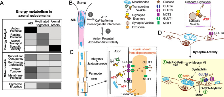
“Studies have shown that glycolytic enzymes are physically associated with fast-moving vesicles through their interaction with Huntingtin and provide onboard ATP synthesis to propel vesicular transport [154, 155] (Fig.”
BackgroundLim, M. A., Selak, M. A., Xiang, Z., Krainc, D., Neve, R. L., Kraemer, B. C., Watts, J. L., & Kalb, R. G. (2012) · Journal of Neuroscience · doi:10.1523/jneurosci.6554-10.2012 · Cited by 102
Howlett, E. H., Jensen, N. O., Belmonte, F. R., Zafar, F., Hu, X., Kluss, J. H., Schüle, B., Kaufman, B. A., Greenamyre, J. T., & Sanders, L. H. (2017) · Human Molecular Genetics · doi:10.1093/hmg/ddx320 · Cited by 101
ATP‐driven molecular chaperone machines
Clare, D. K., & Saibil, H. R. (2013) · Biopolymers · doi:10.1002/bip.22361 · Cited by 100
Matthews, D. C., Lerman, H., Lukic, A., Andrews, R. D., Mirelman, A., Wernick, M. N., Giladi, N., Strother, S. C., Evans, K. C., Cedarbaum, J. M., et al. (2018) · NeuroImage Clinical · doi:10.1016/j.nicl.2018.08.006 · Cited by 100
Burré, J., & Volknandt, W. (2007) · Journal of Neurochemistry · doi:10.1111/j.1471-4159.2007.04453.x · Cited by 99 · cited 1×
“The proteomic characterization of mammalian brain synaptic vesicles also identifies many glycolytic enzymes [232, 233] that physically associate with synaptic vesicles to fuel their transport along axons and their neurotransmitter uptake [154, 155, 234, 235].”
BackgroundNoori, A., Mezlini, A. M., Hyman, B. T., Serrano-Pozo, A., & Das, S. (2021) · Neurobiology of Disease · doi:10.1016/j.nbd.2020.105225 · Cited by 98
González‐Domínguez, R., García‐Barrera, T., Vitórica, J., & Gómez‐Ariza, J. L. (2014) · Biochimica et Biophysica Acta (BBA) - Molecular Basis of Disease · doi:10.1016/j.bbadis.2014.09.014 · Cited by 97
Parkinson disease and the risk of epileptic seizures
Gruntz, K., Bloechliger, M., Becker, C., Jick, S. S., Fuhr, P., Meier, C. R., & Rüegg, S. J. (2018) · Annals of Neurology · doi:10.1002/ana.25157 · Cited by 97 · cited 1×
“Pre-symptomatic neuronal hyperexcitability in disease-vulnerable brain regions has also been documented in PD [365, 366], HD [367], and ALS [368].”
BackgroundOligodendroglial Cells in Alzheimer’s Disease
Butt, A. M., Rocha, I. C. D. L., & Rivera, A. D. (2019) · Advances in experimental medicine and biology · doi:10.1007/978-981-13-9913-8_12 · Cited by 97
White matter alterations in early‐stage Alzheimer's disease: A tract‐specific study
Wen, Q., Mustafi, S. M., Li, J., Risacher, S. L., Tallman, E., Brown, S. A., West, J. D., Harezlak, J., Farlow, M. R., Unverzagt, F. W., et al. (2019) · Alzheimer's & Dementia: Diagnosis, Assessment & Disease Monitoring · doi:10.1016/j.dadm.2019.06.003 · Cited by 96 · cited 1×
“,DTI, NODDI, and q-space) to study white matter alterations in early-stage AD Altered white matter tracts (cingulum, thalamic radiation, and forceps major) in MCI subjects: • Fractional anisotropy↓ (loss of fiber organization) • Radial diffusivity↑ (myelin degeneration or cell membrane deterioration) [52]”
BackgroundMitochondrial dysfunction in astrocytes: A role in Parkinson’s disease?
Bantle, C. M., Hirst, W. D., Weihofen, A., & Shlevkov, E. (2021) · Frontiers in Cell and Developmental Biology · doi:10.3389/fcell.2020.608026 · Cited by 95
Call, C. L., & Bergles, D. E. (2021) · Nature Communications · doi:10.1038/s41467-021-25035-2 · Cited by 95
Angeletti, C., Amici, A., Gilley, J., Loreto, A., Trapanotto, A. G., Antoniou, C., Merlini, E., Coleman, M. P., & Orsomando, G. (2022) · iScience · doi:10.1016/j.isci.2022.103812 · Cited by 95 · cited 1×
“Under normal physiological conditions, when NAD+ and NADP+ levels are actively maintained by NMNAT2 and NAD+ kinase (NADK) [268, 316], SARM1 stays inactive because of substrate inhibitions [313, 317].”
BackgroundCheng, A., Wang, J., Ghena, N., Zhao, Q., Perone, I., King, T. M., Veech, R. L., Gorospe, M., Wan, R., & Mattson, M. P. (2019) · Journal of Neuroscience · doi:10.1523/jneurosci.1446-19.2019 · Cited by 94 · cited 1×
“Thus, it is noteworthy that GABAergic neurons are particularly vulnerable to SIRT3 loss [374].”
BackgroundWang, X., Zhang, Q., Bao, R., Zhang, N., Wang, Y., Polo‐Parada, L., Tarim, A. O., Alemifar, A., Han, X., Wilkins, H., et al. (2017) · Cell Reports · doi:10.1016/j.celrep.2017.08.022 · Cited by 93
Nmnat2 delays axon degeneration in superior cervical ganglia dependent on its NAD synthesis activity
Yan, T., Feng, Y., Zheng, J., Ge, X., Zhang, Y., Wu, D., Zhao, J., & Zhai, Q. (2010) · Neurochemistry International · doi:10.1016/j.neuint.2009.09.007 · Cited by 92 · cited 2×
“NMNAT2 is the most abundant NMNAT in the brain and is mainly expressed in neurons [269, 270].”
BackgroundAge-Related Phasic Patterns of Mitochondrial Maintenance in Adult Caenorhabditis elegans Neurons
Morsci, N. S., Hall, D. H., Driscoll, M., & Sheng, Z.-H. (2016) · The Journal of Neuroscience · doi:10.1523/jneurosci.2799-15.2016 · Cited by 92
NMNATs, evolutionarily conserved neuronal maintenance factors
Ali, Y. O., Li-Kroeger, D., Bellen, H. J., Zhai, R. G., & Lu, H.-C. (2013) · Trends in Neurosciences · doi:10.1016/j.tins.2013.07.002 · Cited by 91
Self-citationSynaptosomal Mitochondrial Dysfunction in 5xFAD Mouse Model of Alzheimer's Disease
Wang, L., Guo, L., Lu, L., Sun, H., Shao, M., Beck, S. J., Lin, L., Ramachandran, J., Du, Y., & Du, H. (2016) · PLoS ONE · doi:10.1371/journal.pone.0150441 · Cited by 90
C9orf72 expansion within astrocytes reduces metabolic flexibility in amyotrophic lateral sclerosis
Allen, S. P., Hall, B., Woof, R., Francis, L. I., Gatto, N., Shaw, A. C., Myszczynska, M. A., Hemingway, J., Coldicott, I., Willcock, A., et al. (2019) · Brain · doi:10.1093/brain/awz302 · Cited by 89
Dynein Light Chain LC8 Regulates Syntaphilin-Mediated Mitochondrial Docking in Axons
Chen, Y., Gerwin, C. M., & Sheng, Z. (2009) · Journal of Neuroscience · doi:10.1523/jneurosci.1472-09.2009 · Cited by 88 · cited 1×
“SNPH also binds to dynein light chain LC8, which then enhances the docking between SNPH and microtubules to further reduce mitochondrial mobility [215].”
BackgroundGlaab, E., & Schneider, R. (2014) · Neurobiology of Disease · doi:10.1016/j.nbd.2014.11.002 · Cited by 86 · cited 2×
“Integrative bioinformatic analysis using brain multi-omics data has illuminated the agedependent molecular and cellular changes in four major NDAs: AD [17], PD [18], HD [19] and amyotrophic lateral sclerosis (ALS) [31].”
BackgroundDecoding the relationship between ageing and amyotrophic lateral sclerosis: a cellular perspective
Pandya, V. A., & Patani, R. (2019) · Brain · doi:10.1093/brain/awz360 · Cited by 86 · cited 13×
“This study observed an increase in upstream glucose catabolism activity, including glycolysis and TCA cycle, as well as the lipid peroxidation in ALS mouse presynapses [353, 354].”
BackgroundInfluentialUemura, N., Uemura, M. T., Lo, A., Bassil, F., Zhang, B., Luk, K. C., Lee, V. M. ‐., Takahashi, R., & Trojanowski, J. Q. (2019) · Journal of Neuropathology & Experimental Neurology · doi:10.1093/jnen/nlz070 · Cited by 86
Liu, W., Zhuo, P., Li, L., Jin, H., Lin, B., Zhang, Y., Liang, S., Wu, J., Huang, J., Wang, Z., et al. (2017) · Free Radical Biology and Medicine · doi:10.1016/j.freeradbiomed.2017.07.024 · Cited by 84
Subcellular compartmentalization of NAD+ and its role in cancer: A sereNADe of metabolic melodies
Zhu, Y., Liu, J., Park, J., Rai, P., & Zhai, R. G. (2019) · Pharmacology & Therapeutics · doi:10.1016/j.pharmthera.2019.04.002 · Cited by 82
Self-citationMarion, C. M., McDaniel, D. P., & Armstrong, R. C. (2019) · Experimental Neurology · doi:10.1016/j.expneurol.2019.113040 · Cited by 82
Mayer, P. R., Huang, N., Dewey, C. M., Dries, D. R., Zhang, H., & Yu, G. (2010) · Journal of Biological Chemistry · doi:10.1074/jbc.m110.178913 · Cited by 82
Synaptic mitochondria: A brain mitochondria cluster with a specific proteome
Völgyi, K., Gulyássy, P., Háden, K., Kis, V., Badics, K., Kékesi, K. A., Simor, A., Györffy, B., Tóth, E. A., Lübec, G., et al. (2015) · Journal of Proteomics · doi:10.1016/j.jprot.2015.03.005 · Cited by 81
Singulani, M. P., Pereira, C. P. M., Ferreira, A. F. F., Garcia, P. C., Ferrari, G. D., Alberici, L. C., & Britto, L. R. (2020) · Experimental Gerontology · doi:10.1016/j.exger.2020.110882 · Cited by 81
R6/2 Huntington's Disease Mice Develop Early and Progressive Abnormal Brain Metabolism and Seizures
Cepeda-Prado, E., Popp, S., Khan, U., Stefanov, D. G., Rodríguez, J., Menalled, L., Dow-Edwards, D. L., Small, S. A., & Moreno, H. (2012) · Journal of Neuroscience · doi:10.1523/jneurosci.0388-12.2012 · Cited by 80
Brain cortex mitochondrial bioenergetics in synaptosomes and non-synaptic mitochondria during aging
Lores‐Arnaiz, S., Lombardi, P., Karadayian, A. G., Orgambide, F., Cicerchia, D., & Bustamante, J. (2016) · Neurochemical Research · doi:10.1007/s11064-015-1817-5 · Cited by 80
Decreased sirtuin deacetylase activity in LRRK2 G2019S iPSC-derived dopaminergic neurons
Schwab, A. J., Sison, S. L., Meade, M. R., Broniowska, K. A., Corbett, J. A., & Ebert, A. D. (2017) · Stem Cell Reports · doi:10.1016/j.stemcr.2017.10.010 · Cited by 80
Heterogeneity of astrocyte and NG2 cell insertion at the node of ranvier
Serwanski, D. R., Jukkola, P. I., & Nishiyama, A. (2016) · The Journal of Comparative Neurology · doi:10.1002/cne.24083 · Cited by 77 · cited 1×
“PNS nodes are solely contacted by the microvilli emanating from Schwann cells [170], while CNS nodes are mostly covered by astrocytic processes and sometimes contacted by oligodendrocyte precursor cells (OPCs) [175].”
BackgroundA quantitative electron microscopic study of the purkinje cell axon initial segment
Somogyu, P., & Hámori, J. (1976) · Neuroscience · doi:10.1016/0306-4522(76)90127-5 · Cited by 76
Proteomic profiling of neuronal mitochondria reveals modulators of synaptic architecture
Graham, L. C., Eaton, S. L., Brunton, P. J., Atrih, A., Smith, C., Lamont, D. J., Gillingwater, T. H., Pennetta, G., Skehel, P., & Wishart, T. M. (2017) · Molecular Neurodegeneration · doi:10.1186/s13024-017-0221-9 · Cited by 76
Graham, S. F., Rey, N. L., Yılmaz, A., Kumar, P., Madaj, Z., Maddens, M. E., Bahado-Singh, R. O., Becker, K., Schulz, E., Meyerdirk, L. K., et al. (2018) · Journal of Proteome Research · doi:10.1021/acs.jproteome.8b00224 · Cited by 75
Axonal degeneration induced by glutamate excitotoxicity is mediated by necroptosis
Hernández, D. E., Salvadores, N., Moya‐Alvarado, G., Catalán, R. J., Bronfman, F. C., & Court, F. A. (2018) · Journal of Cell Science · doi:10.1242/jcs.214684 · Cited by 75 · cited 1×
“Furthermore, excitotoxicity triggered by excessive glutamate release and Ca2+ influx can activate a necroptosis pathway that is reported to non-canonically deplete axonal NMNAT2 and activate SARM1, which destabilizes NAD homeostasis, exacerbates glucose hypometabolism, and eventually drives axonal degeneration and net…”
BackgroundSummers, D. W., Frey, E. N., Walker, L. J., Milbrandt, J., & DiAntonio, A. (2019) · Molecular Neurobiology · doi:10.1007/s12035-019-01796-2 · Cited by 74
Stedehouder, J., Brizee, D., Slotman, J. A., Pascual-Garcia, M., Leyrer, M. L., Bouwen, B. L. J., Dirven, C. M. F., Gao, Z., Berson, D. M., Houtsmuller, A. B., et al. (2019) · eLife · doi:10.7554/elife.48615 · Cited by 74 · cited 1×
“These axonal arbors are mostly located in the grey matter and are rarely myelinated [186, 187].”
BackgroundDifferential presynaptic ATP supply for basal and high-demand transmission
Sobieski, C., Fitzpatrick, M. J., & Mennerick, S. J. (2017) · The Journal of Neuroscience · doi:10.1523/jneurosci.2712-16.2017 · Cited by 74 · cited 23×
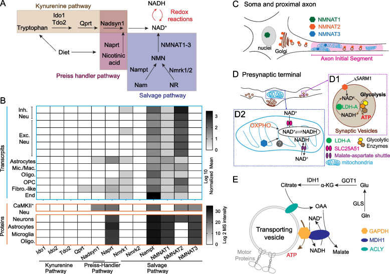
“Pivotally, the NAD redox potential (NAD+/NADH) determines the thermodynamic driving force for the NAD+ or NADH consuming steps in both glycolysis and OXPHO [255].”
BackgroundInfluentialAn emerging role of dysfunctional axon-oligodendrocyte coupling in neurodegenerative diseases
Mot, A. I., Depp, C., & Nave, K. (2018) · Dialogues in Clinical Neuroscience · doi:10.31887/dcns.2018.20.4/amot · Cited by 72 · cited 1×
“The morphological complexity of astrocytes significantly decreases in AD-susceptible brain regions during aging, such as the entorhinal cortex and hippocampal CA1 region [402, 403].”
BackgroundStojakovic, A., Trushin, S., Sheu, A., Khalili, L., Chang, S.-Y., Li, X., Christensen, T., Salisbury, J. L., Geroux, R. E., Gateno, B., et al. (2021) · Communications Biology · doi:10.1038/s42003-020-01584-y · Cited by 72
van Nifterick, A. M., Gouw, A. A., van Kesteren, R. E., Scheltens, P., Stam, C. J., & de Haan, W. (2022) · Alzheimer s Research & Therapy · doi:10.1186/s13195-022-01041-4 · Cited by 72
Glucose consumption of inflammatory cells masks metabolic deficits in the brain
Backes, H., Walberer, M., Ladwig, A., Rueger, M. A., Neumaier, B., Endepols, H., Hoehn, M., Fink, G. R., Schroeter, M., & Graf, R. (2015) · NeuroImage · doi:10.1016/j.neuroimage.2015.12.044 · Cited by 71 · cited 1×
“Furthermore, the remyelinating and immune reactive process in oligodendrocytes seem to be energy demanding [413, 414], potentially competing for the glucose metabolism substrates and therefore worsening the axonal bioenergetics.”
BackgroundSubcellular NAD+ pools are interconnected and buffered by mitochondrial NAD+
Høyland, L. E., VanLinden, M. R., Niere, M., Strømland, Ø., Sharma, S. A., Dietze, J., Tolås, I., Lucena, E., Bifulco, E., Sverkeli, L. J., et al. (2024) · Nature Metabolism · doi:10.1038/s42255-024-01174-w · Cited by 71
Vagnoni, A., Hoffmann, P. C., & Bullock, S. L. (2015) · Journal of Cell Science · doi:10.1242/jcs.179184 · Cited by 70 · cited 1×
“Reduced mitochondrial transport is also observed in Drosophila wing neuron axons [78, 79], and mouse retina nerve fiber layer myelinated axons [80] during aging.”
BackgroundA cAMP/PKA/Kinesin-1 Axis Promotes the Axonal Transport of Mitochondria in Aging Drosophila Neurons
Vagnoni, A., & Bullock, S. L. (2018) · Current Biology · doi:10.1016/j.cub.2018.02.048 · Cited by 70 · cited 1×
“Reduced mitochondrial transport is also observed in Drosophila wing neuron axons [78, 79], and mouse retina nerve fiber layer myelinated axons [80] during aging.”
BackgroundStauch, K. L., Purnell, P. R., & Fox, H. S. (2014) · Aging · doi:10.18632/aging.100657 · Cited by 69 · cited 2×
“In synaptosomal mitochondria samples purified from the whole brain, aging elicits mitochondrial DNA (mtDNA) deletion and activation of nuclear respiratory factor 1 (NRF1) and peroxisome proliferatoractivated receptor gamma coactivator 1 alpha (PGC1A) mediated mitochondrial fitness pathways, accompanied by the adaptive…”
BackgroundTorrens‐Mas, M., Pons, D. G., Sastre‐Serra, J., Oliver, J., & Roca, P. (2020) · Redox Biology · doi:10.1016/j.redox.2020.101505 · Cited by 69
Subcellular proteomics of dopamine neurons in the mouse brain
Hobson, B. D., Choi, S. J., Mosharov, E. V., Soni, R. K., Sulzer, D., & Sims, P. A. (2022) · eLife · doi:10.7554/elife.70921 · Cited by 68 · cited 1×
“Although it is not known whether mammalian brains retain this “glycolytic metabolon” forming capability upon hypoxia, proteomics profiling shows the enrichment of glycolytic enzymes in striatal axon terminals of midbrain dopaminergic neurons in mouse brains [231].”
BackgroundNeuronal structural remodeling: is it all about access?
Chen, J. L., & Nedivi, E. (2010) · Current Opinion in Neurobiology · doi:10.1016/j.conb.2010.06.002 · Cited by 68 · cited 1×
“Furthermore, the robust anatomical plasticity of axons, reflected by the dynamic gain and loss of axonal arbors and presynaptic boutons, requires substantial protein synthesis as well as cytoskeleton remodeling [194].”
BackgroundMoutaux, E., Christaller, W., Scaramuzzino, C., Genoux, A., Charlot, B., Cazorla, M., & Saudou, F. (2018) · Scientific Reports · doi:10.1038/s41598-018-31759-x · Cited by 66
Qu, X., Kumar, A., Blockus, H., Waites, C. L., & Bartolini, F. (2019) · Current Biology · doi:10.1016/j.cub.2019.10.049 · Cited by 66 · cited 3×
“1D), according to an observation in mouse hippocampi upon LTP induction [219].”
BackgroundKim, J., Choi, I.-Y., Michaelis, M. L., & Lee, P. (2011) · NeuroImage · doi:10.1016/j.neuroimage.2011.02.039 · Cited by 65
Rodriguez-Moreno, J., Rollenhagen, A., Arlandis, J., Santuy, A., Merchán-Pérez, Á., DeFelipe, J., Lübke, J. H. R., & Clascá, F. (2017) · Cerebral Cortex · doi:10.1093/cercor/bhx187 · Cited by 65 · cited 1×
“Still, less than 50% of boutons contain mitochondria [202–205], except lemniscal thalamocortical synapses where 92% of them contain mitochondria [206].”
BackgroundDillman, A., Majounie, E., Ding, J., Gibbs, J. R., Hernandez, D. G., Arepalli, S., Traynor, B. J., Singleton, A., Galter, D., & Cookson, M. R. (2017) · Scientific Reports · doi:10.1038/s41598-017-17322-0 · Cited by 65
Carlyle, B. C., Kandigian, S. E., Kreuzer, J., Das, S., Trombetta, B. A., Kuo, Y., Bennett, D. A., Schneider, J. A., Petyuk, V. A., Kitchen, R. R., et al. (2021) · Neurobiology of Aging · doi:10.1016/j.neurobiolaging.2021.04.012 · Cited by 65
Peters, O. M., Lewis, E. A., Osterloh, J. M., Weiss, A., Salameh, J. S., Metterville, J., Brown, R. H., & Freeman, M. R. (2018) · Human Molecular Genetics · doi:10.1093/hmg/ddy260 · Cited by 64
FHL2 anchors mitochondria to actin and adapts mitochondrial dynamics to glucose supply
Basu, H., Pekkurnaz, G., Falk, J. E., Wei, W., Chin, M., Steen, J. A. J., & Schwarz, T. L. (2021) · The Journal of Cell Biology · doi:10.1083/jcb.201912077 · Cited by 64 · cited 1×
“the O-linked-N-acetylglucosaminylation (O-GlcNAcylation) of the mitochondrial adaptor protein, Milton, which allows its interaction with four and a half LIM domains protein 2 (FHL2) to anchor mitochondria onto presynaptic actin filaments, therefore arresting mitochondria in the boutons [217, 218].”
BackgroundSzegő, É. M., Dominguez‐Meijide, A., Gerhardt, E., König, A., Koss, D. J., Li, W., Pinho, R., Fahlbusch, C., Johnson, M., Santos, P. I., et al. (2019) · Cell Reports · doi:10.1016/j.celrep.2019.06.009 · Cited by 63 · cited 1×
“Overexpressed α-synuclein accumulates in presynaptic sites, interacts with the mitochondrial chaperone heat shock protein 10 (HSP10), and prevents its import into mitochondria, resulting in compromised quality control of essential antioxidant machineries within the mitochondrial matrix which eventually leads to synapt…”
BackgroundImplications of NAD metabolism in pathophysiology and therapeutics for neurodegenerative diseases
Hikosaka, K., Yaku, K., Okabe, K., & Nakagawa, T. (2021) · Nutritional Neuroscience · doi:10.1080/1028415x.2019.1637504 · Cited by 62 · cited 1×
“Using preclinical NDA models, numerous studies provide strong evidence supporting the therapeutic benefits of supplementing NAD+ and its precursors [384, 385].”
BackgroundXu, Y., Mei, Y., Shi, X.-Q., Yi-fang, Z., Wang, X., Guan, L., Wang, Q., & Pan, H. (2019) · Brain Research · doi:10.1016/j.brainres.2019.05.037 · Cited by 61
McClory, H., Williams, D., Sapp, E., Gatune, L. W., Wang, P., DiFiglia, M., & Li, X. (2014) · Acta Neuropathologica Communications · doi:10.1186/s40478-014-0178-7 · Cited by 60
Myelination induces axonal hotspots of synaptic vesicle fusion that promote sheath growth
Almeida, R., Williamson, J., Madden, M., Early, J. J., Voas, M. G., Talbot, W. S., Bianco, I. H., & Lyons, D. A. (2021) · Current Biology · doi:10.1016/j.cub.2021.06.036 · Cited by 59 · cited 1×
“These axonal arbors are mostly located in the grey matter and are rarely myelinated [186, 187].”
BackgroundLove, N. R., Pollak, N., Dölle, C., Niere, M., Chen, Y., Oliveri, P., Amaya, E., Patel, S., & Ziegler, M. (2015) · Proceedings of the National Academy of Sciences · doi:10.1073/pnas.1417290112 · Cited by 59 · cited 3×
“It is yet to be determined whether NADP+ synthesis by NADK is required to suppress SARM1 activity and if SARM1 also mediated NADP+ degradation to exacerbate oxidative stress [324].”
BackgroundSantuy, A., Turégano-López, M., Rodríguez, J., Alonso‐Nanclares, L., DeFelipe, J., & Merchán-Pérez, Á. (2018) · Cerebral Cortex · doi:10.1093/cercor/bhy159 · Cited by 59
Stein, L. R., Wozniak, D. F., Dearborn, J. T., Kubota, S., Apte, R. S., Izumi, Y., Zorumski, C. F., & Imai, S. (2014) · Journal of Neuroscience · doi:10.1523/jneurosci.4730-13.2014 · Cited by 58
Toxic Peripheral Neuropathies: Agents and Mechanisms
Valentine, W. M. (2019) · Toxicologic Pathology · doi:10.1177/0192623319854326 · Cited by 57 · cited 1×
“Axons, the longest and the most morphologically complex subcellular compartment of most neurons [57–59], are particularly vulnerable to aging [60, 61], environmental insults [62] and neurodegenerative conditions Abbreviations: ALS amyotrophic lateral sclerosis, APOE Apolipoprotein E, APP Amyloid precursor protein, C9O…”
BackgroundMorphology of mitochondria in spatially restricted axons revealed by cryo-electron tomography
Fischer, T. D., Dash, P. K., Liu, J., & Waxham, M. N. (2018) · PLoS Biology · doi:10.1371/journal.pbio.2006169 · Cited by 57
Wang, X., He, H., Xiong, X., Zhou, S., Wang, W., Feng, L., Han, R., & Xie, C. (2021) · Frontiers in Cell and Developmental Biology · doi:10.3389/fcell.2021.668491 · Cited by 56
Glycolysis selectively shapes the presynaptic action potential waveform
Lujan, B., Kushmerick, C., Banerjee, T. D., Dagda, R. K., & Renden, R. (2016) · Journal of Neurophysiology · doi:10.1152/jn.00629.2016 · Cited by 56 · cited 1×
“In the calyx of Held, glycolysis is preferentially required to shape faithful AP waveforms and sustain synaptic transmission [229].”
BackgroundLafourcade, M., van der Goes, M.-S. H., Vardalaki, D., Brown, N. J., Voigts, J., Yun, D. H., Kim, M. E., Ku, T., & Harnett, M. T. (2022) · Neuron · doi:10.1016/j.neuron.2022.01.025 · Cited by 56
Lores‐Arnaiz, S., & Bustamante, J. (2011) · Neuroscience · doi:10.1016/j.neuroscience.2011.04.060 · Cited by 55
Combined LRRK2 mutation, aging and chronic low dose oral rotenone as a model of Parkinson’s disease
Liu, H., Ho, P. W., Leung, G. C.-T., Lam, C. S.-C., Pang, S. Y.-Y., Li, L., Kung, M. H.-W., Ramsden, D. B., & Ho, S. (2017) · Scientific Reports · doi:10.1038/srep40887 · Cited by 55
Dong, Y., Digman, M. A., & Brewer, G. J. (2019) · GeroScience · doi:10.1007/s11357-019-00052-8 · Cited by 54 · cited 1×
“Noteworthy, assessed by two-photon lifetime imaging in dissociated neurons from mouse hippocampus of different ages, mitochondrial NADH level is significantly lower in 3xTg neurons, and declines with age [382].”
BackgroundHyperexcitable Substantia Nigra Dopamine Neurons inPINK1- andHtrA2/Omi-Deficient Mice
Bishop, M. W., Chakraborty, S., Matthews, G. A., Dougalis, A., Wood, N. W., Festenstein, R., & Ungless, M. A. (2010) · Journal of Neurophysiology · doi:10.1152/jn.00466.2010 · Cited by 53
Axonal and myelinic pathology in 5xFAD Alzheimer’s mouse spinal cord
Chu, T., Cummins, K., Sparling, J. S., Tsutsui, S., Brideau, C., Nilsson, K. P. R., Joseph, J. T., & Stys, P. K. (2017) · PLoS ONE · doi:10.1371/journal.pone.0188218 · Cited by 52
Central presynaptic terminals are enriched in ATP but the majority lack mitochondria
Chavan, V., Willis, J., Walker, S. K., Clark, H. R., Liu, X., Fox, M. A., Srivastava, S., & Mukherjee, K. (2015) · PLoS ONE · doi:10.1371/journal.pone.0125185 · Cited by 52
Presynaptic Mitochondrial Volume and Packing Density Scale with Presynaptic Power Demand
Justs, K. A., Lu, Z., Chouhan, A. K., Borycz, J. A., Lu, Z., Meinertzhagen, I. A., & Macleod, G. T. (2021) · Journal of Neuroscience · doi:10.1523/jneurosci.1236-21.2021 · Cited by 52 · cited 1×
“In the Drosophila motor nerve terminal, the mitochondrial volume and densities within each bouton is positively correlated with the estimated energetic demand of each bouton [213], a correlation presumably caused by the activity-driven mechanism that couples energy consumption and synaptic mitochondrial recruitment.”
BackgroundPAM forms an atypical SCF ubiquitin ligase complex that ubiquitinates and degrades NMNAT2
Desbois, M., Crawley, O., Evans, P. R., Baker, S. T., Masuho, I., Yasuda, R., & Grill, B. J. (2018) · Journal of Biological Chemistry · doi:10.1074/jbc.ra118.002176 · Cited by 50 · cited 1×
“The abundance of NMNAT2, the main axonal NAD+ provider, is tightly regulated by proteosome degradation and mitogen-activated protein kinase (MAPK) signaling [318, 319].”
BackgroundWhitmore, C. A., Haynes, J. R., Behof, W. J., Rosenberg, A. J., Tantawy, M. N., Hachey, B. C., Wadzinski, B. E., Spiller, B. W., Peterson, T. E., Paffenroth, K. C., et al. (2022) · Pharmaceuticals · doi:10.3390/ph15060742 · Cited by 50
Striatal synaptic bioenergetic and autophagic decline in premotor experimental parkinsonism
Merino-Galán, L., Jiménez‐Urbieta, H., Zamarbide, M., Rodríguez-Chinchilla, T., Belloso-Iguerategui, A., Santamaría, E., Fernández‐Irigoyen, J., Aiastui, A., Doudnikoff, É., Bézard, E., et al. (2022) · Brain · doi:10.1093/brain/awac087 · Cited by 48 · cited 2×
“Synaptosomal proteomic analysis with SNpc prepared from a PD mouse model overexpressing mutant human α-synuclein further sheds light on the importance of synaptic metabolic rewiring that precedes mitochondrial damage and synaptic pathology [114, 349].”
BackgroundAzevedo, C., Teku, G., Pomeshchik, Y., Reyes, J. F., Chumarina, M., Russ, K., Savchenko, E., Hammarberg, A., Lamas, N. J., Collin, A., et al. (2022) · Proceedings of the National Academy of Sciences · doi:10.1073/pnas.2111405119 · Cited by 48
Regulation of Myelination by Exosome Associated Retinoic Acid Release from NG2-Positive Cells
Goncalves, M. B. C. V., Wu, Y., Clarke, E. E., Grist, J., Hobbs, C., Trigo, D., Jack, J., & Corcoran, J. P. T. (2019) · Journal of Neuroscience · doi:10.1523/jneurosci.2922-18.2019 · Cited by 48 · cited 1×
“Knowing that OPCs secrete exosomes [283], it will be interesting to investigate whether membranebound NMNAT2 can reach axons through OPC-derived exosomes and facilitate axonal NAD biosynthesis.”
BackgroundPresynaptic Boutons That Contain Mitochondria Are More Stable
Lees, R. M., Johnson, J. D., & Ashby, M. C. (2020) · Frontiers in Synaptic Neuroscience · doi:10.3389/fnsyn.2019.00037 · Cited by 48
SARM1 signaling mechanisms in the injured nervous system
Sambashivan, S., & Freeman, M. R. (2021) · Current Opinion in Neurobiology · doi:10.1016/j.conb.2021.05.004 · Cited by 46
Distribution of mitochondria along small‐diameter myelinated central nervous system axons
Edgar, J. M., McCulloch, M. C., Thomson, C. E., & Griffiths, I. R. (2008) · Journal of Neuroscience Research · doi:10.1002/jnr.21672 · Cited by 44
Jeffries, M. A., McLane, L. E., Khandker, L., Mather, M. L., Evangelou, A. V., Kantak, D., Bourne, J. N., Macklin, W. B., & Wood, T. L. (2021) · Journal of Neuroscience · doi:10.1523/jneurosci.1377-20.2021 · Cited by 44 · cited 1×
“Furthermore, the remyelinating and immune reactive process in oligodendrocytes seem to be energy demanding [413, 414], potentially competing for the glucose metabolism substrates and therefore worsening the axonal bioenergetics.”
BackgroundMitochondria modulate programmed neuritic retraction
Баранов, В. С., Baranova, O. V., Yablonska, S., Suofu, Y., Vazquez, A. L., Kozai, T. D. Y., Cui, X. T., Ferrando, L. M., Larkin, T. M., Tyurina, Y. Y., et al. (2018) · Proceedings of the National Academy of Sciences · doi:10.1073/pnas.1811021116 · Cited by 43 · cited 5×
“These measurements are particularly important for presynaptic boutons where mitochondria support the intense synaptic activities while enduring the potentially incompetent protein turnover and Ca2+ buffering [222, 223].”
BackgroundInfluentialOzaki, E., Gibbons, L., Neto, N. G. B., Kenna, P. F., Carty, M., Humphries, M. M., Humphries, P., Campbell, M., Monaghan, M. G., Bowie, A., et al. (2020) · Life Science Alliance · doi:10.26508/lsa.201900618 · Cited by 43
Metabolic Recruitment in Brain Tissue
Barros, L. F., Ruminot, I., Sotelo-Hitschfeld, T., Lerchundi, R., & Fernández‐Moncada, I. (2022) · Annual Review of Physiology · doi:10.1146/annurev-physiol-021422-091035 · Cited by 43 · cited 1×
“suggest a putative coupling mechanism between astrocyte metabolism and synaptic activity through astrocytic Na+/K+ ATPases and Na/HCO3 cotransporters [250].”
BackgroundAn early axonopathy in a hLRRK2(R1441G) transgenic model of Parkinson disease
Tagliaferro, P. A., Kareva, T., Oo, T. F., Yarygina, O., Kholodilov, N. G., & Burke, R. E. (2015) · Neurobiology of Disease · doi:10.1016/j.nbd.2015.07.009 · Cited by 43
Yakushev, I. Y., Schreckenberger, M., Müller, M. J., Schermuly, I., Cumming, P. D., Stoeter, P., Gerhard, A., & Fellgiebel, A. (2011) · European Journal of Nuclear Medicine and Molecular Imaging · doi:10.1007/s00259-011-1882-1 · Cited by 42
Gatto, R. G., Ye, A. Q., Colon-Perez, L., Mareci, T. H., Lysakowski, A., Price, S. D., Brady, S. T., Karaman, M., Morfini, G., & Magin, R. L. (2019) · Magnetic Resonance Materials in Physics, Biology and Medicine · doi:10.1007/s10334-019-00742-6 · Cited by 42
Comparing 3D ultrastructure of presynaptic and postsynaptic mitochondria
Delgado, T., Petralia, R. S., Freeman, D. W., Sedlacek, M., Wang, Y., Brenowitz, S. D., Sheu, S., Gu, J. W., Kapogiannis, D., Mattson, M. P., et al. (2019) · Biology Open · doi:10.1242/bio.044834 · Cited by 42
Shokhirev, M. N., & Johnson, A. A. (2022) · Ageing Research Reviews · doi:10.1016/j.arr.2022.101721 · Cited by 42
Aberrant upregulation of the glycolytic enzyme PFKFB3 in CLN7 neuronal ceroid lipofuscinosis
López-Fabuel, I., Macia, M. G., Buondelmonte, C., Burmistrova, O., Bonora, N., Alonso-Batan, P., Morant-Ferrando, B., Vicente‐Gutiérrez, C., Jiménez-Blasco, D., Quintana–Cabrera, R., et al. (2022) · Nature Communications · doi:10.1038/s41467-022-28191-1 · Cited by 42 · cited 1×
“pentose phosphate pathway flux and antioxidant capability [239, 240].”
BackgroundBradshaw, D. V., Knutsen, A. K., Korotcov, A. V., Sullivan, G. M., Radomski, K. L., Dardzinski, B. J., Zi, X., McDaniel, D. P., & Armstrong, R. C. (2021) · Acta Neuropathologica Communications · doi:10.1186/s40478-021-01193-8 · Cited by 41
Hong, I., Kang, T., Yoo, Y., Park, R., Lee, J., Lee, S., Kim, J., Song, B., Kim, S.-Y., Moon, M., et al. (2013) · Journal of Alzheimer s Disease · doi:10.3233/jad-130311 · Cited by 41
The Na(+)/K(+) pump dominates control of glycolysis in hippocampal dentate granule cells
Meyer, D. J., Díaz-García, C. M., Nathwani, N., Rahman, M., & Yellen, G. (2022) · eLife · doi:10.7554/elife.81645 · Cited by 40
Dufour, A., Rollenhagen, A., Sätzler, K., & Lübke, J. H. R. (2015) · Cerebral Cortex · doi:10.1093/cercor/bhv270 · Cited by 39
Cable energy function of cortical axons
Ju, H., Hines, M. L., & Yu, Y. (2016) · Scientific Reports · doi:10.1038/srep29686 · Cited by 39 · cited 17×
“Future studies are needed to determine the contribution of glycolysis to AIS energetics.”
BackgroundInfluentialvan der Velpen, V., Rosenberg, N., Maillard, V., Teav, T., Chatton, J., Gallart‐Ayala, H., & Ivanišević, J. (2021) · Journal of Neurochemistry · doi:10.1111/jnc.15362 · Cited by 39 · cited 1×
“Similarly, dysregulated NAD homeostasis was also found in an AD mouse model, 3xTg transgenic mice, in a sex-biased manner [380, 381].”
BackgroundKrell‐Roesch, J., Syrjanen, J. A., Vassilaki, M. E., Lowe, V. J., Vemuri, P., Mielke, M. M., Machulda, M. M., Stokin, G. B., Christianson, T. J. H., Kremers, W. K., et al. (2020) · American Journal of Geriatric Psychiatry · doi:10.1016/j.jagp.2020.06.006 · Cited by 39
Franke, T. N., Irwin, C., Bayer, T. A., Brenner, W., Beindorff, N., Bouter, C., & Bouter, Y. (2020) · Frontiers in Medicine · doi:10.3389/fmed.2020.00529 · Cited by 37
Wade, A., Jacob, P., & Morton, A. J. (2007) · Brain Research · doi:10.1016/j.brainres.2007.06.059 · Cited by 37
Amyloid and FDG PET of Successful Cognitive Aging: Global and Cingulate-Specific Differences
Baran, T. M., Lin, F. V., & Initiative, A. D. N. (2018) · Journal of Alzheimer s Disease · doi:10.3233/jad-180360 · Cited by 37
Márkus, N. M., Hasel, P., Qiu, J., Bell, K. F. S., Heron, S., Kind, P. C., Dando, O., Simpson, T. I., & Hardingham, G. E. (2016) · PLoS ONE · doi:10.1371/journal.pone.0148164 · Cited by 36
Diehl‐Schmid, J., Licata, A. E., Goldhardt, O., Förstl, H., Yakushew, I., Otto, M., Anderl‐Straub, S., Beer, A. J., Ludolph, A. C., Landwehrmeyer, G. B., et al. (2019) · Translational Psychiatry · doi:10.1038/s41398-019-0381-1 · Cited by 36 · cited 2×
“Here they aimed to examine functional changes • Mutation carriers: Glucose uptake in thalamus↓↓ Glucose uptake in frontal and temporal areas, cingulate cortex, Rolandic operculum, caudate nuclei↓ • Non-carriers: Glucose uptake in right thalamus↓ Glucose uptake in frontal and temporal areas, right supplementary motor a…”
BackgroundSynaptic Vesicle-bound Pyruvate Kinase can Support Vesicular Glutamate Uptake
Ishida, A., Noda, Y., & Ueda, T. (2008) · Neurochemical Research · doi:10.1007/s11064-008-9833-3 · Cited by 35 · cited 1×
“The proteomic characterization of mammalian brain synaptic vesicles also identifies many glycolytic enzymes [232, 233] that physically associate with synaptic vesicles to fuel their transport along axons and their neurotransmitter uptake [154, 155, 234, 235].”
BackgroundHering, T., Kojer, K., Birth, N., Hallitsch, J., Taanman, J., & Orth, M. (2016) · Experimental Neurology · doi:10.1016/j.expneurol.2016.10.017 · Cited by 35
Nicotinamide phosphoribosyltransferase secreted from microglia via exosome during ischemic injury
Lu, Y., Chen, C., Huang, J., Tian, Y., Xie, X., Yang, P., Wu, M., Tang, C., & Zhang, W. (2019) · Journal of Neurochemistry · doi:10.1111/jnc.14811 · Cited by 35
Wang, X., Zhang, Z., Zhang, N., Li, H., Zhang, L., Baines, C. P., & Ding, S. (2019) · Journal of Neurochemistry · doi:10.1111/jnc.14878 · Cited by 34
Katsu-Jiménez, Y., & Giménez-Cassina, A. (2019) · Molecular and Cellular Neuroscience · doi:10.1016/j.mcn.2019.103415 · Cited by 34
Ravera, S., Torazza, C., Bonifacino, T., Provenzano, F., Rebosio, C., Milanese, M., Usai, C., Panfoli, I., & Bonanno, G. (2019) · Journal of Neurochemistry · doi:10.1111/jnc.14819 · Cited by 33
Synaptosome as a tool in Alzheimer’s disease research
Ahmad, F., & Liu, P. (2020) · Brain Research · doi:10.1016/j.brainres.2020.147009 · Cited by 33 · cited 1×
“A more detailed summary of synaptosomal mitochondrial deficits in current AD mouse models can be found in [345].”
BackgroundGenetic diversity of axon degenerative mechanisms in models of Parkinson's disease
Peters, O. M., Weiss, A., Metterville, J., Song, L., Logan, R. A., Smith, G. A., Schwarzschild, M. A., Mueller, C., Brown, R. H., & Freeman, M. (2021) · Neurobiology of Disease · doi:10.1016/j.nbd.2021.105368 · Cited by 32 · cited 1×
“However, there are also several reports showing minimal/no therapeutic benefits of SARM1 loss in PD [393] and ALS models [394, 395].”
BackgroundStojakovic, A., Chang, S.-Y., Nesbitt, J. J., Pichurin, N. P., Ostroot, M., Aikawa, T., Kanekiyo, T., & Trushina, E. V. (2020) · Journal of Alzheimer s Disease · doi:10.3233/jad-201015 · Cited by 31
Christodoulou, C. C., Zachariou, M., Tomazou, M., Karatzas, E., Demetriou, C. A., Zamba‐Papanicolaou, E., & Spyrou, G. M. (2020) · International Journal of Molecular Sciences · doi:10.3390/ijms21197414 · Cited by 31 · cited 2×
“Integrative bioinformatic analysis using brain multi-omics data has illuminated the agedependent molecular and cellular changes in four major NDAs: AD [17], PD [18], HD [19] and amyotrophic lateral sclerosis (ALS) [31].”
Backgroundla Fuente-Muñoz, C. E. D., Rosas‐Lemus, M., Moreno‐Castilla, P., Bermúdez‐Rattoni, F., Uribe‐Carvajal, S., & Arias, C. (2020) · International Journal of Molecular Sciences · doi:10.3390/ijms21228727 · Cited by 31
Matching Mitochondria to Metabolic Needs at Nodes of Ranvier
Chiu, S. Y. (2011) · The Neuroscientist · doi:10.1177/1073858410393740 · Cited by 30
Hung, C., & Coleman, M. P. (2016) · Traffic · doi:10.1111/tra.12428 · Cited by 30
Brain-wide projection reconstruction of single functionally defined neurons
Wang, M., Liu, K., Pan, J., Li, J., Sun, P., Zhang, Y., Li, L., Guo, W., Xin, Q., Zhao, Z., et al. (2022) · Nature Communications · doi:10.1038/s41467-022-29229-0 · Cited by 29
Bouter, C., Irwin, C., Franke, T. N., Beindorff, N., & Bouter, Y. (2021) · Frontiers in Medicine · doi:10.3389/fmed.2021.745064 · Cited by 27
Cepeda, C., Oikonomou, K. D., Cummings, D. M., Barry, J., Yazon, V.-W., Chen, D. T., Asai, J., Williams, C. K., & Vinters, H. V. (2019) · Journal of Neuroscience Research · doi:10.1002/jnr.24503 · Cited by 27 · cited 1×
“Pre-symptomatic neuronal hyperexcitability in disease-vulnerable brain regions has also been documented in PD [365, 366], HD [367], and ALS [368].”
BackgroundHigashihara, M., Pavey, N., van den Bos, M. A. J., Menon, P., Kiernan, M. C., & Vucic, S. (2021) · Neurology · doi:10.1212/wnl.0000000000011798 · Cited by 26 · cited 12×
“This study observed an increase in upstream glucose catabolism activity, including glycolysis and TCA cycle, as well as the lipid peroxidation in ALS mouse presynapses [353, 354].”
BackgroundInfluentialLiu, J., Tao, X., Zhu, Y., Li, C. V., Ruan, K., Diaz-Perez, Z., Rai, P., Wang, H., & Zhai, R. G. (2021) · eLife · doi:10.7554/elife.70046 · Cited by 26
Plum, S., Eggers, B., Helling, S., Stepath, M., Theiß, C., Leite, R. E. P., Molina, M., Grinberg, L. T., Riederer, P. F., Gerlach, M., et al. (2020) · Cells · doi:10.3390/cells9122580 · Cited by 26
Ravera, S., Bonifacino, T., Bartolucci, M., Milanese, M., Gallia, E., Provenzano, F., Cortese, K., Panfoli, I., & Bonanno, G. (2018) · Molecular Neurobiology · doi:10.1007/s12035-018-1059-z · Cited by 25
Do Evolutionary Changes in Astrocytes Contribute to the Computational Power of the Hominid Brain?
Bush, N. A. O., & Nedergaard, M. (2017) · Neurochemical Research · doi:10.1007/s11064-017-2363-0 · Cited by 25
Correia, S. C., Machado, N. J., Alves, M. G., Oliveira, P. F., & Moreira, P. I. (2021) · International Journal of Molecular Sciences · doi:10.3390/ijms22010461 · Cited by 25
He, K., Nie, L., Zhou, Q., Rahman, S. U., Liu, J., Yang, X., & Li, S. (2019) · Journal of Proteome Research · doi:10.1021/acs.jproteome.9b00136 · Cited by 24
Differential role of neuronal glucose and PFKFB3 in memory formation during development
Cruz, E., Bessières, B., Magistretti, P. J., & Alberini, C. M. (2022) · Glia · doi:10.1002/glia.24248 · Cited by 24
Fascicle- and Glucose-Specific Deterioration in White Matter Energy Supply in Alzheimer’s Disease
Roy, M., Rheault, F., Croteau, É., Castellano, C., Fortier, M., St‐Pierre, V., Houde, J., Turcotte, É. E., Bocti, C., Fülöp, T., et al. (2020) · Journal of Alzheimer s Disease · doi:10.3233/jad-200213 · Cited by 23 · cited 1×
“Imaging: FDG‐ and 11C‐acetoacetate‐PET for glucose and ketone metabolism with 51 MCI, 13 AD, 14 ctrl subjects Aimed to quantify both glucose and ketone metabolism in specific white matter fascicles associated with MCI and AD compared to ctrl • AD: Glucose uptake in the left posterior cingulate segment↓ • AD: Ketone up…”
MethodATP-citrate lyase promotes axonal transport across species
Even, A., Morelli, G., Turchetto, S., Shilian, M., Bail, R. L., Laguesse, S., Krusy, N., Brisker, A., Brandis, A. S., Inbar, S., et al. (2021) · Nature Communications · doi:10.1038/s41467-021-25786-y · Cited by 23 · cited 1×
“Of note, ACLY is shown to be enriched in vesicles for axonal transports [294].”
BackgroundPosterior cingulate cortex reveals an expression profile of resilience in cognitively intact elders
Kelley, C. M., Ginsberg, S. D., Liang, W. S., Counts, S., & Mufson, E. J. (2022) · Brain Communications · doi:10.1093/braincomms/fcac162 · Cited by 23
NMNAT3 is protective against the effects of neonatal cerebral hypoxia‐ischemia
Galindo, R., Greenberg, M. B., Araki, T., Sasaki, Y., Mehta, N., Milbrandt, J., & Holtzman, D. M. (2017) · Annals of Clinical and Translational Neurology · doi:10.1002/acn3.450 · Cited by 23 · cited 1×
“The literature suggests that NMNAT3 is a mitochondrial-targeted isoform [270, 284].”
BackgroundThe challenges of axon survival: Introduction to the special issue on axonal degeneration
Coleman, M. P. (2013) · Experimental Neurology · doi:10.1016/j.expneurol.2013.06.007 · Cited by 22
Zhang, J., Long, B., Li, A., Sun, Q., Tian, J., Luo, T., Ding, Z., Gong, H., & Li, X. (2020) · Frontiers in Neuroanatomy · doi:10.3389/fnana.2020.608177 · Cited by 22
Zhou, Q., Zheng, H. X., Chen, J., Li, C., Du, Y., Xia, H., & Gao, H. (2018) · Metabolic Brain Disease · doi:10.1007/s11011-018-0274-7 · Cited by 22
Zhu, Y., Uytiepo, M., Bushong, E. A., Haberl, M. G., Beutter, E., Scheiwe, F., Zhang, W., Chang, L., Luu, D., Chui, B., et al. (2020) · Cell Reports · doi:10.1016/j.celrep.2021.108953 · Cited by 22 · cited 1×
“EM data from neocortex and hippocampus showed that mitochondria are rarely present in dendritic spine heads or adjacent to the post-synaptic densities [71, 181, 205].”
BackgroundLundt, S., Zhang, N., Wang, X., Polo‐Parada, L., & Ding, S. (2020) · Scientific Reports · doi:10.1038/s41598-019-57085-4 · Cited by 21
Tjiang, N., & Zempel, H. (2022) · Cellular and Molecular Life Sciences · doi:10.1007/s00018-022-04150-3 · Cited by 21
Finnegan, L. K., Chadderton, N., Kenna, P. F., Palfi, A., Carty, M., Bowie, A., Millington‐Ward, S., & Farrar, G. J. (2022) · International Journal of Molecular Sciences · doi:10.3390/ijms23031606 · Cited by 20
Inhibitory Synapse Formation at the Axon Initial Segment
Nathanson, A. J., Davies, P. A., & Moss, S. J. (2019) · Frontiers in Molecular Neuroscience · doi:10.3389/fnmol.2019.00266 · Cited by 20 · cited 1×
“Inhibitory axo-axonic synapses are selectively enriched onto the AIS region in excitatory pyramidal neurons [165, 166], while ~ 1% of AIS in cortex are associated with microglia [167].”
Background
Research works printed but not linked: 2
- Mc cluskey M. thesis: “Study of vesicular glycolysis in health and Huntington’s Disease” Université Grenoble Alpes [2020]. [GIN] Grenoble Institut des Neurosciences; 2021.
- Magali V, Lena H, Jörn D, Ingvill T, Lars S, Marc N, Camila C-W, van den Barbara H, Øyvind S, Roland S, et al. Chronic depletion of subcellular NAD pools reveals their interconnectivity and a buffering function of mitochondria. Nature Portfolio; 2022.
Cited by
Sorted by citations
White matter damage and degeneration in traumatic brain injury.
Armstrong, R. C., Sullivan, G. M., Perl, D. P., Rosarda, J. D., & Radomski, K. L. (2024) · Trends in Neurosciences · doi:10.1016/j.tins.2024.07.003 · Cited by 62
Mitochondria in Alzheimer’s Disease Pathogenesis
Reiss, A. B., Gulkarov, S., Jacob, B., Srivastava, A., Pinkhasov, A., Gomolin, I. H., Stecker, M. M., Wısnıewskı, T., & Leon, J. D. (2024) · Life · doi:10.3390/life14020196 · Cited by 71
“Upon axonal injury or mitochondrial malfunction, NMNAT2 levels are reduced, and so is the production of NAD+. NMNAT2 loss promotes SARM1 activation, and this combination results in energetic failure in axons [48,49].”
BackgroundVoicu, V. A., Toader, C., Șerban, M., Covache-Busuioc, R.-A., & Ciurea, A. V. (2025) · Biomedicines · doi:10.3390/biomedicines13082025 · Cited by 59
“Neurons with long axonal projections—such as layer V pyramidal neurons, cholinergic brainstem neurons and cerebellar Purkinje cells—are at the greatest risk of cytoskeletal degradation and tau-induced microtubule disassembly [169,170].”
Diabetes-related cognitive impairment: Mechanisms, symptoms, and treatments
Yu, X., He, H., Wen, J., Xu, X., Ruan, Z., Hu, R., Wang, F., & Ju, H. (2025) · Open Medicine · doi:10.1515/med-2024-1091 · Cited by 57
Energy Metabolism and Brain Aging: Strategies to Delay Neuronal Degeneration
Na, D., Zhang, Z., Meng, M., Li, M., Gao, J., Kong, J., Zhang, G., & Guo, Y. (2025) · Cellular and Molecular Neurobiology · doi:10.1007/s10571-025-01555-z · Cited by 44
“Age-related impairments in glucose metabolism have been documented in studies (Yang et al. 2023).”
Metadata
- Authors
- Yang, S., Park, J. H., & Lu, H.-C.
- Affiliations
- Indiana University Bloomington (US)
- Corresponding author
- Lu, Hui-Chen
- Venue
- Molecular Neurodegeneration
- Published
- 2023-07-20
- Volume
- 18
- Issue
- 1
- Pages
- 49-49
- ISSN
- 1750-1326 (ISSN-L)
- PMC
- PMC10357692
- OpenAlex
- W4384925673
- Cited by
- 94
- References
- 428
- Citations by year
- 2026: 25 · 2025: 37 · 2024: 28 · 2023: 4
- FWCI
- 2.54
- Type
- journal article
- Language
- en
- Open access
- gold
- doi.org
- Locations
- Molecular Neurodegeneration (publishedVersion, cc-by)
- PubMed Central (publishedVersion, cc-by)
- PubMed (publishedVersion)
- DOAJ (DOAJ: Directory of Open Access Journals) (submittedVersion, cc-by-sa)
- Funders
- NINDS (NS086794)
- Topics
- Life Sciences › Biochemistry, Genetics and Molecular Biology › Molecular Biology › Mitochondrial Function and Pathology
- Life Sciences › Neuroscience › Cellular and Molecular Neuroscience › Neuroscience and Neuropharmacology Research
- Life Sciences › Neuroscience › Neurology › Neurological Disease Mechanisms and Treatments
- Keywords
- axonal energy metabolism, axonopathy, neurodegenerative diseases, brain aging, glucose hypometabolism, brain connectome, long-range axons, axon initial segment, myelinated axons, axonal arbors, NAD+ homeostasis, NAD biosynthesis, NAD+ recycling, neuroprotection, Alzheimer's disease, Parkinson's disease, Huntington's disease, amyotrophic lateral sclerosis, cognitive decline
- MeSH terms
- Aging, Animals, Axons, Energy Metabolism, Glucose, Humans, NAD, Neurodegenerative Diseases
About these checks
Registry, retraction, Cites and statistics results are computed, not judged, with one exception: a printed reference that no index lists is read once by a model before it counts as a citation error. Claims are extracted by a model and kept only when their quote appears verbatim in the text; each cited source is then checked, with a model’s help, for whether it says what the claim relies on. Those verdicts can be wrong — read the claim’s quote and the cited source before relying on one.
Statistics and arithmetic are recomputed, not judged.
Confidence bands are shown only for fields calibrated on our annotated test set. None is calibrated yet, so no band is shown.
What counts as a claim ›How we measure confidence ›How we check this ›
Are you an author, or do you see a problem with this page? Request a review
