

mixOmics: An R package for ‘omics feature selection and multiple data integration
Open accessPublished version of a preprint
The DOI matches its Crossref record; 58 of the 58 references it cites were found in a registry or an index.
Abstract
The advent of high throughput technologies has led to a wealth of publicly available 'omics data coming from different sources, such as transcriptomics, proteomics, metabolomics. Combining such large-scale biological data sets can lead to the discovery of important biological insights, provided that relevant information can be extracted in a holistic manner. Current statistical approaches have been focusing on identifying small subsets of molecules (a 'molecular signature') to explain or predict biological conditions, but mainly for a single type of 'omics. In addition, commonly used methods are univariate and consider each biological feature independently. We introduce mixOmics, an R package dedicated to the multivariate analysis of biological data sets with a specific focus on data exploration, dimension reduction and visualisation. By adopting a systems biology approach, the toolkit provides a wide range of methods that statistically integrate several data sets at once to probe relationships between heterogeneous 'omics data sets. Our recent methods extend Projection to Latent Structure (PLS) models for discriminant analysis, for data integration across multiple 'omics data or across independent studies, and for the identification of molecular signatures. We illustrate our latest mixOmics integrative frameworks for the multivariate analyses of 'omics data available from the package.
Overview
The claims and statistics of this paper haven’t been checked yet.
Free: it reads the open-access text and checks the first 5 claims that cite a source we can check. Create a free account to check every claim.
Integrity signals
Registry
Cites
Statistics
Figures



Verification
Claims the paper makes
The claims and statistics of this paper haven’t been checked yet.
Free: it reads the open-access text and checks the first 5 claims that cite a source we can check. Create a free account to check every claim.
Self-check
Percentages, sums and reported differences re-computed from the paper’s own numbers.
The self-check needs the paper’s full text.
Citations
Cites
R: A Language and Environment for Statistical Computing
R Core Team (2025) · doi:10.32614/r.manuals · Cited by 352,181
Regression shrinkage and selection via the lasso
Tibshirani, R. (1996) · Journal of the Royal Statistical Society Series B: Statistical Methodology · doi:10.1111/j.2517-6161.1996.tb02080.x · Cited by 51,482 · cited 1×
“Feature selection is performed via l1 regularisation (LASSO, [27]), which is implemented into each method’s statistical criterion to be optimised.”
MethodRegularization Paths for Generalized Linear Models via Coordinate Descent
Friedman, J., Hastie, T., & Tibshirani, R. (2010) · Journal of Statistical Software · doi:10.18637/jss.v033.i01 · Cited by 19,927 · cited 1×
“glmnet, [38], PMA, [39]), mixOmics uses soft-thresholding to improve usability by replacing the l(1) parameter by the number keepX of features to select on each dimension.”
MethodEnterotypes of the human gut microbiome
MetaHIT Consortium (additional members), Arumugam, M., Raes, J., Pelletier, E., Le Paslier, D., Yamada, T., Mende, D. R., Fernandes, G. R., Tap, J., Bruls, T., et al. (2011) · Nature · doi:10.1038/nature09944 · Cited by 7,548
Partial least squares for discrimination
Barker, M., & Rayens, W. (2003) · Journal of Chemometrics · doi:10.1002/cem.785 · Cited by 2,841
Khan, J., Wei, J. S., Ringnér, M., Saal, L. H., Ladanyi, M., Westermann, F., Berthold, F., Schwab, M., Antonescu, C. R., Peterson, C., et al. (2001) · Nature Medicine · doi:10.1038/89044 · Cited by 2,644 · cited 2×
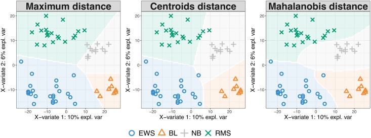
“The study investigates Small Round Blue Cell Tumors (SRBCT, [35]) of 63 tumour samples with the expression levels of 2,308 genes.”
BackgroundMethodLê Cao, K.-A., Boitard, S., & Besse, P. (2011) · BMC Bioinformatics · doi:10.1186/1471-2105-12-253 · Cited by 1,007 · cited 3×
“In single ‘omics analyses this distance achieves best accuracy when the predicted values are close to 0 or 1 [10].”
BackgroundMethodInfluentialPlankton networks driving carbon export in the oligotrophic ocean
Guidi, L., Chaffron, S., Bittner, L., Eveillard, D., Larhlimi, A., Roux, S., Darzi, Y., Audic, S., Berline, L., Brum, J. R., et al. (2016) · Nature · doi:10.1038/nature16942 · Cited by 870 · cited 1×
“(2016), Table 3 and factomineR Husson et al. (2017)). However, very few methods propose feature selection, including sparse CCA (pma package Witten et al. (2013)), sparse PLS (spls package, Chung et al.”
MethodTumor classification by partial least squares using microarray gene expression data
Nguyen, D. V., & Rocke, D. M. (2002) · Bioinformatics · doi:10.1093/bioinformatics/18.1.39 · Cited by 816 · cited 1×
“The assessment is data-driven and similar to the process detailed in Rohart et al. (2016), where one-sided t-tests assess whether there is a gain in performance when adding components to the model.”
MethodPath Models with Latent Variables: The NIPALS Approach
WOLD, H. (1975) · Quantitative Sociology · doi:10.1016/b978-0-12-103950-9.50017-4 · Cited by 802 · cited 1×
“Unsupervised analyses methods include Principal Component Analysis—based on NonLinear Iterative Partial Least Squares for missing values [18], Independent Component Analysis [19], Partial Least Squares regression—PLS, also known as Projection to Latent Structures [20], multi-group PLS [21], regularised Canonical Corre…”
Method
Research works printed but not linked: 3
- A Singh, 2016, DIABLO-an integrative, multi-omics, multivariate method for multi-group classification, bioRxiv, 067611
- F Rohart, 2017, MINT: A multivariate integrative approach to identify a reproducible biomarker signature across multiple experiments and platforms, BMC Bioinformatics, 18
- H Wold, 1966, Estimation of principal components and related models by iterative least squares, J Multivar Anal, 391
Cited by
Items 1,001–1,013 of 1,013
Profiling Soil–Plant–Microbial Communities: DNA and Multi-Omics Techniques
Li, S., Chiodi, C., Maucieri, C., Lucia, M. C. D., Zardinoni, G., Ravi, S., Squartini, A., Concheri, G., Geng, G., Wang, Y., et al. (2026) · Genes · doi:10.3390/genes17030303 · Cited by 27
“Among these approaches, the mixOmics suite provides a versatile framework based on PLS and canonical correlation analysis (CCA), enabling the visualization and interpretation of cross-omics correlation structures [98].”
Design and application of a novel two-amplicon approach for defining eukaryotic microbiota
Popovic, A., Bourdon, C., Wang, P. W., Guttman, D. S., Voskuijl, W. P., Grigg, M. E., Bandsma, R., & Parkinson, J. (2018) · Microbiome · doi:10.1186/s40168-018-0612-3 · Cited by 21
Santos, I. C. D. L., dos Santos, A., Oumar, Z., Soares, M. A., Silva, J. C. C., Zanetti, R., & Zanúncio, J. C. (2019) · Remote Sensing · doi:10.3390/rs11141641 · Cited by 21
“The analyses were performed with the R program [37] and mixOmics [51] and raster [38] packages.”
MethodCulibrk, L., Croft, C. A., Toor, A., Yang, S. J., Singhera, G. K., Dorscheid, D. R., Moore, M. M., & Tebbutt, S. J. (2019) · Frontiers in Cellular and Infection Microbiology · doi:10.3389/fcimb.2019.00016 · Cited by 20
“To further filter our dataset, we used sparse partial least squares (sPLS) (Rohart et al., 2017) to identify highly correlated differentially expressed gene pairs between the human and A.”
MethodM3NetFlow: A multi-scale multi-hop graph AI model for integrative multi-omic data analysis
Zhang, H., Goedegebuure, S. P., Ding, L., DeNardo, D., Fields, R. C., Province, M., Chen, Y., Payne, P., & Li, F. (2025) · iScience · doi:10.1016/j.isci.2025.111920 · Cited by 12
Meade, E. B., Iwanowicz, L. R., Neureuther, N., LeFevre, G. H., Kolpin, D. W., Zhi, H., Meppelink, S. M., Lane, R. F., Schmoldt, A., Mohaimani, A., et al. (2023) · Science of The Total Environment · doi:10.1016/j.scitotenv.2022.159069 · Cited by 12
Model-based joint visualization of multiple compositional omics datasets
Hawinkel, S., Bijnens, L., Cao, K.-A. L., & Thas, O. (2020) · NAR Genomics and Bioinformatics · doi:10.1093/nargab/lqaa050 · Cited by 9
“Partial least squares with canonical mode was applied to raw and clr-transformed data as implemented in the mixOmics package (39).”
Self-citationThanasupawat, T., Glogowska, A., Pascoe, C., Krishnan, S. N., Munir, M., Begum, F., Beiko, J., Krcek, J., Del Bigio, M. R., Pitz, M., et al. (2021) · International Journal of Molecular Sciences · doi:10.3390/ijms22179566 · Cited by 8
Yeo, K., Wu, F., Li, R., Smith, E., Wormald, P.-J., Valentine, R., Psaltis, A. J., Vreugde, S., & Fenix, K. (2024) · Pathogens · doi:10.3390/pathogens13100826 · Cited by 5
“28 R package [76].”
MethodInfrared spectral profiling of demyelinating activity in multiple sclerosis brain tissue
Gakh, O., Wilkins, J. M., Guo, Y., Popescu, B. F., Weigand, S. D., Kalinowska-Lyszczarz, A., & Lucchinetti, C. F. (2024) · Acta Neuropathologica Communications · doi:10.1186/s40478-024-01854-4 · Cited by 4
“The area under the receiver operating characteristic (AUROC) curve was averaged over the cross-validation process using a one-vs-all comparison [50].”
MethodInfluentialApplication of predictive modeling tools for the identification of Ocimum spp. herbal products
Abraham, E. J., Chamberlain, S. J., Perera, W. H., Jordan, R. T., & Kellogg, J. J. (2025) · Analytical and Bioanalytical Chemistry · doi:10.1007/s00216-025-05735-0 · Cited by 2
“PLS‑DA Binary partial least squares—discriminatory analysis (PLS-DA) models were constructed in R with the splsda() function in the mixOmics package [26].”
Cohen, N. J., Lesseur, C., Cardenas, A., Ward-Caviness, C. K., Spring, A. C., Rager, J. E., Fry, R. C., & Eaves, L. A. (2025) · Epigenomics · doi:10.1080/17501911.2025.2533115 · Cited by 2
Wu, Y., Zhou, Y., Fan, Q., Gao, Y., Fang, S., Sun, Y., & Gu, S. (2026) · European Journal of Nutrition · doi:10.1007/s00394-026-03953-5
“PLS-DA (Partial Least Squares Discriminant Analysis) was also introduced as a supervised model to reveal differ - ences in microbial communities between groups, using the “plsda” function in the R package “mixOmics” [25].”
Metadata
- Authors
- Rohart, F., Gautier, B., Singh, A., & Lê Cao, K.-A.
- Affiliations
- Translational Research Institute (AU)
- University of British Columbia (CA)
- Venue
- PLOS Computational Biology
- Publisher
- Public Library of Science (PLoS)
- Published
- 2017-11-03
- Volume
- 13
- Issue
- 11
- Pages
- e1005752
- ISSN
- 1553-734X (ISSN-L)
- 1553-7358
- PMC
- PMC5687754
- OpenAlex
- W2950985821
- Cited by
- 4,056
- References
- 57
- Citations by year
- 2026: 629 · 2025: 612 · 2024: 600 · 2023: 523 · 2022: 496 · 2021: 489 · 2020: 350 · 2019: 241 · 2018: 95 · 2017: 14
- FWCI
- 78.13
- Type
- journal article
- Language
- en
- Update notices
- new_version (2017-11-15)
- Open access
- gold
- doi.org
- Licence
- Locations
- PLoS Computational Biology (publishedVersion, cc-by)
- bioRxiv (Cold Spring Harbor Laboratory) (acceptedVersion, cc-by-nc-nd)
- PubMed Central (publishedVersion, cc-by)
- PubMed (publishedVersion)
- Minerva Access (University of Melbourne) (submittedVersion, cc-by)
- RePEc: Research Papers in Economics (submittedVersion)
- DOAJ (DOAJ: Directory of Open Access Journals) (submittedVersion, cc-by-sa)
- Figshare (submittedVersion, cc-by)
- Funders
- National Health and Medical Research Council (APP1087415)
- Australian Cancer Research Foundation (ACRF) for the Diamantina Individualised Oncology Care Centre
- Topics
- Life Sciences › Biochemistry, Genetics and Molecular Biology › Molecular Biology › Bioinformatics and Genomic Networks
- Life Sciences › Biochemistry, Genetics and Molecular Biology › Molecular Biology › Metabolomics and Mass Spectrometry Studies
- Life Sciences › Biochemistry, Genetics and Molecular Biology › Molecular Biology › Gene expression and cancer classification
- Keywords
- R package, multi-omics integration, feature selection, multivariate analysis, dimensionality reduction, partial least squares regression, discriminant analysis, molecular signatures, systems biology, transcriptomics, proteomics, metabolomics, exploratory data analysis
- MeSH terms
- Data Interpretation, Statistical, Humans, Neoplasms, Software, Computational Biology, Genomics, Systems Biology, Metabolomics
About these checks
Registry, retraction, Cites and statistics results are computed, not judged, with one exception: a printed reference that no index lists is read once by a model before it counts as a citation error. Claims are extracted by a model and kept only when their quote appears verbatim in the text; each cited source is then checked, with a model’s help, for whether it says what the claim relies on. Those verdicts can be wrong — read the claim’s quote and the cited source before relying on one.
Statistics and arithmetic are recomputed, not judged.
Confidence bands are shown only for fields calibrated on our annotated test set. None is calibrated yet, so no band is shown.
What counts as a claim ›How we measure confidence ›How we check this ›
Are you an author, or do you see a problem with this page? Request a review
Data sources
Bibliographic data: Semantic Scholar, OpenAlex, Crossref.
Contains information from the Semantic Scholar Academic Graph Datasets, made available under the ODC Attribution License (ODC-BY 1.0).
Semantic Scholar’s data is described in: Kinney, R., et al. (2023). The Semantic Scholar Open Data Platform. arXiv:2301.10140.
